That Bind the Same Site within the N-Terminal Domain of the Viral
CA Protein
Christopher T. Lemke,aSteve Titolo,b* Uta von Schwedler,cNathalie Goudreau,aJean-François Mercier,bElizabeth Wardrop,b Anne-Marie Faucher,a* René Coulombe,aSoma S. R. Banik,b* Lee Fader,aAlexandre Gagnon,a* Stephen H. Kawai,a* Jean Rancourt,a Martin Tremblay,aChristiane Yoakim,aBruno Simoneau,aJacques Archambault,b* Wesley I. Sundquist,cand Stephen W. Masonb*
Department of Chemistryaand Department of Biological Sciences,bBoehringer Ingelheim (Canada) Ltd., Research & Development, Laval, Quebec, Canada, and Department of Biochemistry,cUniversity of Utah, Salt Lake City, Utah, USA
The emergence of resistance to existing classes of antiretroviral drugs necessitates finding new HIV-1 targets for drug discovery. The viral capsid (CA) protein represents one such potential new target. CA is sufficient to form mature HIV-1 capsids in vitro, and extensive structure-function and mutational analyses of CA have shown that the proper assembly, morphology, and stability of the mature capsid core are essential for the infectivity of HIV-1 virions. Here we describe the development of an in vitro cap-sid assembly assay based on the association of CA-NC subunits on immobilized oligonucleotides. This assay was used to screen a compound library, yielding several different families of compounds that inhibited capsid assembly. Optimization of two chemi-cal series, termed the benzodiazepines (BD) and the benzimidazoles (BM), resulted in compounds with potent antiviral activity against wild-type and drug-resistant HIV-1. Nuclear magnetic resonance (NMR) spectroscopic and X-ray crystallographic analy-ses showed that both series of inhibitors bound to the N-terminal domain of CA. These inhibitors induce the formation of a pocket that overlaps with the binding site for the previously reported CAP inhibitors but is expanded significantly by these new, more potent CA inhibitors. Virus release and electron microscopic (EM) studies showed that the BD compounds prevented vi-rion release, whereas the BM compounds inhibited the formation of the mature capsid. Passage of virus in the presence of the inhibitors selected for resistance mutations that mapped to highly conserved residues surrounding the inhibitor binding pocket, but also to the C-terminal domain of CA. The resistance mutations selected by the two series differed, consistent with differences in their interactions within the pocket, and most also impaired virus replicative capacity. Resistance mutations had two modes of action, either directly impacting inhibitor binding affinity or apparently increasing the overall stability of the viral capsid with-out affecting inhibitor binding. These studies demonstrate that CA is a viable antiviral target and demonstrate that inhibitors that bind within the same site on CA can have distinct binding modes and mechanisms of action.
T
he current antiretroviral arsenal against HIV-1 comprises more than 26 FDA-approved drugs from six mechanistic classes that target one of the three viral enzymes or viral entry (5). In spite of this array of drugs and targets and the simplification of therapies, drug resistance can still occur due to lack of adherence, often owing to toxicities associated with the lifelong therapy re-quired for sustained viral suppression (28,36). Moreover, cross-resistance within mechanistic classes and the emergence of multi-drug-resistant isolates can have considerable impact on treatment options and disease outcomes, underscoring the need to discover new classes of HIV inhibitors.The HIV-1 capsid (CA) protein plays essential roles in viral replication and as such represents an attractive new therapeutic target (11,18). CA is initially synthesized as the central region of the 55-kDa Gag polyprotein, which is the protein that mediates the assembly and budding of the immature virion. In this context, CA provides key protein-protein interactions required for imma-ture virion assembly (18,40). During viral maturation, proteolytic cleavage of Gag releases CA, allowing the protein to assemble into the cone-shaped central capsid that surrounds the viral RNA ge-nome and its associated enzymes, reverse transcriptase (RT) and integrase (IN) (34,35). The capsid is stabilized by multiple weak protein-protein interactions, and CA mutations that impair the assembly and/or stability of the capsid typically inhibit viral rep-lication (10,17,40). Thus, HIV-1 CA plays essential roles during
the assembly of both the immature virion and the mature viral capsid.
CA is composed of two highly helical domains, the N-terminal domain (CANTD, residues 1 to 146) and the C-terminal domain
(CACTD, residues 151 to 231), which are separated by a short
flex-ible linker. Solution nuclear magnetic resonance (NMR) and
Received 25 February 2012 Accepted 30 March 2012 Published ahead of print 11 April 2012
Address correspondence to Christopher T. Lemke,
christopher.lemke@boehringer-ingelheim.com, or Stephen W. Mason, Stephen.mason@bms.com.
* Present address: S. Titolo, AL-G Technologies, Lévis, Quebec, Canada; A.-M. Faucher, Université de Montréal, Département de Chimie, Montréal, Quebec, Canada; S. S. R. Banik, Integral Molecular, Philadelphia, Pennsylvania, USA; A. Gagnon, Université du Québec a` Montréal (UQÀM), Département de Chimie, Montréal, Quebec, Canada; S. H. Kawai, Department of Chemistry and Biochemistry, Concordia University, Montréal, Quebec, Canada; J. Archambault, Institut de Recherches Cliniques de Montréal (IRCM) and Department of Biochemistry, Université de Montréal, Montréal, Quebec, Canada; S. W. Mason, Bristol-Myers Squibb, Virology, Wallingford, Connecticut, USA.
Supplemental material for this article may be found athttp://jvi.asm.org/. Copyright © 2012, American Society for Microbiology. All Rights Reserved. doi:10.1128/JVI.00493-12
on May 18, 2016 by UNIV DU QUEBEC A MONTREAL
http://jvi.asm.org/
high-resolution X-ray crystal structures have been reported for both isolated domains (4,13,14,19,41). Conical HIV-1 capsids belong to a class of geometric structures called fullerene cones, which comprise hexagonal lattices with 12 pentagonal defects that allow the cones to close at both ends. Although individual HIV-1 capsids differ in size and shape, they typically contain⬃250 CA hexagons and have 7 CA pentagons at the wide end and 5 CA pentagons at the narrow end of the cone (15).
The recent availability of high-resolution structures of CA hexagons and pentagons has enabled molecular modeling of the viral capsid (29,30). The capsid lattice is stabilized by four differ-ent types of intermolecular CA-CA interactions: a CANTD/CANTD
interaction that creates the hexameric (or pentameric) rings (29, 30), a CANTD/CACTDinteraction that forms a “girdle” that
rein-forces the rings (16,29), dimeric CACTD/CACTDinteractions that
link adjacent hexamers across local 2-fold axes (1,4,22,41), and trimeric CACTD/CACTDinteractions that link adjacent hexamers
across local 3-fold axes. Each of these different interfaces has been characterized structurally, although the interactions that stabilize the CACTD/CACTDtrimer are not yet known in atomic detail (4).
Moreover, several distinct but related CACTD/CACTDdimers have
been observed (1,4,22,41), and it is not yet certain how these different dimers are used to connect the CA hexamers and pen-tamers within authentic viral capsids (22). Although capsid-like conical assemblies can form in vitro, most conditions that drive CA assembly favor CA hexamerization over pentamerization such that recombinant CA proteins typically assemble into long helical tubes composed exclusively of CA hexamers (4,26).
CA-binding inhibitors of HIV-1 capsid assembly have been reported, thus providing evidence that CA may be a viable drug target (2,24,32, 33, 37,38). These inhibitors have collectively defined three independent inhibitor binding sites on CA. How-ever, each of these sites ultimately appears to interfere with the formation of the CANTD/CACTDinterface, suggesting that this
in-teraction may be an inhibitory “Achilles’ heel” for capsid assem-bly. The small-molecule inhibitor CAP-1 binds to an induced hy-drophobic pocket at the base of the CANTDhelical bundle (24,37).
The pocket is located at the junction of␣-helices 1, 2, 4, and 7 and is normally occupied by the aromatic side chain of Phe-32 in structures of uninhibited CA hexamers, pentamers, and mono-mers. Binding of CAP-1 distorts the loop between helices 3 and 4, which may inhibit the formation of the CANTD/CACTDinterface
(16,24). A second CA inhibitor, termed the CAI peptide, binds to a conserved hydrophobic cleft within the CACTDfour-helix
bun-dle and inhibits both Gag and CA assembly in vitro (33,38). Su-perposition of the CACTD-CAI complex onto the CANTD/CACTD
interface of assembled CA suggested that binding of the peptide would sterically hinder the CANTD/CACTDinteraction (16,29).
The peptide may also act allosterically by altering the geometry of CACTDdimers such that propagation of the mature CA lattice is
prevented. More recently, a third small-molecule binding site on CA was identified via a high-throughput screen for inhibitors of HIV replication. PF-3450074 and related compounds bind be-tween helix 4 and helix 7 of the CANTD. This latest class of CA
inhibitors disrupts the stability of the viral capsid in both the early and late stages of viral replication, again probably by altering CANTD/CACTDinteractions (2,32).
Here we describe two new and highly potent families of CA inhibitors that were identified by a novel high-throughput screen-ing (HTS) assay. Inhibitors from both families bind to the CANTD
by further expanding the CAP-binding pocket. Moreover, al-though these two classes of inhibitors target the same binding pocket, they have distinct binding modes, select for unique pat-terns of resistance mutations, and have different effects on virion morphology.
MATERIALS AND METHODS
Immobilized capsid assembly assay. The 5=-biotin-labeled (TG)25 oligo-nucleotide (Integrated DNA Technology Inc.) was immobilized on Re-acti-Bind neutravidin-coated black 384-well plates (Pierce catalog no. 15402) that were washed with 80l/well of buffer C {50 mM Tris (pH 8.0), 350 mM NaCl, 10M ZnSO4, 0.0025% (wt/vol) 3-[(3-cholamidopro-pyl)-dimethylammonio]-1-propanesulfonate (CHAPS), 50 g/ml bo-vine serum albumin (BSA), 1 mM dithiothreitol (DTT)} prior to the ad-dition of 50l/well of a 25 nM solution of oligonucleotide in buffer C plus 5 mg/ml BSA, followed by overnight incubation. Unbound material was removed by two washes with buffer C. Assembly reactions were per-formed in 60-l/well reaction mixtures comprising 100 nM 5=-fluoresce-in-labeled (TG)25oligonucleotide (Integrated DNA Technology Inc.), 2 M CA-NC protein, and various concentrations of test compounds di-luted in buffer C with a final dimethyl sulfoxide (DMSO) concentration of 1%. Assembly reaction mixtures were incubated for 2 h at room temper-ature, followed by two washes with buffer C, the addition of 80l/well of buffer C plus 0.1% sodium dodecyl sulfate (SDS), and a 15-min incuba-tion prior to quantificaincuba-tion of captured fluorescence on a Victor2plate reader (Perkin-Elmer Life Sciences) equipped with fluorescein excitation and emission filters. The amount of captured fluorescence is proportional to the level of assembly. The concentrations of compound required for 50% inhibition of assembly (IC50s) were generated by fitting inhibition curves from 10-point dilution series to the following equation: % inhibi-tion⫽ (Imaxn⫻ [I]n)/([I]n⫹ IC50n)⫻ 100, where Imaxis the maximal percent inhibition, [I] is the corresponding concentration of inhibitor, and the superscript n denotes the Hill coefficient.
Protein expression and purification. pET-11a vectors were used to
express the HIV-1NL4-3CA-NC (WISP-98-68, Gag residues 133 to 432) carrying the CA G94D mutation, full-length CA (WISP-98-85), CANTD (WISP-96-19, CA residues 1 to 146), and CACTD(WISP-97-07, CA resi-dues 146 to 231) (17,26). Point mutations were introduced using the QuikChange II site-directed mutagenesis kit (Stratagene) according to the manufacturer’s instructions.
All proteins were expressed in Escherichia coli BL21(DE3) cells (Nova-gen). Briefly, LB medium was inoculated with overnight precultures, which were grown at 37°C until mid log-phase (A600,⬃0.6). Protein ex-pression was induced with 0.5 to 1 mM isopropyl--D
-thiogalactopyra-noside (IPTG) for 4 to 6 h at 30°C. Cells were harvested by centrifugation, and pellets were stored at⫺80°C until purification. For NMR studies, 15N-labeled proteins were produced using Spectra 9 (15N, 98%) medium (Cambridge Isotope Laboratories Inc.).
Purification of CA-NC (and all mutants) was carried out as follows. Five to 10 g of cell paste was lysed by sonication in 40 ml of buffer A (20 mM Tris [pH 7.5], 1M ZnCl2, 10 mM-mercaptoethanol) supple-mented with 0.5 M NaCl and Complete EDTA-free protease inhibitor tablets (Roche). Nucleic acids and cell debris were removed by adding 0.11 volume of 0.2 M ammonium sulfate and an equivalent volume of 10% poly(ethyleneimine) (pH 8.0), stirring the sample for 20 min at 4°C, and centrifuging at 30,000⫻ g for 20 min. CA-NC protein was recovered from the supernatant by adding 0.35 volume of saturated ammonium sulfate solution, followed by centrifugation at 10,000⫻ g for 15 min. The pellet was dissolved in 10 ml of buffer A plus 0.1 M NaCl and was dialyzed overnight in buffer A plus 0.05 M NaCl. The sample was cleared by cen-trifugation and was chromatographed on a 1-ml HiTrap SP HP column (GE Healthcare) preequilibrated with dialysis buffer. CA-NC protein was eluted with buffer A plus 0.5 M NaCl. Fractions containing the protein were pooled, and the concentration was determined by absorbance at 280
on May 18, 2016 by UNIV DU QUEBEC A MONTREAL
http://jvi.asm.org/
nm using the calculated molar extinction coefficient (ε ⫽ 40,220 M⫺1 cm⫺1).
Purification of CANTD(wild type [WT], all mutants, and15N labeled) was similar to that of CA-NC, but lysis was performed in buffer B (20 mM morpholineethanesulfonic acid [MES] [pH 6.5], 10 mM -mercaptoeth-anol) supplemented with 0.5 M NaCl and Complete EDTA-free protease inhibitor tablets. Nucleic acids and cell debris were removed as described above. CANTDwas recovered from the supernatant by the addition of 0.6 volume of saturated ammonium sulfate. The pellet was dissolved in 10 ml of buffer B and was dialyzed in the same buffer using dialysis tubing with a 10,000-molecular-weight cutoff. The sample was clarified by centrifu-gation and was sequentially passed through HiTrap SP HP and Q HP columns (GE Healthcare) preequilibrated in buffer B. CANTDwas recov-ered in the flowthrough and wash fractions and was concentrated, and the protein concentration was determined by the absorbance at 280 nm using the calculated molar extinction coefficient (ε ⫽ 25,320 M⫺1cm⫺1).
Cells. SupT1 and 293FT cells were obtained from the ATCC
(CRL-1942) and Invitrogen (R700-07), respectively. C8166 cells were obtained from J. Sullivan, University of Massachusetts Medical Center. C8166-LTR-Luc cells were produced by stable transfection of C8166 cells with an HIV long terminal repeat (LTR)-luciferase construct followed by selec-tion with 5g/ml blasticidin S-HCl through three consecutive limiting dilutions. SupT1 and C8166 cells were maintained in RPMI medium (Wisent) supplemented with 10% fetal bovine serum (FBS; HyClone). C8166-LTR-Luc cells were maintained in the same medium supple-mented with 5g/ml blasticidin S-HCl. Antibiotic was removed for all antiviral activity assays. 293FT cells were maintained in Dulbecco’s mod-ified Eagle medium (DMEM; Wisent) supplemented with 10% FBS (37°C, 5% CO2).
Antiviral activity (EC50) determinations. Inhibitors were prepared in
10 to 20 mM stocks in 100% DMSO and were serially diluted in RPMI medium plus 10% FBS. C8166-LTR-luciferase cells were infected at a multiplicity of infection (MOI) of 0.005 with HIV-1 NL4-3 for 1.5 h and were seeded at 25,000 cells/well in 96-well black microtiter plates, in wells already containing inhibitors or an equivalent concentration of DMSO (0.5%). Plates were incubated for 3 days (37°C, 5% CO2), and luciferase expression levels were determined by the addition of 50l per well of SteadyGlo (Promega) and measurement on the BMG LUMIstar Galaxy luminometer. The inhibitor concentration needed to produce a 50% re-duction of viral replication activity (50% effective concentration [EC50]) was determined by nonlinear regression analysis using SAS software (SAS Institute, Cary, NC).
Single-cycle assays for evaluating early- versus late-stage inhibition of HIV-1 replication. Three constructs for the generation of vesicular
stomatitis virus G glycoprotein (VSV-G)-pseudotyped HIV-1 were made as follows. HIV-1 helper virus was amplified from SODk1CG2 cells (21) and was cloned into pcDNA 3.1. The helper plasmid, which lacks both LTRs as well as functional gp120 and Nef, has all HIV coding sequences expressed from the immediate-early cytomegalovirus (CMV) promoter. Gag-Pol, Vif, and Vpr were derived from NL4-3, while all other sequences were derived from HXB2. The pTV-linker self-inactivating transfer vector (23) was obtained through the AIDS Research and Reference Reagent Program, Division of AIDS, NIAID, NIH, from Lung-Ji Chang. pTV-linker was further modified by inserting the EF-1␣ promoter and firefly luciferase gene, creating pTV-Luc. The VSV-G expression plasmid was obtained from Ivan Lessard (Boehringer Ingelheim [Canada] Ltd.).
The activity of the CA compounds in the early phase of the replication cycle was evaluated by transducing SupT1 cells with VSV-G-pseudotyped HIV-1 in the presence of test compounds. VSV-G-pseudotyped HIV-1 was prepared by batch transfection of 293FT cells in a T-75 flask with 2.2 g of VSV-G plasmid, 6.7 g of HIV-1 helper plasmid, and 9 g of pTV-luc by using FuGene (Roche Applied Science). The 293FT superna-tant was harvested 48 h posttransfection and was centrifuged for 5 min at 2,000 rpm. p24 levels were quantified using an HIV-1 p24 enzyme-linked immunosorbent assay (ELISA) kit (Beckman Coulter). Viral supernatants
were stored at⫺80°C. In a 96-well microtiter plate, 40,000 SupT1 cells (25 l) were infected with an amount of virus corresponding to 4 ng of p24 (25l) in the presence of 50 l of the test compound (in 1% DMSO). Forty-eight hours postinfection, 25l Steady Glo (Promega) was added to each well, and luciferase activity was measured using a TopCount plate reader (Perkin-Elmer).
Inhibitory activity during the late phase of the replication cycle was evaluated by adding test compounds to 293FT cells during the production of VSV-G-pseudotyped HIV-1. Cells were batch transfected (as described above) in the absence of compounds by using FuGene (Roche Applied Science) for 4 to 6 h in a T-75 (75-cm2) flask, after which cells were dislodged by pipetting, and⬃40,000 cells in 50 l were transferred to a 96-well microtiter plate (Corning Costar) containing an equivalent vol-ume of test compound from 10-point dose-response curves (in 1% DMSO). Forty-eight hours posttransfection, 10l viral supernatant was diluted 1:10 in RPMI medium supplemented with 10% FBS in a second 96-well microtiter plate and was then frozen at⫺80°C for at least 1 h. Following thawing, 10l of diluted viral supernatant was used to infect 40,000 SupT1 cells in a final volume of 100l. Thus, compounds were diluted 100-fold in order to minimize any potential inhibitory activity in the early phase of the replication cycle. Forty-eight hours postinfection, firefly luciferase activity was measured as described above. The cytotoxic-ity of the test compounds was evaluated by adding 50l of CellTiter Glo (Promega) to the microplates containing the transfected 293FT cells. Lu-minescence was measured using a TopCount plate reader.
ITC. Isothermal titration calorimetry (ITC) was performed at 25°C in
50 mM Tris (pH 8.0), 350 mM NaCl, and 1% DMSO by using a VP-ITC microcalorimeter (MicroCal Inc.; GE Health Sciences). Titrations were performed using 200M CANTDin the syringe and approximately 20M compound in the sample cell. After degassing, the compound solution was centrifuged at 15,000⫻ g for 15 min and was loaded in the sample cell. The precise compound concentration in the sample cell was assessed by high-performance liquid chromatography (HPLC) using a reference DMSO solution of the compound. Each titration consisted of 19 injections of 15 l at 280-s intervals. Thermodynamic parameters were derived by fitting the binding isotherms to the single-site binding model algorithm, with stoichiometries (n), enthalpies (⌬H), and equilibrium dissociation con-stants (KD) allowed to float during nonlinear least-squares fits of the data. Typically, stoichiometries (n) were between 0.9 and 1.1. The starting con-centrations of more-potent compounds were reduced to 5M in the sample cell for better assessment of the KD.
Selection of HIV-1 variants resistant to capsid inhibitors. C8166
cells were infected at an MOI of 0.1 with WT HIV-1 2.12 (7) in complete RPMI medium (RPMI 1640, 10% FBS, 10g/ml gentamicin, and 10 M -mercaptoethanol) with twice to 5 times the EC50of the capsid inhibitor. At each passage (3 to 4 days), microscopic evaluation of the cytopathic effect (CPE) was performed, and the culture supernatant was used to infect fresh C8166 cells, which were then maintained at the same or a higher concentration of inhibitor depending on the CPE. At passages where viral breakthrough was evident, the genomic DNA was isolated using the DNeasy Blood and Tissue kit (Qiagen). The CA gene was am-plified by PCR, and the fragments were cloned into the Zero Blunt TOPO plasmid (Invitrogen) and sequenced by automated sequencing.
Construction of recombinant HIV-1. To create viruses carrying CA
mutations, site-directed mutagenesis was carried out using the QuikChange site-directed mutagenesis kit (Stratagene). All mutations were confirmed by sequencing on an ABI Prism 3100 genetic analyzer (Applied Biosystems Inc.). Sequenced DNA fragments containing the confirmed mutation(s) were subcloned into the NL4-3 provirus by stan-dard molecular biology techniques. Viral stocks were produced by trans-fecting 293 cells by the calcium phosphate method (20). The culture su-pernatant was collected after 3 days (37°C, 5% CO2); cell debris was removed; and aliquots were frozen at⫺80°C. The 50% cell culture infec-tive dose (CCID50) was determined by monitoring the formation of syn-cytia in C8166 cells.
on May 18, 2016 by UNIV DU QUEBEC A MONTREAL
http://jvi.asm.org/
Jurkat cell replication capacity assay. The replication capacity assay
was carried out with Jurkat-LTR luciferase cells as described previously (7). Briefly, Jurkat cells expressing an HIV LTR luciferase construct were infected with virus at an MOI of 0.05 for 2 h at 37°C, washed, and seeded at 1⫻ 105cells/well in 200l RPMI 1640 supplemented with 10% FBS and 10g/ml gentamicin in clear-bottom black 96-well microtiter plates. Every 3 to 4 days, the cells were mixed, and 100l medium was removed and replaced with fresh medium. Luciferase levels were determined by adding 50l/well BrightGlo (Promega) at days 7, 10, 12, and 14 postin-fection and reading on the LUMIstar galaxy plate reader (BMG). Replica-tion was not assessed at later times owing to the potential for variability over longer periods.
Assays for inhibition of virion release, infectivity, and assembly.
293T cells were transfected with the proviral HIV-1NL4-3R9 expression construct in the presence of benzodiazepine (BD) and benzimidazole (BM) inhibitors (50-fold over the EC50s). Viral Gag expression, process-ing, virion release, and viral titers were assayed as described in detail else-where (40). Released virions were fixed with gluteraldehyde and osmium tetroxide, stained with uranyl acetate, embedded in epoxy resin, thin sec-tioned (thickness, 60 to 90 nm), poststained with Reynold’s lead citrate, and imaged on a Hitachi H-7100 transmission electron microscope at a magnification of⫻50,000, as described in detail elsewhere (39).
X-ray crystallography models. A full description of the crystallization
and diffraction data will be reported elsewhere (manuscript in prepara-tion). For the superposition shown in Fig. 3, monomer A of the CANTD-BD 3 structure and monomer B of the CANTD-BM 4 structure were superposed on the unliganded CANTD(residues 1 to 146) of the stabilized hexameric CA structure (PDB identification code [ID] 3H47) (29). Monomer A of the CANTD-BD 3 structure was selected because it is the only one of the two monomers of the asymmetric unit that bound the BD 3 inhibitor (the other monomer is apo). Monomer B of the CANTD-BM 4 structure was selected because the bound inhibitor appears to be less affected by neighboring molecules of the crystal lattice. The alignments were based on 300 atoms constituting the backbone of 75 CANTDresidues: Ala47 to Asn57, Ala64 to Leu83, Arg97 to Met118, and Ile124 to Tyr145. This selection focuses on the most immutable portions of CANTD, avoiding helices 1 and 2 and other highly flexible regions. The calculated root mean square distances (RMSDs) for the fitted CANTD-BD 3 and CANTD-BM 4 structures to the unliganded CANTDwere both 0.44 Å. For the liganded hexameric model shown inFig. 9, monomer B of the CANTD-BM 4 structure was superposed on the unliganded CA monomer of the hexameric CA structure (PDB ID 3H47) (29). The superposition was based on 440 atoms constituting the backbone of 110 CA residues: residues 1 to 3, 10 to 24, 33 to 57, 64 to 82, and 97 to 144. This more comprehensive selection of residues includes the major secondary struc-tural elements while avoiding highly flexible regions. A hybrid CA mole-cule was then made by fusing residues 1 to 140 of the CANTD-BM 4 struc-ture with residues 141 to 219 of 3H47. The hexamer of this hybrid molecule was then generated by applying the appropriate crystallographic symmetry operations based on the hexagonal (P6) space group of 3H47.
Protein structure accession numbers. The PDB entries for
CANTD-BD 3 and CANTD-BM 4 are 4E91 and 4E92, respectively.
RESULTS
Efficient HTS assay for inhibitors of HIV-1 CA assembly. In
principle, inhibition of CA tube assembly can be used to screen for small molecules that block capsid assembly, but this method gen-erally requires high protein concentrations and must be per-formed under high-ionic-strength conditions (25,26). We there-fore developed an in vitro CA assembly assay that was more amenable to high-throughput screening (HTS).
Our assay employed a Gag fragment that spanned the CA and NC regions and took advantage of the ability of the nucleocapsid (NC) to bind tightly to TG-rich deoxyoligonucleotides (9). The use of d(TG)25as a “scaffold” enabled CA tubes to form at much
lower protein and salt concentrations than in previously reported assays. Assembly reactions were performed in neutravidin-coated 384-well plates using both biotin- and fluorescein-labeled d(TG25). The biotin-labeled d(TG25) oligonucleotide bound on
the surface acts to nucleate the assembly of complexes, and further “polymerization” of CA-NC is driven by soluble fluorescein-la-beled d(TG25). Unbound and unassembled species are washed
away from captured assembly products at the end of the assembly reaction.
As shown inFig. 1A, titration of increasing quantities of wild-type (WT) CA-NC in this assay led to a dose-dependent increase in the fluorescence signal up to a level of saturation that corre-sponded to complete loading of the oligonucleotide with CA-NC protein. In contrast, two control CA-NC proteins with amino acid substitutions previously shown to block capsid assembly in vitro and in vivo (A42D and W184A/M185A) (17,40) were inactive in this assay.
The sensitivity of the capsid assembly assay to known inhibi-tors was confirmed using the CAI peptide as a positive control (33). As shown inFig. 1B, increasing concentrations of the CAI peptide abrogated the assay signal, with an IC50of 0.6M (Fig.
1B). In contrast, a control peptide with the same amino acid com-FIG 1 Validation of the capsid assembly assay. (A) Assay showing that
wild-type HIV-1 CA-NC assembles in a concentration-dependent fashion, whereas the assembly-defective CA-NC mutants (the A42D and W184A M185A mu-tants) do not. CA-NC assembly was detected by capture and immobilization of a fluorescent oligodeoxynucleotide. (B) CAI, a known peptide inhibitor of HIV-1 capsid assembly (33), inhibits CA-NC assembly in a concentration-dependent fashion, whereas a scrambled peptide control (ctrl) does not. The assay was performed with wild-type CA-NC under standard assay conditions, and activities are reported as the percentage of inhibition of the control fluo-rescence signal.
on May 18, 2016 by UNIV DU QUEBEC A MONTREAL
http://jvi.asm.org/
position as CAI, but with a scrambled sequence, failed to inhibit CA-NC assembly, even at concentrations as high as 25M (Fig. 1B). Taken together, these results validate the assay and indicate that it recapitulates the formation and inhibition of capsid-like assemblies in vitro.
Identification of two classes of HIV-1 CA assembly inhibi-tors. The CA assembly assay was used to screen the Boehringer
Ingelheim corporate compound collection, producing⬎50 active hit clusters. Hits were triaged based on several criteria, including relative potency in the capsid assembly assay, lack of activity in an analogous assay based on assembly of a Gag CA-NC protein from Rous sarcoma virus (RSV Gag⌬MBD ⌬PR protein), cytotoxicity, and assessment of chemical tractability. Compounds that survived this triage showed direct binding to CANTDas analyzed by NMR
chemical shift perturbations and cocrystallization (see below) (data not shown). Based on these hit-to-lead studies, two struc-turally distinct families of compounds were chosen for lead opti-mization (Table 1): the benzodiazepines (BDs) and the benzimi-dazoles (BMs). Significant medicinal chemistry efforts (8; also manuscripts in preparation) led to the synthesis of the most po-tent inhibitors from the BD and BM series, BD 1 (half-maximal antiviral effective concentration [EC50], 70⫾ 30 nM [n ⫽ 21];
half-maximal cytotoxic concentration [CC50],⬎28 M) and BM
1 (EC50, 62⫾ 23 nM [n ⫽ 53]; CC50,ⱖ20 M), both of which
displayed potent antiviral activity and a large window relative to cytotoxicity (CC50/EC50,⬎300-fold). These inhibitors, along with
related compounds within the same two families (Table 1), were characterized further to establish their mechanisms of action.
Mechanistic studies of the BD and BM inhibitors. To ensure
that the BD and BM compounds did not also inhibit the same targets as currently available antiretroviral agents, representative compounds from both the BD and BM series were tested for ac-tivity against HIV-1 NL4-3 strains that were resistant to the four major classes of antiretroviral inhibitors: nonnucleoside reverse transcriptase inhibitors (NNRTI) (RT mutations Y188L and V106A), nucleoside reverse transcriptase inhibitors (NRTI) (RT mutations K65R and M184V), protease (PR) inhibitors (PI) (PR mutations V32I I47V and L33F I54L), and integrase strand trans-fer inhibitors (INSTI) (IN mutation G140S Q148H). As shown in Table 2, BD 1, BM 2, and BM 3 inhibited the replication of the wild-type and all mutant viruses with equal potencies. In contrast, control experiments showed the expected pattern of reduced sus-ceptibility to well-characterized antiretrovirals. These data indi-cate that the mechanism of action of the BD and BM compounds differs from that of the four major classes of antiretroviral agents. To determine the stage of the viral replication cycle targeted by the BM and BD compounds, we tested their effects on the effi-ciency of HIV-1 vector transduction when the compounds were applied either during virus production (late phase) or during in-fection (early phase). A vesicular stomatitis virus G glycoprotein (VSV-G)-pseudotyped HIV-1 vector containing a luciferase re-porter under the control of the HIV-1 LTR was used in these studies, allowing discrimination between the inhibition of early replication events (i.e., virus entry through transcription) and late events (i.e., viral transcription through virus maturation). Con-trol compounds behaved as expected in our assays: nevirapine, which inhibits the early-stage process of reverse transcription, re-duced the reporter activity only when added to target cells. In contrast, lopinavir, which inhibits the late-stage processes of Gag proteolysis and viral maturation, inhibited vector transduction
TABLE 1 Compounds used in this study and their antiviral activities
Compound Structure EC50(M)a BD compounds BD 1 0.070 BD 2 1.1 BD 3 0.48 BD 4 0.13 BM compounds BM 1 0.062 BM 2 0.26 BM 3 0.11 BM 4 ⬎46b BM 5 2.4c a
Activity determined in a standard antiviral replication assay (see Materials and Methods).
b
BM 4 was inactive in the viral replication assay but active in the capsid assembly assay. cBM 5 was active in the capsid assembly assay.
on May 18, 2016 by UNIV DU QUEBEC A MONTREAL
http://jvi.asm.org/
only when added to producer cells (Table 3). By these criteria, the BD and BM compounds both acted primarily at the late stage of the viral replication cycle, since both BD 1 and BM 2 inhibited replication when added during virus production. Furthermore, the EC50s obtained in the late-stage assay for BD 1 and BM 2 were
similar to those obtained in standard, multiple-round HIV-1 rep-lication assays (Table 3). In contrast, EC50s for these compounds
were at least 10-fold higher when they were applied during the infection of target cells (early phase). Both compounds did exhibit weak inhibitory activity in the early phase (EC50,⬃3 M), which
could be due to effects on viral core stability and disassembly or, alternatively, to cytotoxicity. Regardless, the data establish that both the BD and BM compounds act primarily by inhibiting the late stage(s) of viral replication.
BD and BM inhibitors bind the N-terminal domain of the HIV-1 CA protein. Isothermal titration calorimetry (ITC) and
2-dimensional (2D)-NMR chemical shift perturbation analyses were performed to test whether the BD and BM compounds bound purified recombinant HIV-1 CA proteins and to map their binding sites. The NMR chemical shift perturbation analyses demonstrated that BD and BM inhibitors bound CANTD, with
shifted residues clustered at the bases of helices 1, 2, 4, and 7 and at the flexible loops between helices 1 and 2 and helices 3 and 4 (data not shown). ITC binding isotherms showed that inhibitors from both families of compounds bound CANTDwith 1:1
stoichiome-tries and submicromolar dissociation constants. For example, two BM compounds, BM 2 and BM 3, and the weak-binding BD com-pound BD 2 (seeTable 1) bound CANTDwith dissociation
con-stants of 210 nM, 87 nM, and 690 nM, respectively (Table 4). These dissociation constants correlated well with the antiviral
EC50s for these compounds (260, 110, and 1,100 nM, respectively
[Table 1]). Binding of the BM compounds was enthalpically driven (⌬H, ⫺18.3 and ⫺18.7 kcal/mol, respectively), which is a favorable property for viral inhibitors (12). Unfortunately, ITC analyses could not be performed with more-potent compounds in the BD series owing to their limited solubility. Nevertheless, our data establish that both compound classes bind directly to the CANTD, independently of the CA C-terminal domain, CA
dimerization, or higher-order CA assembly.
Crystal structures of BD and BM inhibitors bound to the
HIV-1 CANTD. To explore the inhibitor binding modes in greater
detail, representative compounds from each inhibitor family, BD 3 and BM 4 (seeTable 1), were cocrystallized in complex with CANTD. The structures of both complexes were determined at
high resolutions and were refined fully (for CANTD-BD 3, 1.7 Å
resolution, R⫽ 22.0%, and Rfree⫽ 24.3%; for CANTD-BM 4, 1.8 Å
resolution, R⫽ 22.0%, and Rfree⫽ 26.4%). Complete details of
the crystallography will be reported elsewhere.
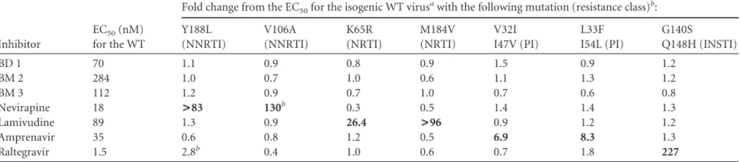
As shown inFig. 2, both inhibitor types bind to a pocket lo-cated at the base of the central four-helix bundle of the CANTD,
which is the same site described previously for CAP-1 (24). As was the case for the CAP inhibitors, binding of the BD or BM com-pounds displaces the side chain of Phe32 from the helical bundle to create a new pocket within the hydrophobic core. Although their binding sites overlap, there are interesting differences in the ways in which the two compounds interact with the protein and affect its structure (Fig. 2and3). Specifically, the BD compounds bind more deeply and enlarge the pocket to a greater extent, re-sulting in significant disruptions of the CANTDtertiary structure
(Fig. 3A). In particular, the trifluoromethyl group of BD 3 binds very deeply, displacing the CA Trp23 side chain at the top of the pocket by more than 3 Å, which in turn contributes to a⬎1.5-Å displacement of the CA␣1 helix. The BM compound BM 4 also perturbs the CA tertiary structure, but in this case the native TABLE 2 Relative activities of capsid assembly inhibitors against drug-resistant viruses
Inhibitor
EC50(nM)
for the WT
Fold change from the EC50for the isogenic WT virusawith the following mutation (resistance class)b:
Y188L (NNRTI) V106A (NNRTI) K65R (NRTI) M184V (NRTI) V32I I47V (PI) L33F I54L (PI) G140S Q148H (INSTI) BD 1 70 1.1 0.9 0.8 0.9 1.5 0.9 1.2 BM 2 284 1.0 0.7 1.0 0.6 1.1 1.3 1.2 BM 3 112 1.2 0.9 0.7 1.0 0.7 0.6 0.8 Nevirapine 18 >83 130b 0.3 0.5 1.4 1.4 1.3 Lamivudine 89 1.3 0.9 26.4 >96 0.9 1.2 1.2 Amprenavir 35 0.6 0.8 1.2 0.5 6.9 8.3 1.3 Raltegravir 1.5 2.8b 0.4 1.0 0.6 0.7 1.8 227
aAll values are averages for two independent experiments except for the values for V106A with nevirapine and Y188L with raltegravir, for which experiments were performed once. Significant fold change values are in boldface.
bResistance targets were RT for Y188L, V106A, K65R, and M184V; PR for V32I I47V and L33F I54L; and IN for G140S Q148H.
TABLE 3 Inhibition of postentry (early) versus postintegration (late)
stages of virus replication
Compound
Value for single-cycle assaysa
EC50(M) for multiple-cycle assays Early EC50 (M) Late EC50 (M) Cytotoxicityb (CC50[M]) BD 1 2.8 0.2 ⬎7.0 0.06 BM 2 3.2 0.2 6.2 0.26 Lopinavir ⬎0.5 0.01 ⬎0.5 0.009 Nevirapine 0.08 ⬎2.2 ⬎2.2 0.02
aBoldface values denote the expected result for late- and early-stage activities of Lopinavir and Nevirapine, respectively.
bCytotoxicity was determined on virus producer cells (late phase).
TABLE 4 Energetics of inhibitor binding to CANTDa
Compound KD (M) ⌬G (kcal/mol) ⌬H (kcal/mol) ⫺T⌬S (kcal/mol) BM 2 0.210 ⫺9.12 ⫺18.3 9.21 BM 3 0.087 ⫺9.64 ⫺18.7 9.06 BD 2 0.690 ⫺8.40 ⫺7.50 ⫺0.90 a
Inhibitor binding was quantified by isothermal titration calorimetry (see Materials and Methods).
on May 18, 2016 by UNIV DU QUEBEC A MONTREAL
http://jvi.asm.org/
CA interactions at the top of the pocket are much less disrupted (Fig. 3B).
Additionally, both compounds reposition the flexible loop linking CA helices 3 and 4 (Fig. 3B), but in considerably different
ways. BD 3 hydrogen bonds with the backbone NH of His62, thus positioning the imidazole side chain away from the pocket (Fig. 2A), while BM 4 interacts with the His62 imidazole, positioning it nearer to the pocket (Fig. 2B). Both inhibitor-bound loop confor-FIG 2 X-ray crystal structures of CANTDwith the BD 3 (A) and BM 4 (B) inhibitors bound within an induced pocket at the base of the HIV-1 CANTDfour-helix
bundle. Note that BD 3 binds deeply within the pocket and forms two direct H-bonds (green) with the backbone nitrogen atoms of Phe32 and His62, as well as water-mediated hydrogen bonds with the backbone carbonyl oxygen atoms of Val24 and Val59. BM 4 binds less deeply and forms direct hydrogen bonds with the backbone nitrogen atom of Phe32 and with N␦ of His62. The inhibitors are shown in stick models with carbons in white, nitrogens in blue, oxygens in red, and fluorines in green. CA backbones and key side chains are shown in orange (BD 3 complex) and blue (BM 4 complex).
FIG 3 Structural comparisons illustrating the differing effects of BD and BM inhibitor binding on the CANTDconformation. (A) Overlay of the BD 3 (orange)
and BM 4 (blue) complex structures. van der Waals surfaces illustrate how BM 4 protrudes further into the solvent, whereas BD 3 binds more deeply and induces the formation of a larger pocket, primarily by shifting CA-NC helices 1 and 2 and the Trp23 side chain. The “protruding region,” or the substituents on the inhibitors that extend outside the pocket, is indicated (see the text). (B) Overlay of the apo (white)-, BD 3 (orange)-, and BM 4 (blue)-bound CANTDstructures
detailing shifts of backbones and key residues. The bound inhibitors have been removed for clarity. Residues 1 to 146 of the stabilized hexameric CA structure (PDB ID 3H47) (29) were used as the reference apo structure. Residues of helices 1 and 2 and highly flexible portions of CANTDwere omitted from the
superposition calculations, as described in Materials and Methods.
on May 18, 2016 by UNIV DU QUEBEC A MONTREAL
http://jvi.asm.org/
mations differ substantially from that of the apo-CA hexamer structure (29) and would be expected to perturb or inhibit the formation of the CANTD/CACTDinterface.
Finally, the two classes of compounds protrude from the CANTDwith significantly different trajectories, and the BM
com-pounds extend much further outside the pocket than do the BD compounds (Fig. 3A). Structure-activity relationships within the protruding groups of the BM compounds revealed their impor-tance for potency, suggesting that this region of the inhibitor may make additional interactions within the assembled capsid (or Gag) lattices. In contrast, the activities of the BD inhibitors were not significantly altered by chemical modifications beyond the directly attached phenyl group, implying that this part of the in-hibitor does not make other significant contacts. Thus, although both classes of inhibitors bind the same pocket on CA, differences in the details of their binding modes suggest the possibility that they may have distinct effects on capsid assembly.
Differential effects of the BD and BM inhibitors on virus as-sembly and maturation. The effects of the BD and BM inhibitors
on Gag production, processing, and virus release were analyzed by Western blotting of cell- and virion-associated Gag proteins (Fig. 4). Virus was produced in the presence of high concentrations (50-fold over the EC50) of two different inhibitors from each
fam-ily. As expected, in all cases, viral titers were dramatically reduced from that with the control DMSO treatment (Fig. 4A). Neither class of inhibitor had a significant effect on Gag expression or processing, as analyzed by Western blotting of intracellular and cell-associated viral proteins with antibodies that detected the MA and CA proteins (and their Gag precursors) (Fig. 4B). Virus re-lease was analyzed by Western blotting to detect virion-associated CA proteins released into the culture supernatant. In this case, the two classes of inhibitors exhibited quite different effects. The BM compounds reduced virion release only modestly from that with the DMSO control, whereas the BD inhibitors reduced virion re-lease to nearly undetectable levels. We therefore conclude that the BD inhibitors function primarily by blocking Gag assembly and virion release, whereas the BM inhibitors have a different mode of action.
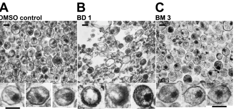
Electron microscopic (EM) analyses were performed to exam-ine the morphologies of viral particles released in the presence of the two different classes of inhibitors. As expected, control virus produced in the presence of DMSO (Fig. 5A) yielded virions with a mixture of different core morphologies, but a high proportion had discernible conical cores (44%⫾ 9%, based on 1,005 virions scored in multiple fields). Consistent with the Western blot anal-ysis, very few virions were produced in the presence of BD inhib-itors, and most of the visible structures corresponded to amor-phous cellular vesicles (Fig. 5B). The few virions that were present had a variety of gross morphological defects, including partially formed immature Gag shells and acentric electron-dense “cores” (see enlargements of individual virions belowFig. 5Bfor three examples). In contrast, virions produced in the presence of the BM series were much more prevalent and homogeneous (Fig. 5C) but only rarely had conical cores (2%⫾ 0.2% of 543 virions scored in multiple fields). Instead, they typically contained acentric, elec-tron-dense complexes that lacked discernible capsids. These data indicate that the BM inhibitors act primarily by inhibiting the assembly of mature conical capsids.
Selection of inhibitor resistance mutations. To confirm that
CA was the target of the inhibitors and to learn more about how
the virus could adapt to inhibition, we selected for viruses that were resistant to BD or BM compounds. To select for resistance, HIV-1NL4-3was cultured in C8166 cells in the presence of
increas-ing concentrations of two representative compounds from each series for as many as 11 passages (see Table S1 in the supplemental material). The development of viral resistance resulted in in-creased virus replication, which led to cytopathicity. At later pas-FIG 4 BM and BD compounds inhibit different stages of HIV-1 assembly
and maturation. (A) HIV-1NL4-3titers produced from cells treated with the
designated inhibitors (concentration, 50-fold over the EC50) or with a
DMSO control. (B) Western blots showing levels of virion-associated CA proteins released into the supernatant (top) or levels of Gag, CA, and MA proteins being produced in cells (center). Cellular tubulin levels are shown as a control for toxicity and loading levels (bottom). Note that virus release was nearly normal in cells treated with the two BM inhibitors but was severely impaired in cells treated with the two BD inhibitors. The experi-ments were performed twice, and similar results were obtained in both cases, although the absolute viral titers differed in the two experiments.
on May 18, 2016 by UNIV DU QUEBEC A MONTREAL
http://jvi.asm.org/
sages (between passages 7 and 11), whenever cytopathicity became evident, proviral DNA was cloned; the capsid gene was sequenced to identify candidate resistance mutations; and the inhibitor con-centration was raised in subsequent passages to elicit additional resistance mutations.
In most cases, multiple different CA mutations were present when high-level resistance developed. Perhaps not surprisingly,
many of the selected amino acid substitutions mapped to the in-hibitor binding site (Fig. 6). Interestingly, however, distinct pat-terns of amino acid substitutions were selected with potent versus weaker inhibitors and with BD versus BM inhibitors. Specifically, the V36T and G61E substitutions were obtained only upon selec-tion with BD inhibitors (Fig. 6A), whereas the K30R and S33G substitutions were obtained only upon selection with BM inhibi-FIG 5 EM analyses showing the different viral assembly and maturation defects induced by BM and BD inhibitors. Images show transmission electron
micrographs of thin-sectioned concentrated virions released from cells treated with DMSO (control) (A), BD 1 (B), or BM 3 (C). Enlargements of 3 individual virions are shown below each representative field. Inhibitor treatments were performed as for the experiments shown inFig. 4. Similar results were also obtained with BD 4 (rare, highly aberrant virions) and BM 1 (acentric, nonconical cores). The experiments were performed twice, and similar results were obtained in both cases. Bars, 100 nm.
FIG 6 Identities and structural locations of viral resistance mutations selected by treatment with BD (A) and BM (B) inhibitors. Two compounds from each
chemical series were used for the selection of resistant virus: BD 1 and BD 2 from the BD series and BM 2 and BM 3 from the BM series (seeTable 1for compound structures and activities). Resistance substitutions (highlighted in red; side chains shown in stick form) selected by these compounds are mapped onto the CANTD
C␣ trace (gray), with bound BD and BM inhibitors shown in orange (BD 3) and blue (BM 4).
on May 18, 2016 by UNIV DU QUEBEC A MONTREAL
http://jvi.asm.org/
tors (Fig. 6B). The G61E substitution was selected only by a weaker inhibitor from the BD series, BD 2, and was never obtained with the more potent compound BD 1. T58I was selected by both inhibitors from the BM series (BM 2 and BM 3) and was also observed as a minor substitution selected by BD 1. Finally, Val27 alterations to Ala or Ile were observed as minor substitutions for the BD series only. Table S1 in the supplemental material summa-rizes all major and minor CA amino acid substitutions obtained during selection. All major substitutions, as well as several of the minor substitutions, were chosen for further characterization, as described below.
Interestingly, inhibitors from both families also selected for amino acid substitutions in the CACTD, including G208R (major
for BD 1 and BM 3; minor for BM 2) and E213G (a minor substi-tution for BM 2 only). At later passages and higher inhibitor con-centrations, these CACTDsubstitutions were often found together
with binding pocket substitutions, and the following double mu-tants were observed in different clones: the K30R/G208R, S33G/ G208R, T58I/G208R, or T58I/E213G (BM) and V36T/G208R (BD) mutants.
Characterization of key inhibitor resistance mutations. To
quantify the degree of inhibitor resistance induced by different amino acid substitutions, all major single and double substitu-tions were introduced separately into the NL4-3 proviral clone, and the mutant viruses were tested for their sensitivities to capsid assembly inhibitors BD 1, BM 2, and BM 3. As shown inTable 5, all of the single substitutions that were selected by BD inhibitors resulted in significant BD drug resistance, ranging from 3-fold (G208R) to 25-fold (V36T). Similarly, single substitution muta-tions that were selected by BM inhibitors resulted in significant BM inhibitor resistance, ranging from 4-fold (K30R versus BM 2) to 22-fold (T58I versus BM 3). In most cases, resistance mutations selected against one inhibitor family also conferred resistance to the other, presumably reflecting their common binding sites. The demonstration that resistance is engendered by clonal mutations at a series of different CA residues provides formal proof that CA is the functional target of both the BD and the BM inhibitors.
As has been seen in other drug resistance profiles, multiple mutations often synergized to produce high-level drug resistance. For example, although the most prevalent CACTDresistance
sub-stitution, G208R, conferred only modest (3- to 7-fold) resistance to either inhibitor family, this substitution synergized with V36T to produceⱖ35-fold resistance to both inhibitor series. Interest-ingly, CA residue 208 is also highly variable among different virus isolates. Although Arg is found at this position in only 3 of 2,240 reported HIV-1 sequences, Ala substitutions are quite common, particularly in subtype B (44% of all sequences and 90% of sub-type B sequences in the Los Alamos National Laboratory HIV Database [http://www.hiv.lanl.gov]). We found, however, that the
G208A substitution in HIV-1NL4-3, a subtype B-derived virus, did
not confer resistance to any of the capsid assembly inhibitors, implying that viral strains with Ala at position 208 will not exhibit intrinsic resistance to BD or BM inhibitors (Table 5).
Most BD and BM resistance mutations reduce viral replica-tive capacity. Drug resistance mutations often reduce viral fitness
(31). We therefore tested whether this was also the case for the BD and BM resistance substitutions. Viral fitness was assessed in a Jurkat-LTR-luciferase reporter T cell line that provided a more stringent test of replicative capacity than the highly permissive cell lines used for the selection of resistant virus. As shown inFig. 7, BD and BM resistance substitutions invariably reduced viral rep-lication capacity from that of the wild-type virus, showing impair-ments that were modest (40 to 80% of wild-type replication for V27I), moderate (5 to 40% for V36T, K30R, T58I, K30R/T58I, and E213G), or profound (less than 5% for G208R, V36T/G208R, and K30R/G208R). The S33G resistance substitution could not be tested in this assay because infectious virus was not produced from the mutated proviral clone, implying that this substitution se-verely impairs viral replicative capacity. Thus, the development of resistance to either family of capsid inhibitors can be expected to reduce viral fitness.
Effects of resistance mutations on inhibitor binding. To test
whether inhibitor resistance mutations reduced inhibitor bind-ing, recombinant CANTDproteins carrying representative CA
in-hibitor resistance substitutions were purified and used in ITC ex-periments to quantify the binding of the BM 2, BM 5, BM 6, and BM 7 inhibitors. As shown in Table 6, and in Table S2 in the supplemental material, three of the substitutions; S33G, V27A, and V36T, reduced the inhibitor binding affinity between 6- and 21-fold. For BM 2, the decrease in binding energy was driven by TABLE 5 Susceptibilities of viruses with resistance mutations to inhibitors of capsid assembly
Compound
Fold change from the EC50of the wild-type virus with the following mutationa:
V27I (BD) V36T (BD) K30R (BM) T58Ia (BM and BD) G208R (BM and BD) G208A K30R G208R (BM) V36T G208R (BD) BD 1 5 25 1 6 3 1 4 ⬎36 BM 2 1 8 4 16 7 1 16 55 BM 3 1 7 5 22 5 1 5 35
aThe class(es) of compound with which each mutation was selected is given in parentheses. T58I was a major substitution selected with BM compounds but a minor substitution selected with BD compounds. G208A is a natural polymorphism.
FIG 7 Inhibitor resistance substitutions reduce viral fitness. Shown are levels
of luciferase produced by Jurkat LTR-luciferase reporter cells 14 days after infection with wild-type HIV-1 or with viruses carrying the designated muta-tions within the CA protein that confer resistance to BD inhibitors or BM inhibitors. The major G208A polymorphism was also tested. Luciferase activ-ities are expressed as relative replicative capacactiv-ities, with that for the wild-type virus set at 100%. Replicative capacities were measured at days 7, 12, and 14. The results for each mutant were similar on days 12 and 14. Replicative capac-ity was not assessed at additional times owing to the potential for variabilcapac-ity over longer periods. Each value represents the average for 6 replicates from a typical experiment.
on May 18, 2016 by UNIV DU QUEBEC A MONTREAL
http://jvi.asm.org/
reductions in binding enthalpy, indicating that these substitutions removed favorable interactions (see Table S3 in the supplemental material). Similar reductions in binding affinity were seen for each mutant with all related BM compounds that were tested (see Table S2). Given that all three of these residues are located within the BM inhibitor binding site, it is likely that the mutations confer resis-tance, at least in part, by directly reducing inhibitor binding. Two other resistance substitutions, K30R and T58I, were of interest because they did not reduce the CANTDbinding affinities of the
BM compounds (Table 6; see also Table S2). Thus, the 4- to 22-fold levels of resistance conferred by these substitutions (Table 5) were not attributable to reduced inhibitor binding affinity, imply-ing that they act via an indirect mechanism.
Indirect resistance mutations promote CA-NC assembly in vitro. We hypothesized that resistance mutations that did not
di-rectly affect inhibitor binding might act indidi-rectly by increasing the efficiency of capsid assembly and/or capsid stability, thereby counteracting the detrimental destabilizing effects of inhibitor binding. To investigate this possibility, we took advantage of pre-vious reports that amino acid substitutions that increase HIV-1 core stability often reduce the ionic strength requirements for CA-NC assembly in vitro (6,10). We therefore examined the as-sembly properties of purified recombinant CA-NC proteins that carried either the aforementioned T58I resistance substitution or the G208R substitution, which is located in the CACTD and is
therefore distant from the inhibitor binding site. The wild-type and mutant CA-NC proteins were tested for their abilities to as-semble in vitro under suboptimal (150 mM) salt concentrations, and the “direct” S33G resistance substitution was used as a con-trol.
As predicted, the wild type CA-NC protein assembled effi-ciently in buffers that contained 350 mM NaCl (Fig. 1), but not at 150 mM NaCl (Fig. 8). Similarly, the control CA-NC mutant (the S33G mutant) also failed to assemble detectably under these low-salt conditions. In contrast, low-low-salt CA-NC assembly was dra-matically stimulated by both of the resistance mutations located outside the inhibitor binding pocket (T58I and G208R). Thus, both of these substitutions enhanced CA-NC assembly efficiency and/or stability in vitro, consistent with the idea that they impart inhibitor resistance by “counteracting” the capsid destabilization that accompanies BM inhibitor binding.
DISCUSSION
There is a growing need for drugs that act against new HIV-1 targets. Toward this end, we developed a high-throughput in vitro capsid assembly assay and used it to screen a compound library to identify potential inhibitors of capsid assembly. Subsequent opti-mization of two distinct chemical series produced very potent inhibitors of viral replication. These compounds (i) bind CANTD,
(ii) are fully active against viruses that are resistant to other classes of antiretroviral drugs, (iii) select for resistance mutations that map to CA, (iv) act during the late phase of the viral replication cycle, and (v) inhibit virus assembly. We therefore conclude that both compound classes are bona fide capsid assembly inhibitors.
X-ray crystal structures of CANTDin complex with
representa-tive compounds from the BD and BM families revealed that these inhibitors bind to the same site, but with distinct binding modes that lead to different effects on virus assembly. BD compounds bind more deeply within the four-helix bundle, and the trifluo-romethyl group induces a large displacement of the␣1 helix, which normally makes intersubunit contacts within the mature CANTDhexameric and pentameric rings (29,30). This
displace-ment may, therefore, be incompatible with mature CA ring for-mation. The␣1 displacement may also be incompatible with the formation of Gag hexamers that make up the immature virion lattice. Although a high-resolution structure of the Gag hexamer is lacking, modeling studies have suggested that the Gag and CA hexamers may be similar (3,42), and recent H/D exchange studies support the idea that intersubunit packing within the mature and immature hexamers is similar, albeit with some reorientation of ␣1 within the hexamer interface (27). The dramatic effects of the BD compounds on both virion production and capsid morphol-ogy (Fig. 5B) are consistent with the idea that these inhibitors affect the formation of both immature Gag and mature CA hex-amers.
In contrast, BM inhibitors exert their effects primarily by in-hibiting the assembly of the mature viral capsid. These inhibitors insert less deeply into the helix bundle and disrupt the surround-ing tertiary structure less extensively, but they protrude signifi-cantly further outside the CANTD. The high-resolution structures
of CA hexamers and pentamers (29) reveal that this region of the CANTDmakes contact with the CACTDof the neighboring unit
within the hexamer. Indeed, when a BM compound is modeled into the hexamer structure (Fig. 9), the protruding portion of the inhibitor clashes with the neighboring CACTDdomain,
particu-larly with the conserved residues Arg162, Asp163, and Asp166. Moreover, BM inhibitor binding also displaces the CANTDPhe32
and His62 side chains and reorients the loop between helices 3 and 4. All of these changes are predicted to destabilize the CANTD/
FIG 8 Inhibitor resistance mutations can enhance CA-NC assembly in vitro.
The assembly of wild-type and mutant CA-NC proteins was assayed as shown inFig. 1, except that the assembly conditions were more stringent (150 mM NaCl). CA-NC/oligonucleotide ratios were maintained across the 2-fold dilu-tion series, and “1⫻” represents the concentrations used in the standard as-sembly assay.
TABLE 6 Reduction in affinity for BM compounds by resistance
substitutions
Compound
Fold change from the KDof the compound for wild-type
CANTDawith the following mutation:
S33G V27A V36T T58I K30R
BM 2 13 6 16 0.9 NDb
BM 5 14 10 18 1.1 0.9
aThe K
Ds of BM 2 and BM 5 for wild-type CANTDwere 210 and 170 nM, respectively.
b
ND, not determined.
on May 18, 2016 by UNIV DU QUEBEC A MONTREAL
http://jvi.asm.org/
CACTDinterface, and therefore it is likely that this is how the BM
compounds prevent or disrupt mature capsid assembly (Fig. 5C). In summary, our data show that the BM and BD capsid assembly inhibitors have distinct binding modes within the four-helix bun-dle of CANTDthat apparently translate into distinct effects on
vi-rion morphology.
Consistent with their differing binding modes, the profile of resistance substitutions selected by the BM and BD inhibitors are also different. Mutations conferring resistance to both classes of compound had two different types of effects. Not surprisingly, substitutions within the inhibitor binding site decreased the affin-ity of inhibitor binding, consistent with the notion that these mu-tations exert their effects directly by reducing inhibitor binding. Most of these resistance mutations reduced viral fitness, implying that the integrity of the inhibitor binding pocket is important for viral replication.
More interestingly, other resistance substitutions were located slightly outside (T58I) or much more distant (G208R) from the inhibitor binding site and apparently did not affect inhibitor bind-ing affinity. Remarkably, both these substitutions enhanced CA-NC assembly in vitro, implying that these changes conferred resistance by counteracting the destabilizing effects of the binding of the inhibitor to the viral capsid. We speculate that the G208R mutation may stabilize the capsid by making favorable contacts across the local 3-fold axis between adjacent hexamers (4). Al-though a precise structure of the trimer interface is not yet avail-able, it is believed that the Gly208 residue sits near the Glu231 residue of a neighboring subunit within the trimer. An E231Q mutation stabilizes the viral capsid (4), and it is therefore conceiv-able that the G208R mutation also exerts a similar stabilizing effect by helping to neutralize the Glu231 side chain. Increases in capsid stability have been reported to inhibit viral replication (10), and both of the indirect resistance substitutions reduced viral replica-tion, with a⬎95% decrease observed for G208R (Fig. 7).
Viral fitness was reduced by each of the BM and BD resistance mutations tested (Fig. 7), suggesting that there may be a high barrier to the development of resistance against capsid assembly inhibitors binding within this site in vivo. It is also noteworthy that most of the resistance substitutions appear to be rare in natural HIV-1 isolates. For example, S33G, E213G (both selected by BM compounds only), V27A, and G208R (selected by both chemo-types) are each present in fewer than 5 of the 2,240 HIV-1 se-quences in the Los Alamos database, and the K30R substitution is present in only approximately 2% of sequences. Two of the resis-tance substitutions did correspond to known polymorphisms: V27I (selected by BD compounds only) is present in 49%, and T58I (selected primarily by BM compounds) is present in 18%, of the database sequences. However, V27I conferred only low-level BD resistance and did not confer cross-resistance to BM com-pounds (Table 5). Interestingly, T58I was not observed among subtype B sequences and appears to cosegregate with the I54M polymorphism in subtype C viruses. The significance of this ob-servation is not known, but it may represent a compensatory change necessary for efficient replication because, at least in the NL4-3 background, the T58I mutant replicated poorly (Fig. 7). Finally, although the BD and BM compounds selected different patterns of resistance mutations, those selected against one inhib-itor class typically also conferred moderate resistance to the other compound class. This is presumably because the inhibitor binding sites overlap and because inhibitor binding likely destabilizes the viral capsid in both cases.
Compounds that bind to a different site on the outer face be-tween helices 4 and 7 of the CANTDwere recently shown to reduce
capsid stability and inhibit HIV-1 replication (2,32). These inhib-itors promote the disassembly of purified capsids in vitro and dur-ing early stages of virus entry (32). They therefore exhibit posten-try defects in single-cycle replication assays, in addition to inhibiting late stages of viral replication (2). Our BD and BM FIG 9 Models showing that BM inhibitors are expected to interfere with the formation of CANTD/CACTDinterfaces in the CA hexamer. (A) Global view of BM
4 (sticks) bound to the CA hexamer (surface model, with different CA subunits in different colors, and N-terminal domains shown in darker shades). (B) Close-up view of a single CANTD/CACTDinterface (boxed in panel A) showing how a BM 4 inhibitor bound to the CANTD(yellow) would be positioned to perturb
the interface with an adjacent CACTD(red). The model was generated by superimposing the structure of the CANTDBM 4 complex onto the crystal structure of
the HIV-1 CA hexamer (PDB ID 3H47) as described in Materials and Methods.