by
Wenqing Xu B.S., Biology
University of Science and Technology of China, 1985
M.S., Biochemistry and Crystallography Chinese Academy of Science, 1988 Submitted to the Department of Biology
in Partial Fulfillment of the Requirements for the Degree of DOCTOR OF PHILOSOPHY
at the
Massachusetts Institute of Technology July 1995
© 1995 by Wenqing Xu. All rights reserved.
The author hereby grants to MIT permission to reproduce and to
distribute copies of this thesis document in whole or in part.
Signature of Author... ... ... .... .... ... ... ... ... . D partment of Biology, July 18, 1995
Certified by...; .
Certified by ... , ... ...
Carl O. Pabo, Professor of Biophysics and Structural Biology
Thesis Supervisor
///
Accepted by... ... ... ...
Frank Solomon, Professor of Biology
MASSA HUSETTSINSTlsu:Chairman, Biology Graduate Committee OF TECHNOIL OYn
AUG 10 1995
PAIRED DOMAIN - DNA COMPLEX
by
Wenqing
Xu
submitted to the Department of Biology in partial fulfillment of the
requirements for the degree of Doctor of Philosophy
ABSTRACT
This thesis describes the determination of a paired domain-DNA complex crystal structure (involving the paired domain of the Drosophila Prd protein), and discusses the structural basis of DNA binding specificity of the paired domain and the structural basis of
Pax developmental mutations. It also describes the
co-crystallization of the human PAX6 paired domain-DNA complex.
Chapter 1 provides an introduction to paired domains and the
Pax family. Pax genes play very important roles for vertebrate development. Mutations in several Pax genes have been associated
with mouse and human congenital disorders. The paired domain, a
highly conserved DNA-binding domain, is critical for Pax protein
function.
Chapter 2 describes the purification of Drosophila Prd paired
domain, the crystallization of the Prd paired domain-DNA complex, and the determination of the crystal structure of this complex.
Chapter 3 describes the structure of the Prd paired domain
-DNA complex. The crystal structure shows that the paired domain folds as two independent sub-domains, each containing a helical structure that is very similar to the homeodomain. The N-terminal
domain makes extensive DNA contacts. It has a novel -turn motif that fits in the minor groove and a HTH unit that contacts the major groove. The -turn makes base specific contacts in the minor
groove, and is critical for both DNA binding and for Pax in vivo function. The HTH unit folds like a homeodomain but docks on DNA
like repressor. The C-terminal domain of the Prd paired domain
does not contact the optimized DNA binding site, and other
Most Pax developmental mutations are found at the protein-DNA interface. This chapter was published as "Crystal Structure of a Paired Domain-DNA Complex at 2.5 A Resolution Reveals Structural
Basis for Pax Developmental Mutations" (Xu, W., Rould, M. A., Jun, S., Desplan, C. and Pabo, C. 0. (1995). Cell 80, 639-650).
Chapter 4 further discusses the structural basis of paired
domain DNA-binding specificity and Pax developmental mutations.
Chapter 5 describes the purification of PAX6 paired domain
and the cocrystallization trials of PAX6 paired domain-DNA complex. Several promising cocrystal forms have been obtained.
ACKNOWLEDGMENT
The work presented in this thesis relied on help and support from many people. I would like to thank my thesis supervisor, Dr.
Carl 0. Pabo, for his support, advice, patience and generosity. During
the six years I have spent in his lab, Carl has been an excellent teacher and a mentor for my development as a scientist. His
example as a scientist and his leadership made his laboratory a very
exciting place to work. I have appreciated the opportunity to work
here.
I wish to thank members of my thesis committee, Dr.
Alexander Rich, Dr. Robert Sauer, Dr. Richard Maas and Dr. Stephen Bell, who offered me advice and encouragement while I worked on
this project.
My collaborators Dr. Claude Desplan (on the Prd structure, Rockefeller University) and Dr. Richard Maas (on the PAX6 project,
Harvard Medical School) were my constant sources of advice and
scientific insight on the biology of the paired domain and Pax family. I am especially grateful to Dr. Mark Rould. We worked together closely on solving the Prd structure. He patiently taught me the
techniques for solving the structure, and was a constant source of help for interpreting the structure and learning crystallography.
Susie Jun in Dr. Desplan's lab was my collaborator in solving the Prd structure. Her unpublished results on the roles of paired
C-terminal domain were very helpful for interpreting our Prd
structure. Jonathan Epstein, my collaborator in Dr. Maas' group, was a constant source of help and comments. Guojun Sheng in Dr.
Desplan's group deserves my special thanks. It was the discussion
with him that led me to the Pax field.
Within the Pabo lab, I have benefited from the help and expertise of many members. I owe a lot to my baymate Ernest
Fraenkel. He gave invaluable comments for many of my English
writings. Lena Nekludova has been very helpful in analysing the DNA structure and making graphics images. Cindy Limb purified many
DNA oligomers that I used for PAX6 cocrystallization. Monicia
Elrod-Erikson took care of my troublesome vacuum pumps many
times. I must offer thanks to Juli Klemm, Kristen Chambers, Eric Xu, Beishan Liu, Harvey Greisman, Edward Rebar, Lisa Tucker-Kellogg,
Philip Ma, Jin-Soo Kim, Cynthia Wolberger, Neil Clarke and Chuck Kissinger, for putting up with me, for many stimulating discussions
and many other matters. My former baymate, Nikola Pavletich, was a
source of inspiration for me, especially in his scientific intensity.
I appreciated the many kinds of help from Amy Dunn, Kristine Kelly
and Kathleen Kolish.
Finally, I thank my wife, Hongkui Zeng, for her love, support
TABLE OF CONTENTS
Abstract
Dedication
Acknowledgment
Table of Contents
List of Figures and Tables
Chapter1:
Chapter 2:
Chapter 3:
Chapter 4:
Chapter 5:
Paired Domain and PAX Family
Purification, Crystallization and Structural Determination of the Prd Paired Domain-DNA Complex
Crystal Structure of a Paired Domain-DNA
Complex at 2.5 A Resolution Reveals Structural Basis for Pax Developmental Mutations
Structural Basis of Specificity: Pax Binding Sites, Protein-DNA Contacts, and PAX
Developmental Mutations
Purification and Crystallization of Human PAX6 Paired Domain-DNA Complex
References 2 4 5 7 8 11
27
43 79 107122
LIST OF FIGURES AND TABLES
Note: Legends of figures and tables are at the end of each chapter.
Chapter 1
Figure 1: Sub-family classification and structural features
of PAX proteins.
Table 1: Functions of PAX genes and phenotypes of PAX
developmental mutations.
Chapter 2
Figure 1:
Figure 2:
Isomorphous difference Patterson map.
Ramachandran plot. Chapter 3
Figure
la:
Figure b:. Figure c:. Figure d:. Figure 2: Figure 3: Figure 4:The sequence and secondary structure of the paired
domain.
Missense mutations in paired domains.
DNA binding sites of paired domains.
DNA oligonucleotide used for cocrystallization.
Overview of the paired domain-DNA complex.
DNA recognition in the minor groove by the -turn. Hydrogen bonds between the N-terminal helical unit
Sketch summarizing hydrogen bonding interactions
between the Prd paired domain and DNA.
Original 2.5 A resolution solvent-flattened MIR electron density map.
Model indicating how the C-terminal domain of Pax-5 and Pax-6 may contact DNA.
N-terminal HTH unit of paired domain folds like
homeodomain, but docks on DNA like repressor. Chapter 4
Stereo overview of the prd paired domain - DNA
complex with protein sidechains
Stereo side view of the paired N-terminal domain.
Superposition of N-terminal paired domain with engrailed homeodomain and Hin recombinase.
Binding sites of Pax-2/5/8.
Overview of the locations of PAX missense mutations in the structure.
Structural basis for mutation G48A.
Structural basis for mutation R23L and R23G.
Structural basis for mutation G15S.
Structural environment of residue Phe 12.
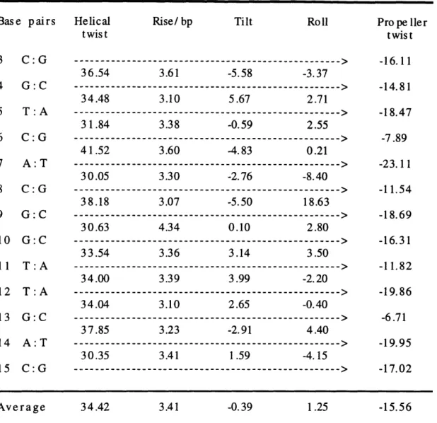
Table 1: DNA structure parameters.
Figure 5: Figure 6: Figure 7: Figure 8: Figure 1: Figure 2: Figure 3: Figure 4: Figure 5: Figure 6: Figure 7: Figure 8: Figure 9:
Chapter 5
Figure 1:
Figure 2:
Flow chart for PAX6-PD purification.
Photographic image of the PAX6-PD and DNA
Chapter 1
DNA-binding Protein Families
DNA-binding proteins are critical for many biological
processes, such as transcriptional regulation, DNA recombination,
genome replication, repairing damaged DNA, and responding to
environment signals. Transcription factors that regulate gene
expression comprise one of the largest and most diverse classes of
DNA-binding proteins. Among other fields, transcription factors play central roles in the field of development biology --- regulating cell development, differentiation, and cell growth, by binding to specific DNA sites and thereafter activating or inhibiting gene
expression.
One of the most important observations of the DNA-binding
protein studies is that most DNA-binding proteins can be grouped into classes that use structurally related DNA-binding domains or
motifs. Some families, such as the helix-turn-helix family, were
recognized by structural similarities. More families were first identified by sequence comparisons and later characterized by
structural studies. Some of the largest families include helix-turn-helix proteins, zinc-finger proteins, homeodomain-containing
proteins, helix-loop-helix proteins, and leucine-zipper proteins. Structural and recognition aspects of transcription factor families
were review by Pabo and Sauer (1992), and Harrison (1991) - more
references can be found therein. Structural studies with one family member can usually provide basic information for the whole family.
Cloning and Characterization of Pax Genes
The 384 bp long paired box was first identified in three
Drosophila segmentation genes paired (prd), gooseberry (gsb) and
gooseberry neuro (gsb-n) (Bopp et al., 1986; Baumgartner et al., 1987), and subsequently in two tissue-specific genes, Pox meso and Pox neuro (Bopp et al., 1989). Paired boxes have been detected in such divergent organisms as mouse, human, nematode, zebra fish,
frog, turtle, and chicken, and very recently in C. elegans (Deutsch et
al., 1988; Dressier et al., 1988; Burri et al., 1989; Walther et al. 1991; Martin et al. 1992; Krauss et al., 1991; Stapleton et al., 1993; Wallin et al., 1993; Chisholm and Horvitz, submitted). So far at least 30-40 paired-box genes have been cloned based on the sequence
homology in the paired-box, including 9 murine Pax genes (Pax-1 to
Pax-9) and 9 human PAX genes (PAX1 to PAX9), where Pax refers to
paired-box-containing
genes.
Unlike the developmental regulatory homeobox (Hox) genes, which were found clustered on particular chromosomes, each of nine
human PAX genes is located on an entirely different chromosome.
The most important clue leading to our current understanding of Pax
biology was the association between Pax genes and several
previously known mouse and human developmental phenotypes. For example, mutations in human PAX3 and PAX6 genes were found to be
responsible for Waardenburg syndrome type 1 and type 3 (Tassabehji
et al., 1992; Baldwin et al., 1992; Farrer et al., 1994) and aniridia (Ton et al., 1991; Glaser et al., 1992, 1994), respectively. Mutations in the mouse Pax-1, Pax-3 and Pax-6 genes are associated with
undulated, Splotch, and Small eye phenotypes, respectively (Balling
et al., 1988; Epstein et al., 1992; Hill et al., 1991).
The 128 amino acid paired domain encoded by the paired-box is the only region common to all PAX proteins. The DNA binding
activity of paired domain was first demonstrated between the
Drosophila paired protein (Prd) and the e5 DNA sequence in the even-skipped promoter (Treisman et al., 1991; Chalepakis et al., 1991).
All Pax protein showed specific binding to this e5 sequence, and
thus it has been used to study Pax protein-DNA interactions. The
inference that Pax proteins act as transcription factors is based on their being localized in the nucleus (Dressler and Douglass, 1992;
Glaser et al., 1995) and the presence of DNA-binding domain. This has been verified for Pax-5, Pax-6 and Pax-8, which have been
shown to regulate cell type-specific gene transcription (Pax-5:
Rothman et al., 1991; Williams and Maziels, 1991; Liao et al., 1992; Pax-8: Zannini, 1992; Pax-6: Cvekl et al., 1994, 1995; Chalepakis et al., 1994b; Richardson et al., 1995; Plaza et al., 1995; see figure 4 of Chapter4). In addition, the Pax-1, Pax-2, Pax-5, Pax-6 and Pax-8 proteins have been shown to activate reporter gene expression upon
binding to modified e5 sites in transfection experiments (Czerny et
al., 1993; Fickenscher et al., 1993; Kozmik et al., 1993; Zannini et al., 1992). Recently, it has also been shown that Pax-3 contains
domains for both transcriptional activation and transcriptional
inhibition (Chalepakis et al., 1994; Czerny and Busslinger, 1995). Pax Gene Structure and Classification
Although Pax genes are operationally defined by the presence of a paired domain, they also share overall structural features. Pax genes were grouped into at least four subfamilies (Figure 1),
initially based on the degree of homology in the paired domain, in
conjunction with subfamily-specific amino acids at certain
positions of the paired domain (Walther et al., 1991; Figure la of
Chapter 3). This grouping is consistent with a classification based
on the presence or absence of three structural features: 1) a characteristic octapeptide sequence (OP in Figure 1); 2) an intact paired-type homeodomain (HD); or 3) a partial paired-type
homeodomain containing only the N-terminal arm and first helix (Hill and Hanson, 1992). The first Pax subfamily, which includes
Pax-1 and Pax-9, encodes the paired domain and a conserved octapeptide sequence but lacks a homeodomain. The second
subfamily consists of Pax-3 and Pax-7 and, in addition to the paired domain and octapeptide, also encodes a full-length paired-type
homeodomain. Drosophila paired and gooseberry genes also belong to this subfamily. The third class, represented by Pax-2, Pax-5 and
Pax-8, encodes paired domain, octapeptide and a partial
homeodomain . Pax-4 and Pax-6 represent the fourth subfamily, which encodes the paired domain and homeodomain but lacks the
octapeptide. The subfamilies and their structural features are
Additional support for this subgrouping can also be found in
the genomic organization of Pax genes. For example, genes within a
given subfamily share specific intron/exon boundaries (Stapleton et
al.,1993). Moreover, some Pax proteins in the same subfamily have
been shown to have very similar DNA-binding activities (Czerny et
al., 1993, 1995; Epstein et al., 1994a).
Pax Gene Expression Pattern
Mouse Pax genes are expressed with a distinct spatiotemporal
pattern beginning between day 8 and day 9.5 of embryogenesis.
Although several Pax genes are also expressed in adult tissues, the
primary expression of all known functional Pax genes is in the
embryo. All Pax genes (except Pax-1 and Pax-9 which are expressed in the developing vertebral column) are expressed in the developing neural tube and brain, and contribute to early nervous system
development (Chalepakis et al.,1993; Noll, 1993; Stoykova and Gruss, 1994). Unlike Hox genes, which are characterized by region-specific expression along the anterior-posterior axis, Pax genes can show expression along the full length of this axis, but often with a progressive reduction as development proceeds.
Individual Pax genes are also expressed at high levels in
tissues outside the central nervous system, such as Pax-2 and Pax-8 expression in the developing kidney (Dressier et al., 1990; Plachov et
al., 1990), Pax-5 expression in B-lymphocytes (Adams et al., 1993),
Pax-3 expression in paraspinal mesoderm (Goulding et al., 1991), and Pax-8 expression in the thyroid gland (Plachov et al., 1990).
Pax Gene Developmental Mutations
At least seven phenotypes are known to be associated with
loss-of-function mutations in three human PAX genes. Mutations in
human PAX3 gene cause Waardenburg syndrome (WS) type 1, type 3 and Craniofacial-deafness-hand syndrome (summarized in Farrer et
al., 1995). Mutations in PAX6 are associated with familial and sporadic aniridia, Peters' anomaly and cataracts (Ton et al., 1991;
Glaser et al., 1992,1994,1995). More recently, mutations in PAX2
gene have been associated with human kidney and retinal defects (Sanyanusin et al., 1995). In addition, mutations in three mouse Pax genes, Pax-1, Pax-3 and Pax-6 are known to produce the undulated,
Splotch and Small-eye mutant phenotypes, respectively (Balling et
al., 1988; Epstein et al., 1992; Hill et al., 1991).
Waardenburg syndrome and Aniridia are the best studied of the above syndromes. Waardenburg syndrome type 1 (Waardenburg,
1951) is a heritable autosomal dominant trait occurring with a frequency of approximately 1 in 100,000 of the population
(Tassabehji et al.,1993) and is characterized by white forehead, premature graying of the hair, different colored eyes , and an outward displacement of the inner canthii of the eye (da-Silva,
1991). Of the patients with Waardenburg syndrome, approximately
one third are deaf, representing 2% of all adult cases of congenital deafness (Hoth et al., 1993). Klein-Waardenburg syndrome or WS type 3 has been described as combination of WS type 1 and limb abnormalities (Goodman et al., 1982). Splotch (mouse Pax-3
mutation) and WS 1 (human PAX3 mutation) have similar neural crest
deficiency-associated phenotypes (Tassabehji et al., 1994).
The human congenital eye disease aniridia is characterized by
hypoplasia of the iris and affects the iris, lens, cornea, filtration apparatus, and retina, leading to cataracts, corneal opacification,
and glaucoma that worsen with age (Glaser et al.,1995). It is an important cause of blindness and a paradigm among human
geneticists as a Mendelian autosomal dominant disorder. It occurs
because of a decreased dosage of PAX6, a gene which controls early events in the morphogenesis of the brain and eye (Glaser et al., 1994). PAX6 mutations have been detected in both sporadic and
familial aniridia. PAX6 mutations have also been described in Peters' anomaly, a congenital defect of the anterior chamber of the eye, that is usually a central corneal opacity overlying a defect in
the posterior layers of the cornea (Hanson et al., 1994). A broad
spectrum of PAX6 mutations have been found in Aniridia / Peters' anomaly. Large deletions may extend to neighboring genes, including
the WT1 Wilms' tumor gene, causing the WAGR contiguous gene
syndrome (Wilms tumor, aniridia, genito-urinary abnormalities and mental retardation). The Small eye mouse mutants (associated with mouse Pax-6 mutations) display phenotypes that include eye
defects, primarily complete absence of eye structure or defects of
the lens, cornea and retina and of the nose and associated olfactory structures (Hogan et al., 1988).
The human PAX2 gene is expressed in primitive cells of the kidney, ureter, eye, ear and central nervous system (CNS) (Dressler et al., 1990; Nornes et al., 1990). A mutational analysis of PAX2 in a
family with optic nerve colobomas, renal hypoplasia, mild
proteinuria and vesicoureteral reflux revealed a single nucleotide
deletion, which cause a frameshift of PAX2 coding region in the octapeptide (Sanyanusin et al., 1995). The phenotype resulting from
PAX2 mutation in this family was very similar to abnormalities that have been reported in Krd mutant mice (Keller et al., 1994).
Mouse Pax-1 mutations are associated with undulated
phenotypes (Balling et al., 1988). The undulated mouse shows
reduction of the posterior portion of the vertebrae, with increased intervertebral disk spaces, causing a "wavy" spine (Wright, 1947; Carter, 1947).
A property of Pax mutations in both human and mouse is that
abnormal phenotypic effects accompany the disruption of only one of
the normal pair of genes (Hill and Hanson, 1992). Therefore in
human, these disorders segregate as autosomal dominant. In mouse, such heterozygous effects are referred to as semidominant, because homozygotes show increased phenotypic severity. These mutations are assumed to be loss-of-function mutations, as the majority of Pax mutations are large scale truncations or frameshift that exhibit
haploinsufficiency has been used to describe this aspect of the
PAX2, PAX3 and PAX6 mutations (Glaser et al.,1994, 1995; reviewed by Read, 1995).
Pax Gene Oncogenic Potential
Not only can an insufficient Pax dosage lead to a variety of
phenotypes, but over-dosage or gain-of-function Pax mutations can also cause developmental defects, often tumorigenesis. So far,
murine Pax genes have been demonstrated to induce tumorigenesis in mice, and various human PAX genes have been tentatively implicated in a variety of human cancers.
When Pax genes are expressed in fibroblasts under the control of the cytomegalovirus (CMV) promoter/enhancer, the observed Pax
protein overexpression is accompanied by an uncontrolled increase
of cell growth in vitro. When injected into nude athymic mice, cells
that constitutively overexpress Pax proteins develop into solid
tumors. The oncogenic potential of murine Pax genes appears to be dependent on the presence of a functionally active paired domain. For example, the murine Pax-1 undulated point mutation in the
paired box, which results in a DNA-binding deficient protein, does
not have the transformation activity. The absence of the
octapeptide or homeodomain does not affect transforming potential. Although Pax genes induce transformation that results in
vascularized tumor formation, metastasis was not demonstrated
(Maulbecker and Gruss, 1993).
Wilms' tumor, a pediatric renal carcinoma, is a common malignancy in children, occurring in approximately 1 in 10,000 of the population (Hustie, 1993). The presence of both the PAX2 protein
and Wilms' tumor suppresser protein WT1 has been observed in primary Wilms' tumor (Dressler and Douglass, 1992). It has been demonstrated that WT1 can bind to three high affinity sites in 5'
untranslated PAX2 leader sequence with high affinity, and repress
demonstrated to be expressed in Wilms' tumor (Poleev et al., 1992).
A frequent site of chromosomal rearrangement in pediatric
alveolar rhabdomyosarcoma maps to the PAX3 locus. It has been
shown that the common translocation in this type of
rhabdomyosarcoma results in a portion of PAX3 being translocated
and forming a fusion protein with a portion of a forkhead gene FKHR (Galili et al., 1993; Shapiro et al., 1993). The fusion protein retains the entire PAX3 DNA-binding domains and only 55% of the forkhead
domain. Since the activity of forkhead proteins is dependent on the
presence of an intact forkhead domain (Lai et al., 1990), the activity of the PAX3-FKHR fusion protein would appear to be due to the PAX3 DNA binding domains, which may or may not be modulated by the forkhead region of the fusion protein. It has been shown that the
PAX3-FKHR fusion protein is a more potent transcriptional activator
than the intact PAX3 protein (Fredericks et al., 1995). PAX5 has also been implicated in the progression of
astrocytomas (which account for 60% of all tumors of the human central nervous system) to their most malignant and prognostically unfavored form - glioblastoma multiforme (Stuart et al., 1994).
Clearly, vertebrate development is sensitive to the precise
dosage of PAX protein. Why has natural selection managed such a
fragile mechanism?
Functions of Pax Genes
Like homeobox (Hox) genes, Pax genes encode transcription
factors that play important roles in development, as demonstrated
by the abundance of mouse and human congenital defects associated
with Pax gene mutations.
In Drosophila, paired-box-containing genes may have a role in
segmentation. For example, the three earliest characterized genes containing paired box, paired (prd), gooseberry (gsb) and gooseberry
neuro (gsbn), are segmentation genes of the pair-rule and
segment-polarity class. The initial activation of the segment-polarity genes
engrailed (en), wingless (wg), and gsb has been shown to depend on prd at least in every other stripe (Noll, 1993). In addition, gsbn and pox neuro (poxn) are involved in neurogenesis. Most interestingly, the critical role of the eyeless (ey) gene, the Drosophila homolog of
PAX6, in controlling Drosophila eye formation has been clearly
demonstrated. Ectopic eyeless expression induces formation of full-fledged eyes in Drosophila wings, legs and other tissues. This
suggests it may be a "master control gene" for eye development (Halder et al., 1995).
Mouse Pax genes are expressed after somite formation has established the initial segmentation pattern. Therefore, vertebrate
Pax genes are unlikely to be involved in primary segmentation of the
body axis. Instead, they appear to have tissue-specific roles in specifying positional information (Strachan and Read, 1994). Analysis of Pax mutational phenotypes and murine Pax expression
patterns may lead to a better understanding of the primary functions of Pax genes. Pax-1 and Pax-9 should have a role in the development of the vertebral column (Dietrich and Gruss, 1995). All other Pax
genes have a potential role in CNS development (Stuart et al., 1994).
In addition, Pax-2 is important in kidney and eye development
(Sanyanusin et al., 1995); Pax-3 should be involved in neural crest
cell patterning and may inhibit myogenic differentiation (Epstein et
al., 1995); Pax-5 is associated with B lymphocyte development and
midbrain/hindbrain boundary patterning (Adams et al., 1992); Pax-6
plays an important role in eye morphogenesis (Halder et al., 1995);
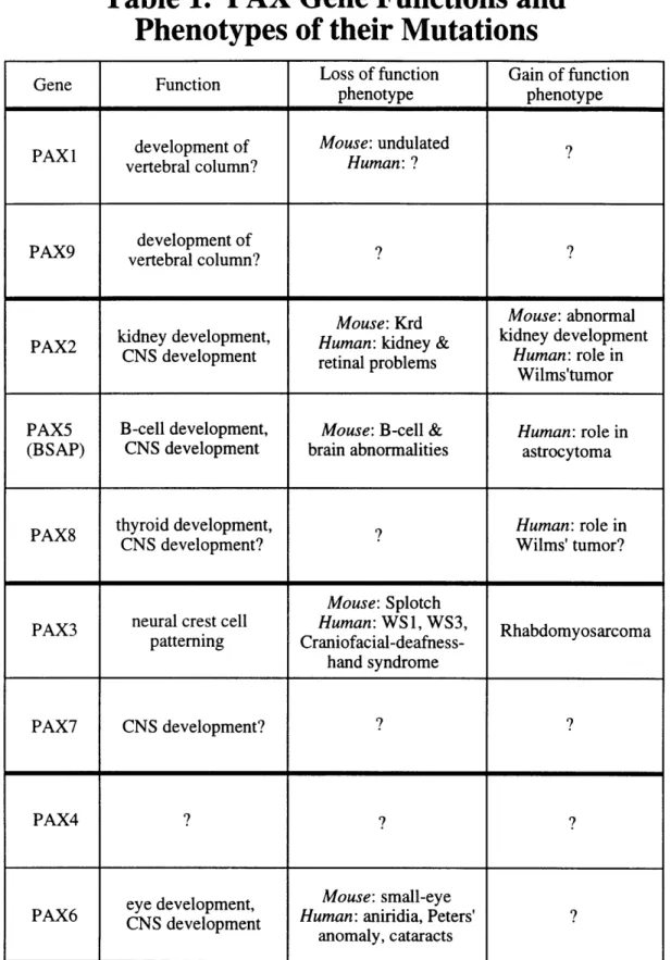
Pax-8 is associated with thyroid development (Zannini et al., 1992). Pax mutational phenotypes and functions are summarized in Table 1.
Although the physiological importance of Pax genes have been
clearly demonstrated, little is known concerning their molecular mechanisms, such as the up-stream regulators or down-stream targets of Pax proteins. Some functional target sequences for
1989; Kozmik et al., 1992; Waters et al., 1989; Rothman et al., 1991; Williams and Maziels, 1991; Liao et al., 1992; Pax-8: Zannini, 1992;
Pax-6: Cvekl et al., 1994, 1995; Chalepakis et al., 1994b; Richardson et al., 1995; Plaza et al., 1995; see figure 4 of Chapter 4). Pax-5
was identified as a B-cell-specific transcription factor and it potentially regulates the CD19 gene, which encodes a B-cell-specific surface-protein. The sea-urchin Pax-5 homolog, TSAP,
regulates two pairs of non-allelic histone genes, H2A-2 and H2B-2.
Pax-8, which is expressed in the thyroid, binds to and regulates the
thyroperoxidase and thyroglobulin genes. Recently, crystallin genes
have been proposed to be Pax6 targets (Cvekl et al., 1994, 1995; Richardson et al., 1995). The study of Pax protein-DNA interactions
will provide important information for understanding the molecular
mechanism of Pax proteins.
Paired Domain Is Critical for Pax Functioning
Pax proteins vary from 360 to 480 amino-acids in length. The
highly conserved 128 amino acid paired domain is located near the
N-terminal end of Pax proteins. The functional importance of the paired domain is well demonstrated by the clustering of Pax
missense mutations inside this domain. Although the majority of
Pax mutations are large-scale truncating mutations (gene deletion,
frameshifting deletion or insertion, splicing site alteration and nonsense mutation), a variety of Pax missense mutations has been reported. Most known missense mutations occur in the N-terminal
region of the paired domain (Strachan and Read, 1994). In addition, the oncogenic potential of Pax proteins is also dependent on the DNA
binding activity of the paired domain, as the Pax-1 undulated mutant protein, which carries a point mutation in the paired domain that impairs DNA binding, can not induce tumor formation (Maulbecker
and Gruss, 1993).
Of the nine mouse Pax proteins (Figure 1), five do not encode any other known DNA binding motifs and thus may exclusively use paired domain to bind DNA. The other four Pax proteins
(Pax-3/4/6/7) also contain an intact paired-type homeodomain. The
binding of PAX3 and Pax-6 homeodomains to a series of DNA sites
containing a single TAAT core, in different sequence contexts, was
not detected (Chalepakis et al. 1994; Czerny and Busslinger, 1995). Paired type homeodomains can form dimer upon binding to
palindromic DNA sites, which significantly improves its DNA binding activity (Wilson et al., 1993). However, using the optimal binding sites for both paired domain and homeodomain (palindromic site), in the context of full-length Pax-6 protein, the paired domain proved to
be more effective, by about 2 orders of magnitude, in DNA binding than the homeodomain (Czerny and Busslinger, 1995). It seems that paired domain plays a dominant role in determining the DNA binding
activity of Pax proteins.
The paired domain may also have roles other than specific DNA binding. For example, it has been shown that a region responsible
for a strong transcription inhibition activity is located in the first 90 N-terminal amino acids of the mouse Pax-3 protein, which
includes the first 57 residues of the paired domain. This region can
function as a transcriptional inhibitor independent of the remaining
portions of the Pax-3 protein, as it can be transferred onto a heterologous GAL4 DNA-binding domain (Chalepakis et al., 1994). Paired Domain Is a Novel DNA-binding Motif
Paired domain is a highly conserved DNA-binding domain that does not share any obvious sequence homology with any other DNA
binding protein. Thus the study of paired domain-DNA interactions can provide new perspective for understanding the general principles of protein-DNA interactions, in addition to laying the groundwork for
understanding the mechanisms Pax proteins use to regulate gene
expression during development.
There is evidence indicating that the paired domain is
composed of two sub-domains that bind to two half sites in adjacent
binds to 5' half site), and that the N-terminal domain plays a
dominant role in the paired domain-DNA interaction (Czerny et al.,
1993; Epstein et al., 1994). When aligning the Pax-5 recognition sequences to obtain a binding site consensus, none of the naturally
occurring Pax-5 binding sites completely conform to the long consensus sequence. A subset of Pax-5 binding sites, that match
better to the consensus in the 5' half than the rest of Pax-5 sites and do not match the 3' sub-site, can be bound by truncated Pax-5
paired domain lacking 36 C-terminal amino acids. The rest of Pax-5 binding sites that match the 5' half of the consensus sequence less
well, match better to the 3' half to the consensus sequence (see Figure 4 of Chapter 4). The bipartite structure of both the Pax-5
paired domain and its binding site was directly demonstrated by Pax-5 methylation interference analysis and in vitro mutagenesis of
both the Pax-5 paired domain and its recognition sequence. Thus
Pax-5 DNA binding sites contain compensatory base changes in their half sites that explain the versatile and seemingly degenerate DNA
sequence recognition of Pax-5 protein (Czerny et al., 1993). What are the structures of the two subdomains of paired
domain? What is the relationship of these two domains? What is the structural basis for the specificity of paired domain-DNA interaction? How could single missense mutations in the paired
domain lead to the observed phenotypes? Could paired domain-DNA
interactions provide new information for understanding the general principles of protein-DNA interactions? These are the questions we hoped to answer by solving a paired domain-DNA complex crystal
structure.
Figure Legends
Figure 1
Sub-family classification and structural features of PAX proteins.
PD denotes the paired domain, OP the octapeptide and HD the paired-type homeodomain. The three helices in homeodomain are
highlighted. The length of proteins and the distance between the structural features are not drawn in proportion. The classification
is based on the overall sequence organization (presence of a paired-type homeodomain and an octapeptide motif, location of introns and overall sequence identity) and especially on comparison of the
paired box sequences (Walther et al. 1991; Wallin et al. 1993; Stapleton et al. 1993).
Table 1
This table summarize the functions of PAX genes and phenotypes of PAX developmental mutations. Pax proteins in the same subfamily are clustered. WS denotes Waardenburg syndrome. CNS denotes the
Figure 1.
Classification and Structural Features of Pax Proteins
PD
Pax-I
PaH-9
PaH-2Pax-5
Pax-8
L
Pax-3
Pap-7
Pax-6
Pax-4
OP HD p - --p0 -p E L p E D-r rll_
A_~~~
11111_
I mll I I I~~~~I
m~~~
I IIIIII I II r I I r rTable 1. PAX Gene Functions and
Phenotypes of their Mutations
Loss of function Gain of function
phenotype phenotype
development of Mouse: undulated ?
vertebral column? Human: ?
PAX9 development of ?
vertebral column?
Mouse: Krd Mouse: abnormal
PAX2 kidney development, Human: kidney & kidney development CNS development retinal problems Human: role in
Wilms'tumor
PAX5 B-cell development, Mouse: B-cell & Human: role in
(BSAP) CNS development brain abnormalities astrocytoma
thyroid development, v Human: role in
CNS development? Wilms' tumor?
Mouse: Splotch
PAX3 neural crest cell Human: WS 1, WS3,arcoma
patterning Craniofacial-deafness-hand syndrome
PAX7 CNS development? ? ?
PAX4 | ? ?
eye development, Mouse: small-eye
PAX6 CNS development Human: aniridia, Peters' ? anomaly, cataracts
Chapter 2
Purification, Crystallization and Structural Determination
of Prd Paired Domain - DNA complex
When we started to try to solve the structure of paired domain by means of crystallography, very little information about the DNA
binding site of Pax proteins was available. Susie Jun and Claude
Desplan (Rockefeller University) defined a optimal DNA binding site
of Drosophila Prd paired domain by using in vitro selection and
amplification of randomized DNA sequences, and that set the basis for our collaboration.
A.
Purification
Purification of Drosophila prd Paired Domain
Prd paired domain was initially prepared as a C-terminal
fusion with gluthione S-transferase. The chimeric protein was over-expressed in E. coli and purified on gluthione-agarose column. However it was very difficult to obtain specific cleavage between gluthione S-transferase and paired domain. Non-specific cleavage also imposed difficulty in purification. Although correct cleavage
rate could reach 30% in solutions containing specific DNA site and 50% glycerol (high glycerol may help to stabilize loose domain structure, as reviewed by Sousa, R, and Lafer, E.M., 1990), the final
recovery yield was below 0.2-0.3 mg per litre E. coli culture. Thus we tried several new plasmid expression vectors. Among them, a vector with an N-terminal polyhistidine tag, pET14bprdPDB, gave high expression level in soluble phase and its polyhistidine tag could
be specifically cleaved, and so was later used to express the
Drosophila Prd paired domain in E. coli strain BL21(DE3). The protein
used in our crystallographic study contains the whole Prd paired domain and and four additional residues (Gly-Ser-His-Met) on the N-terminal end that was introduced from expression vector as part of
the polyhistidine tag. All plasmid vectors I have tested were
constructed by our collaborator Susie Jun (Rockefeller University)
Cells were grown at 370 and were induced with 0.4 mM
isopropyl-13-D-thiogalactoside (IPTG) when they reached OD600=0.8. Cells were harvested 3 hours after induction, washed with
prechilled phosphate-buffered saline buffer, frozen in a
dry-ice/ethanol bath and stored at -80°C. Sonication was carried out in a buffer containing 25 mM Hepes pH 7.6, 0.1M KCI, 0.1% NP-40, 0.3 mg/ml lysozyme, 7 mM 2-mercaptoethanol, 1 g/ml aprotinin, 1
gg/ml pepstatin, 1 g/ml benzamidine, and 1 g/ml sodium
metabisulfite. The cell lysate was diluted with solution A (25 mM Hepes pH 7.9, 0.1 M NaCI, 5 mM MgCI2, 15% glycerol, 0.1% NP-40, 7 mM 2-mercaptoethanol) and loaded onto a Ni-NTA column (Novagen). The column was extensively washed with 8 mM imidazole (pH 8.0) in solution A, and then with 40 mM imidazole in solution A; the Prd paired domain was eluted with 100 mM imidazole in solution A. The
eluted protein was treated with 0.25U/Il thrombin at 30°C for
15-20 hours to remove the N-terminal polyhistidine tag, and the
reaction was stopped by adding 1 mM PMSF to the solution. The Prd
paired domain was purified with a Mono-S column (Pharmacia), using
a gradient of 0.3 M to 0.7 M NaCI in 40 mM phosphate buffer (pH 6.6), containing 1 mM DTT. Prd paired domain was eluted out by 0.5-0.55
M NaCI. The purified protein gave a single band on an overloaded SDS gel in the absence of reductant. The protein used for crystallization was then purified by gel filtration on a superdex-75 column
(Phamacia), with a buffer containing 10 mM bis-tris-propane (pH7.0)
and 1 mM DTT. Protein was concentrated by Centricon-3, then frozen
by liquid nitrogen and stored at -80°C. In later stage of
crystallization, protein purified in this way was further purified by
preparative reverse phase HPLC on a Vydac C4 column, and then was
lyophilized. Lyophilized proteins were then resuspended by a buffer
containing 10mM bis-tris-propane (pH7.5), 1mM DTT, aliquoted, frozen by liquid nitrogen and stored at -80°C. The HPLC/
lyophilization step also function as a concentration step, in this way
protein could be concentrated to 22 mg/ml, while it was hard to concentrate protein up to 10 mg/ml by Centriprep-3 or Centricon-3
(Amicon). The HPLC purified protein could produce crystals more
reproducibly. The final yield of purification is about 5 mg per litre of E. coil culture.
paired domain was further confirmed by N-terminal sequencing, amino acid composition analysis, high resolution mass spectrometry
(Harvard MicroChem facility), and gel shift experiments.
Purification of DNA oligomers used for crystallization
We used solid-phase phosphoramidite method on an Applied Biosystems DNA/RNA synthesizer 392 for producing all of the DNA
oligonucleotides used for crystallization. Individual DNA
oligonucleotide strands containing 5-dimethoxytrityl (DMT)-group were purified by preparative reverse-phase HPLC on a Vydac C4 column, using an acetonitrile gradient in 50 mM triethylammonium
acetate (pH6.5). The trityl group was cleaved by treatment with
1.1% trifluroacetic acid for 10 min, and the solution was
immediately neutrilized by 1.4% triethylamine. Oligomers were then dialyzed extensively against 10 mM triethylammonium bicarbonate (pH7.0) and were then lyophilized. The detrityled oligonucleotides
were purified a second time by a C4 reverse-phase column and
dialyzed extensively against 10 mM triethylammonium bicarbonate
(pH7.0). DNA strands were annealed by heating at 90°C for 10 min
and cooling slowly to room temperature. DNA duplexes were stored as freeze-dried aliquots.
The uncoupled failure products were capped by acetylation in each synthesis cycle, and the capped oligos could be easily separated
from DNA oligomers with DMT group in reverse-phase HPLC. Thus We
kept DMT protecting group after last cycle and then purified
oligomers by two runs of reverse-phase HPLC as described above, in
order to totally get rid of those uncoupled failure products. However for short DNA oligomers (15mer or shorter) used for crystallization trials, we expected that one-step purified DNA should be
sufficiently pure. For example, I obtained paired-DNA complex
single crystals with a 15mer DNA oligo. While crystal with
DMT-on/two-step purified DNA oligo diffracted 2.5
A,
DMT-off/single step purified diffracted to at least 2.8 A.B. Co-crystallization of a Prd Paired Domain - DNA
Complex
Selection of DNA Sites and Results of Co-crystallization Trial
When we started our cocrystallization trials, little
information was available about the DNA binding specificity of
paired domain. The in vitro optimal DNA-binding site of Prd paired
domain was deduced from selection and amplification experiments
with randomized DNA sequences. The binding site consensus is 12 base pairs long, CGTCACG(G/C)TT(G/C)(A/G). Considering the
footprinting of Prd paired domain is 15 base paired long, we decided to search cocrystallization conditions with 14 to 21 base pairs long
DNA oligomers, which contains the whole binding site consensus.
It has been repeatly shown that the sequence and length of the
DNA oligo used in cocrystallization trials have significant effects
on the quality of the cocrystals produced (Jordan et al., 1985; Schultz et al., 1990; Liu et al., 1990; Wolberger et al., 1991). The differences in the DNA length as little as one base pair can
dramatically effect the crystal quality. The sequence identity at the
5' and 3' ends of the DNA, in particular the overhanging bases, if any, can also have a large effect on the quality of the crystals. Thus we decided to test a variety of different DNA sequences and lengths in
our cocrystallization trials. I first tested the effect of DNA length
on the crystallizability of prd paired domain - DNA complex. I
synthesized and purified 8 DNA duplexes with different lengths from
14 mer to 21 mer. I was able to obtain microcrystals only with the 15 mer DNA oligomer, after using volatile salt ammonium acetate in the droplet that is neccesary to keep the protein - DNA complex
soluble and to obtain any sort of microcrystal. Then I tried 4 other 15 mers with different end bases and/or overhanging bases. With one of the 4 oligomers, which has two overhanging bases (AA/TT), I
obtained nice crystals that diffracted to 2.5 A resolution.
In low ionic, neutral pH, the solubility of prd paired domain
-DNA complex is low (lower than 1 mg PrdPD/ml), even with the
presence of excessive DNA (which slightly improved the complex solubility). Preliminary studies revealed that the solubility of the Prd paired domain-DNA complex was sensitive to several factors,
including ionic strength and pH. High ionic (> 0.25 M NaCI) or alkaline pH (pH> 8.0) can dramatically increase the solubility to above 10 mg PrdPD/ml, with a DNA:protein ratio of 1.5:1.0. However, I was not be able to obtain any ordered solid form, in the high salt (> 0.25 M NaCI) or high pH (> pH 8.0) conditions, with any DNA oligomers
I have tried.
At this point, the dynamic light scattering experiment (Ferre-D'Amare and Burley, 1994) indicated that Prd protein-DNA complex
is mono-dispersive in solutions containing up to 0.2 M NaCI. In many
cases, monodispersity suggests conformational homogeneity. Empirical observations suggest that macromolecules that are monodispersive under "normal" conditions crystallize readily,
whereas randomly aggregating or polydispersive systems rarely, if ever, yield crystals (Ferre-D'Amare and Burley, 1994). This result is both encouraging and informative. In the early crystallization
trials, the drops initially contains high salt (> 0.25 M NaCI), the salt concentration would go even higher upon equilibrating with reservoir solution containing precipitant. This could cause partial
disassociation of protein-DNA complex as indicated by gel-shift. However it seems possible to achieve a soluble mono-dispersive system by using volatile salts.
I then extensively searched the possibility of using volatile salt ammonium acetate and ammonium bicarbonate to cocrystallize prdPD - DNA complex. Ammonium bicarbonate seems not suitable for cocrystallization, because the pH of the droplets containing
ammonium bicarbonate tends to go up. The pH of droplets containing ammonium acetate can keep stable around pH 7.0, in a period of
neutral pH. Evaporation of ammonium acetate from droplets decreases the ionic strength in the droplet, and thus drive the PrdPD-DNA complex into supersaturation. The rate of this process
depends on the ammonium acetate concentration in the drop solution and in the well solution, as well as the size of the droplet and
temperature. Ionic strength is an important determinant of the
strength of electrostatic interaction and hydrophobic interaction. It is pretty common that salt can influence the solubility of protein or protein-DNA complex. We expect volatile salt could also be useful for crystallizing other protein or protein-DNA complex. In fact, we have lately obtained several crystal forms of PAX6 paired domain -DNA complex using volatile salt ammonium acetate.
Crystallization Condition
It was interesting that co-crystals could grow in similar
conditions and to similar morphology and size in both MPD and PEGs.
However crystals grew from MPD could only diffract to about 8 A
resolution, while crystal grew from PEG400 diffracted to 3.2 A, and
crystals from PEG1000 were able to diffract to 2.5
A
resolution.Crystals with the DNA oligo shown in Figure d of chapter 3 were
grown by the evaporation of volatile salts from the hanging drops.
Extensively lyophilized DNA oligomers were resuspended with 10 mM bis-tris-propane (pH 7.0) at a concentration of 1 O.D.2 60 per
microlitre. Then 1.76 pl of above DNA solution was mixed with 1.81 ,ul "7.5X buffer" containing 2.25 M ammonium acetate (pH7.0), 0.15 M MgCI2, 37.5 mM DTT, 0.75 mM EDTA. Then 5 l 22 mg/ml PrdPD was
slowly added to above DNA-containing solution while stirring with a pippete tip. Adding DNA to protein or adding protein too quickly would results in some irreversible precipitation. Above DNA-protein
mixture was then mixed with equal volume of reservoir solution as
the hanging drops and these drops were equilibrated against a
reservoir containing 10% PEG 1000 and 5 mM DTT. Crystals grew in 4 to 5 days. Co-crystals diffracting to 2.5 A resolution grow in orthorhombic space group P212121, with a=39.6 A, b=68.6 A,
C.
Structure
Determination
Preparation of Heavy Atom Derivative Crystals
We used multiple isomorphous replacement method to solve phase problem. Heavy atoms were introduced into isomorphous crystal by replacing thymine with 5-iodouracil during DNA
synthesis.
Iodine atoms in the DNA are not stable upon exposure to light
and alkaline conditions. We took special care with the handling of
the iodinated DNA oligomers. First we tried to keep oligomers in a
dark environment whenever possible, in the whole process of synthesis, purification and crystallization. Secondly, we used
milder condition for oligomer deprotection. lodinated DNA oligomers
was deprotected in fresh saturated ammonium hydroxide at room temperature for 20 to 24 hours, then the cap of the vials was opened
and kept at room temperature for another 12 hours (to allow ammonium hydroxide to evaporate and to prepare for speed-vac). Thirdly, the trityl-off reaction was controlled with great care. After incubation with 1.1% of trichloracetic acid for 8 minutes, the reaction solution was neutralized immediately with 1.2%
triethylamine, and then one tenth volume of 0.5 M bis-tris-propane
buffer (pH7.0). The solution was then extensively dialysed against
10 mM TEAB before the second step of HPLC purification. The purity
of final iodine-substituted DNA oligomer was confirmed with
Mono-Q anion exchange column (Pharmacia LKB, Piscataway, New Jersey), which showed that the molar ratio of iodinated DNA and DNA that has lost iodine was 100 to 1 or higher. I found that it is not necessary to use the more expensive FOB (fast oligonucleotide
deprotecting) protection reagent, which uses different protecting
groups than the standard CE protection method. First the
CE-protected DNA oligomers purified in the way decribed above were fully suitable for making isomorphous heavy atom derivative crystal. Secondly, FOB-protected column was not commercially available.
Thus the 3' end nucleotide is usually CE-protected, and DNA
oligomers synthesized with FOB reagent still have to be deprotected in CE-deprotection condition.
We tested a number of these modified DNA oligomers in
crystallization trials, and found that substitution of thymine by 5-iodouracil in base pairs 11, 12 or 14 produced isomorphous crystals which were suitable for phasing. (After the structure was solved, we noticed that these three thymine bases are neither contacted by protein from major groove, nor involved in crystal packing). All three derivative crystal forms were isomorphous to native crystal
and diffracted to 2.5 A resolution, same as native crystals. Data Collection and Reduction
During crystal data collection, there appeared to be gradual changes in the cell dimensions. Most severe changes occured to cell
dimension b, which can change from 64.7 A to 69.3 A (thus
increasing by 7.1%). It is first considered that the change may be
caused by temperature fluctuation. We then tried to collect data at
constant 10°0C and 2C respectively. The problem persisted. Then we noticed some relationship between the age of the crystal and the the length of b axis. We surmised that the cell dimension change may be caused by the existence of trace amount of ammonium
acetate. We eventually solved the cell dimension change problem by the following steps: first, "aging" the crystals for at least two weeks before data collection; second, improving crystallization
condition so that the well solution does not contain any ammonium
acetate which was originally used for controlling degree of
supersaturation; third, mounting crystals without adding any well
solution.
All data finally used for structure determination were
collected at room temperature on the R-axis image plate system of our laboratory. The crystal have unit cell dimensions of a = 39.6 A, b
Initial determination of the lattice parameters was done by
collecting 30 frames of oscillation data (Aphi = 1). The diffraction pattern of these 30 frames were converted to positions in reciprocal
space using the conversion programs developed by Mark Rould
(extract_peaks.for, peaksto_reciprocal_space_coordinates.for).
By
measuring the distances and angles of individual spots in the
reciprocal space, using the crystallography graphics program FRODO (Jones, 1978), the primary reciprocal lattice parameters were
determined. Then the real space crystal unit cell parameters were deduced. We found at this point that all unit cell angles were very close to 90°, and surmised the crystal belong to a primitive
orthorhombic space group. The existence of 2-fold axes in all three directions were confirmed by testing for the presence of the two-fold symmetry operators. Thus we could obtain a full data set by collecting 900 data. After a full native data set was collected, we examined for systematic absences which indicated that we had a
space group P 212i21. The space group is further confirmed by the
solution of the difference Patterson map.
All data sets were reduced using the program DENZO (Z.
Otwinowski). Crystal and camera parameters were refined, and intensity mesurements were made by using profile fitting of the recorded spots. Partially recorded reflections were merged and integrated from successive oscillation frames (merge-denzo.for, M.
Rould). Data were then scaled using the program SCALEPACK (Z.
Otwinowski). We devided merged oscillation frames by 5 wedges, then applied a single scale factor for each wedge. No explicit corrections were made for absorption or crystal decay. Derivative data sets were local scaled to the native data set using the program
MAXSCALE (M. Rould).
Structure Determination by MIR Method
Reflections with large intensity differences (>7a) between the native and derivative data sets were removed from the reflection list (Exorcise.for, M. Rould), because those few reflections strongly
biased the difference Patterson maps. Derivative data sets were
local scaled against the native data set again, using MAXSCALE (M. Rould). Isomorphous difference Patterson maps and anomalous
difference Patterson maps were calculated for each derivative using
the program PROTEIN (Steigemann, 1974). Exorcise and MAXSCALE
significantly improved the quality of Patterson maps. At this point our isomorphous difference Patterson maps and anomalous
difference Patterson maps showed clearly the heavy atom peaks in
the Harker sections (Figure 1). We then picked initial heavy atom
sites corresponding to heavy atom peaks in the Harker sections using
the program HASSP (Terwilliger et al., 1987). HASSP is an
independent program which systematically searches the difference Patterson function and pick up potential heavy atom sites with large values for both self- and cross-vector positions.
The refinement of heavy atom parameters was carried out
using the program REFINE from CCP4 package (The SERC
Collaborative Computing Project No.4, a Suite of Programs for
Protein Crystallography [Distributed from Daresbury Laboratory,
Warrington WA4 4AD, UK, 1979]). After refining the heavy atom
parameters (positions, occupancy, and thermal parameters) for every derivative, we used the refined heavy-atom positions to fix the
origin of the unit cell with respect to the positions of the heavy
atom sites from the three derivatives. With the heavy atom sites
initially refined for every derivative, we used difference Fourier
methods to check the correctness of the heavy atom sites
(Henderson and Moffat, 1971). After refining the heavy atom parameters for every derivative individually, we did cross-phased
refinement using the program PHARE from CCP4 package (SERC, 1979) in order to reduce bias (Blow and Mattews, 1973). With this program, the parameters of one derivatives are refined while the
other two derivatives are used to calculate phases. After every derivative was refined twice by cross-phase refinement, we
generated an initial MIR map with the mean figure of merit 0.59 at 2.5 A resolution. We then used a procedure (Rould et al., 1992) to
parameter refinement from the calculation of parent phases by first
solvent-flattening (Wang, 1985) the initial MIR map in order to
generate new solvent-flattened phases. These new phases, in turn,
are used in the second round of refinement of heavy atom
parameters. New MIR phases are not updated until the convergence of the refinement. The new MIR map (mean fom = 0.71) was subject
to another round of solvent flattening to give the final MIR electron
density map (Figure 6 of chapter 3, mean fom = 0.79). All of the DNA was clearly resolved in this map, as were almost all the sidechains and mainchain carbonyl groups of the N-terminal domain of the protein (Figure 6 of chapter 3). The electron density for the C-terminal domain was not as good (it is packed less rigidly in the
crystal), but about half of the sidechains of this globular
sub-domain were clear. The initial model was built using TOM FRODO in Silicon Graphics computer (Israel, M., Chirino, A. J. and Cambillau, C. M., personal communication). The initial idealized B-form DNA was generated using the program Insight.
Structure Refinement
The initial model was subjected to multiple rounds of
positional refinement (Bringer, 1992a) and manual adjustment. Refinement was monitored by following the free R-factor to avoid overbuilding (Bringer, 1992b). In later stages of refinement, tightly restrained individual B-factors were used. Local scaling of the observed and calculated structure factors (using a minimum
neighborhood of 100 reflections and excluding the reflection being scaled) was also done to correct for absorption and anisotropic diffraction. In the final cycle, 13 water molecules were included in
the model. Every water molecule added forms at least two hydrogen bonds with the paired-DNA complex, and has a B-factor lower than
50.0. Although most structural features including water molecules
were cleared resolved in the initial unbiaed MIR map, all of the key contacts and the key features of the complex were further confirmed by checking simulated annealing omit maps (Hodel et al., 1992). About 30% of the sidechains of the C-terminal domain could not be
built with confidence and were modeled as alanines; the first 5 and
last 4 residues of the polypeptide also were omitted. (A few of these N-terminal residues were ones introduced during cloning, and thus our model includes residues 2-124 of the paired domain.) Our
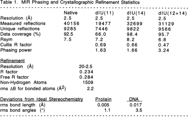
current model has an R factor of 23.4% and a free R factor of 28.4% with good stereochemistry (Table 1). All phi and psi angles, except for residues 78 (in the linker) and 91 (in an extended loop), are in allowed regions of the Ramachandran plot (Figure 2).
Figure Legends
Figure 1
This figure shows three sections of the isomorphous difference Patterson map, corresponding to Harker section u=1/2, v=1/2, and w=1/2, calculated from the native data set and the dlU(11) data set
(see Chapter 3 Table 1). The map was generated by the program PROTEIN (Steigemann, 1975), and used data after local scaling, from 20 to 2.8
A
resolution. The contours of the maps start at 1 sigma and are in increment of 1 sigma. The peaks representing the single iodine atom are clear in this map.Figure 2
Ramachandran plot. This figure shows the location in Phi, Psi space of each amino acid residue from the final model of the Prd paired
domain-DNA complex. The angle phi (the dihedral angle about N-Ca
bond) is shown on the abscissa, and the angle psi (the dihedral angle about the Ca-C bond) is shown on the ordinate. All phi () and psi (p) angles, except for residue 78 (in the linker) and 91 (in the extended loop), are in allowed regions of the Ramachandran plot. Coordinates
RRHRCHRNORRN PLOT PRO -a. . _ _ _ _ _ a I . I ' X ' ' I -1 0 , -180 -120 a ...
-
-60 a ..-.-. a 0 . 60 .a 120 a . 180'~a
a -120 0 ---.---. ! ! _ _ >! , .. , .. _ ._ ., ... _ .. ... ... ... -180 - 1,
-180
...
,,,,,~,
...
J
- 180 -120 -60 0 60 120 180Figure 2
180 120 60 '1' 0 t I I I I I .. _ _ _ _ _ __ . . l:---: I~~~~~ . . I _an ..J ---- ---vu I I I I IChapter
3
Crystal Structure of a Paired Domain-DNA Complex
at 2.5 A Resolution Reveals Structural Basis
Summary
The 2.5 A resolution structure of a co-crystal containing the paired domain from the Drosophila Paired protein and a 15 bp site
shows structually independent N-terminal and C-terminal
sub-domains. Each of these domains contains a helical region resembling
the homeodomain and the Hin recombinase. The N-terminal domain
makes extensive DNA contacts, using a novel f3-turn motif that binds
in the minor groove and a helix-turn-helix unit with a docking
arrangement surprisingly similar to that of the X repressor. The
C-terminal domain is not essential for Prd binding and does not contact the optimized site. All known developmental missense
mutations in the paired box of mammalian Pax genes map to the Nterminal subdomain, and most of them are found at the protein
-DNA interface.
Introduction
The paired domain is a conserved DNA-binding domain (Treisman et al., 1991; Chalepakis et al., 1991) found in a set of
transcription factors (Pax proteins, Figure la) that play important
roles in development (Gruss and Walther, 1992). This 128 amino
acid domain was first identified in the Drosophila paired (prd) and
gooseberry genes (Bopp et al., 1986) and often is found in
association with a homeodomain (Walther et al., 1991). Numerous paired domain proteins are known, and nine PAX genes have been identified in the human genome (Walther et al., 1991; Stapleton et al., 1993; Wallin et al., 1993; Figure la). A number of murine and human developmental mutants are known to have alterations in
specific Pax genes, and several of these involve missense mutations
in the paired domain (reviewed by Gruss and Walther, 1992; Strachan and Read, 1994; Figure b). Mutations in the human PAX3 and PAX6
genes cause Waardenburg's syndrome (Tassabehji et al., 1992;