HAL Id: hal-02613808
https://hal.sorbonne-universite.fr/hal-02613808
Submitted on 20 May 2020
HAL is a multi-disciplinary open access
archive for the deposit and dissemination of
sci-entific research documents, whether they are
pub-lished or not. The documents may come from
teaching and research institutions in France or
abroad, or from public or private research centers.
L’archive ouverte pluridisciplinaire HAL, est
destinée au dépôt et à la diffusion de documents
scientifiques de niveau recherche, publiés ou non,
émanant des établissements d’enseignement et de
recherche français ou étrangers, des laboratoires
publics ou privés.
Usefulness of morphometric image analysis with Sirius
Red to assess interstitial fibrosis after renal
transplantation from uncontrolled circulatory death
donors
Myriam Dao, Christelle Pouliquen, Alyette Duquesne, Katia Posseme,
Charlotte Mussini, Antoine Durrbach, Catherine Guettier, Hélène François,
Sophie Ferlicot
To cite this version:
Myriam Dao, Christelle Pouliquen, Alyette Duquesne, Katia Posseme, Charlotte Mussini, et al..
Use-fulness of morphometric image analysis with Sirius Red to assess interstitial fibrosis after renal
trans-plantation from uncontrolled circulatory death donors. Scientific Reports, Nature Publishing Group,
2020, 10 (1), pp.6894. �10.1038/s41598-020-63749-3�. �hal-02613808�
Usefulness of morphometric image
analysis with Sirius Red to assess
interstitial fibrosis after renal
transplantation from uncontrolled
circulatory death donors
Myriam Dao
1,2,8, Christelle pouliquen
3,8, Alyette Duquesne
4, Katia posseme
5,
charlotte Mussini
5, Antoine Durrbach
6, Catherine Guettier
5, Hélène françois
2,7,8✉ &
Sophie ferlicot
5,8Early interstitial fibrosis (IF) correlates with long-term renal graft dysfunction, highlighting the need for accurate quantification of IF. However, the currently used Banff classification exhibits some limitations. The aim of our study was to precisely describe the progression of IF after renal transplantation using a new morphometric image analysis method relying of Sirius Red staining. The morphometric analysis we developed showed high inter-observer and intra-observer reproducibility, with ICC [95% IC] of respectively 0.75 [0.67–0.81] (n = 151) and 0.88 [0.72–0.95] (n = 21). We used this method to assess IF (mIF) during the first year after the kidney transplantation from 66 uncontrolled donors after circulatory death (uDCD). Both mIF and interstitial fibrosis (ci) according to the Banff classification significantly increased the first three months after transplantation. From M3 to M12, mIF significantly increased whereas Banff classification failed to highlight increase of ci. Moreover, mIF at M12 (p = 0.005) correlated with mean time to graft function recovery and was significantly associated with increase of creatininemia at M12 and at last follow-up. To conclude, the new morphometric image analysis method we developed, using a routine and cheap staining, may provide valuable tool to assess IF and thus to evaluate new sources of grafts.
Chronic kidney disease (CKD) is a burden for Public Health and concerns millions of individuals worldwide. Kidney transplantation remains the optimal treatment for CKD, offering a better survival than dialysis and being cost-effectiveness1–3.
In order to increase the pool of available donors in a setting of organ shortage, grafts from uncontrolled donors after circulatory death (uDCD) have been used in France since 2006. Several studies have shown that kidneys from DCD provide almost an equal function as kidneys from donation after brain death (DBD)4–13. Moreover, as
DBD kidney transplantation, DCD kidney transplantation is associated with increased survival of patients who have end-stage renal disease (ESRD) and are on the transplant waiting list14. Whereas early reports of uDCD
showed good renal outcome and no increase in IF/TA compared to extended criteria DBD, other reported a very early and more severe development of IF/TA15 than in DBD.
Whatever the donor’s status, chronic allograft dysfunction (CAD), which is the final result of different etio-logical and pathogenetic conditions, remains the first cause of graft loss16–20. CAD corresponds to the irreversible
1AP-HP, Service de Néphrologie adulte, Hôpital Necker, 75015, Paris, France. 2Inserm UMR_S 1155, Hôpital Tenon, 75020, Paris, France. 3Service d’Anatomie pathologique, Hôpital Foch, 92150, Suresnes, France. 4Service de Néphrologie, CHI André Grégoire, 93100, Montreuil, France. 5AP-HP, Service d’Anatomie et de Cytologie Pathologiques, Hôpital de Bicêtre, 94270 Le Kremlin Bicêtre, France, Hôpitaux Universitaires Paris-Saclay, Le Kremlin-Bicêtre, France. 6AP-HP, Service de Néphrologie, Hôpital de Bicêtre, 94270 Le Kremlin Bicêtre, France, Hôpitaux Universitaires Paris-Saclay, Le Kremlin-Bicêtre, France. 7AP-HP, Unité de Néphrologie et de Transplantation rénale, Hôpital Tenon, 4 rue de la Chine, 75020 Paris, Sorbonne Université, Paris, France. 8These authors contributed equally: Myriam Dao, Christelle Pouliquen, Hélène François and Sophie Ferlicot. ✉e-mail: helene.francois@aphp.fr
www.nature.com/scientificreports
www.nature.com/scientificreports/
replacement of functional renal tissue by extracellular matrix (ECM) proteins, leading to the progressive impair-ment of renal graft function. Among the pathological lesions observed during CAD, one of the most prominent is interstitial fibrosis and tubular atrophy (IF/TA). Other histological damages include glomerulosclerosis, splitting of glomerular capillary basement membranes and vascular intimal hyperplasia21. Many mechanisms which are of
immunological origins or not, are involved in this multifactorial and complex process22–26. The current concept
states that many processes leading to graft fibrosis, i.e. IF/TA, occur early after the transplantation, especially within the first few months27. IF/TA involves about 40% of kidney grafts at 3–6 months and up to 50% at 1 year,
while renal function remains stable28,29, which suggests that renal function is a suboptimal and late marker of
kidney graft dysfunction. Up to now, the only reliable method to assess IF/TA is the histological evaluation of a graft biopsy30,31 which also allows to determine specific lesions and pathogenic processes affecting the graft22,23.
It is also well-known that early IF correlates with long-term graft dysfunction32–34, highlighting the need for
accurate quantification of IF to better identify patients requiring specific therapeutic interventions and to deter-mine the efficacity of such interventions. IF in kidney graft is usually graded using the Banff classification35.
However, the Banff classification exhibits some limitations. The semi-quantitative evaluation in only 4 grades (0 to 3) prevents the precise evaluation of IF evolution and may be not sensitive enough in the early stages. Furthermore, studies have shown a wide interobserver variation in the assessment of renal graft biopsies using the Banff classification28,36–38.
The aim of the study was to assess the initial progression of interstitial fibrosis and tubular atrophy in kidney grafts from uDCD formerly classified as “Maastricht II” non heart beating donors, using a new image analysis method based on Sirius Red staining.
In the first part of our work, we will evaluate the accuracy, the robustness and reproducibility of our comput-erized analysis method (mIF) to assess IF. In the second part, mIF will be applied to assess IF during the first year after kidney transplantation from uDCD and mIF score will be correlated with clinico-biological data.
Results
Validation and reliability tests of the image analysis method for the quantification of
inter-stitial fibrosis (mIF).
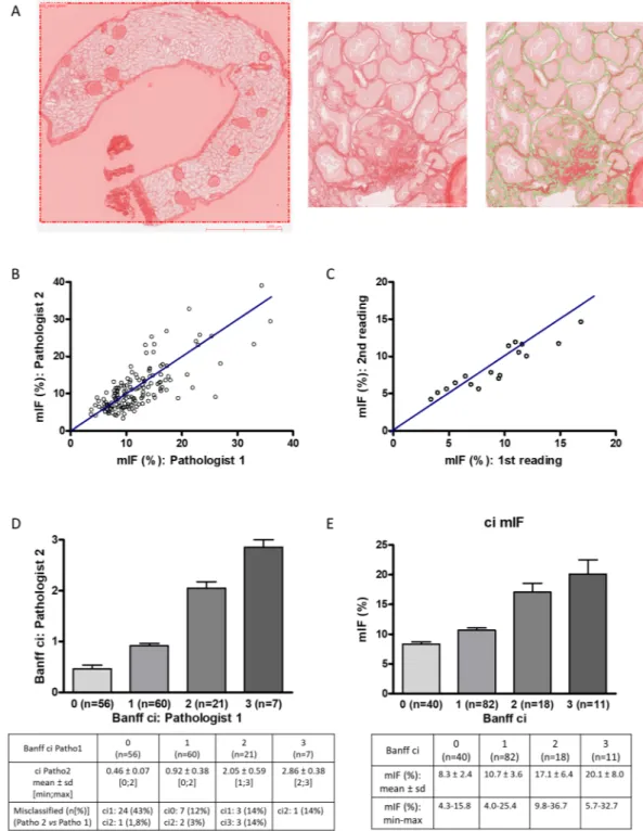
Among the 166 graft biopsies, 15 were rejected for image analysis because specimen adequacy was unsatisfactory (<7 glomeruli and/or no artery on the sample) according to the Banff criteria39 orbecause the paraffin block was worn out. Measurement of interstitial fibrosis by image analysis (mIF) was per-formed on the 151 biopsies independently by two pathologists (CP and SF) (Fig. 1A). The value of inter-operator intraclass correlation coefficient (ICC) was 0.75 with its 95% confidence interval (95% CI) equal to [0.67–0.81] (p = 8.4.10-29) (Fig. 1B). To assess the intra-observer reproducibility, 21 consecutive graft biopsies were analyzed
again by one of the pathologists (CP) 6 months later. The value of ICC was 0.88 with 95% CI of [0.72–0.95] (p = 5.5.10-8) (Fig. 1C). Analysis of interstitial fibrosis (ci) according to the Banff criteria (using Masson’s
tri-chrome staining) exhibited inter-operator ICC of 0.78 with 95% CI of [0.70–0.84] (p = 5.6.10-25) (Fig. 1D). Then,
we compared morphometric quantification of interstitial fibrosis and semi-quantitative analysis performed by an expert pathologist (SF) according to Banff criteria (Fig. 1E). The mIF score significantly increased (p < 0.05) between the four groups defined by the Banff classification. Mean mIF (%) was respectively 8.3 ± 2.4 (ranges: 4.3– 15.8) in the ci0 group (n = 40), 10.7 ± 3.6 (ranges: 4.0–25.2) in the ci1 group (n = 82), 17.1 ± 6.4 (ranges: 9.8–36.6) in the ci2 group (n = 18) and 20.1 ± 8.0 (ranges: 5.7–32.7) in the ci3 group (n = 11). The correlation between mIF and Banff ci was 0.62 with 95% CI of [0.51–0.71] (p < 0.001). Discrepancies between ci and mIF were observed in only 2 cases. These cases were carefully reanalyzed: interstitial edema and inflammation were the main drawback to accurately quantify ci in Massons’ trichrome staining (Supplemental Fig. 1).
Clinical characteristics of the patients.
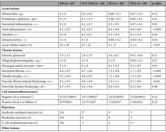
The 66 patients who received kidney graft from uDCD included 14 females and 52 males. Mean age at the time of kidney transplantation was 44.7 ± 9.8 years old (median: 46; range: 20–59). Indications for kidney transplantation were hypertensive nephroangiosclerosis (n = 16), IgA nephropathy (n = 7), autosomal dominant polycystic kidney disease (n = 6), Alport syndrome (n = 4), dia-betic nephropathy (n = 3), other glomerulopathies (n = 5), tubulo-interstitial nephritis (n = 3) and uropa-thy (n = 4). Nephropauropa-thy remained undetermined in 18 patients. All patients received induction therapy with anti-lymphocyte serum. They also received mycophenolate mofetil, corticosteroids and tacrolimus per local prac-tice. Characteristics of the recipient patients are summarized in Table 1.Uncontrolled DCD: Baseline characteristics, transplantation and clinical outcome.
The 66 patients received kidney graft from 48 uDCD (Table 1). Donors included 9 females and 39 males and were 39.7 ± 8.5 years old (median: 40.5; range: 19–53). None of them had history of arterial hypertension or diabetes mellitus. Mean total warm ischemic time (tWIT) from cardiac arrest to in situ preservation was 145.9 ± 15.2 min-utes (median: 148.5; ranges: 115–188). Mean cold ischemia was 16.2 ± 3.7 hours (median: 17.0, ranges: 10–23.8). Delayed graft function occurred in 54/66 patients (82%). Mean time between transplantation and renal graft recovery was 22.6 ± 9.8 days (median: 20, ranges: 1–58). Non-primary graft function occurred in 2 patients. Biopsy-proven acute rejection occurred in eight patients (12%). Mean LDH level reached a pic at Day 3 post transplantation (2539 ± 1089 IU/L) which was significantly associated with time to graft recovery (time to recov-ery according to LDH at D3: y = 15.2 + 0.0025x, p = 0.016, R² = 0.1028).Histological features.
Among the 48 uDCD, 43 underwent kidney biopsies (so called D0) before organ donation. Thereafter, 20 patients underwent kidney graft biopsy between 15 and 30 days (D15–30) post-transplantation for delayed graft function, 28 at 3 months (M3) and at 12 months (M12) for routine evalu-ation. Socio-demographic and clinical data were not significantly different between the patients who underwent biopsies at D15–30, M3 and M12. Histological features of biopsies are summarized in Table 2. Acute tubularFigure 1. Quantification of interstitial fibrosis by image analysis: validation and reliability tests of the
morphometric quantification of IF (mIF). (A) Morphometric analysis. Renal biopsy sections stained with Sirius red were captured by a ScanScope Aperio scanner (CS), using 20X objective. For each biopsy, the cortical section was manually selected. Glomeruli and medium-sized arteries were deleted by the operator. The red positive area was expressed as a percentage of the entire cortical kidney section using a computer-based morphometric analysis software (Calopix, Tribvn, Montrouge, France). (B) The value of inter-operator intraclass correlation coefficient (ICC) using mIF was 0.75 with its 95% confidence interval (95% CI) equal to [0.67–0.81] (p < 10-3) (n = 151). (C) The value of intra-observer ICC of mIF was 0.88 with 95% CI of
[0.72–0.95] (p < 10-3) (21 consecutive graft biopsies were analyzed again 6 months later). (D) The value of
inter-operator ICC using Banff criteria was 0.78 with 95% CI of [0.70–0.84] (p < 10-3) (n = 151). (E) mIF according
to Banff ci. Pearson correlation between mIF and Banff ci was 0.62 with 95% CI of [0.51–0.71] (p < 10-3).
www.nature.com/scientificreports
www.nature.com/scientificreports/
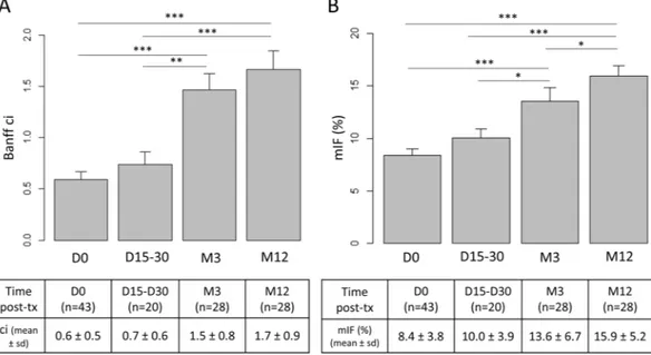
injury significantly decreased from D0 to D15-D30 (63% ± 28% versus 47% ± 22%, p = 0.036), then to M3 (9% ± 11%, p < 10-3) and M12 (4% ± 6%, p = 0.016). Conversely, chronic lesions, especially interstitial
fibro-sis and tubular atrophy, significantly increased over time. According to the Banff staging system, interstitial fibrosis remained unchanged from D0 to D15/30 (0.6 ± 0.5 at D0, 0.7 ± 0.6 at D15/30), increased from D0 to M3 (0.6 ± 0.5 versus 1.5 ± 0.8, p < 10-3) and remained stable between M3 and M12 (1.7 ± 0.9 versus 1.5 ± 0.8, p = 0.37) (Fig. 2A).
mIF significantly increased after the transplantation and correlated with clinical outcome.
Image analysis IF was 8.4% ± 3.8% at D0, 10.0% ± 3.9% at D15-D30, 13.6% ± 6.7% at M3 and 15.9% ± 5.2% at M12 (Fig. 2B). Increase was significant from D0 to M3 (p < 10-3). Conversely to ci according to the Banff scoring
system, increase of mIF remained significant from M3 to M12 (p = 0.021). Fibrosis tended to increase as early as the first month after renal transplant (p = 0.056). Most of the patients underwent more than one kidney biopsy. Among them, seven patients underwent early kidney graft biopsy between D7 and D30 and protocol biopsies at M3 and at M12 (Supplemental Fig. 2). In these paired cases, mIF significantly increased from first month to M3 (respectively 7.8% ± 1.8% and 11.4% ± 2.7%, p < 0.01) then from M3 to M12 (respectively 11.4% ± 2.7% and 17.7 ± 4.8%, p = 0.02). In these cases, Banff classification failed to demonstrate early increase of ci from first month to M3 (respectively 0.88 ± 0.35 and 1.3 ± 0.46, p = 0.08).
Baseline characteristics of uDCD (sex, age), tWIT and cold ischemia were not associated with mIF at any time. mIF at M12 (R² = 0.29, p = 0.005) but not at D0, D15–30 and M3 correlated with mean time to renal graft func-tion recovery. In addifunc-tion, mIF at M12 correlated with increase of creatininemia at M12 (R² = 0.32, p = 0.013) and at last follow-up (R² = 0.29, p = 0.005). We found the same correlation using the Banff scoring system, however, whereas there was no correlation between ci according to the Banff scoring system at M3 and creatininemia at M12 (R² = 0.15, p = 0.18), mIF at M3 tended to be associated with increase of creatininemia at M12 (R² = 0.31,
p = 0.057).
Donors characteristics (n = 48)
Age (years)* 39.7 ± 8.5
Sex: male [n(%)]/female [n(%)] 39(81%)/9(19%)
BMI (kg/m²)* 26.0 ± 4.5
Smoke history [n(%)] 28(58%)
Diabete mellitus [n(%)] 0(0%)
High blood pressure [n(%)] 0(%)
Dyslipidemia [n(%)] 3(6%)
Ischemic times
No flow time (min)* 6.9 ± 8.0 Low flow time (min)* 138.7 ± 14.4 Total warm ischemic time (min)* 145.9 ± 15.2 In situ cold perfusion time (min)* 148.7 ± 54.1
Recipients characteristics (n = 66)
Age (years)* 44.7 ± 9.8
Sex: male [n(%)]/female [n(%)] 52(79%) / 14(21%)
ESRD cause
Nephroangiosclerosis [n(%)] 16(24%) Diabetes mellitus [n(%)] 3(5%) Glomerulopathies [n(%)] 16(24%) Tubulo-interstitial nephritis [n(%)] 4(6%) Polycystic kidney disease [n(%)] 6(9%) Malformative uropathies [n(%)] 4(6%)
Unknown [n(%)] 17(26%)
Dialysis duration (months)*,a 32.2 ± 28.8
PRA < 10% [n(%)] 66(100%)
Graft characteristics (n = 66)
HLA mismatches (A + B + DR)* 4.5 ± 1.4
Machine perfusion [n(%)] 66(100%)
Rc lifeport at 30 min 0.21 ± 0.07
Cold ischemic time (hours)* 16.2 ± 3.7
Clinical outcome (n = 66)
Delayed graft function [n(%)] 54(82%)
Time before graft recovery (days)*,b 22.6 ± 9.8
Biopsy-proven acute rejection [n(%)] 8(12%)
Table 1. Characteristics of donors, recipients and grafts. Abbreviations: ESRD, End-stage renal disease; HLA,
Human Leukocytes Antibodies; PRA, panel reactive antibodies. *Numeric data are expressed by mean ± standard deviation. aPreemptive transplantation: n = 3. bNon-primary graft function: n = 2.
Discussion
The aim of this study was to precisely describe the evolution of IF after renal transplantation with uDCD, using a new computerized image analysis method. Indeed, IF is one of the most prominent pathological lesions of CAD and correlated with renal graft prognosis32–34 but the currently used Banff classification is a semi-quantitative
system which prevents precise description of IF evolution and may not be sensitive enough in early stages. Slight but significant increase of IF on renal graft may be missed. Grimm et al. demonstrated that a precise quantifica-tion of IF by computerized image analysis provides a better surrogate marker for time to graft failure than any combination of chronic lesion scoring using the Banff schema34. Moreover, some studies also exhibited a wide
inter-observer variation in the IF assessment according to the Banff classification36,37. The morphometric analysis
we developed exhibited high inter-observer and intra-observer reproducibility, with ICC [95% IC] of respectively 0.79 [0.74–0.83] and 0.88 [0.72–0.95]. A variety of techniques have been used to measure renal fibrosis, each with its own advantages and disadvantages. We decided to assess IF with unpolarized Sirius red staining, firstly because Sirius red staining is commonly used in pathological laboratories since the 1980s40, especially to assess
liver fibrosis41–45, secondly because to its high specificity for collagen fibers and its previously demonstrated
supe-riority compared to polarized Sirius Red and trichrome staining46–49. Indeed, IF is typically considered to be an
excess accumulation of fibrillary collagen. Types I and III collagen represent almost all collagens synthetized by fibroblasts and often predominate in fibrotic scars. Sirius red dye molecules intercalate into the tertiary groove in the structure of types I and III collagen and are strongly birefringent when observed under polarized light. Polarized Sirius Red analysis exhibited good results in IF assessment34,49,50 but it requires individual fields to be
photographed in polarized light and analyzed or cost-expensive whole slide scanners capable of polarized light. Similarly, trichrome staining, that is commonly used to the visual assessment of collagen content in the interstit-ium, has also be used to quantify IF by image analysis, with good results51,52. However, studies demonstrated than
unpolarized Sirius Red had not only the best correlation with estimated glomerular filtration rate (GFR)47,48
com-pared to polarized Sirius Red and trichrome staining, but also significant correlation with the decrease of GFR33.
In our uDCD cohort, both mIF and ci according to the Banff classification significantly increased the first three months after renal transplantation From M3 to M12, mIF significantly increased whereas Banff classifica-tion failed to highlight increase of ci. Then, mIF may be more accurate than semiquantitative Banff evaluaclassifica-tion to identify the effects of antifibrotic therapeutic interventions. Morphometric IF tended to increase in the first month after renal transplantation but without reaching significance, due to the limited size of sample, graft biop-sies being not systematically performed during the first month. However, this early and sensitive quantification of IF may be a very valuable surrogate marker to study therapeutic intervention aiming the early development if CAD in kidney transplantation. Moreover, whereas increase of creatininemia at M12 was not associated with ci
D0 (n = 43) D15-D30 (n = 20) M3 (n = 28) M12 (n = 28) p value Acute lesions: Glomerulitis « g » 0 ± 0 0.2 ± 0.4 0.08 ± 0.3 0.07 ± 0.3 0.12 Peritubular capillaritis « ptc » 0 ± 0 0.1 ± 0.3 0.08 ± 0.3 0.04 ± 0.2 0.41 Interstitial inflammation « i » 0 ± 0 0.2 ± 0.7 0.3 ± 0.5 0.07 ± 0.3 0.01 Total inflammation « ti » 0.1 ± 0.3 0.2 ± 0.7 0.6 ± 0.8 0.8 ± 0.8 <0.001 Tubulitis « t » 0 ± 0 0.2 ± 0.7 0.2 ± 0.4 0.1 ± 0.4 0.03 Intimal arteritis « v » 0 ± 0 0 ± 0 0.04 ± 0.2 0.04 ± 0.2 0.54
Acute tubular injury (%) 63 ± 28 47 ± 22 9 ± 11 4 ± 6 <0.05
Chronic lesions:
Sclerotic glomeruli 3.5 ± 5.1 2.0 ± 3.5 4.4 ± 6.7 9.0 ± 14.0 0.11
Allograft glomerulopathy « cg » 0 ± 0 0 ± 0 0 ± 0 0.04 ± 0.2 0.37
Mesangial matrix increase « mm » 0 ± 0 0 ± 0 0.1 ± 0.3 0.2 ± 0.5 0.01 Interstitial fibrosis « ci » 0.6 ± 0.5 0.7 ± 0.6 1.5 ± 0.8 1.7 ± 0.9 <0.001 Tubular atrophy « ct » 0.2 ± 0.5 0.4 ± 0.5 1.2 ± 0.8 1.5 ± 0.9 <0.001 Vascular fibrous intimal thickening « cv » 0.3 ± 0.5 0.8 ± 0.9 1.0 ± 1.1 0.7 ± 0.9 <0.01 Arteriolar hyaline thickening « ah » 0.3 ± 0.5 0.2 ± 0.4 0.4 ± 0.6 0.4 ± 0.6 0.49
C4d (immunofluorescence):
Negative (0 or minimal 1) 37/37(100%)a 15/17(88%)b 24/25(96%)b 25/26(96%)c 0.22
Positive (focal 2 or diffuse 3) 0/37(0%)a 2/17(12%)b 1/25(4%)b 1/26(4%)c 0.22 Rejection:
Antibody-mediated rejection (n) NA 2d 1e 1e
Borderline rejection (n) NA 0 4 3
T-cell-mediated rejection (n) NA 1 f 0 1 g
Other lesions:
Table 2. Histological features of renal biopsies at D0, D15-D30, M3 and M12. Digital data are means ±
standard deviation. aNA = 6. bNA = 3. cNA = 2. dAntibody-mediated rejection included: 1 acute
antibody-mediated rejection, and 1 “suspicious” for acute antibody-antibody-mediated rejection. eacute antibody-mediated
www.nature.com/scientificreports
www.nature.com/scientificreports/
according to the Banff scoring system at M3, it tended to be associated with mIF at M3, suggesting usefulness of a more sensitive quantification analysis.
In our study, mIF at M12 was significantly associated with increase of creatininemia at M12 and at last follow-up, which confirms the need for a precise assessment of IF.
IF, which corresponds to the replacement of renal functional tissue by ECM, is not only a major concern after kidney transplantation, but also during CKD in native kidneys. Indeed, renal fibrosis still represents the final target to treat CKD53. Surprisingly, few studies have focused on morphometric quantification of IF on native
kidneys54,55. Hunter et al.54 demonstrated than high collagen matrix index and fibrillary collagen index, both
assessed by quantitative morphometry after Sirius Red staining, predicted relapse and progression to ESRD dur-ing lupus nephritis. Similarly, Gibyeli Genek et al.55 performed a quantitative evaluation of IF with Sirius Red in
IgA nephritis and highlighted that such evaluation might serve as an effective novel method to determine the prognosis in IgA nephritis.
To conclude, the new morphometric image analysis method we developed, using a routine and cheap staining, may provide valuable tool to assess IF during chronic allograft dysfunction and thus to evaluate new sources of grafts. The use of this method to describe CKD on native kidney should also be evaluated.
Methods
Patients.
After institutional review board, we retrospectively included all the 66 patients who received a kid-ney graft from uDCD between 2007 and 2012 in Bicêtre hospital. Kidkid-ney donation followed the 2008 Declaration of Istanbul principles and the French Agence Nationale de la Biomédecine regulation. Research was approved by the committee of the Centre de Ressources Biologiques (CRB, Paris Sud University). Informed written consent was given by all the patients for the scientific use of the graft biopsies in the CRB, Paris Sud University. All proce-dures and the use of tissues were performed in accordance with the Declaration of Helsinki principles. Clinical reports and biological data were collected from the associated clinical database.Histological review of kidney graft biopsies.
A total of 166 kidney graft biopsies was performed in the 66 patients. The biopsies were processed for routine light microscopy, as we previously described56. Biopsysam-ples were fixed in formalin, acetic acid and alcohol (AFA), paraffin-embedded and sliced 2.5 µm thick. Slides were stained with HES (hematoxylin, eosin and saffron), Masson’s trichrome (3 sections), periodic acid Schiff, Jones methenamine silver and Sirius red. Third Masson’s trichrome and Sirius red staining were consecutive. Kidney graft biopsies were reviewed independently by 2 pathologists (CP and SF) for histological features according to Banff recommendations35 in a blind manner from clinical data. Immunostaining for C4d was performed using a
rabbit monoclonal A24-T anti-human C4d antibody (DB Biotech, Kosice, Slovak Republic; dilution 1/100) and a Leica BOND-MAX
™
autostainer (Leica Biosystems Newcastle Ltd, UK). Epitope retrieval was achieved using the ready-to-use Bond Epitope Retrieval Solution 1 (Leica Biosystems Newcastle Ltd, UK) after 30 min heating.Figure 2. Interstitial fibrosis (IF) significantly increased after renal graft transplantation. (A) According to the
Banff criteria, ci remained unchanged from D0 to D15/30, increased from D0 to M3 (p < 10-3) and remained
stable between M3 and M12 (p = 0.37). (B) Using morphometry analysis, mIF tended to increase as early as the first month after renal transplant (p = 0.056), increase was significant from D0 to M3 (p < 10-3) and remained
significant from M3 to M12 (p = 0.021). Abbreviations: ci = interstitial cortical fibrosis; mIF, morphometric interstitial fibrosis; sd, standard deviation; tx, renal transplantation; *p < 0,05; ***p< 10-3. Figure was
Quantification of interstitial fibrosis by image analysis.
Renal biopsy sections sliced 2.5 µm sliced and stained with Sirius red were digitalized by a ScanScope Aperio scanner (CS), using 20X objective. For each biopsy, the cortical section, defined as the part inside the renal capsule and outside the medulla, was manually selected on digital slides. Glomeruli and medium-sized arteries were deleted by the operator.Renal-cortex fibrosis was quantified using a computer-based morphometric analysis software (Calopix, Tribvn Healthcare, Châtillon France) as previously published56,57. Briefly, the operator manually selects internal Sirius
Red negative areas and positive areas and next runs the software that show the final selection of the Red area. The internal negative control step allows the comparison of slides that are sequentially stained in various batches of Sirius Red since staining usually may vary. Finally, the result was expressed as a percentage of the red positive area on the total cortical surface. Morphometric analyses were performed twice for each biopsy in an independant manner by two pathologists (CP and SF) who had no knowledge of the clinical data.
Statistical analyzes.
Descriptive statistical methods (means, medians, standard deviations and ranges) were used to assess the distributions of variables. Wilcoxon rank sum and t tests for continuous variables, Fisher’s exact and chi-squared tests for categorical variables were performed. The intraclass correlation coefficient (ICC) with its 95% CI was used to study the reproducibility of morphometric IF (mIF) measures. Correlations between quan-titative variables were assessed with Pearson product-moment correlation coefficient. For all analyses, a p value <0.05 was regarded as significant. Analyses were performed using R software (version 3.2.0)58,59, InStat 3 software(GraphPad Software, San Diego, CA) and Prism 4 (GraphPad Software).
Data availability
The datasets generated during and/or analyzed during the current study are available from the corresponding author on reasonable request.
Received: 17 October 2019; Accepted: 19 March 2020; Published: xx xx xxxx
References
1. Wolfe, R. A. et al. Comparison of mortality in all patients on dialysis, patients on dialysis awaiting transplantation, and recipients of a first cadaveric transplant. N. Engl. J. Med. 341, 1725–1730 (1999).
2. Savoye, E., Tamarelle, D., Chalem, Y., Rebibou, J.-M. & Tuppin, P. Survival benefits of kidney transplantation with expanded criteria deceased donors in patients aged 60 years and over. Transplantation 84, 1618–1624 (2007).
3. Bayat, S., Kessler, M., Briançon, S. & Frimat, L. Survival of transplanted and dialysed patients in a French region with focus on outcomes in the elderly. Nephrol. Dial. Transplant. 25, 292–300 (2010).
4. Wijnen, R. M. et al. Outcome of transplantation of non-heart-beating donor kidneys. Lancet 345, 1067–1070 (1995).
5. Cho, Y. W., Terasaki, P. I., Cecka, J. M. & Gjertson, D. W. Transplantation of kidneys from donors whose hearts have stopped beating.
N. Engl. J. Med. 338, 221–225 (1998).
6. Weber, M., Dindo, D., Demartines, N., Ambühl, P. M. & Clavien, P.-A. Kidney transplantation from donors without a heartbeat. N.
Engl. J. Med. 347, 248–255 (2002).
7. Brook, N. R., Waller, J. R. & Nicholson, M. L. Nonheart-beating kidney donation: current practice and future developments. Kidney
Int. 63, 1516–1529 (2003).
8. Koffman, G. & Gambaro, G. Renal transplantation from non-heart- beating donors: a review of the European experience. J. Nephrol.
16, 334–341 (2003).
9. Arias-Diaz, J., Alvarez, J., del Barrio, M. R. & Balibrea, J. L. Non-heart-beating donation: current state of the art. Transplant. Proc. 36, 1891–1893 (2004).
10. Alonso, A. et al. Renal transplantation from non-heart-beating donors: a single-center 10-year experience. Transplant. Proc. 37, 3658–3660 (2005).
11. Gagandeep, S. et al. Expanding the donor kidney pool: utility of renal allografts procured in a setting of uncontrolled cardiac death.
Am. J. Transplant. 6, 1682–1688 (2006).
12. Kokkinos, C. et al. Outcome of kidney transplantation from nonheart-beating versus heart-beating cadaveric donors.
Transplantation 83, 1193–1199 (2007).
13. Sánchez-Fructuoso, A. I. et al. Victims of cardiac arrest occurring outside the hospital: a source of transplantable kidneys. Ann.
Intern. Med. 145, 157–164 (2006).
14. Snoeijs, M. G. et al. Kidneys from donors after cardiac death provide survival benefit. J. Am. Soc. Nephrol. 21, 1015–1021 (2010). 15. Viglietti, D. et al. Kidney allograft fibrosis after transplantation from uncontrolled circulatory death donors. Transplantation 99,
409–415 (2015).
16. Lamb, K. E., Lodhi, S. & Meier-Kriesche, H.-U. Long-term renal allograft survival in the United States: a critical reappraisal. Am. J.
Transplant. 11, 450–462 (2011).
17. Haas, M. Chronic allograft nephropathy or interstitial fibrosis and tubular atrophy: what is in a name? Curr. Opin. Nephrol.
Hypertens. 23, 245–250 (2014).
18. Cosio, F. G., El Ters, M., Cornell, L. D., Schinstock, C. A. & Stegall, M. D. Changing Kidney Allograft Histology Early Posttransplant: Prognostic Implications of 1-Year Protocol Biopsies. Am. J. Transplant. 16, 194–203 (2016).
19. Hart, A. et al. OPTN/SRTR 2015 Annual Data Report: Kidney. Am. J. Transplant. 17(Suppl 1), 21–116 (2017).
20. Stegall, M. D., Cornell, L. D., Park, W. D., Smith, B. H. & Cosio, F. G. Renal Allograft Histology at 10 Years After Transplantation in the Tacrolimus Era: Evidence of Pervasive Chronic Injury. Am. J. Transplant. https://doi.org/10.1111/ajt.14431 (2017).
21. Solez, K. et al. Banff ’05 Meeting Report: differential diagnosis of chronic allograft injury and elimination of chronic allograft nephropathy (‘CAN’). Am. J. Transplant 7, 518–526 (2007).
22. Racusen, L. C. & Regele, H. The pathology of chronic allograft dysfunction. Kidney Int. Suppl. S27–32, https://doi.org/10.1038/ ki.2010.419 (2010).
23. Pascual, J., Pérez-Sáez, M. J., Mir, M. & Crespo, M. Chronic renal allograft injury: early detection, accurate diagnosis and management. Transplant. Rev. 26, 280–290 (2012).
24. Sellarés, J. et al. Understanding the causes of kidney transplant failure: the dominant role of antibody-mediated rejection and nonadherence. Am. J. Transplant. 12, 388–399 (2012).
25. Heemann, U. & Lutz, J. Pathophysiology and treatment options of chronic renal allograft damage. Nephrol. Dial. Transplant. 28, 2438–2446 (2013).
www.nature.com/scientificreports
www.nature.com/scientificreports/
26. Maluf, D. G. et al. Evaluation of molecular profiles in calcineurin inhibitor toxicity post-kidney transplant: input to chronic allograft dysfunction. Am. J. Transplant. 14, 1152–1163 (2014).
27. Torres, I. B., Moreso, F., Sarró, E., Meseguer, A. & Serón, D. The Interplay between inflammation and fibrosis in kidney transplantation. BioMed Res. Int. 2014, 750602 (2014).
28. Serón, D. et al. Reliability of chronic allograft nephropathy diagnosis in sequential protocol biopsies. Kidney Int. 61, 727–733 (2002). 29. Melk, A., Schmidt, B. M. W., Vongwiwatana, A., Rayner, D. C. & Halloran, P. F. Increased expression of senescence-associated cell
cycle inhibitor p16INK4a in deteriorating renal transplants and diseased native kidney. Am. J. Transplant. 5, 1375–1382 (2005). 30. Serón, D. et al. Protocol renal allograft biopsies and the design of clinical trials aimed to prevent or treat chronic allograft
nephropathy. Transplantation 69, 1849–1855 (2000).
31. Yilmaz, S. et al. Protocol core needle biopsy and histologic Chronic Allograft Damage Index (CADI) as surrogate end point for long-term graft survival in multicenter studies. J. Am. Soc. Nephrol. 14, 773–779 (2003).
32. Serón, D. et al. Early protocol renal allograft biopsies and graft outcome. Kidney Int. 51, 310–316 (1997).
33. Pape, L. et al. Computer-assisted quantification of fibrosis in chronic allograft nephropaty by picosirius red-staining: a new tool for predicting long-term graft function. Transplantation 76, 955–958 (2003).
34. Grimm, P. C. et al. Computerized image analysis of Sirius Red-stained renal allograft biopsies as a surrogate marker to predict long-term allograft function. J. Am. Soc. Nephrol. 14, 1662–1668 (2003).
35. Roufosse, C. et al. A 2018 Reference Guide to the Banff Classification of Renal Allograft Pathology. Transplantation, https://doi. org/10.1097/TP.0000000000002366 (2018).
36. Marcussen, N., Olsen, T. S., Benediktsson, H., Racusen, L. & Solez, K. Reproducibility of the Banff classification of renal allograft pathology. Inter- and intraobserver variation. Transplantation 60, 1083–1089 (1995).
37. Furness, P. N. & Taub, N. & Convergence of European Renal Transplant Pathology Assessment Procedures (CERTPAP) Project. International variation in the interpretation of renal transplant biopsies: report of the CERTPAP Project. Kidney Int. 60, 1998–2012 (2001).
38. Furness, P. N. et al. International variation in histologic grading is large, and persistent feedback does not improve reproducibility.
Am. J. Surg. Pathol. 27, 805–810 (2003).
39. Solez, K. et al. Banff 07 classification of renal allograft pathology: updates and future directions. Am. J. Transplant. 8, 753–760 (2008). 40. Junqueira, L. C., Bignolas, G. & Brentani, R. R. Picrosirius staining plus polarization microscopy, a specific method for collagen
detection in tissue sections. Histochem. J. 11, 447–455 (1979).
41. López-De León, A. & Rojkind, M. A simple micromethod for collagen and total protein determination in formalin-fixed paraffin-embedded sections. J. Histochem. Cytochem. 33, 737–743 (1985).
42. Moragas, A. et al. Cirrhotic changes in livers from children undergoing transplantation. Image analysis. Anal. Quant. Cytol. Histol.
14, 359–366 (1992).
43. Manabe, N. et al. Interferon-alpha 2b therapy reduces liver fibrosis in chronic non-A, non-B hepatitis: a quantitative histological evaluation. Hepatol. 18, 1344–1349 (1993).
44. Calvaruso, V. et al. Computer-assisted image analysis of liver collagen: relationship to Ishak scoring and hepatic venous pressure gradient. Hepatol. 49, 1236–1244 (2009).
45. Huang, Y. et al. Image analysis of liver collagen using sirius red is more accurate and correlates better with serum fibrosis markers than trichrome. Liver Int. 33, 1249–1256 (2013).
46. De Heer, E. et al. Morphometry of interstitial fibrosis. Nephrol. Dial. Transplant. 15(Suppl 6), 72–73 (2000).
47. Diaz Encarnacion, M. M. et al. Correlation of quantitative digital image analysis with the glomerular filtration rate in chronic allograft nephropathy. Am. J. Transplant. 4, 248–256 (2004).
48. Farris, A. B. et al. Morphometric and visual evaluation of fibrosis in renal biopsies. J. Am. Soc. Nephrol. 22, 176–186 (2011). 49. Street, J. M. et al. Automated quantification of renal fibrosis with Sirius Red and polarization contrast microscopy. Physiol. Rep. 2,
(2014).
50. Sund, S., Grimm, P., Reisaeter, A. V. & Hovig, T. Computerized image analysis vs semiquantitative scoring in evaluation of kidney allograft fibrosis and prognosis. Nephrol. Dial. Transplant. 19, 2838–2845 (2004).
51. Servais, A. et al. Quantification of interstitial fibrosis by image analysis on routine renal biopsy in patients receiving cyclosporine.
Transplantation 84, 1595–1601 (2007).
52. Servais, A. et al. Interstitial fibrosis evolution on early sequential screening renal allograft biopsies using quantitative image analysis.
Am. J. Transplant 11, 1456–1463 (2011).
53. François, H. & Chatziantoniou, C. Renal fibrosis: Recent translational aspects. Matrix Biol. J, https://doi.org/10.1016/j. matbio.2017.12.013 (2017).
54. Hunter, M. G., Hurwitz, S., Bellamy, C. O. C. & Duffield, J. S. Quantitative morphometry of lupus nephritis: the significance of collagen, tubular space, and inflammatory infiltrate. Kidney Int. 67, 94–102 (2005).
55. Gibyeli Genek, D. et al. Quantitative evaluation of interstitial fibrosis with Sirius Red in IgA nephritis. Ren. Fail. 36, 73–77 (2014). 56. Dao, M. et al. The cannabinoid receptor 1 is involved in renal fibrosis during chronic allograft dysfunction: Proof of concept. J. Cell.
Mol. Med. https://doi.org/10.1111/jcmm.14570 (2019).
57. Lecru, L. et al. Cannabinoid receptor 1 is a major mediator of renal fibrosis. Kidney Int. 88, 72–84 (2015).
58. R Core Team. R. A Language and Environment for Statistical Computing. (R Foundation for Statistical Computing, 2017). 59. Gamer, M., Lemon, J. & Singh, I.F.P. Various Coefficients of Interrater Reliability and Agreement. (R package version 0.84.1, 2019).
Author contributions
M.D.+: conception of the work, data collection, data analysis and interpretation, drafting the article; C.P.+:
conception of the work, data collection, data analysis and interpretation, critical revision of the article; A.D.: conception of the work, data collection, data analysis and interpretation, critical revision of the article; K.P.: data collection; C.M.: data analysis and interpretation, critical revision of the article; A.D.: revision of the article and Head of the Division of the Bicetre nephrology and transplantation department from which the patients were recruited; C.G.: conception of the work, critical revision of the article; HF§: conception of the work, data
collection, data analysis and interpretation, drafting the article; S.F.§: conception of the work, data collection, data
analysis and interpretation, drafting the article. All authors reviewed the manuscript.
competing interests
The authors declare no competing interests.
Additional information
Supplementary information is available for this paper at https://doi.org/10.1038/s41598-020-63749-3.
Reprints and permissions information is available at www.nature.com/reprints.
Publisher’s note Springer Nature remains neutral with regard to jurisdictional claims in published maps and
institutional affiliations.
Open Access This article is licensed under a Creative Commons Attribution 4.0 International
License, which permits use, sharing, adaptation, distribution and reproduction in any medium or format, as long as you give appropriate credit to the original author(s) and the source, provide a link to the Cre-ative Commons license, and indicate if changes were made. The images or other third party material in this article are included in the article’s Creative Commons license, unless indicated otherwise in a credit line to the material. If material is not included in the article’s Creative Commons license and your intended use is not per-mitted by statutory regulation or exceeds the perper-mitted use, you will need to obtain permission directly from the copyright holder. To view a copy of this license, visit http://creativecommons.org/licenses/by/4.0/.