Contents lists available atScienceDirect
DNA Repair
journal homepage:www.elsevier.com/locate/dnarepair
The MRE11 complex: A versatile toolkit for the repair of broken DNA
Giordano Reginato
a,b, Petr Cejka
a,b,*
aInstitute for Research in Biomedicine, Università della Svizzera Italiana (USI), Faculty of Biomedical Sciences, Bellinzona, Switzerland bDepartment of Biology, Institute of Biochemistry, Eidgenössische Technische Hochschule (ETH), Zürich, Switzerland
A R T I C L E I N F O Keywords:
DNA damage and repair Homologous recombination End-joining
DNA end resection Checkpoint Cancer
A B S T R A C T
When DNA breaks, the ends need to be stabilized and processed to facilitate subsequent repair, which can occur by either direct but error-prone end-joining with another broken DNA molecule or a more accurate homology-directed repair by the recombination machinery. At the same time, the presence of broken DNA triggers a signaling cascade that regulates the repair events and cellular progression through the cell cycle. The MRE11 nuclease, together with RAD50 and NBS1 forms a complex termed MRN that participates in all these processes. Although MRE11 wasfirst identified more than 20 years ago, deep insights into its mechanism of action and regulation are much more recent. Here we review how MRE11 functions within MRN, and how the complex is further regulated by CtIP and its phosphorylation in a cell cycle dependent manner. We describe how RAD50, NBS1 and CtIP convert MRE11, exhibiting per se a 3′→5′ exonuclease activity, into an ensemble that instead degrades primarily the 5′-terminated strand by endonucleolytic cleavage at DNA break sites to generate 3′ overhangs, as required for the initiation of homologous recombination. The unique mechanism of DNA end resection by MRN-CtIP makes it a veryflexible toolkit to process DNA breaks with a variety of secondary structures and protein blocks. Such a block can also be the Ku heterodimer, and emerging evidence suggests that MRN-CtIP may often need to remove Ku from DNA ends before initiating homologous recombination. Misregulation of DNA break repair results in mutations and chromosome rearrangements that can drive cancer development. Therefore, a detailed understanding of the underlying processes is highly relevant for human health.
1. A brief introduction to DNA double-strand break repair pathway choice
DNA double-strand breaks (DSBs) arise from cellular exposure to exogenous sources such as radiation and chemicals [1,2], or from en-dogenous metabolic processes [3] (Fig. 1). The most common en-vironmental source of ionizing radiation is the radon gas [1]. Ionizing radiation mainly damages DNA indirectly via the formation of radicals that attack the sugar-phosphate backbone resulting in extensive base damage, numerous single-strand DNA breaks (SSBs) and approximately 10-times fewer DSBs, which nevertheless account for most of the ra-diation-induced toxicity [4].
Chemicals capable to trigger DSBs are often used as chemother-apeutics in cancer therapy, as they preferentially target rapidly-dividing tumor cells. These drugs include topoisomerase inhibitors such as eto-poside or camptothecin, DNA alkylating agents such as methyl metha-nesulfonate or temozolomide, crosslinking agents such as mitomycin C or cisplatin, the radiomimetic drug bleomycin, and inhibitors of DNA replication such as hydroxyurea and aphidicolin [1,5]. Most of these
chemicals do not cause DSBs directly, but rather create initial lesions that are converted into DSBs upon further processing in cells. The most common source of endogenous DSBs are abortive topoisomerase reac-tions [6] or errors during DNA replication, such as when replication forks encounter DNA lesions, or during replication-transcription con-flicts [7].
Depending on the nature of the DSB formation, the DNA breaks can be chemically "clean" or "dirty". Radiation-induced DSBs are often de-scribed as "dirty", as they often contain modified chemistry that pre-vents ligation, which differs from canonical 5′-phosphate and 3′-hy-droxyl groups at nuclease-induced "clean" breaks. Likewise, DNA breaks resulting from abortive topoisomerase reactions contain covalently at-tached topoisomerases (protein blocks). The chemistry of the DSB, as well as the availability of a second broken DNA end, determines the subsequent pathway choice in DSB repair.
Generally, cells possess two main pathways for DSB repair, com-prising end-joining and homology-directed repair [8]. The canonical non-homologous end-joining (NHEJ), dependent on the Ku hetero-dimer, as well as the Ku-independent pathway termed
microhomology-https://doi.org/10.1016/j.dnarep.2020.102869
Received 31 March 2020; Received in revised form 30 April 2020; Accepted 4 May 2020
⁎Corresponding author at: Institute for Research in Biomedicine, Via Vincenzo Vela 6, 6500 Bellinzona, Switzerland.
E-mail address:petr.cejka@irb.usi.ch(P. Cejka).
DNA Repair 91–92 (2020) 102869
Available online 15 May 2020
1568-7864/ © 2020 The Author(s). Published by Elsevier B.V. This is an open access article under the CC BY-NC-ND license (http://creativecommons.org/licenses/BY-NC-ND/4.0/).
mediated end-joining (MMEJ), link the two broken DNA molecules with no or short microhomologies at the DNA ends [9,10]. The repair is fast, cell cycle stage independent, and responsible for joining the majority of DSBs in human cells. However, this process is primarily capable to join DNA ends that require little processing prior to ligation (Fig. 1).
Instead, homology-directed repair requires extensive homology be-tween the recombining DNA molecules, which in most cases guarantees an accurate repair (Fig. 1). To reveal this homology, the 5′-terminated DNA strand at a DSB must be resected by specialized nucleases or nu-clease complexes, creating a 3′-overhang. The ssDNA overhang is bound and protected by the single-strand DNA binding replication protein A (RPA). RPA must be subsequently replaced by the strand exchange protein RAD51, which is facilitated by BRCA2 and other recombination mediators. RAD51 forms together with ssDNA a nucleoproteinfilament capable to identify and invade homologous DNA [8,11]. To prevent mutagenesis, in vegetative cells the template for recombination is in most cases the sister chromatid.
Both end-joining and homologous recombination, as well as pro-cesses that regulate the pathway choice, have been reviewed ex-tensively [8,10,12–14], and will not be comprehensively covered here. The most important point relevant for understanding the text below is that extended DNA end resection generally commits DSB repair to the homologous recombination pathways, and prevents end-joining. The pathway choice depends on a number of proteins including the pro end-joining and anti-resection factor 53BP1 and its effectors such as the Shieldin complex [15–21]. In contrast, BRCA1 is a pro-resection factor that favors homologous recombination over end-joining [19–22]. It was believed that the decision whether and how extensively to resect DSBs ultimately determines the pathway choice in DSB repair [23,24]. However, it was recently discovered that the 5′-terminated DNA strand that has been partially resected may be resynthesized by the Shieldin
complex together with DNA polymeraseα [25], indicating that resec-tion andfill-in can be two competing processes, and thus, the pathway choice may be moreflexible than previously believed.
In the text below, we will focus on mechanistic insights into the function of the MRN complex. MRN stands at the crossroads between homologous recombination, end-joining and DNA damage signaling, and it is one of thefirst factors that is recruited to broken DNA. MRN consists of the MRE11 nuclease, the ATPase RAD50 containing ex-tended coiled-coils and a protein with a structural function termed NBS1 in human cells or Xrs2 (forming the MRX complex) in the budding yeast Saccharomyces cerevisiae. The MRN/X complex can dimerize via the globular domains of MRE11 and the ATPase domain of RAD50 that form the base of the structure in contact with DNA [26], as well as via a zinc-hook at the end of the coiled-coils of RAD50 at the apex of the complex [27]. We will focus on mechanisms inferred from studying both yeast and mammalian homologues, as this is very informative about the evolutionary conservation and importance of the underlying processes. We will use the mammalian nomenclature (i.e. MRE11) when discussing a mammalian protein or making more general state-ments, and the yeast nomenclature (i.e. Mre11) when referring speci-fically to experiments with the yeast factors.
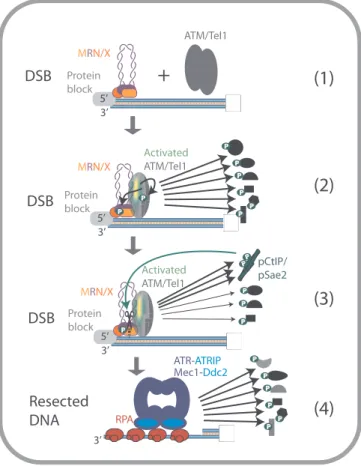
2. The function of the MRN/X complex in checkpoint signaling Cells possess factors that sense and signal the presence of DSBs (Fig. 2) [28]. MRN/X very rapidly localizes to DSBs [29], and physically and functionally associates with the ataxia-telangiectasia-mutated (ATM, human) or Tel1 (yeast) kinase [30,31]. This relationship is un-derlined by defects in human MRE11 linked to ataxia-telangiectasia-like disorder (ATLD), affecting the musculoskeletal and central nervous systems, which is similar to ataxia-telangiectasia (AT) caused by defects in ATM [32,33]. Notably, disorders caused by defects in the other components of the MRN complex share features with AT and ATLD such as spontaneous genomic instability [34,35].
DNA and MRN/X together activate ATM/Tel1, which phosphor-ylates a large number of cellular targets. Among them, there are factors that function in DNA end resection, both in the initial (short-range) and subsequent (long-range) processing. ATM/Tel1 phosphorylates all three components of MRN/X, so that the complex both activates and becomes a target of the DNA damage-induced checkpoint (Fig. 2) [36–39]. Be-yond MRN/X, ATM/Tel1 also phosphorylate BRCA1 and CtIP/Sae2 (Fig. 2) [40–43]. In particular, ATM-dependent phosphorylation of CtIP at T859 is required for thefirst DNA end resection step in human cells [40], while in contrast, the MRX-Tel1 axis appears dispensable for the activation of short-range resection by MRX-Sae2 in yeast [44]. Evidence suggests that ATM/Tel1 regulates also enzymes of the downstream long-range resection pathway both positively and negatively. This in-cludes phosphorylation of EXO1 in human cells [45], resulting in re-section inhibition. On the other hand, ATM phosphorylates the Bloom (BLM) helicase to increase its accumulation at DSB sites [46], and the Werner (WRN) helicase to promote recovery from replication stress [47]. Activated ATM/Tel1 also phosphorylate chromatin proteins in the vicinity of the DSBs such as the histone variant H2AX (in humans) [48] or H2A (in yeast) [49]– the respective phosphorylated forms are de-noted as γH2AX/γH2A. This signaling cascade triggers changes in chromatin conformation and facilitates the recruitment of multiple factors including DSB repair proteins belonging to both recombination and end-joining pathways [50].
The detailed mechanism of ATM/Tel1 activation by MRN/X at DSBs was examined using both human and yeast reconstituted complexes in vitro, respectively. The nuclease activity of MRE11 is dispensable for ATM activation [51,52], while the presence of yeast Mre11 was not absolutely required for Tel1 activation [53]. The presence of RAD50 is essential in both yeast and human systems, with ATP hydrolysis by Rad50 being indispensable for Tel1 activation in the yeast system [53], whereas ATP binding by RAD50 was found sufficient for ATM Fig. 1. An overview of the main DNA double-strand break repair pathways.
Exogenous (environmental) and endogenous (cellular) insults directly or in-directly cause DNA double-strand breaks. Homologous recombination is a template-directed and therefore largely accurate DSB repair pathway, which is active in the S and G2 phases of the cell cycle. In contrast, end-joining pathways are template and cell cycle independent, and result in a direct ligation of the broken ends. Recombination depends on extended DNA end resection to reveal ssDNA needed for homology search, while the end-joining pathways require only limited end processing. Non-homologous end-joining links DNA breaks with no or only limited microhomologies at the broken ends, which micro-homology-mediated end-joining typically utilizes microhomologies of 2–20 nucleotides in length. Both end-joining pathways are highly mutagenic.
activation when using purified human proteins [51]. Xrs2 was found partially dispensable for Tel1 activation in the yeast reconstituted system [53], and NBS1 provided a clear stimulatory function in the reconstituted system with the human proteins [51].
NBS1/Xrs2 carries a nuclear localization signal, and is therefore required for the nuclear import of the MR complex and indirectly for all its functions in both yeast [54] and human cells [55]. However, ex-periments where the nuclear localization signal was artificially placed on yeast Mre11 instead of Xrs2 revealed that Xrs2 per se was still im-portant for Tel1 activation [44]. These results agree with other data that identified the FHA and the C-terminal domains of Xrs2 to be re-quired for Tel1 activation [56]. Xrs2 phosphorylation appears to ne-gatively regulate Tel1 [57]. On the other hand, using a murine model, most of NBS1 was found dispensable for ATM activation except a very small fragment that represents its interaction interface with MRE11 [58], suggesting that ATM activation in mice is largely mediated by MRE11 and RAD50. Therefore, while the MR complex is clearly critical for ATM/Tel1 activation, the function of NBS1/Xrs2 may not be fully required under all circumstances.
Yeast mre11 mutants with point mutations in the nuclease active site (mre11-nd) exhibit checkpoint hyperactivation upon exposure to DNA damage [59], likely due to the persistence of MRX at DNA ends, which prolongs Tel1 activation. CtIP/Sae2 functions to promote the nuclease
activity of MRE11/Mre11 within the MRN/X complex (see below). To this point, it was intriguing that yeast sae2Δ mutants exhibit more se-vere sensitivity to DNA damaging drugs compared to mre11-nd [59,60]. These results suggested that Sae2 has additional function(s) on top of stimulating the Mre11 nuclease. Later, it was found that sae2Δ mutants activated checkpoint signaling to even higher levels than mre11-nd cells, leading to a permanent cell cycle arrest [59,61–64], thereby ex-plaining the strong sensitivity of sae2Δ cells. Indeed, the DNA damage sensitivity of mre11-nd and sae2Δ cells in a checkpoint-deficient back-ground is identical [62,65]. The specific function of Sae2 in checkpoint attenuation was found to be dependent on Tel1 phosphorylation sites in Sae2, and Sae2 was thus proposed to reduce checkpoint signaling by competing with other Tel1 targets (Fig. 2) [65,66]. In human cells, these relationships have not been studied in detail yet, although CtIP depletion similarly leads to hyperactivation of ATM-dependent sig-naling [67].
Downstream of the MRN-ATM/MRX-Tel1 signaling circuit, both short-range and long-range DNA end resection generate ssDNA that is bound by RPA, which is then sensed by ATR-ATRIP/Mec1-Ddc2 pro-teins (Fig. 2) [68]. These proteins activate both local and cell-wide responses [69].
3. The function of the MRN/X and CtIP/Sae2 ensemble in DNA end resection
3.1. A brief introduction to DNA end resection
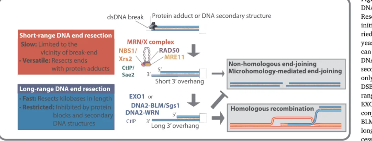
DNA end resection can be a two-step process, in particular in human cells. The initial short-range resection by MRN/X and its co-factor CtIP (in humans) or Sae2 (in yeast) is slow and limited to the vicinity of the DNA end (Fig. 3). At the same time, the short-range resection has the capacity to process DNA ends with secondary DNA structures and bound protein blocks. Accordingly, in yeast, the Mre11 nuclease and Sae2 are essential for the repair of meiotic DNA breaks with covalently attached Spo11, but are partially dispensable for the repair of nuclease-induced clean breaks in vegetative cells [70–73]. The MRE11 nuclease appears to be much more important in mammals even for the proces-sing of clean breaks. This may result from a high concentration of the Ku heterodimer, which needs to be removed from DSBs prior to re-section [23,74], as will be further detailed below.
Downstream of the initial processing by MRE11 and its co-factors, two long-range DNA end resection nucleases, namely the exonuclease EXO1 and the helicase-nuclease DNA2, are capable to resect kilobase lengths of DNA (Fig. 3). EXO1 resects 5′-terminated DNA within dsDNA [75–79]. In contrast, DNA2 degrades 5′-terminated ssDNA, and per se is incapable to unwind dsDNA [80,81]. Therefore, DNA2 requires a lead helicase partner, which can be either BLM or WRN in human cells [82–84] or Sgs1 in yeast [85]. Both DNA2 and EXO1-dependent re-section pathways are fast and processive, but sensitive to secondary DNA structures and protein blocks [74,86–88].
Despite the short- and long-range resection pathways are catalyzed by distinct nucleases, the processes are not entirely independent. The MRN/X complex additionally has a structural (nuclease independent) role to promote the EXO1 and DNA2-dependent pathways in both yeast [85,87,89–92] and humans [83,88,93]. Likewise, in addition to its role to promote MRN, CtIP also stimulates the DNA2-dependent pathway [94,95], showing that it can function as a co-factor of nucleases in both short- and long-range resection. The division of labor between versatile but slow (short-range), and fast but inflexible (long-range) resection pathways guarantees optimal DNA end processing under most circum-stances. In the next sections, we will focus on the mechanism of func-tion and regulafunc-tion of the MRN/X nuclease in DNA end resecfunc-tion. 3.2. The MRE11 nuclease polarity paradox
Recombinant MRE11 per se is a potent exonuclease with a strict 3′→
Protein block 5’ 3’ MRN/X P P Protein block 5’ 3’
+
ATM/Tel1 Activated ATM/Tel1 P P P P P P P Protein block 5’ 3’ Activated ATM/Tel1 P P P P MRN/X MRN/X 3’ RPA ATR-ATRIP Mec1-Ddc2DSB
DSB
DSB
Resected
DNA
P P P P P P P pCtIP/ pSae2(1)
(2)
(3)
(4)
Fig. 2. An overview of the MRN/X function in the activation of ATM/Tel1-mediated DNA damage response. MRN/X localizes at DSB and recruits ATM (human) or Tel1 (yeast), which transforms from an inactive dimer to an active monomer (1–2). Activated ATM/Tel1 phosphorylates many cellular targets, such as DNA repair factors, chromatin and checkpoint proteins, including MRN/X, initiating the DNA damage response (2). Phosphorylation of CtIP by ATM helps activate the endonuclease activity of MRN, initiating resection (see later for details). At the same time, CtIP/Sae2 likely attenuates checkpoint activation by competing for ATM/Tel1 substrates (3). Resected DNA is coated by RPA, which is detected by ATR-ATRIP (human) or Mec1-Ddc2 (yeast), which continue checkpoint signaling by phosphorylating an overlapping set of cellular targets (4).
5′ polarity (Fig. 4) [96–98]. This polarity of DNA degradation was counterintuitive, as resection of the opposite, 5′-terminated strands, is required for homologous recombination [8]. Early genetic studies in yeast instead identified Mre11 to be responsible for 5′→3′ DNA end
resection in vivo, leading to an apparent polarity paradox [77,99,100]. It was unclear (1) how is the 3′→5′ exonuclease of MRE11 attenuated, as 3′-end degradation at DSB ends observed in vivo was minimal [77,101], and (2) how MRE11 can instead contribute to the degrada-tion of the opposite, 5′-terminated DNA strands.
In cells, MRE11 appears to be always bound to the RAD50 ATPase, forming a tight MR complex [98]. Under physiological conditions when ATP is present, Rad50 was found to strongly limit the exonuclease of Mre11 (Fig. 4) [102]. This inhibition required ATP binding by Rad50, but not ATP hydrolysis. Similar results were found for the Pyrococcus furiosus MR complex [103]. Due to the low ATPase activity of Rad50, these results likely explain why Mre11 does not extensively degrade 3 ′-terminated DNA in vivo.
RAD50, with its ATP hydrolytic activity, instead facilitates the en-donucleolytic cleavage of DNA by MRE11 (Fig. 4), which was initially mainly observed on circular ssDNA, at the base of secondary structures such as hairpins and at junctions of single and double-stranded DNA [96–98,104], and accordingly in genetic assays [105]. NBS1 further potentiates this endonucleolytic activity [106,107]. While NBS1/Xrs2 is required for the nuclear import of MR [54,55], its contribution to DNA end resection per se significantly differs between yeast and mammalian cells. In yeast, Xrs2 is not required for endonucleolytic cleavage of DNA in vitro [44], although a modest stimulatory effect of Xrs2 can be re-vealed under restrictive experimental conditions [108]. Consistently with this notion, in vivo, DNA end resection was only minimally im-paired in xrs2Δ cells where the nuclear localization signal was placed on Mre11 [44], suggesting that Xrs2 has only a minor function in DNA end resection in yeast. In contrast, in the human system, NBS1 is much more important. NBS1, in fact, potentiates MRE11 endonucleolytic activity and was shown to be necessary for the cleavage of dsDNA [106,107,109]. The more prominent role of NBS1 in the function of the MRN complex will be discussed in the next sections.
When using linear DNA substrates with protein-blocked DNA ends, the endonuclease activity of MRE11 (within the MRN/X complex) was found to be strongly promoted by CtIP/Sae2 [107,109,110]. CtIP/Sae2 specifically stimulates the cleavage of the 5′-terminated DNA strand by MRN/X at sites internal to the protein-blocked DSB. The endonuclease activity of the ensemble thus preferentially degrades 5′-terminated DNA [102,107,109,110], showing how a 3′ overhang can be created. Therefore, the MRE11 polarity paradox can be explained by (1) proteins that bind DNA ends and physically block the 3′→5′ exonuclease of MRE11; (2) RAD50 limiting the 3′→5′ exonuclease of MRE11, and (3) RAD50, CtIP/Sae2 and NBS1 turning MRE11 into an endonuclease that preferentially targets the 5′ strand at internal sites past protein blocks. dsDNA break
or
Protein adduct or DNA secondary structure
MRE11 RAD50 NBS1/ Xrs2 EXO1 DNA2-BLM/Sgs1 3’ 5’ Short-range DNA end resection
3’ 5’ Long-range DNA end resection
DNA2-WRN Slow: Limited to the
vicinity of break-end • Versatile: Resects ends with protein adducts
• Fast: Resects kilobases in length • Restricted: Inhibited by protein blocks and secondary DNA structures Short 3’ overhang Long 3’ overhang Homologous recombination Non-homologous end-joining Microhomology-mediatedend-joining MRN/X complex CtIP/ Sae2 CtIP
Fig. 3. An overview of the two steps of DNA end resection in eukaryotic cells. Resection of the 5′ strand at DSBs is initiated by the short-range step, car-ried out by the MRN complex (MRX in yeast). This pathway is versatile as it can overcome various obstacles at the DNA ends such as protein blocks or secondary structures, but can resect only up to a limited distance from the DSB. The second phase is the long-range resection, carried out either by EXO1/Exo1 or by DNA2/Dna2 in conjunction with a helicase (WRN or BLM in humans and Sgs1 in yeast). The long-range resection is fast and pro-cessive, but it is inhibited by non-ca-nonical structures at the DNA ends. DNA ends that underwent limited resection may still be repaired by end-joining (possibly uponfill-in DNA synthesis), whereas, extensive resection generally commits the DSB repair to homologous recombination.
MRE11
RAD50
AT
P
ADP
Exonuclease (3’ - 5’)
Exonuclease (3’ - 5’)
5’ 5’ Exonuclease 3’ - 5’ 5’ ATPEndonuclease
MRE11
MRE11
MRE11
MRE11
ExonucleaseLong-range resection
5’-3’
3’ - 5’MRE11 alone
(not ph
y
siological)
MRE11 with R
AD50 (MRN/X) and CtIP/S
ae2
Degradation of 3’-terminated strand
Degradation of 5’-terminated strand
P P
CtIP/Sae2
NBS1/Xrs2
Fig. 4. The MRE11 nuclease polarity paradox. MRE11 by itself, in reconstituted reactions in vitro, is a 3′→5′ exonuclease (top part). In contrast in cells, the 3′→ 5′ MRE11 exonuclease within the MRN/X complex is restricted by multiple mechanisms, and MRE11 instead preferentially catalyzes 5′ strand degradation (bottom part). First, ATP binding to the MRE11 partner RAD50 restricts the MRE11 exonuclease. Second, the presence of protein blocks at DSBs (not shown) directly inhibits the 3′ end degradation. Furthermore, ATP hydrolysis by RAD50, coupled with the stimulation by phosphorylated CtIP (Sae2 in yeast) activates the MRE11 endonuclease activity, which preferentially targets the 5′-terminated DNA strand. NBS1 further promotes the endonucleolytic cleavage of the 5′ strand. After the first incision, the exonuclease activity of MRE11 (still within the MRN/X complex) degrades the 5′-terminated strand towards the end, creating an entry site for the long-range resection nucleases.
3.3. Protein blocks in DNA end resection
Protein blocks at DSBs can be removed by MRN-CtIP or other pro-cessing complexes and the strand integrity restored by end-joining or homologous recombination, depending on the type of the block and cellular context (Fig. 3). Examples of covalent protein blocks at DSBs are meiotic SPO11 and stalled topoisomerase DNA cleavage complexes. The initial processing of Spo11-bound breaks appears to be entirely dependent on the MRX-Sae2 ensemble and consequently repaired ex-clusively by the homologous recombination machinery in yeast [71,73,111]. Similarly in mouse spermatocytes, the resection pattern of meiotic SPO11-bound DSBs is consistent with MRE11 initiating the resection process [112].
DSBs induced by aberrant topoisomerase II (TopoII) activity were recently described to result in frequent spontaneous genome re-arrangements of the human genome [6]. Topoisomerase complexes can be directly removed by TDP1/Tdp1 or TDP2, depending on whether the topoisomerase is attached to the 5′ end (TopoII, repaired by TDP2 in human cells [113]) or the 3′ end (TopoI, repaired by TDP1 [114,115]). In yeast, the majority of topoisomerase-induced breaks is however re-paired by the recombination pathway dependent on the Mre11 nu-clease, as supported by an increase in the level of Top2 covalent com-plexes upon deletion of Mre11 in both S. cerevisiae [116] and S. pombe [117]. Only a subset of topoisomerase-induced breaks is repaired by end-joining in yeast [118]. On the other hand, the repair of topoi-somerase-linked breaks heavily relies on NHEJ in human and chicken cells [119,120]. MRN and CtIP were observed to be necessary to re-move the TopoII adducts prior to the ligation process [121,122]. This agrees with the profound sensitivity of CtIP-depleted cells to the to-poisomerase poisons etoposide and camptothecin [123,124]. The ad-ditional NHEJ-related role of the MRN complex in the repair of topoi-somerase-linked DSBs will be discussed more in detail in a dedicated section of this review. The text immediately below focuses on the re-moval of protein blocks by the MRE11 nuclease, in the context of DSB repair by the recombination pathway.
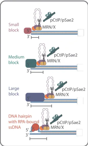
The ability of the MRE11 complex to cleave 5′-terminated DNA past protein blocks is conserved in evolution. Already in bacteria, the Escherichia coli’s SbcCD (MRE11-RAD50-like complex) exhibits this activity, which however cleaves both DNA strands past blocks [125,126]. As there are no Sae2/CtIP and NBS1/Xrs2 homologues in E. coli, this endonucleolytic activity does not appear to be strictly regu-lated in prokaryotes. Experiments with eukaryotic recombinant MRN/X and linear DNA substrates with streptavidin-bound ends showed that a DNA cleavage activity past protein blocks was strongly stimulated by CtIP/Sae2 [107,109,110]. The endonuclease incision sites were located ∼15−20 nucleotides away from the streptavidin-bound DNA end on the 5′ strand (Fig. 5). These cleavage positions roughly correspond to the length of DNA fragments (10–40 nucleotides) found attached to Spo11 during yeast meiotic recombination [72]. Importantly, in con-trast to the E. coli enzymes, the 5′-terminated strand was preferentially cleaved [109,110], although there is evidence that MRN/X can also cleave both strands in some cases [107]. The molecular mechanism that determines the strand cleavage preference remains unclear.
Curiously, the endonucleolytic cleavage by MRX-Sae2 was also ob-served when streptavidin was placed in the middle of an oligonucleo-tide some distance away from the DNA end; in this case, the incision sites were again ∼15−20 nucleotides away from the internal block [108]. Likewise, MRX-Sae2 could cleave DNA near oligonucleotide-bound nucleosomes [108], although the complex was also seen to by-pass nucleosomes while sliding [93]. However, the efficacy of the DNA cleavage of internal blocks on oligonucleotide-based substrates was lower compared to end-bound blocks. Similarly to the oligonucleotide-based substrates, the MRX-Sae2 ensemble could efficiently cleave the 5′-terminated strands of plasmid-length DNA, when the DNA ends were bound by streptavidin [102]. However, no cleavage was observed when streptavidin was attached to a plasmid in its circular covalently-closed
form (our unpublished observations). Such behavior makes sense as it would restrict the endonucleolytic cleavage by MRN/X to the vicinity of DSBs, and thus prevent unscheduled DNA degradation. Structural stu-dies showed that the MRE11 enzymatic ensembles undergo notable changes in conformation upon ATP binding, hydrolysis, and during DNA cleavage [26,125,127]. One possibility is that these structural changes are only permissible at DNA ends. Furthermore, the MRN/X was shown to melt DNA [106,128], but it remains to be established whether this activity is used to sense DNA ends. The mechanism that guarantees MRN/X-CtIP/Sae2-dependent DNA cleavage preferentially near DSB-bound blocks thus remains to be elucidated.
What is the identity of the physiological protein blocks that direct the MRN/X-CtIP/Sae2 endonuclease? The initial observations with streptavidin-bound DSBs suggested that there is no strict requirement for cognate protein-protein interactions to direct cleavage by the en-semble [107,109,110]. This lack of specificity makes the MRN/X-CtIP/ Sae2 complex very versatile, as DSBs can be linked to a variety of proteins. Beyond meiotic SPO11 and stalled topoisomerase complexes [115,129], MRN/X in concert with CtIP/Sae2 also processes DSBs with RPA-bound ssDNA at secondary structures [86,108], as well as DSBs with random proteins crosslinked to DNA [129].
The protein component of covalent protein-DNA crosslinks may be
Small
block
Medium
block
Large
block
DNA hairpin
with RPA-bound
ssDNA
5’
5’
5’
5’
3’
3’
3’
3’
MRN/X
MRN/X
MRN/X
MRN/X
P PpCtIP/pSae2
P PpCtIP/pSae2
P PpCtIP/pSae2
P PpCtIP/pSae2
Fig. 5. Determining the site of the endonucleolytic DNA cleavage by MRN/X and phosphorylated CtIP/Sae2. Depending on the DNA footprint of the protein block or the size of the secondary DNA structure, the incision mediated by MRN/X in conjunction with phosphorylated CtIP/Sae2 can be closer or farther from the DNA end. Small blocks, for example Streptavidin (used as a tool in reconstituted reactions), result in DNA cleavage close to the end (∼15–20 nucleotides). In the case of medium-sized blocks, such as yeast Ku, the nick forms further away (∼30 nucleotides). Even larger blocks, such as human DNA-PK, induce DNA cleavage up to∼45 nucleotides away from the DSB. DNA secondary structures at the DNA ends, possibly bound by the ssDNA binding protein RPA, may similarly direct the endonucleolytic DNA cleavage by MRN/ X-CtIP/Sae2.
processed by specialized proteases such as SPARTAN/Wss1 [130–132]. The function of these enzymes is mostly linked to DNA replication and therefore not specific for DNA ends. SPARTAN/Wss1 however cannot remove the entire protein component of the crosslink, which necessi-tates downstream processing. Indeed, SPARTAN and TDP1 may func-tion in the same pathway in mammalian cells [133]. Any potential functional interplay of SPARTAN/Wss1 and MRN/X has not been de-fined yet. As the activity of SPARTAN/Wss1 can lead to intermediates with various degrees of proteolytic cleavage, any downstream proces-sing step must involve a mechanism that does not require a specific interaction between the lesion and the nuclease machinery. The lack of specificity requirement between MRN/X-CtIP/Sae2 and the end-bound polypeptide would thus guarantee efficient processing of any type of protein-blocked DSB (Fig. 5).
The MRX-Sae2 ensemble can also cleave near Ku-bound DSBs [86,108]. When using the Ku heterodimer instead of streptavidin in vitro, the cleavage positions on the 5′-strand were further away from the end,∼30 nucleotides for yeast Ku as opposed to ∼15−20 nucleotides away for streptavidin (Fig. 5) [86,108]. Similar results were obtained with human MRN/CtIP on Ku and DNA-PKcs bound DSBs, with the cleavage positions even further at∼45 nucleotides away from the end (Fig. 5) [74]. This difference in cleavage sites is likely due to the larger footprint of the respective factors that shield DNA ends [134], as op-posed to streptavidin that was attached to the end of the DNA molecule by a biotin linkage, but does not otherwise bind DNA (Fig. 5). Ac-cordingly, the MRN complex was found to diffuse along DNA strands in single-molecule assays [93]. Such a movement could explain how the ensemble can cleave near protein blocks: physical impediments could stall the sliding of MRN and thus enhance the residency of the nuclease ensemble near protein blocks, which could, in turn, facilitate cleavage [93].
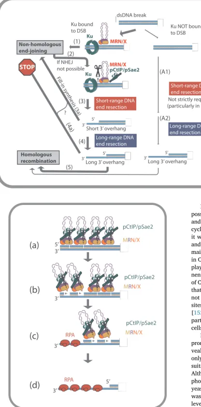
The Ku heterodimer as a protein block deserves special attention. Ku is very abundant, particularly in human cells [135]. It has a very strong affinity to DSBs, where it is recruited within seconds in vivo [136]. Importantly, however, Ku is a non-homologous end-joining factor; in the absence of Ku, the frequency of homologous recombination is ty-pically increased, while non-homologous end-joining is abrogated [137,138]. Therefore, Ku is not a factor that is required to direct DNA cleavage by MRN-CtIP. Current data rather support a model where Ku rapidly binds DNA ends and attempts tofirst facilitate DSB repair by the non-homologous end-joining pathway, likely with the assistance of MRN/X (the role of the MRN/X complex in NHEJ will be discussed in a later section) (Fig. 6). If this is not possible, such as in the absence of the second DNA end, or when the DNA end contains altered chemistry that prevents ligation, CtIP/Sae2 gets recruited and activated by phos-phorylation (see next section). The ensemble then cleaves the 5′-ter-minated DNA strand past Ku (Fig. 6). The break can then be repaired by end-joining or resected further for processing by the recombination pathway (Fig. 6) [23,24,74,86].
The capacity of MRN/X to cleave past Ku-bound ends is supported by in vivo data: yeast mre11 nuclease-deficient cells are sensitive to DNA damage, which can be alleviated by the deletion of Ku, and the cellular sensitivity can be instead enhanced by Ku overexpression [60]. This indicates that the Mre11 nuclease needs to remove Ku to promote cellular resistance to DNA damage, most likely by facilitating resection and repair. However, in most cases in yeast, the Mre11 nuclease is not required for the processing of clean DSBs [70,75,77]. In contrast, the MRN-CtIP axis is essential for all DNA end resection events in high eukaryotes [123], where Ku is very abundant [135]. It remains to be established whether the high concentrations of Ku in mammalian cells can explain this difference. Support for this model comes from experi-ments in murine embryonic cells, where CtIP is normally required for resection [139,140], as in other cell types [123,124]. Upon deletion of Ku however, DSB resection became largely CtIP independent (Fig. 6) [139]. Further evidence for this model was demonstrated by bio-chemical and single molecule experiments with human MRN-CtIP and
Ku-DNA-PKcs proteins [74]. Therefore, the requirement of CtIP and the MRE11 nuclease for resection in vertebrates may be caused by the necessity to remove Ku from DNA ends, prior to further processing (Fig. 6).
3.4. A unified model for exo- and endo-nuclease activities of MRE11 in DNA end resection
In the previous sections we clarified how the 3′→5′ exonuclease activity of MRE11 is restricted to allow 5′ DNA end resection (Fig. 4). This raised a question why MRE11 possesses such an exonucleolytic activity at all. In order to accommodate for the seemingly wrong po-larity of the MRE11 exonuclease, the so-called bi-directional DNA end resection model was proposed [23,72,141]. Upon initial en-donucleolytic cleavage by MRN-CtIP, the 3′→5′ exonuclease activity of the complex then catalyzes DNA degradation back toward the DNA end, while at the same time the endonucleolytic incision sites serve as entry points for 5′→3′ DNA degradation by the long-range resection machi-neries (Fig. 4). This model was initially proposed based on the analysis of resection end-points during yeast meiotic recombination [72,141–143], and later substantiated in time-course experiments in vitro [86,102]. In this model, the 3′→5′ exonuclease activity of Mre11 ultimately helps to remove the protein block, possibly with the help of the dsDNA melting capacity of the MRN/X complex [106,128]. This "jump over" endo/exonucleolytic processing is ideally suited to process DNA ends with non-canonical structures as it allows to bypass the end-blocking lesions.
In yeast strains lacking the long-range resection enzymes, the Mre11-dependent resection can reach up to several hundred nucleo-tides upon several hours (Fig. 7) [75,77,142,143]. Experiments with recombinant proteins revealed a similar behavior: although slow, the MRX-Sae2 ensemble can process the 5′-terminated DNA end at DSB sites in a stepwise manner [102]. Upon thefirst incision of the 5′-ter-minated DNA by MRX-Sae2, thefirst end-bound ensemble likely serves as a structure that directs cleavage by the adjacent MRX-Sae2 complex (Fig. 7). The 3′→5′ exonucleolytic activity of the complex would then be limited to degrade DNA between the endonucleolytic incision sites (Fig. 7). The progressive sequential DNA cleavage is consistent with the ability of the human MR complex to form oligomers at the end of DNA molecules observed by scanning force microscopy [144] and the pos-sibility to readily detect MRX foci in yeast [98]. The distance between the subsequent incision sites may correspond to the site size of the MRX-Sae2 complex on DNA [102]. In meiosis, MRX-Sae2-dependent incision sites up to∼300 nucleotides away from the DNA end were observed [141], raising the possibility that DNA looping or internal blocks may also direct cleavage at distances further away from the DNA break. How exactly the incision sites are determined thus remains to be established. In summary, it seems very likely that the DNA end pro-cessing by MRN/X-CtIP/Sae2 is initiated by endonucleolytic cleavage of the 5′ strand, and the rather weak exonucleolytic DNA degradation is then restricted to DNA fragments between the endonucleolytic cleavage sites in the direction back toward the DNA end.
3.5. Regulation of MRN by phosphorylation of CtIP
A key feature of homologous recombination is that it is template-directed. The genetic information stored in the DNA template is used to recover missing sequence information in the vicinity of the broken end. In order to guarantee the largely error-free repair, vegetatively dividing cells must use a template with the matching sequence, which is only represented by the sister chromatid upon DNA replication in the S and G2 phases of the cell cycle [8]. In the absence of such a template, the repair could in principle proceed from the homologous chromosome, potentially leading to the loss of heterozygosity. Likewise, aberrant repair from ectopic sites, such as in repetitive sequences, may lead to large chromosomal rearrangements [2,145].
In order to prevent genome instability due to resection in G1, cells possess regulatory mechanisms that allow extensive DNA end resection and hence homologous recombination only in the S-G2 phase of the cell cycle when the sister chromatid DNA is available (Fig. 8). To this point, it was observed that CDK inhibition limits homologous recombination and DNA end resection [146,147]. Later, CtIP/Sae2 was found as the main target of this regulatory mechanism, with the key sites being T847 in CtIP [148] and S267 in Sae2 [149] (Fig. 8). CDK regulates also other players in the resection pathway, including DNA2 [150] and compo-nents of the MRN complex such as NBS1 [151], but the phosphorylation of CtIP/Sae2 appears to be the most important. Later studies established that the phosphorylation of T847/S267 in CtIP/Sae2 is necessary, but not sufficient to fully promote DNA end resection. Multiple additional sites were identified in both the human CtIP [40] and the yeast Sae2 [152,153] proteins that further promote their resection capacity. In particular, the ATM kinase is important for CtIP activation in human cells upon DNA damage [40].
How does phosphorylation regulate the capacity of CtIP/Sae2 to promote DNA end resection? In vitro reconstitution experiments re-vealed that CtIP/Sae2 promotes end resection by the MRN/X complex only in its phosphorylated state, and the reconstituted system was thus suitable to reveal the underlying mechanism [86,107,109,110,152]. Although Xrs2 contains the FHA domains that interact with phos-phorylated Sae2 [154], the resection related functions of the minimal yeast ensemble do not depend on Xrs2, as stated above [44]. Instead, it was found that the phosphorylation of Sae2 controls resection on two levels. First, it regulates transitions between inactive hypo-phosphory-lated Sae2 multimers (large complexes) and active phosphoryhypo-phosphory-lated tetramers (Fig. 8) [152,153]. Second, phosphorylation of the C-terminal region of Sae2 was found to be required for its physical interaction with Rad50, while it does not affect the interaction with Mre11 (Fig. 8) [152]. Therefore, in yeast, Rad50 appears to be the main sensor of Sae2 phosphorylation. Rad50, in turn, hydrolyzes ATP that triggers the Mre11 endonuclease (Fig. 8) [152]. To this point, it is possible that Sae2 helps coupling ATP hydrolysis by Rad50 with productive en-donucleolytic DNA cleavage by Mre11 [102], although the detailed underlying mechanism remains to be clarified. It should be pointed out that the rad50S alleles, initially identified as mutations that separate the dsDNA break 3’ Short-range DNA end resection Long-range DNA end resection If NHEJ not possible Ku Ku 3’ 5’ Short 3’ overhang Homologous recombination 3’ 5’ Long 3’ overhang Short-range DNA end resection Short-range DNA end resection
Not strictly required (particularly in yeast) 3’ 5’ Long 3’ overhang 3’ Long-range DNA end resection Ku bound to DSB Ku NOT bound to DSB MRN/X P P MRN/X pCtIP/pSae2 Non-homologous end-joining
STOP
Fill-in synthesis (3a)
?
(1)
(2)
(3)
(4)
(4a)
(5)
(A1)
(A2)
Fig. 6. A model for the interplay of Ku and short-range re-section. DSBs can be bound by Ku, which together with MRN/X promotes NHEJ (1). If the ligation is not successful (2), phosphorylated CtIP/Sae2 (pCtIP/pSae2) is recruited to the ensemble and helps remove Ku by activating the MRE11 endonuclease activity (3). Partially resected DSBs can be in some casesfilled-in by DNA synthesis, bound again by Ku and channeled to NHEJ (3a), as may occur in NHEJ-mediated repair of non-readily ligatable DNA ends. Extensive resection (4) instead generally inhibits end-joining (4a), and commits the DSB to repair to HR (5). In the absence of Ku, the re-section of “clean” DSBs can be independent of the short-range resection (A1) and instead entirely catalyzed by the long-range pathway (A2), especially in yeast.
P P P P P P P P 5’ 3’ P P P P 3’ 5’ 3’ MRN/X pCtIP/pSae2 MRN/X pCtIP/pSae2 MRN/X pCtIP/pSae2 RPA 3’ RPA P P P P P P P P
(a)
(b)
(c)
(d)
Fig. 7. A model for 5′ DNA end resection by MRN/X and phosphorylated CtIP/ Sae2 in the absence of the long-range resection. Multiple MRN/X and phos-phorylated CtIP/Sae2 (pCtIP/pSae2) complexes are recruited and perform multiple endonucleolytic incisions of the 5′-terminated strand (a). DNA nicking is followed by exonucleolytic degradation in the 3′ to 5′ direction between the incision sites (b). After exonucleolytic degradation, the resected DNA is pro-gressively covered by RPA (c). In the end, the short-range resection results in RPA-coated 3′-ended ssDNA tail, which can be used for the next step of the recombination pathway (d).
functions of Rad50 in meiosis (and hence resection/recombination) from its other functions such as Tel1 activation [155,156], do not physically and functionally interact with phosphorylated Sae2 [102,152]. Accordingly, the phenotypes of rad50S, mre11-nd and sae2Δ yeast cells in checkpoint-deficient background are indistinguishable from each other [65].
In human cells, the regulation of MRN by phosphorylated CtIP is more complex. Although, like Sae2, CtIP is also mostly active only as a tetramer in vivo [157], whether and how phosphorylation regulates its oligomeric state has not been defined yet. Most importantly, the sensor of CtIP phosphorylation in the human system is not RAD50, but NBS1, through its FHA and BRCT domains (Fig. 8) [40,158]. Once NBS1 senses that CtIP is phosphorylated, it promotes endonucleolytic clea-vage by the human MR complex, which is dependent on a short region in NBS1 that mediates interaction with MRE11 (Fig. 8) [58,158]. Si-milarly as in yeast, RAD50 also needs to hydrolyze ATP to promote the human MRE11 endonuclease in conjunction with CtIP (Fig. 8) [107,109]. Evidence suggests that CtIP phosphorylation also regulates its interaction with CCAR2 [159], a negative regulator of DNA end resection, uncovering another layer of regulation of the MRN-CtIP en-semble activity in higher eukaryotes.
The regulatory control of the MRE11 nuclease thus significantly differs in the various kingdoms of life. In bacteria, the MRE11-RAD50-like SbcCD complex cleaves both DNA strands near protein blocks without the need of co-factors such as NBS1/Xrs2 or CtIP/Sae2 [126]. Similar reactions without co-factors were observed in archaea, although only the 5′ strand was cleaved [160]. In yeast, both Sae2 and Xrs2 are present and only Sae2 plays an important function while Xrs2 is partly dispensable [44,108,110]. In contrast, in human cells, both CtIP and NBS1 critically control the MRE11 nuclease [58,107,109,158]. The more complex regulatory control of the human MRN-CtIP complex compared to yeast could reflect a possible need to more tightly control the MRE11 nuclease in high eukaryotic organisms due to the larger genome size and cancer risk.
3.6. Interplay of short and long-range DNA end resection
The resection of clean, nuclease-induced DSBs in mre11 or rad50 yeast mutants is delayed [70,77,99,161]. However, once resection commences, it proceeds with the same rate as in wild type cells [77]. Furthermore, the resection defect of mre11-null cells is more severe compared to mre11-nd cells [71]. The impaired resection in mre11 and rad50 cells can be explained by a structural role of the components of the short-range resection pathway to recruit and stimulate long-range resection, in addition to their nuclease-dependent function in the re-moval of protein blocks. To this point, it was observed in vitro that MRX, independently of its nuclease activity, promotes resection by both Exo1 and Sgs1-Dna2 (primarily DNA unwinding by Sgs1 was stimulated) [85,87,89–92]. Additionally, in reconstituted reactions that combined short- and long-range resection, Exo1 was much more efficient to in-itiate long-range resection downstream of endonucleolytic incisions by MRX-Sae2 compared to a non-related bacterial nuclease with the same specific activity [86]. Accordingly, in the human system, MRN has a structural function to promote both DNA2-BLM and EXO1-dependent resection pathways [83,88,93].
A potential mechanism for this stimulation, in addition to a re-cruitment function, could be the local melting of the DNA ends by the MRN/X complex independently of its nuclease activity [90,128]. This is consistent with the ability of the MRN complex to unwind short frag-ments of DNA [96] and melt the ends of a double-stranded DNA mo-lecule in FRET based assays [128]. Molecular dynamics simulations of yeast Mre11 with a 30 bp DNA molecule also suggested a possible melting of the DNA ends by Mre11 [90]. The strand separation could then facilitate DNA access to the long-range resection machinery. In accord with this model, the Mre11-R10T mutant, which promotes hyper-resection by Exo1 in vivo, displays persistent melting of the DNA ends in molecular dynamics simulations [90].
Similarly, although CtIP mainly functions as a co-factor of the MRE11 nuclease in short-range resection, it was also found to promote long-range resection. Specifically, CtIP stimulates the DNA2-dependent Fig. 8. A model for the activation of the MRE11/Mre11 en-donuclease within human MRN or yeast MRX by phosphory-lated CtIP/Sae2. Top panel: in the budding yeast, Sae2 phos-phorylation by CDK in S/G2 promotes the formation of active tetramers (1). Sae2 phosphorylation is sensed by Rad50 (2) which in turn activates Mre11 endonuclease activity via ATP hydrolysis (3). Xrs2 appears dispensable for the activation. Bottom panel: human CtIP is activated by CDK during S/G2 and by ATM upon DNA damage (1). In contrast to yeast, the sensor of CtIP phosphorylation is NBS1 (2) that in turn acti-vates MRE11 nicking activity (3a). As in the yeast system, ATP hydrolysis by RAD50 is also necessary for the activation of the endonuclease (3b). Phosphorylation of CtIP affects its inter-action with additional resections regulators (not shown) pro-viding another level of control.
sub-pathway, where it enhances DNA unwinding by BLM, as well as the activity of DNA2 [94,95]. Yeast Sae2 was also observed to stimulate the ssDNA degradation by Dna2 in vitro but failed to stimulate DNA un-winding by Sgs1 [94]. The functional interaction between EXO1/Exo1 and CtIP/Sae2 seems to be less conserved. While Sae2 is able to sti-mulate yeast Exo1 [91], CtIP was shown to inhibit human EXO1 activity in reconstituted reactions [162]. The positive relationships between components of the short-range and long-range resection likely help to coordinate both steps to make them more effective, and facilitate a more efficient regulation.
4. The function of the MRN/X complex in end-joining
In S. cerevisiae, the MRX complex has a well-defined function to promote non-homologous end-joining (Fig. 6). NHEJ is dependent on the Ku heterodimer, and other components including the DNA Ligase IV-Lif1 complex [10,163,164]. The pro-NHEJ functions of MRX likely involve its DNA end tethering capacity that helps to keep both ends together in vivo [165,166]. Additionally, Xrs2 directly interacts with Lif1, and the MRX complex consequently promotes ligation of DNA ends in reconstituted reactions [167]. Accordingly, yeast cells lacking MRX are deficient in NHEJ [168].
Vertebrate cells involve additional NHEJ components including the catalytic subunit of the DNA-dependent protein kinase (DNAPK-cs), as well as end-processing enzymes such as Artemis [10,163]. Although the functions of MRN in mammalian end-joining are less defined, possibly due to redundant activities, MRN plays at least a supportive role [169–171]. MRN may promote NHEJ by two mechanisms. First, MRN stimulates NHEJ in a way that does not involve its nuclease activity, and its nuclease function is consequently partly dispensable for the canonical NHEJ repair of“clean” DSBs in mammals. Second, there is evidence that the nuclease of MRE11, assisted by CtIP, is additionally involved in the repair of DSBs with “dirty” ends by NHEJ (Fig. 6) [24,170]. The MRE11 nuclease also appears to be involved in the processing of DSBs with topoisomerase adducts, which are subsequently channeled to NHEJ [121]. This nuclease-dependent function of MRN remains however unclear, in particular as it invokes MRE11 nuclease activity in the G1 phase of the cell cycle, when it is normally restricted. Although the endonuclease activity of the MRN-CtIP ensemble gen-erally requires NBS1, there is a residual nuclease activity in the absence of NBS1. The NBS1-independent MRE11-RAD50 nuclease requires CtIP but not strictly its phosphorylation [158]. Whether this mechanism could explain the function of MRE11 nuclease activity in G1 remains to be clarified.
Upon ionizing radiation damage, PLK3 was shown to phosphorylate CtIP at S327 in G1 [24,172]. Phosphorylation of S327 in CtIP is critical for its interaction with BRCA1 [173], which is also involved in the non-homologous end-joining mediated repair of DSBs alongside with CtIP itself [24]. It should be pointed out that the S327 site phosphorylation in CtIP was also identified in S-G2, when it becomes a target of CDK, and was observed to promote homologous recombination [40,122,173–176], although the importance of this interaction for re-combination was questioned [124,177,178].
In yeast, in the absence of canonical NHEJ, cells employ an end-joining alternative termed microhomology-mediated end-joining (MMEJ) [179]. In mammalian cells, MMEJ appears more frequent than in the low eukaryotes [180]. MMEJ is Ku and DNA Ligase IV-in-dependent [181], and instead involved DNA Ligase III and Polθ [182–185]. Like NHEJ, MMEJ can also operate in any phase of the cell cycle, including G1 [180,186]. MMEJ generally requires short micro-homologies (4–20 nucleotides) at the DNA ends to facilitate ligation [9]. The DNA overhangs are generated by DNA end resection enzymes, including MRN-CtIP [9,169,170,175,180,187–189]. As with NHEJ, the mechanism of activation of DNA cleavage by MRN-CtIP in MMEJ re-mains undefined.
5. Conclusion and future perspectives
DSB repair is of crucial importance in the maintenance of genome stability and perturbations of the underlying pathways are often asso-ciated with cancer development. The MRN/X complex plays a pivotal role in the cellular decision of which mechanism will be used to repair broken DNA. For this reason, a detailed understanding of how MRE11 functions in healthy cells, and how its functions integrate with the other repair pathways is important in the context of cancer treatment. Furthermore, in cells lacking recombination factors such as BRCA1 or BRCA2, misregulated MRE11 activity was found to degrade nascent DNA at challenged replication forks, leading to cellular lethality [190]. Most likely, MRE11 initiates the degradation of replication forks that have undergone reversal, forming a DSB-like structure upon annealing of the two nascent DNA strands [191,192]. How the MRE11 activity is regulated in this process is not known. The degradation of reversed replication forks may resemble some but not all aspects of the DSB processing described above. The key distinctive feature is the function of RAD51. In the context of canonical DSB repair, RAD51 only functions downstream of resection to initiate recombination. In contrast, RAD51 acts upstream to protect nascent DNA from degradation by MRE11 [190] and downstream nucleases during replication stress [193]. BRCA1 and BRCA2 mutations are common in breast and ovarian can-cers, and as the cellular resistance to therapy was linked to abrogated DNA degradation by MRE11 [194], mechanistic insights into these processes, largely missing to date, can lead to novel therapeutic stra-tegies.
Beyond the academic and clinical interests to understand the MRE11 nuclease, the balance between the DSB repair pathways is known to affect the outcome of gene editing. With the advent of Cas9-based technologies, there is considerable interest in improving the ef-ficacy of homology-directed repair, in order to facilitate precise and efficient introduction or correction of mutations in the human genome [195–197]. This includes understanding the processes that negatively regulate resection in G1, as non-dividing cells are generally refractory to these modifications. Overcoming the block of resection in G1 might ultimately make gene editing possible in a much broader variety of cell types.
Declaration of Competing Interest
The authors declare no conflict of interest. Acknowledgments
We would like to thank the members of the Cejka laboratory (A. Acharya, E. Cannavo, I. Ceppi, V. Mengoli, A. Sanchez) for comments on the manuscript. We apologize to colleagues whose work could not be cited due to length constraint. The research in the Cejka laboratory is supported by the European Research Council (ERC, HRMECH, 681630) and the Swiss National Science Foundation (31003A_175444). References
[1] S.P. Jackson, J. Bartek, The DNA-damage response in human biology and disease, Nature 461 (2009) 1071–1078.
[2] A. Ciccia, S.J. Elledge, The DNA damage response: making it safe to play with knives, Mol. Cell 40 (2010) 179–204.
[3] A. Tubbs, A. Nussenzweig, Endogenous DNA damage as a source of genomic in-stability in Cancer, Cell 168 (2017) 644–656.
[4] J.F. Ward, DNA damage produced by ionizing radiation in mammalian cells: identities, mechanisms of formation, and reparability, Prog. Nucleic Acid Res. Mol. Biol. 35 (1988) 95–125.
[5] C.J. Lord, A. Ashworth, The DNA damage response and cancer therapy, Nature 481 (2012) 287–294.
[6] A. Canela, Y. Maman, S. Jung, N. Wong, E. Callen, A. Day, K.R. Kieffer-Kwon, A. Pekowska, H. Zhang, S.S.P. Rao, S.C. Huang, P.J. McKinnon, P.D. Aplan, Y. Pommier, E.L. Aiden, R. Casellas, A. Nussenzweig, Genome organization drives chromosome fragility, Cell 170 (2017) 507–521 e518.
[7] T. Garcia-Muse, A. Aguilera, R. Loops, From physiological to pathological roles, Cell 179 (2019) 604–618.
[8] L. Ranjha, S.M. Howard, P. Cejka, Main steps in DNA double-strand break repair: an introduction to homologous recombination and related processes, Chromosoma (2018).
[9] A. Sfeir, L.S. Symington, Microhomology-mediated end joining: a back-up survival mechanism or dedicated pathway? Trends Biochem. Sci. 40 (2015) 701–714. [10] H.H.Y. Chang, N.R. Pannunzio, N. Adachi, M.R. Lieber, Non-homologous DNA end
joining and alternative pathways to double-strand break repair, Nat. Rev. Mol. Cell Biol. 18 (2017) 495–506.
[11] R.B. Jensen, A. Carreira, S.C. Kowalczykowski, Purified human BRCA2 stimulates RAD51-mediated recombination, Nature 467 (2010) 678–683.
[12] S.C. Kowalczykowski, An overview of the molecular mechanisms of recombina-tional DNA repair, Cold Spring Harb. Perspect. Biol. 7 (2015).
[13] A.K. Chaplin, T.L. Blundell, Structural biology of multicomponent assemblies in DNA double-strand-break repair through non-homologous end joining, Curr. Opin. Struct. Biol. 61 (2019) 9–16.
[14] R. Scully, A. Panday, R. Elango, N.A. Willis, DNA double-strand break repair-pathway choice in somatic mammalian cells, Nat. Rev. Mol. Cell Biol. 20 (2019) 698–714.
[15] S.M. Noordermeer, S. Adam, D. Setiaputra, M. Barazas, S.J. Pettitt, A.K. Ling, M. Olivieri, A. Alvarez-Quilon, N. Moatti, M. Zimmermann, S. Annunziato, D.B. Krastev, F. Song, I. Brandsma, J. Frankum, R. Brough, A. Sherker, S. Landry, R.K. Szilard, M.M. Munro, A. McEwan, T. Goullet de Rugy, Z.Y. Lin, T. Hart, J. Moffat, A.C. Gingras, A. Martin, H. van Attikum, J. Jonkers, C.J. Lord, S. Rottenberg, D. Durocher, The shieldin complex mediates 53BP1-dependent DNA repair, Nature 560 (2018) 117–121.
[16] F. Ochs, G. Karemore, E. Miron, J. Brown, H. Sedlackova, M.B. Rask, M. Lampe, V. Buckle, L. Schermelleh, J. Lukas, C. Lukas, Stabilization of chromatin topology safeguards genome integrity, Nature 574 (2019) 571–574.
[17] H. Dev, T.W. Chiang, C. Lescale, I. de Krijger, A.G. Martin, D. Pilger, J. Coates, M. Sczaniecka-Clift, W. Wei, M. Ostermaier, M. Herzog, J. Lam, A. Shea, M. Demir, Q. Wu, F. Yang, B. Fu, Z. Lai, G. Balmus, R. Belotserkovskaya, V. Serra, M.J. O’Connor, A. Bruna, P. Beli, L. Pellegrini, C. Caldas, L. Deriano, J.J.L. Jacobs, Y. Galanty, S.P. Jackson, Shieldin complex promotes DNA end-joining and coun-ters homologous recombination in BRCA1-null cells, Nat. Cell Biol. 20 (2018) 954–965.
[18] R. Gupta, K. Somyajit, T. Narita, E. Maskey, A. Stanlie, M. Kremer, D. Typas, M. Lammers, N. Mailand, A. Nussenzweig, J. Lukas, C. Choudhary, DNA repair network analysis reveals shieldin as a key regulator of NHEJ and PARP inhibitor sensitivity, Cell 173 (2018) 972–988 e923.
[19] S.F. Bunting, E. Callen, N. Wong, H.T. Chen, F. Polato, A. Gunn, A. Bothmer, N. Feldhahn, O. Fernandez-Capetillo, L. Cao, X. Xu, C.X. Deng, T. Finkel, M. Nussenzweig, J.M. Stark, A. Nussenzweig, 53BP1 inhibits homologous re-combination in Brca1-deficient cells by blocking resection of DNA breaks, Cell 141 (2010) 243–254.
[20] P. Bouwman, A. Aly, J.M. Escandell, M. Pieterse, J. Bartkova, H. van der Gulden, S. Hiddingh, M. Thanasoula, A. Kulkarni, Q. Yang, B.G. Haffty, J. Tommiska, C. Blomqvist, R. Drapkin, D.J. Adams, H. Nevanlinna, J. Bartek, M. Tarsounas, S. Ganesan, J. Jonkers, 53BP1 loss rescues BRCA1 deficiency and is associated with triple-negative and BRCA-mutated breast cancers, Nat. Struct. Mol. Biol. 17 (2010) 688–695.
[21] L. Cao, X. Xu, S.F. Bunting, J. Liu, R.H. Wang, L.L. Cao, J.J. Wu, T.N. Peng, J. Chen, A. Nussenzweig, C.X. Deng, T. Finkel, A selective requirement for 53BP1 in the biological response to genomic instability induced by Brca1 deficiency, Mol. Cell 35 (2009) 534–541.
[22] B.P. Schlegel, F.M. Jodelka, R. Nunez, BRCA1 promotes induction of ssDNA by ionizing radiation, Cancer Res. 66 (2006) 5181–5189.
[23] A. Shibata, D. Moiani, A.S. Arvai, J. Perry, S.M. Harding, M.M. Genois, R. Maity, S. van Rossum-Fikkert, A. Kertokalio, F. Romoli, A. Ismail, E. Ismalaj, E. Petricci, M.J. Neale, R.G. Bristow, J.Y. Masson, C. Wyman, P.A. Jeggo, J.A. Tainer, DNA double-strand break repair pathway choice is directed by distinct MRE11 nuclease activities, Mol. Cell 53 (2014) 7–18.
[24] R. Biehs, M. Steinlage, O. Barton, S. Juhasz, J. Kunzel, J. Spies, A. Shibata, P.A. Jeggo, M. Lobrich, DNA double-strand break resection occurs during non-homologous end joining in G1 but is distinct from resection during non-homologous recombination, Mol. Cell 65 (2017) 671–684 e675.
[25] Z. Mirman, F. Lottersberger, H. Takai, T. Kibe, Y. Gong, K. Takai, A. Bianchi, M. Zimmermann, D. Durocher, T. de Lange, 53BP1-RIF1-shieldin counteracts DSB resection through CST- and Polalpha-dependentfill-in, Nature 560 (2018) 112–116.
[26] R.A. Deshpande, G.J. Williams, O. Limbo, R.S. Williams, J. Kuhnlein, J.H. Lee, S. Classen, G. Guenther, P. Russell, J.A. Tainer, T.T. Paull, ATP-driven Rad50 conformations regulate DNA tethering, end resection, and ATM checkpoint sig-naling, EMBO J. 33 (2014) 482–500.
[27] H. Tatebe, C.T. Lim, H. Konno, K. Shiozaki, A. Shinohara, T. Uchihashi, A. Furukohri, Rad50 zinc hook functions as a constitutive dimerization module interchangeable with SMC hinge, Nat. Commun. 11 (2020) 370.
[28] K. Finn, N.F. Lowndes, M. Grenon, Eukaryotic DNA damage checkpoint activation in response to double-strand breaks, Cell. Mol. Life Sci. 69 (2012) 1447–1473. [29] M. Lisby, J.H. Barlow, R.C. Burgess, R. Rothstein, Choreography of the DNA
da-mage response: spatiotemporal relationships among checkpoint and repair pro-teins, Cell 118 (2004) 699–713.
[30] T. Uziel, Y. Lerenthal, L. Moyal, Y. Andegeko, L. Mittelman, Y. Shiloh, Requirement of the MRN complex for ATM activation by DNA damage, EMBO J. 22 (2003) 5612–5621.
[31] E. Gobbini, D. Cesena, A. Galbiati, A. Lockhart, M.P. Longhese, Interplays between ATM/Tel1 and ATR/Mec1 in sensing and signaling DNA double-strand breaks, DNA Repair (Amst) 12 (2013) 791–799.
[32] A.M. Taylor, A. Groom, P.J. Byrd, Ataxia-telangiectasia-like disorder (ATLD)-its clinical presentation and molecular basis, DNA Repair (Amst.) 3 (2004) 1219–1225.
[33] M.F. Lavin, Y. Shiloh, The genetic defect in ataxia-telangiectasia, Annu. Rev. Immunol. 15 (1997) 177–202.
[34] R. Waltes, R. Kalb, M. Gatei, A.W. Kijas, M. Stumm, A. Sobeck, B. Wieland, R. Varon, Y. Lerenthal, M.F. Lavin, D. Schindler, T. Dork, Human RAD50 defi-ciency in a Nijmegen breakage syndrome-like disorder, Am. J. Hum. Genet. 84 (2009) 605–616.
[35] R. Varon, C. Vissinga, M. Platzer, K.M. Cerosaletti, K.H. Chrzanowska, K. Saar, G. Beckmann, E. Seemanova, P.R. Cooper, N.J. Nowak, M. Stumm, C.M. Weemaes, R.A. Gatti, R.K. Wilson, M. Digweed, A. Rosenthal, K. Sperling, P. Concannon, A. Reis, Nibrin, a novel DNA double-strand break repair protein, is mutated in Nijmegen breakage syndrome, Cell 93 (1998) 467–476.
[36] A.W. Kijas, Y.C. Lim, E. Bolderson, K. Cerosaletti, M. Gatei, B. Jakob, F. Tobias, G. Taucher-Scholz, N. Gueven, G. Oakley, P. Concannon, E. Wolvetang, K.K. Khanna, L. Wiesmuller, M.F. Lavin, ATM-dependent phosphorylation of MRE11 controls extent of resection during homology directed repair by signalling through Exonuclease 1, Nucleic Acids Res. 43 (2015) 8352–8367.
[37] D.S. Lim, S.T. Kim, B. Xu, R.S. Maser, J. Lin, J.H. Petrini, M.B. Kastan, ATM phosphorylates p95/nbs1 in an S-phase checkpoint pathway, Nature 404 (2000) 613–617.
[38] M. Gatei, B. Jakob, P. Chen, A.W. Kijas, O.J. Becherel, N. Gueven, G. Birrell, J.H. Lee, T.T. Paull, Y. Lerenthal, S. Fazry, G. Taucher-Scholz, R. Kalb, D. Schindler, R. Waltes, T. Dork, M.F. Lavin, ATM protein-dependent phosphor-ylation of Rad50 protein regulates DNA repair and cell cycle control, J. Biol. Chem. 286 (2011) 31542–31556.
[39] D. D’Amours, S.P. Jackson, The yeast Xrs2 complex functions in S phase check-point regulation, Genes Dev. 15 (2001) 2238–2249.
[40] H. Wang, L.Z. Shi, C.C. Wong, X. Han, P.Y. Hwang, L.N. Truong, Q. Zhu, Z. Shao, D.J. Chen, M.W. Berns, J.R. Yates 3rd, L. Chen, X. Wu, The interaction of CtIP and Nbs1 connects CDK and ATM to regulate HR-mediated double-strand break repair, PLoS Genet. 9 (2013) e1003277.
[41] E. Baroni, V. Viscardi, H. Cartagena-Lirola, G. Lucchini, M.P. Longhese, The functions of budding yeast Sae2 in the DNA damage response require Mec1- and Tel1-dependent phosphorylation, Mol. Cell. Biol. 24 (2004) 4151–4165. [42] D. Cortez, Y. Wang, J. Qin, S.J. Elledge, Requirement of ATM-dependent
phos-phorylation of brca1 in the DNA damage response to double-strand breaks, Science 286 (1999) 1162–1166.
[43] M. Gatei, B.B. Zhou, K. Hobson, S. Scott, D. Young, K.K. Khanna, Ataxia tel-angiectasia mutated (ATM) kinase and ATM and Rad3 related kinase mediate phosphorylation of Brca1 at distinct and overlapping sites. In vivo assessment using phospho-specific antibodies, J. Biol. Chem. 276 (2001) 17276–17280. [44] J. Oh, A. Al-Zain, E. Cannavo, P. Cejka, L.S. Symington, Xrs2 dependent and
in-dependent functions of the Mre11-Rad50 complex, Mol. Cell 64 (2016) 405–415. [45] E. Bolderson, N. Tomimatsu, D.J. Richard, D. Boucher, R. Kumar, T.K. Pandita,
S. Burma, K.K. Khanna, Phosphorylation of Exo1 modulates homologous re-combination repair of DNA double-strand breaks, Nucleic Acids Res. 38 (2010) 1821–1831.
[46] M. Ababou, S. Dutertre, Y. Lecluse, R. Onclercq, B. Chatton, M. Amor-Gueret, ATM-dependent phosphorylation and accumulation of endogenous BLM protein in response to ionizing radiation, Oncogene 19 (2000) 5955–5963.
[47] F. Ammazzalorso, L.M. Pirzio, M. Bignami, A. Franchitto, P. Pichierri, ATR and ATM differently regulate WRN to prevent DSBs at stalled replication forks and promote replication fork recovery, EMBO J. 29 (2010) 3156–3169.
[48] S. Burma, B.P. Chen, M. Murphy, A. Kurimasa, D.J. Chen, ATM phosphorylates histone H2AX in response to DNA double-strand breaks, J. Biol. Chem. 276 (2001) 42462–42467.
[49] R. Shroff, A. Arbel-Eden, D. Pilch, G. Ira, W.M. Bonner, J.H. Petrini, J.E. Haber, M. Lichten, Distribution and dynamics of chromatin modification induced by a defined DNA double-strand break, Curr. Biol. 14 (2004) 1703–1711. [50] E.R. Foster, J.A. Downs, Histone H2A phosphorylation in DNA double-strand
break repair, FEBS J. 272 (2005) 3231–3240.
[51] J.H. Lee, T.T. Paull, Direct activation of the ATM protein kinase by the Mre11/ Rad50/Nbs1 complex, Science 304 (2004) 93–96.
[52] J.H. Lee, T.T. Paull, ATM activation by DNA double-strand breaks through the Mre11-Rad50-Nbs1 complex, Science 308 (2005) 551–554.
[53] S. Hailemariam, S. Kumar, P.M. Burgers, Activation of Tel1(ATM) kinase requires Rad50 ATPase and long nucleosome-free DNA but no DNA ends, J. Biol. Chem. 294 (2019) 10120–10130.
[54] Y. Tsukamoto, C. Mitsuoka, M. Terasawa, H. Ogawa, T. Ogawa, Xrs2p regulates Mre11p translocation to the nucleus and plays a role in telomere elongation and meiotic recombination, Mol. Biol. Cell 16 (2005) 597–608.
[55] J.P. Carney, R.S. Maser, H. Olivares, E.M. Davis, M. Le Beau, J.R. Yates 3rd, L. Hays, W.F. Morgan, J.H. Petrini, The hMre11/hRad50 protein complex and Nijmegen breakage syndrome: linkage of double-strand break repair to the cellular DNA damage response, Cell 93 (1998) 477–486.
[56] D. Iwasaki, K. Hayashihara, H. Shima, M. Higashide, M. Terasawa, S.M. Gasser, M. Shinohara, The MRX complex ensures NHEJfidelity through multiple pathways including Xrs2-FHA-Dependent Tel1 activation, PLoS Genet. 12 (2016) e1005942. [57] A. Simoneau, X. Robellet, A.M. Ladouceur, D. D’Amours, Cdk1-dependent
reg-ulation of the Mre11 complex couples DNA repair pathways to cell cycle pro-gression, Cell Cycle 13 (2014) 1078–1090.