HAL Id: hal-03116030
https://hal.archives-ouvertes.fr/hal-03116030
Submitted on 20 Jan 2021
HAL is a multi-disciplinary open access
archive for the deposit and dissemination of
sci-entific research documents, whether they are
pub-lished or not. The documents may come from
teaching and research institutions in France or
abroad, or from public or private research centers.
L’archive ouverte pluridisciplinaire HAL, est
destinée au dépôt et à la diffusion de documents
scientifiques de niveau recherche, publiés ou non,
émanant des établissements d’enseignement et de
recherche français ou étrangers, des laboratoires
publics ou privés.
Distributed under a Creative Commons Attribution - NoDerivatives| 4.0 International
License
Remodeling during Chondrogenic Differentiation of
Mesenchymal Stem Cells
Marc Mathieu, Mathieu Iampietro, Paul Chuchana, David Guérit, Farida
Djouad, Danièle Noël, Christian Jorgensen
To cite this version:
Marc Mathieu, Mathieu Iampietro, Paul Chuchana, David Guérit, Farida Djouad, et al.. Involvement
of Angiopoietin-like 4 in Matrix Remodeling during Chondrogenic Differentiation of Mesenchymal
Stem Cells. Journal of Biological Chemistry, American Society for Biochemistry and Molecular
Biol-ogy, 2014, 289 (12), pp.8402-8412. �10.1074/jbc.M113.539825�. �hal-03116030�
Involvement of Angiopoietin-like 4 in Matrix Remodeling
during Chondrogenic Differentiation of Mesenchymal Stem
Cells
*
Received for publication, December 2, 2013, and in revised form, January 16, 2014Published, JBC Papers in Press, February 6, 2014, DOI 10.1074/jbc.M113.539825
Marc Mathieu‡§1, Mathieu Iampietro‡§2, Paul Chuchana‡§, David Guérit‡§, Farida Djouad‡§, Danièle Noël‡§,
and Christian Jorgensenद
From‡Inserm, U844, Hôpital Saint-Eloi, Montpellier F-34091, France,§Université Montpellier 1, UFR de Médecine, Montpellier
F-34967, France, and¶Service d’Immuno-Rhumatologie, Hôpital Lapeyronie, Montpellier F-34295, France
Background:Due to their ability to differentiate into chondrocytes, mesenchymal stem cells (MSCs) are candidates for cartilage repair.
Results:During chondrogenic differentiation of MSCs, angiopoietin-like 4 (ANGPTL4) triggers degradation and reduced synthesis of the cartilage matrix.
Conclusion:ANGPTL4 promotes cartilage matrix remodeling.
Significance:In the perspective of MSC-based cartilage engineering, inhibiting ANGPTL4 expression or action could help to stabilize cartilage formation.
Mesenchymal stem cells (MSCs) are considered for cartilage engineering given their ability to differentiate into chondro-cytes. Chondrogenic differentiation of MSCs is currently trig-gered by micromass culture in the presence of a member of the TGF-superfamily. However, the main constituents of the carti-laginous matrix, aggrecan and type II collagen, are degraded at the end of the differentiation process through induction of matrix met-allopeptidase (MMP)13. We hypothesized that MSCs undergoing chondrogenic differentiation produce an intermediate cytokine that triggers this matrix remodeling. Analysis of transcriptomic data identified angiopoietin-like 4 (ANGPTL4) as one of the most strongly up-regulated gene encoding a secreted factor during TGF--induced chondrogenesis. To gain insight into the role of ANGPTL4 during chondrogenesis, we used recombinant ANGPTL4 as well as a RNA interference approach. Addition of exogenous ANGPTL4 during the course of TGF--induced differentiation reduced the mRNA levels of aggrecan and type II collagen, although it increased those of MMP1 and MMP13. Accordingly, deposition of aggrecan and total collagens was diminished, whereas release of MMP1 and MMP13 was increased. Con-versely, transfection of MSCs with an siRNA targeting ANGPTL4 prior to induction of chondrogenesis increased expression of type II collagen and aggrecan, whereas it repressed that of MMP1, MMP3, and MMP13. A neutralizing antibody against integrin ␣V5, a known receptor for ANGPTL4, mimicked some of the effects observed after siRNA-mediated ANGPTL4 silencing. Our data provide
evi-dence that ANGPTL4 promotes cartilage matrix remodeling by inhibiting expression of its two key components and by up-regulating the level of certain MMPs.
In osteo-articular diseases, the articular cartilage is often irreversibly damaged. Excessive degradation of cartilage matrix components by proteinases is key to the destructive process (1). Mesenchymal stem cells, also called multipotent mesenchymal stromal cells (MSCs)3, are promising candidates for cell therapy
to regenerate cartilage. Indeed, these have the capacity to dif-ferentiate into various lineages, including chondrocytes, osteo-blasts, and adipocytes, are easily isolated from bone marrow or adipose tissue and can be rapidly expanded in vitro. Moreover, MSCs are potent modulators of immune responses, increase the healing capacities of injured tissues, and prevent fibrosis (reviewed in Refs. 2 and 3).
The chondrogenic differentiation process that occurs during development can be recapitulated in vitro with adult MSCs by culturing these cells in micromass or pellets in the presence of an inducer belonging to the TGF- superfamily (4). In this sys-tem, condensed MSCs differentiate progressively into mature chondrocytes that produce a cartilaginous matrix composed typi-cally of type II collagen and aggrecan, which is a large aggregating proteoglycan. These and other matrix components confer to the cartilage its unique biomechanical properties (5). However, with usual protocols, the differentiation process evolves toward a ter-minal stage that is reminiscent of endochondral bone forma-tion (6, 7). At this stage, the cartilage matrix is mineralized and degraded, in particular through the induction of matrix metal-lopeptidase (MMP)13 expression. Indeed, this MMP preferen-tially digests type II collagen among interstitial collagens (8) and also degrades aggrecan (9, 10). Interestingly, MMP1 is also
*This work was supported by the Inserm Institute, the University of Montpel-lier I, and the European Community’s seventh framework program (FP7/ 2007-2013) for the collaborative project: “ADIPOA: Adipose-derived stro-mal cells for osteoarthritis treatment.”
1To whom correspondence should be addressed: Inserm, U844, Hôpital Saint
Eloi, Bâtiment INM, 80 rue Augustin Fliche, F-34091 Montpellier cedex 5, France. Tel.: 33-499-636122; Fax: 33-499-636020; E-mail: marc.mathieu@ inserm.fr.
2Present address: Centre de recherche en Rhumatologie et Immunologie,
Centre Hospitalier Universitaire de Québec, 2705 boulevard Laurier, local T1-49, G1V 4G2 Québec, Canada.
3The abbreviations used are: MSC, mesenchymal stem cell; BMP, bone
mor-phogenic protein; Ct, threshold cycle; FLD, fibrinogen-like domain; qPCR, quantitative PCR; MMP, matrix metallopeptidase.
THE JOURNAL OF BIOLOGICAL CHEMISTRY VOL. 289, NO. 12, pp. 8402–8412, March 21, 2014 © 2014 by The American Society for Biochemistry and Molecular Biology, Inc. Published in the U.S.A.
able to digest these matrix components (11, 12), and both proMMP1 and proMMP13 can be activated by MMP3 (11, 13). However, the regulation of MMP1 and MMP3 in the course of TGF--induced chondrogenesis is not precisely known.
We hypothesized that an intermediate factor secreted by differentiating MSCs induces MMP13 expression and hence matrix remodeling. We selected angiopoietin-like 4 (ANGPTL4) as a potential candidate because of its strong up-regulation during TGF--mediated chondrocytic differentiation. Using recombi-nant ANGPTL4 and a RNA interference approach, we provide evidence that ANGPTL4 decreases expression of type II collagen and aggrecan and promotes their degradation through induction of MMP1, MMP3, and MMP13.
EXPERIMENTAL PROCEDURES
Isolation and Culture of MSCs—Human MSCs were isolated from bone marrow of patients undergoing hip replacement sur-gery, after informed consent, and expanded as described previ-ously (14). MSCs were shown to be positive for CD44, CD73, CD90, and CD105 and negative for CD14, CD34, and CD45. Cells were maintained in ␣-minimum essential medium sup-plemented with 10% fetal bovine serum, 1 ng/ml basic fibro-blast growth factor, 2 mM L-glutamine, 100 units/ml penicillin
and 100 g/ml streptomycin, and used at the third or fourth passage. MSCs were differentiated into chondrogenic, adipo-genic, and osteogenic lineages as described previously (4). Briefly, to induce chondrogenic differentiation, cells were cul-tured in micromass in serum-free chondrogenic medium con-taining either 10 ng/ml TGF--3 or 100 ng/ml bone morpho-genic protein (BMP)-2. These two chondromorpho-genic inducers were initially used for the transcriptomic analysis to select genes associated with the differentiation process and discard those that are regulated only by one of these two factors. To induce adipogenesis and osteogenesis, cells were cultured in mono-layer in specific medium containing serum. Day 0 refers to the day at which differentiation was initiated. When required, recombinant human ANGPTL4 (R&D Systems) was added at 100 nMfinal concentration. This recombinant protein corre-sponds to the processed C-terminal form of ANGPTL4 con-taining the fibrinogen-like domain. Neutralizing experiments was performed by incubating the cells with 10g/ml of mono-clonal antibody to integrin␣V5 (catalog no. MAB2528, R&D Systems) or control IgG1during chondrogenic differentiation.
For hypoxic treatment, cell monolayers were cultured in chon-drogenic medium in an atmosphere containing 2% O2using a
Scientific Innova CO-48 incubator with O2 control option
(New Brunswick, NJ).
Microarray Analysis—Hybridization of Affymetrix HG-U133 (version plus 2.0) arrays has been described in a previous study (15). Raw gene expression data were processed for normalization and signal calculation with the Expression Variation software described previously (16). To determine differentially expressed genes, comparative occurrence analysis was performed using a recently described approach (17).
RNA Extraction, Reverse Transcription, and Quantitative PCR—Total RNA was isolated using RNeasy mini kit (Qiagen) and reverse-transcribed using GeneAmp Gold RNA PCR Core kit (Applied Biosystems). Quantitative PCR (qPCR) was per-formed using LightCycler 480 SYBR Green I Master mix and real-time PCR instrument (Roche Diagnostics). Reaction con-ditions were 95 °C for 5 min followed by 40 cycles of 15 s at 95 °C, 10 s at 64 °C and 20 s at 72 °C. For each reaction, a single amplicon with the expected melting temperature was obtained. Primer pairs are listed in Table 1. These were designed using the web-based applications, Primer3 (18) and BLAST at the National Center for Biotechnology Information. Of note, for ␣-1 type II collagen, primers amplify specifically transcript var-iant 2, which is the splice form expressed by mature chondro-cytes. Expression of the housekeeping gene encoding ribosomal protein S9 (RPS9) was measured for normalization. The thresh-old cycle (Ct) of each amplification curve was calculated by Roche Diagnostics LightCycler 480 software using the second derivative maximum method. The relative amount of a given mRNA was calculated using the⌬⌬Ct method (19).
siRNA-mediated Gene Silencing of ANGPTL4—The siRNA that targets ANGPTL4 (siANGPTL4; sense sequence, 5 ⬘-ACU-UGUGGACAGAGAAGAAtt-3⬘) was designed using a soft-ware provided online by the company Eurofins MWG Operon. Of note, siANGPTL4 targets both splice variants of ANGPTL4. The siRNA control (sense sequence, 5 ⬘-UAAGGCUAUGAA-GAGAUACtt-3⬘) does not target any known sequence and is unable to activate the RNA-induced silencing complex. Trans-fection with 50 nMsiRNA duplex was performed using
Oligo-fectamine according to the manufacturer’s recommendations (Invitrogen). Cells were transfected twice 3 days and 1 day before induction of differentiation.
Western Blot Analysis—Cell culture supernatants were pre-cipitated in acetone and resuspended in Laemmli buffer. Pro-teins were separated by electrophoresis on 12% polyacrylamide gel in denaturing conditions and analyzed by Western blotting. TABLE 1
Primers used for qPCR analyses
mRNA NCBI accession no. Sense primer Antisense primer
ACAN NM_013227, NM_001135 TCGAGGACAGCGAGGCC TCGAGGGTGTAGCGTGTAGAGA ANGPTL4 NM_139314, NM_001039667 GCAGGATCCAGCAACTCTTC GGTCTAGGTGCTTGTGGTCC ANGPTL4 variant 1 NM_139314 CCATTTTTGGTGAACTGCAA TATGCACCTTCTCCAGACCC ANGPTL4 variant 3 NM_001039667 CCTGCACCATGGAGGCT CTTGTAGGCTTCCCAGGGC COL2A1 variant 2 NM_033150 CAGACGCTGGTGCTGCT TCCTGGTTGCCGGACAT COL10A1 NM_000493 TGCTGCCACAAATACCCTTT GTGGACCAGGAGTACCTTGC
HAPLN1 NM_001884 TTCCACAAGCACAAACTTTACACAT GTGAAACTGAGTTTTGTATAACCTCTCAGT MMP1 NM_002421 AGGCCCAGGTATTGGAGGGGAT GCCGATGGGCTGGACAGGATTT
MMP13 NM_002427 TAAGGAGCATGGCGACTTCT GTCTGGCGTTTTTGGATGTT MMP3 NM_002422 GGAGCCAGGCTTTCCCAAGCAAA ACTCGAGTCACAGCACAGGCAGG RPS9 NM_001013 GATTACATCCTGGGCCTGAA ATGAAGGACGGGATGTTCAC
Antibodies recognizing the fibrinogen-like domain of human ANGPTL4 (catalog no. ALX-804-723, Alexis Biochemicals) were used at a 1:2000 dilution. Blots were then probed with peroxidase-conjugated secondary antibodies. For signal rev-elation, blots were incubated for 1 min in freshly prepared solution of 100 mM Tris-HCl, pH 8.5, containing 0.2 mM
coumaric acid, 1.25 mM3-aminophthalhydrazide (luminol),
and 0.009% hydrogen peroxide mixture and exposed to a film for autoradiography.
Immunohistochemistry—Samples were fixed with 4% para-formaldehyde and embedded in paraffin using standard proce-dures. Serial sections of 4-m thickness were deparaffinized and stained by the avidin-biotin-peroxidase method using the Ultravision Detection System (Lab Vision Corp.). Primary anti-bodies to type II collagen (catalog no. AF5710, Acris Antibod-ies), integrin␣V5 (catalog no. MAB2528, R&D Systems), and aggrecan (catalog no. AB1031, Chemicon) were used at a 1:50, 1:100, and 1:1000 dilution, respectively. The latter antibody recognizes an epitope in the chondroitin sulfate-2 glycosamino-glycan attachment domain and was found to react with human aggrecan as shown in a previous study (20). Nonimmune IgG served as controls to check for nonspecific staining. Prior to incubation with antibodies, hyaluronidase treatment of 1 h at 37 °C was performed for epitope unmasking. Reactions were visualized with the chromogenic substrate 3,3 ⬘-diaminobenzi-dine, and sections were counterstained with hematoxylin.
ELISA—Concentrations of ANGPTL4, MMP1, and MMP3 in culture supernatants were measured by ELISA following the manufacturer’s recommendations (R&D Systems).
Measurement of MMP13 Activity—Activity of MMP13 was quantified in cell culture supernatants collected 72 h after the last medium change via a fluorimetric assay (AnaSpec). Briefly, MMP13 was specifically captured on an antibody-coated microti-ter plate. To measure endogenous active MMP13 alone, assay was performed without p-aminophenylmercuric acetate. In the
pres-ence of p-aminophenylmercuric acetate, the activity resulting from activatable pro-MMP13 was also measured. The cleaved fluorogenic substrate was excited at 490 nm, and emission was read at 520 nm, end point mode. Concentrations of active MMP13 were extrapolated from standard curves generated with recombinant human MMP13.
Collagen Content—Pellets were homogenized and digested overnight at 4 °C in 0.5Macetic acid containing 0.1 mg/ml of
pep-sin. Total soluble collagens were then quantified using a Sirius Red-binding assay according to the manufacturer’s instructions (Biocolor). Colorimetry was performed at 550 nm.
Quantitation of DNA—Pellets were homogenized and ultra-sonicated in PBS. Samples and the PicoGreen reagent were diluted in 10 mMTris, 1 mMEDTA, pH 7.5, following the
man-ufacturer’s recommendations (Molecular Probes). Fiftyl of sample or DNA standard were incubated for 5 min with an equal volume of PicoGreen reagent in a microtiter plate. Fluo-rescence was then excited at 480 nm and recorded at 520 nm.
Statistical Analysis—Data are expressed as the mean⫾ S.E. of at least three independent experiments. Student’s t test was used to compare two treatment groups. Statistical significance was set up at p⬍ 0.05.
RESULTS
ANGPTL4 Expression Is Strongly Up-regulated Specifically During Chondrogenic Differentiation of MSCs—Transcriptomic analysis was performed using Affymetrix gene chips. MSCs obtained from three donors were differentiated into chondro-cytes using either TGF--3 or BMP-2 as inducers and their mRNA analyzed at different time points. In silico analysis was performed by varying the stringency for selection of regulated genes. Parameters that were adjustable included normalized fold change over control value at day 0, occurrence in the sam-ples depending on the chondrogenic inducer, the donor, or the time point. Interestingly, ANGPTL4 was singled out as the sole FIGURE 1. Expression of ANGPTL4 is up-regulated during chondrogenic differentiation of MSCs. A, MSCs were differentiated into chondrocytes by micromass culture in the presence of BMP-2 or TGF--3 for the indicated days. Transcriptomic analysis was performed using DNA microarrays. Data obtained with the two probe sets corresponding to ANGPTL4 variant 1 are shown (black and white bars) and correspond to the mean values of the three donors. B, MSCs were differentiated into the chondrogenic lineage by micromass culture in the presence of TGF--3, and expression of the two ANGPTL4 variants was analyzed by qPCR. C, MSCs were differen-tiated into the chondrogenic, adipogenic, or osteogenic lineage. Relative expression of ANGPTL4 was measured by qPCR. D, cumulative release of ANGPTL4 in culture supernatants of MSCs undergoing chondrogenic differentiation was determined by ELISA. *, p⬍ 0.05 versus value at day 0.
gene encoding a secreted factor to be up-regulated with a fold change⬎ 6 by both inducers in at least two donors and one time point. Data obtained on the three donors indicated that ANGPTL4 expression was greatly increased at the early time points (days 1 and 3) and to a lesser extent at the later time points (days 7 and 21) (Fig. 1A). Of note, the two ANGPTL4 probe sets spotted on Affymetrix arrays correspond to script variant 1 encoding ANGPTL4 isoform a. The other tran-script of ANGPTL4, known as variant 3, lacks an alternate in-frame exon and encodes a shorter protein named ANGPTL4 isoform b. We therefore performed qPCR analyses with prim-ers that amplify each splicing variant individually. Data showed that both variants shared a similar regulation profile during TGF--3-induced chondrogenesis with a sharp rise at day 2 and progressive decrease until day 21 (Fig. 1B). Thus, all subsequent qPCR experiments were performed with primers that amplify a region common to both variants to measure overall expression of ANGPTL4. Early up-regulation of ANGPTL4 mRNA level during chondrogenic differentiation was confirmed (Fig. 1C). Of note, basal expression of the ANGPTL4 gene in undifferen-tiated MSCs at day 0, that is in proliferating condition, was relatively high (mean Ct⫽ 24.77 ⫾ 0.40 cycles), similar to that of the RPS9 gene encoding a ribosomal protein (mean Ct⫽ 24.96⫾ 0.40 cycles). However, its expression throughout chon-drogenic differentiation remained always higher than basal level. Accordingly, ANGPTL4 protein progressively accumu-lated in the culture supernatant, reaching⬃ 94 nMat day 21 (Fig. 1D).
We next checked whether expression of ANGPTL4 was reg-ulated when MSCs were directed toward the two other main lineages, osteoblasts and adipocytes. In the course of osteogenic differentiation, expression of ANGPTL4 was not significantly affected, whereas it increased slightly during adipogenesis (Fig. 1C). Differentiation into the three lineages was checked by measuring the expression of various markers (data not shown).
Induction of ANGPTL4 Expression in MSCs Requires Hypoxia or Micromass Culture—Hypoxia has been shown to occur inside three-dimensional culture systems (21) and is a well known inducer of ANGPTL4 expression in various cell types (22, 23). Therefore, we examined the contribution of hypoxia, three-dimensional culture, and TGF--3 in the induction of ANGPTL4 expression in MSCs. Cells were cultured in monolayers in either 21 or 2% O2 for up to 7 days. In comparison with
normoxic conditions, the hypoxic environment strongly induced expression of ANGPTL4 at the RNA and protein level (Figs. 2, A and B). TGF--3 treatment produced some stimulating effect on ANGPTL4 release at day 2 of culture in both conditions (Fig. 2B). Expression of ANGPTL4 was also induced by cobalt, an hypoxia mimetic agent (data not shown). Culture in micromass was sufficient to induce ANGPTL4 expression, and concomitant treatment with TGF--3 pro-duced an additive effect (Fig. 2C).
Exogenous ANGPTL4 Decreases the Formation of a Mature Cartilaginous Matrix by Differentiating MSCs—The specific regulation of ANGPTL4 during chondrogenesis suggested that it could play a role in this differentiation process. Importantly, during chondrogenic differentiation, concentrations of secreted soluble ANGPTL4 reached values that were in the same range as
its reported affinity (KD) for integrins (Fig. 1D). Indeed, ANGPTL4 has been shown to bind integrins1 and 5 with a KDof⬃10 nM
or 100 nM, depending on the study, and to modulate
integrin-mediated signaling (24, 25). The use of recombinant ANGPTL4 at a concentration of 100 nMwas thus expected to be
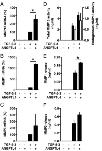
appropri-ate to examine the role of this protein in the present study. We first tested whether ANGPTL4 behaves as an inducer of chon-drogenic differentiation. MSCs were cultured in micromass in the presence of recombinant human ANGPTL4 instead of TGF--3 for 21 days. In this setting, recombinant ANGPTL4 did not induce expression of any chondrogenic markers as shown by qPCR analyses (Figs. 3, A–D). However, when exog-enous ANGPTL4 was added in the course of TGF--3-induced differentiation, expression at day 21 of aggrecan (ACAN),␣-1 type II collagen, transcript variant 2 (COL2A1), HAPLN1 (hya-luronan and proteoglycan link protein 1) and␣-1 type X colla-gen (COL10A1) was inhibited (Fig. 3, A–D). Accordingly, accu-mulation of aggrecan and of total soluble collagens was decreased (Fig. 3, E and F). In contrast, cell number as measured by DNA quantification was not significantly affected (Fig. 3G), whereas gene expression of MMP13 and MMP1 was super-FIGURE 2. Culture conditions required for induction of ANGPTL4
expres-sion. Monolayer cultures of MSCs were subjected to normoxic (21% O2) or
hypoxic (2% O2) conditions in the absence or presence of TGF--3. A, relative
ANGPTL4 mRNA levels were determined by qPCR. B, the amounts of ANGPTL4 in culture supernatants were measured by ELISA. C, cells were cultured in micromass in the absence or presence of TGF--3. Expression of ANGPTL4 was analyzed by qPCR. *, p⬍ 0.05.
induced (Fig. 4, A and B). MMP13 activity and protein level of MMP1 were increased in corresponding culture superna-tants (Fig. 4, D and E). However, the effect on MMP13 activ-ity and on MMP3 expression did not reach statistical signif-icance (Fig. 4, C, D, and F).
ANGPTL4 siRNA Knockdown Stimulates the Formation of a Mature Cartilaginous Matrix by Differentiating MSCs—We next analyzed the role of endogenous ANGPTL4 produced dur-ing TGF--3-induced chondrogenesis using a RNA interfer-ence approach. Cells were transfected with an siRNA-targeting ANGPTL4 (siANGPTL4) or a control siRNA prior to initiation of differentiation. At day 2 of the differentiation process, the increase in ANGPTL4 mRNA abundance was inhibited by 85% (Fig. 5A), and the level of the corresponding protein in culture supernatants was also markedly decreased (Fig. 5, B and C). Western blots performed with antibodies recognizing the C-terminal region of ANGPTL4 detected native ANGPTL4 as well as the C-terminal processed form containing the fibrino-gen-like domain. Isoforms a and b could not be clearly distin-guished because of their small difference in size (Fig. 5B). Extinction of ANGPTL4 protein expression was maintained throughout the whole differentiation period, ranging from 88% at day 2 to 57% at day 21, as shown by ELISA (Fig. 5C). The RNA
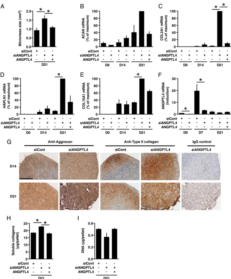
interference approach had a macroscopically visible effect because pellets of siANGPTL4-transfected cells were of greater size than those of control cells, suggesting an increase in the accumulation of extracellular matrix components (Fig. 6A). Culture with recombinant ANGPTL4 significantly inhibited siANGPTL4-induced increase in micromass size (Fig. 6A) but did not affect the micromass size of control siRNA-transfected or untransfected MSCs (data not shown). Quantitative PCR analyses revealed that, in siANGPTL4-transfected cells, expres-sion of the cartilage matrix components aggrecan, COL2A1, HAPLN1, and COL10A1 was superinduced (Fig. 6, B–E). Effi-ciency of ANGPTL4 siRNA knockdown was confirmed in these experiments (Fig. 6F). Immunohistochemistry analyses revealed the presence of chondrocytes appearing as cells surrounded by lacunae in samples transfected with either control siRNA or siANGPTL4 (Fig. 6G). However, aggrecan and type II collagen were more abundant in micromasses of cells transfected with siANGPTL4 (Fig. 6G). Similarly, the amount of total soluble collagens was higher after silencing ANGPTL4 expression (Fig. 6G), whereas DNA content was not significantly altered, indi-cating that the increased accumulation of these matrix constit-uents was not due to an increase in cell number (Fig. 6I). In contrast, expression of MMP13, MMP1, and MMP3 was inhib-FIGURE 3. Exogenous ANGPTL4 inhibits expression of cartilaginous matrix components by differentiating MSCs. Pellets of MSCs were cultured for 21 days in the presence or absence of TGF--3 or ANGPTL4 as indicated. A–D, relative mRNA levels of aggrecan (ACAN), COL2A1 (␣-1 type II collagen, transcript variant 2), HAPLN1 (hyaluronan and proteoglycan link protein 1), and␣-1 type X collagen (COL10A1) were measured by qPCR. Level of mRNA in TGF--3-treated cells was given the nominal value 100%. E, pellets were sectioned and analyzed by immunohistochemistry using aggrecan antibodies. Nonimmune IgG served as negative control to check for specific staining. Representative illustrations are shown. Scale bar, 300m. F, acid and pepsin-soluble collagens were quantified by colorimetry. G, the amount of DNA was determined by a fluorimetric assay. *, p⬍ 0.05.
ited after transfection with siANGPTL4 (Figs. 7, A–C). Analysis of culture supernatants indicated that these cells produced much less MMP13, MMP1, and MMP3 than those transfected with the con-trol siRNA (Figs. 7, D–F). Recombinant ANGPTL4 partially res-cued the effect of siANGPTL4 on micromass size, total soluble collagens, and on expression of aggrecan, COL2A1, HAPLN1, COL10A1, MMP1, and MMP13 (Figs. 6, A–E and H, and 7A). These data ruled out an off-target effect of siANGPTL4.
Blocking Antibody to Integrin␣V5 Stimulates the Forma-tion of Cartilaginous Matrix by Differentiating MSCs—Integrin ␣V5 is one of the transmembrane receptors for ANGPTL4 (24, 25). Quantitative PCR showed that the genes encoding both chains forming this integrin, namely ITGAV and ITGB5, were expressed in undifferentiated MSCs (mean Ct⫽ 28.20 ⫾ 1.53 cycles and 26.83⫾ 1.07 cycles, respectively, compared with
Ct⫽ 24.96 ⫾ 0.40 cycles for the housekeeping gene RPS9) and
throughout TGF--3-induced chondrogenesis in a relative constant manner (data not shown). To gain insight into its implication in cartilaginous matrix remodeling, we examined the effects of an integrin␣V5 blocking antibody added during the differentiation process. We observed that micromasses of
cells treated with the integrin␣V5 antibody were of greater size than those of control cells (1.164 ⫾ 0.073 mm3 versus
0.527⫾ 0.052 mm3, p⬍ 0.05) (Fig. 8A). Expression of aggrecan
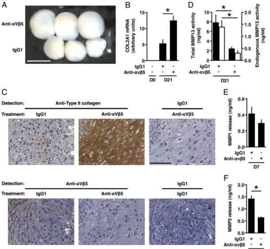
and MMP1 remained unaffected by treatment with the integrin ␣V5 antibody (data not shown). Nevertheless, type II collagen was more abundantly expressed in micromasses treated with the blocking antibody as shown by qPCR and immunohisto-chemistry analyses (Fig. 8, B and C). In contrast, integrin ␣V5 was no longer detected, suggesting that it was degraded and/or down-regulated after recognition by the antibody (Fig. 8C). Analysis of culture supernatants indi-cated that cells cultured with the integrin antibody released significantly less MMP13 and MMP3 than those treated with control IgG1, whereas the level of MMP1 remained unaf-fected (Fig. 8, D–F), in agreement with mRNA expression data (data not shown).
FIGURE 4. Exogenous ANGPTL4 up-regulates expression of MMPs. Pellets of MSCs were cultured for 21 days in the presence or absence of TGF--3 or ANGPTL4 as indicated. A–C, relative mRNA levels of MMPs were determined by qPCR. Level of mRNA in TGF--3-treated cells was given the nominal value 100%. D, activity corresponding to activatable pro-MMP13 and endogenous active MMP13 (black bars) or endogenous active MMP13 only (white bars) was measured in culture supernatants using a fluorimetric assay. E and F, the amounts of MMP1 and MMP3 released in culture supernatants were quanti-fied by ELISA. *, p⬍ 0.05.
FIGURE 5. Extinction of ANGPTL4 expression by RNA interference. MSCs were transfected with negative control siRNA (siCont) or ANGPTL4-targeted siRNA (siANGPTL4) and induced into chondrogenesis. A, expression of ANGPTL4 was measured by qPCR at various time points after induction of differentiation as indicated. Level of ANGPTL4 mRNA in control siRNA-transfected cells at day 2 (D2) of differentiation was given the nominal value 100%. B, culture superna-tants at day 2 of differentiation were analyzed by Western blotting using an antibody to ANGPTL4. Full-length ANGPTL4 and the C-terminal fragment containing the fibrinogen-like domain (FLD) are indicated. C, concentration of ANGPTL4 in supernatants collected at the indicated days of differentiation was determined by ELISA. *, p⬍ 0.05.
FIGURE 6. ANGPTL4 siRNA knockdown increases expression of cartilaginous matrix components by differentiating MSCs. MSCs were transfected with negative control siRNA (siCont) or ANGPTL4-targeted siRNA (siANGPTL4) and induced into chondrogenesis in the absence or presence of recombinant ANGPTL4 as indicated. A, the volume of the micromasses was calculated by considering that these have the shape of ellipsoids. B–E, relative mRNA levels of aggrecan (ACAN), COL2A1 (␣-1 type II collagen, transcript variant 2), HAPLN1 (hyaluronan and proteoglycan link protein 1), and ␣-1 type X collagen (COL10A1) were determined by qPCR and represented as percentage of maximum. F, expression of ANGPTL4 was measured by qPCR and represented as induction over value at day 0 (D0). G, pellets were analyzed by immunohistochemistry at days 14 and 21 with antibodies to aggrecan and type II collagen as indicated. Nonimmune IgG served as negative control to check for specific staining. Scale bar, 200m. H, acid and pepsin-soluble collagens were quantified by colorimetry. I, the amount of DNA was determined by a fluorimetric assay. *, p⬍ 0.05.
DISCUSSION
Owing to their capacity to differentiate into chondrocytes, human MSCs are increasingly considered for cartilage engi-neering. Usual differentiation protocols, however, lead to ter-minal differentiation, a stage at which the cartilage matrix is remodeled, especially by MMP13. This process is reminiscent of what happens in vivo at the growth plate and at the junction of cartilage with subchondral bone, where the cartilage matrix is pro-gressively replaced by bone (6, 7). Terminal chondrogenic differ-entiation should therefore be avoided for cartilage repair. In this study, we identified ANGPTL4 as the factor responsible, at least partly, for the induction of MMP13 during chondrogenic differen-tiation of human MSCs. We focused our interest on ANGPTL4 because it singled out as one of the top up-regulated gene encoding a secreted factor in transcriptomic analysis of MSCs undergoing TGF--3 or BMP-2-induced chondrogenesis.
Using recombinant ANGPTL4 and a RNA interference approach targeting this factor, we show that extracellular matrix deposition of type II collagen and aggrecan, two major components of the cartilage, is markedly decreased by ANGPTL4 during chondrogenic differentiation. We provide evidence that this regulation occurs not only through repres-sion of type II collagen and aggrecan expresrepres-sion but also through induction of MMP13, MMP1, and MMP3 synthesis. Indeed, both MMP13 and MMP1 are known to digest type II collagen and aggrecan (8 –10, 12), whereas MMP3 activates the precursor form of these MMPs (11). The possible involvement of ADAMTS aggrecanases in the degradation of aggrecan dur-ing chondrogenesis was unlikely because no aggrecanase ity was detected in any medium samples using a sensitive activ-ity assay (data not shown). Our results are consistent with those obtained by others who detected MMP-generated aggrecan neoeptitope but not ADAMTS-generated neoeptitope in cell pellets during chondrogenic differentiation of MSCs (26). Nev-ertheless, a current model of aggrecan proteolysis proposes that MMP-mediated aggrecanolysis is not responsible for the release of osmotically active aggrecan from the cartilage (Ref. 27 and references therein). Therefore, the ANGPTL4-induced reduction in aggrecan content observed here may primarily be the result of the decrease in aggrecan gene expression. In addi-tion, ANGPTL4 may have indirectly enhanced diffusional release of intact or MMP-cleaved aggrecan by disrupting the type II collagen network. The amount of soluble endogenous ANGPTL4 released by differentiating MSCs reached⬃ 94 nM, a
value similar to or above reported affinities for integrin sub-units1 and 5 (24, 25). Importantly, the total amount of ANGPTL4 produced by MSCs is likely to be even greater than that found in the supernatant alone because this protein was shown to be also retained in the extracellular matrix in a bio-logically active form bound to heparan sulfate proteoglycans, vitronectin, or fibronectin (28, 29). This may explain why we obtained only a partial effect or no detectable effect on some parameters with the recombinant ANGPTL4 at 100 nM.
We show here that ANGPTL4 triggers matrix remodeling during in vitro chondrogenesis to diminish the prochondro-genic effect of TGF--3. Interestingly, a previous report has shown that ANGPTL4 enhances the expression of MMP1 and MMP3 in chondrocytes (30). In MSCs undergoing chondro-genic differentiation, we observed that endogenous ANGPTL4 up-regulated expression of MMP13 as well as that of MMP1 and MMP3. However, induction of the latter MMPs occurred at an earlier time point than that of MMP13, which is in agree-ment with the time-course of ANGPTL4 expression during chondrogenesis.
MMP13 plays a key role in physiological tissue remodeling at the growth plate and in the pathological destruction of the car-tilage as evidenced by analyses of clinical samples of osteoar-thritis and rheumatoid arosteoar-thritis (31–33), phenotypes of trans-genic and knock-out mice (34, 35), and the identification of the mutation causing the Missouri type of human spondyloepime-taphyseal dysplasia genetic disorder (36). Analysis of MMP1 and MMP3 levels in synovial fluid or serum of rheumatoid arthritis patients argues for their involvement in pathological joint destruction (37, 38). We describe here a regulatory path-FIGURE 7. ANGPTL4 siRNA knockdown inhibits expression of MMP13,
MMP1, and MMP3 in MSCs undergoing chondrogenic differentiation.
MSCs were transfected with negative control siRNA (siCont) or ANGPTL4-tar-geted siRNA (siANGPTL4) and induced into chondrogenesis in the absence or presence of recombinant ANGPTL4 as indicated. A–C, relative mRNA levels of MMP13, MMP1, and MMP3 were measured by qPCR and represented as per-centage of maximum. D, MMP13 activity corresponding to activatable and endogenously active MMP13 was quantified in culture supernatants using a fluorimetric assay. E and F, the amounts of MMP1 and MMP3 released in cul-ture supernatants were measured by ELISA. *, p⬍ 0.05. D0, day 0.
way in which ANGPTL4 lies upstream of MMP1, MMP3, and MMP13, suggesting a role for this cytokine in osteoarticular diseases. In agreement with such role, others have reported high expression of ANGPTL4 in synovial tissue at an early stage in murine collagen-induced arthritis, a condition characterized by cartilage resorption (39). Also, transcriptomic data obtained by Geyer and colleagues have shown that ANGPTL4 is up-reg-ulated in damaged cartilage of a subset of osteoarthritic patients (see supplementary data in Ref. 40). In addition, ANGPTL4 was shown to stimulate osteoclast-mediated bone resorption (41).
Interestingly, we obtained similar results by down-regulating ANGPTL4 expression through RNA interference and by block-ing one of its receptor, namely integrin␣V5 (24). Indeed, a diminished release of MMP3 and MMP13 and an increased accumulation of cartilaginous matrix and type 2 collagen was observed after treatment with an integrin␣V5 neutralizing antibody. However, MMP1 and aggrecan expression remained unaffected. These data suggest that integrin␣V5 partly medi-ates ANGPTL4 signaling in MSCs undergoing chondrogenic differentiation. Together, our results suggest the existence of an autocrine/paracrine regulatory pathway initiated in MSCs
dur-ing TGF--induced chondrogenesis whereby ANGPTL4 dif-ferentially regulate key matrix components and MMPs to trigger destruction of cartilage matrix. Because of the delay observed in the present study between the onset of ANGPTL4 up-regulation and some of its biological effects, we cannot exclude that ANGPTL4 may act in concert with other factors or receptors appearing at a later stage of the differentiation pro-cess. Nevertheless, in the perspective of MSC-based cartilage engineering, inhibiting ANGPTL4 expression or action could help to stabilize cartilage formation.
Acknowledgments—Histological procedures and immunohistological analyses were performed using the facilities at the “Réseau d’Histologie Expérimentale de Montpellier” and the “Plate-forme Régionale d’Imagerie du Languedoc Roussillon”. We thank Gilles Carnac for help in setting up the hypoxia experiment.
REFERENCES
1. Burrage, P. S., Mix, K. S., and Brinckerhoff, C. E. (2006) Matrix metallo-proteinases: role in arthritis. Front Biosci. 11, 529 –543
2. Caplan, A. I. (2009) Why are MSCs therapeutic? New data: new insight. FIGURE 8. Blocking antibody to integrin␣V5 stimulates the formation of a mature cartilaginous matrix by differentiating MSCs. MSCs were induced into chondrogenesis in the presence of an integrin␣V5-neutralizing antibody or control IgG1 as indicated. A, pellets were photographed at day 21 of differentiation using a camera linked to magnifying binocular glasses. Top pellets, integrin␣V5 antibody-treated cells; bottom pellets, control IgG1-treated cells. Scale bar, 1 mm. B, relative mRNA level of COL2A1 (␣-1 type II collagen, transcript variant 2) was determined by qPCR. C, pellets were analyzed by immunohistochemistry at day 21 with antibodies to type II collagen or integrin␣V5 as indicated. Nonimmune IgG1 served as control to check for nonspecific staining. Scale bar, 100m. D, culture supernatants were collected at day 21 (D21) of differentiation to measure activity corresponding to activatable pro-MMP13 and endogenous active pro-MMP13 (black bars) or endogenous active pro-MMP13 only (white bars). E and F, culture supernatants were collected at day 7 (D7) of differentiation to measure release of MMP1 and MMP3 by ELISA. *, p⬍ 0.05.
J. Pathol. 217,318 –324
3. Jorgensen, C., and Noël, D. (2011) Mesenchymal stem cells in osteoarticu-lar diseases. Regen. Med. 6, 44 –51
4. Pittenger, M. F., Mackay, A. M., Beck, S. C., Jaiswal, R. K., Douglas, R., Mosca, J. D., Moorman, M. A., Simonetti, D. W., Craig, S., and Marshak, D. R. (1999) Multilineage potential of adult human mesenchymal stem cells. Science 284, 143–147
5. Poole, A. R., Kojima, T., Yasuda, T., Mwale, F., Kobayashi, M., and Laverty, S. (2001) Composition and structure of articular cartilage: a template for tissue repair. Clin. Orthop. Relat. Res. 391, S26 –33
6. Pelttari, K., Winter, A., Steck, E., Goetzke, K., Hennig, T., Ochs, B. G., Aigner, T., and Richter, W. (2006) Premature induction of hypertrophy during in vitro chondrogenesis of human mesenchymal stem cells corre-lates with calcification and vascular invasion after ectopic transplantation in SCID mice. Arthritis Rheum. 54, 3254 –3266
7. Mueller, M. B., and Tuan, R. S. (2008) Functional characterization of hy-pertrophy in chondrogenesis of human mesenchymal stem cells. Arthritis
Rheum. 58,1377–1388
8. Knäuper, V., López-Otin, C., Smith, B., Knight, G., and Murphy, G. (1996) Biochemical characterization of human collagenase-3. J. Biol. Chem. 271, 1544 –1550
9. Fosang, A. J., Last, K., Knäuper, V., Murphy, G., and Neame, P. J. (1996) Degradation of cartilage aggrecan by collagenase-3 (MMP-13). FEBS Lett.
380,17–20
10. Deng, S. J., Bickett, D. M., Mitchell, J. L., Lambert, M. H., Blackburn, R. K., Carter, H. L., 3rd, Neugebauer, J., Pahel, G., Weiner, M. P., and Moss, M. L. (2000) Substrate specificity of human collagenase 3 assessed using a phage-displayed peptide library. J. Biol. Chem. 275, 31422–31427 11. McCawley, L. J., and Matrisian, L. M. (2001) Matrix metalloproteinases:
they’re not just for matrix anymore! Curr. Opin. Cell Biol. 13, 534 –540 12. Imai, K., Dalal, S. S., Hambor, J., Mitchell, P., Okada, Y., Horton, W. C., and
D’Armiento, J. (2007) Bone growth retardation in mouse embryos ex-pressing human collagenase 1. Am. J. Physiol. Cell Physiol. 293, C1209 – C1215
13. Klein, T., and Bischoff, R. (2011) Physiology and pathophysiology of ma-trix metalloproteases. Amino Acids 41, 271–290
14. Djouad, F., Bony, C., Häupl, T., Uzé, G., Lahlou, N., Louis-Plence, P., Apparailly, F., Canovas, F., Rème, T., Sany, J., Jorgensen, C., and Noël, D. (2005) Transcriptional profiles discriminate bone marrow-derived and synovium-derived mesenchymal stem cells. Arthritis Res. Ther. 7, R1304 – R1315
15. Mrugala, D., Dossat, N., Ringe, J., Delorme, B., Coffy, A., Bony, C., Char-bord, P., Häupl, T., Daures, J. P., Noël, D., and Jorgensen, C. (2009) Gene expression profile of multipotent mesenchymal stromal cells: Identifica-tion of pathways common to TGFbeta3/BMP2-induced chondrogenesis.
Cloning Stem Cells 11,61–76
16. Chuchana, P., Marchand, D., Nugoli, M., Rodriguez, C., Molinari, N., and Garcia-Sanz, J. A. (2007) An adaptation of the LMS method to determine expression variations in profiling data. Nucleic Acids Res. 35, e71 17. Chuchana, P., Holzmuller, P., Vezilier, F., Berthier, D., Chantal, I., Severac,
D., Lemesre, J. L., Cuny, G., Nirdé, P., and Bucheton, B. (2010) Intertwin-ing threshold settIntertwin-ings, biological data and database knowledge to optimize the selection of differentially expressed genes from microarray. PLoS One
5,e13518
18. Rozen, S., and Skaletsky, H. J. (2000) Primer3 on the WWW for general users and for biologist programmers in Bioinformatics Methods and
Pro-tocols: Methods in Molecular Biology(Krawetz, S., and Misener, S., eds.) pp. 365–386, Humana Press, Totowa, NJ
19. Livak, K. J., and Schmittgen, T. D. (2001) Analysis of relative gene expres-sion data using real-time quantitative PCR and the 2(-⌬⌬CT) method. Methods 25,402– 408
20. Bouffi, C., Thomas, O., Bony, C., Giteau, A., Venier-Julienne, M. C., Jor-gensen, C., Montero-Menei, C., and Noël, D. (2010) The role of pharma-cologically active microcarriers releasing TGF-3 in cartilage formation
in vivoby mesenchymal stem cells. Biomaterials 31, 6485– 6493 21. Volkmer, E., Drosse, I., Otto, S., Stangelmayer, A., Stengele, M.,
Kallu-kalam, B. C., Mutschler, W., and Schieker, M. (2008) Hypoxia in static and dynamic 3D culture systems for tissue engineering of bone. Tissue Eng.
Part A 14,1331–1340
22. Belanger, A. J., Lu, H., Date, T., Liu, L. X., Vincent, K. A., Akita, G. Y., Cheng, S. H., Gregory, R. J., and Jiang, C. (2002) Hypoxia up-regulates expression of peroxisome proliferator-activated receptor␥ angiopoietin-related gene (PGAR) in cardiomyocytes: role of hypoxia inducible factor 1alpha. J. Mol. Cell Cardiol. 34, 765–774
23. Le Jan, S., Amy, C., Cazes, A., Monnot, C., Lamandé, N., Favier, J., Philippe, J., Sibony, M., Gasc, J. M., Corvol, P., and Germain, S. (2003) Angiopoietin-like 4 is a proangiogenic factor produced during ischemia and in conven-tional renal cell carcinoma. Am. J. Pathol. 162, 1521–1528
24. Goh, Y. Y., Pal, M., Chong, H. C., Zhu, P., Tan, M. J., Punugu, L., Lam, C. R., Yau, Y. H., Tan, C. K., Huang, R. L., Tan, S. M., Tang, M. B., Ding, J. L., Kersten, S., and Tan, N. S. (2010) Angiopoietin-like 4 interacts with integ-rins1 and 5 to modulate keratinocyte migration. Am. J. Pathol. 177, 2791–2803
25. Zhu, P., Tan, M. J., Huang, R. L., Tan, C. K., Chong, H. C., Pal, M., Lam, C. R., Boukamp, P., Pan, J. Y., Tan, S. H., Kersten, S., Li, H. Y., Ding, J. L., and Tan, N. S. (2011) Angiopoietin-like 4 protein elevates the prosurvival intracellular O2(-):H2O2ratio and confers anoikis resistance to tumors. Cancer Cell 19,401– 415
26. Boeuf, S., Graf, F., Fischer, J., Moradi, B., Little, C. B., and Richter, W. (2012) Regulation of aggrecanases from the ADAMTS family and aggre-can neoepitope formation during in vitro chondrogenesis of human mes-enchymal stem cells. Eur. Cell Mater. 23, 320 –332
27. Sandy, J. D. (2006) A contentious issue finds some clarity: on the inde-pendent and complementary roles of aggrecanase activity and MMP ac-tivity in human joint aggrecanolysis. Osteoarthritis Cartilage 14, 95–100 28. Chomel, C., Cazes, A., Faye, C., Bignon, M., Gomez, E., Ardidie-Robouant,
C., Barret, A., Ricard-Blum, S., Muller, L., Germain, S., and Monnot, C. (2009) Interaction of the coiled-coil domain with glycosaminoglycans protects angiopoietin-like 4 from proteolysis and regulates its antiangio-genic activity. Faseb J. 23, 940 –949
29. Goh, Y. Y., Pal, M., Chong, H. C., Zhu, P., Tan, M. J., Punugu, L., Tan, C. K., Huang, R. L., Sze, S. K., Tang, M. B., Ding, J. L., Kersten, S., and Tan, N. S. (2010) Angiopoietin-like 4 interacts with matrix proteins to modulate wound healing. J. Biol. Chem. 285, 32999 –33009
30. Murata, M., Yudo, K., Nakamura, H., Chiba, J., Okamoto, K., Suematsu, N., Nishioka, K., Beppu, M., Inoue, K., Kato, T., and Masuko, K. (2009) Hypoxia upregulates the expression of angiopoietin-like-4 in human ar-ticular chondrocytes: role of angiopoietin-like-4 in the expression of ma-trix metalloproteinases and cartilage degradation. J. Orthop. Res. 27, 50 –57
31. Mitchell, P. G., Magna, H. A., Reeves, L. M., Lopresti-Morrow, L. L., Yo-cum, S. A., Rosner, P. J., Geoghegan, K. F., and Hambor, J. E. (1996) Clon-ing, expression, and type II collagenolytic activity of matrix metalloprotei-nase-13 from human osteoarthritic cartilage. J. Clin. Invest. 97, 761–768 32. Ståhle-Bäckdahl, M., Sandstedt, B., Bruce, K., Lindahl, A., Jiménez, M. G.,
Vega, J. A., and López-Otín, C. (1997) Collagenase-3 (MMP-13) is ex-pressed during human fetal ossification and re-exex-pressed in postnatal bone remodeling and in rheumatoid arthritis. Lab. Invest. 76, 717–728 33. Shlopov, B. V., Lie, W. R., Mainardi, C. L., Cole, A. A., Chubinskaya, S., and
Hasty, K. A. (1997) Osteoarthritic lesions: involvement of three different collagenases. Arthritis Rheum. 40, 2065–2074
34. Neuhold, L. A., Killar, L., Zhao, W., Sung, M. L., Warner, L., Kulik, J., Turner, J., Wu, W., Billinghurst, C., Meijers, T., Poole, A. R., Babij, P., and DeGennaro, L. J. (2001) Postnatal expression in hyaline cartilage of con-stitutively active human collagenase-3 (MMP-13) induces osteoarthritis in mice. J. Clin. Invest. 107, 35– 44
35. Little, C. B., Barai, A., Burkhardt, D., Smith, S. M., Fosang, A. J., Werb, Z., Shah, M., and Thompson, E. W. (2009) Matrix metalloproteinase 13-de-ficient mice are resistant to osteoarthritic cartilage erosion but not chon-drocyte hypertrophy or osteophyte development. Arthritis Rheum. 60, 3723–3733
36. Kennedy, A. M., Inada, M., Krane, S. M., Christie, P. T., Harding, B., López-Otín, C., Sánchez, L. M., Pannett, A. A., Dearlove, A., Hartley, C., Byrne, M. H., Reed, A. A., Nesbit, M. A., Whyte, M. P., and Thakker, R. V. (2005) MMP13 mutation causes spondyloepimetaphyseal dysplasia, Mis-souri type (SEMD(MO). J. Clin. Invest. 115, 2832–2842
37. Peake, N. J., Khawaja, K., Myers, A., Jones, D., Cawston, T. E., Rowan, A. D., and Foster, H. E. (2005) Levels of matrix metalloproteinase (MMP)-1 in paired sera and synovial fluids of juvenile idiopathic arthritis patients: rela-tionship to inflammatory activity, MMP-3 and tissue inhibitor of metallopro-teinases-1 in a longitudinal study. Rheumatology 44, 1383–1389
38. Green, M. J., Gough, A. K., Devlin, J., Smith, J., Astin, P., Taylor, D., and Emery, P. (2003) Serum MMP-3 and MMP-1 and progression of joint damage in early rheumatoid arthritis. Rheumatology 42, 83– 88 39. Hermann, L. M., Pinkerton, M., Jennings, K., Yang, L., Grom, A., Sowders,
D., Kersten, S., Witte, D. P., Hirsch, R., and Thornton, S. (2005)
Angiopoi-etin-like-4 is a potential angiogenic mediator in arthritis. Clin. Immunol.
115,93–101
40. Geyer, M., Grässel, S., Straub, R. H., Schett, G., Dinser, R., Grifka, J., Gay, S., Neumann, E., and Müller-Ladner, U. (2009) Differential transcriptome analysis of intraarticular lesional vs intact cartilage reveals new candidate genes in osteoarthritis pathophysiology. Osteoarthritis Cartilage 17, 328 –335
41. Knowles, H. J., Cleton-Jansen, A. M., Korsching, E., and Athanasou, N. A. (2010) Hypoxia-inducible factor regulates osteoclast-mediated bone re-sorption: role of angiopoietin-like 4. FASEB J. 24, 4648 – 4659