HAL Id: pasteur-03263221
https://hal-pasteur.archives-ouvertes.fr/pasteur-03263221
Submitted on 17 Jun 2021
HAL is a multi-disciplinary open access
archive for the deposit and dissemination of sci-entific research documents, whether they are pub-lished or not. The documents may come from teaching and research institutions in France or abroad, or from public or private research centers.
L’archive ouverte pluridisciplinaire HAL, est destinée au dépôt et à la diffusion de documents scientifiques de niveau recherche, publiés ou non, émanant des établissements d’enseignement et de recherche français ou étrangers, des laboratoires publics ou privés.
epithelial cells for persistent infection
Chak Hon Luk, Camila Valenzuela, Magdalena Gil, Léa Swistak, Perrine
Bomme, Yuen-Yan Chang, Adeline Mallet, Jost Enninga
To cite this version:
Chak Hon Luk, Camila Valenzuela, Magdalena Gil, Léa Swistak, Perrine Bomme, et al.. Salmonella enters a dormant state within human epithelial cells for persistent infection. PLoS Pathogens, Public Library of Science, 2021, 17 (4), pp.e1009550. �10.1371/journal.ppat.1009550�. �pasteur-03263221�
human epithelial cells for persistent infection
Chak Hon LukID1,2, Camila ValenzuelaID1☯, Magdalena GilID1☯, Le´a SwistakID1,2☯,Perrine Bomme3, Yuen-Yan ChangID1, Adeline MalletID3, Jost EnningaID1,2* 1 Dynamics of Host-Pathogen Interactions Unit and UMR3691 CNRS, Institut Pasteur, Paris, France, 2 Universite´ de Paris, Sorbonne Paris Cite´, Paris, France, 3 Ultrastructural Bioimaging UTechS, C2RT, Institut Pasteur, Paris, France
☯These authors contributed equally to this work.
Abstract
Salmonella Typhimurium (S. Typhimurium) is an enteric bacterium capable of invading a
wide range of hosts, including rodents and humans. It targets different host cell types show-ing different intracellular lifestyles. S. Typhimurium colonizes different intracellular niches and is able to either actively divide at various rates or remain dormant to persist. A compre-hensive tool to determine these distinct S. Typhimurium lifestyles remains lacking. Here we developed a novel fluorescent reporter, Salmonella INtracellular Analyzer (SINA), compati-ble for fluorescence microscopy and flow cytometry in single-bacterium level quantification. This identified a S. Typhimurium subpopulation in infected epithelial cells that exhibits a unique phenotype in comparison to the previously documented vacuolar or cytosolic S. Typhimurium. This subpopulation entered a dormant state in a vesicular compartment dis-tinct from the conventional Salmonella-containing vacuoles (SCV) as well as the previously reported niche of dormant S. Typhimurium in macrophages. The dormant S. Typhimurium inside enterocytes were viable and expressed Salmonella Pathogenicity Island 2 (SPI-2) vir-ulence factors at later time points. We found that the formation of these dormant S. Typhi-murium is not triggered by the loss of SPI-2 effector secretion but it is regulated by (p) ppGpp-mediated stringent response through RelA and SpoT. We predict that intraepithelial dormant S. Typhimurium represents an important pathogen niche and provides an alterna-tive strategy for S. Typhimurium pathogenicity and its persistence.
Author summary
Salmonella Typhimurium is a clinically relevant bacterial pathogen that causes
Salmonel-losis. It can actively or passively invade various host cell types and reside in a
Salmonella-containing vacuole (SCV) within host cells. The SCV can be remodeled into a replicative niche with the aid ofSalmonella Type III Secretion System 2 (T3SS2) effectors or else, the
SCV is ruptured for the access of the nutrient-rich host cytosol. Depending on the infected host cell type,S. Typhimurium undertake different lifestyles that are distinct by their
sub-cellular localization, replication rate and metabolic rate. We present here a novel
a1111111111 a1111111111 a1111111111 a1111111111 a1111111111 OPEN ACCESS
Citation: Luk CH, Valenzuela C, Gil M, Swistak L, Bomme P, Chang Y-Y, et al. (2021) Salmonella enters a dormant state within human epithelial cells for persistent infection. PLoS Pathog 17(4): e1009550.https://doi.org/10.1371/journal. ppat.1009550
Editor: Jean Celli, Washington State University, UNITED STATES
Received: November 17, 2020 Accepted: April 8, 2021 Published: April 30, 2021
Peer Review History: PLOS recognizes the benefits of transparency in the peer review process; therefore, we enable the publication of all of the content of peer review and author responses alongside final, published articles. The editorial history of this article is available here:
https://doi.org/10.1371/journal.ppat.1009550
Copyright:© 2021 Luk et al. This is an open access article distributed under the terms of theCreative Commons Attribution License, which permits unrestricted use, distribution, and reproduction in any medium, provided the original author and source are credited.
Data Availability Statement: All relevant data are within the manuscript and itsSupporting Informationfiles.
fluorescent reporter system that rapidly detectsS. Typhimurium lifestyles using
fluores-cence microscopy and flow cytometry. We identified a dormantS. Typhimurium
popula-tion within enterocyte that displays capacities in host cell persistence, dormancy exit and antibiotic tolerance. We deciphered the (p)ppGpp stringent response pathway that sup-pressesS. Typhimurium dormancy in enterocytes while promoting dormancy in
macro-phages, pinpointing a divergent physiological consequence regulated by the same set ofS.
Typhimurium molecular mediators. Altogether, our work demonstrated the potential of fluorescent reporters in facile bacterial characterization, and revealed a dormantS.
Typhi-murium population in human enterocytes that are phenotypically distinct from that observed in macrophages and fibroblasts.
Introduction
Salmonella enterica serovar Typhimurium (S. Typhimurium) is an enteric bacterium that
closely associates with global food-borne illnesses. The prevalence ofS. Typhimurium has
placed a severe burden on the global food and healthcare industry, leading to millions of cases, hundreds of casualties and costing billions of dollars per annum [1,2].S. Typhimurium resides
in different natural reservoirs and is transmitted to humans through contaminated food. Upon arrival in the human intestine after ingestion, a portion of the luminalS.
Typhimur-ium expresses the Type III Secretion System 1 (T3SS1) encoded withinSalmonella
Pathogenic-ity Island 1 (SPI-1) and its cognate effectors to induce its active entry into non-phagocytic epithelial cells.S. Typhimurium also targets other cell types, such as fibroblasts and
macro-phages. During these events, it induces local tissue injuries and eventually breaches the intesti-nal barrier to reach the lamina propria and tissue-resident immune cells. Then,S.
Typhimurium is carried by macrophages to mesenteric lymph nodes and eventually to the liver and spleen for persistent infection [3].
Within enterocytesS. Typhimurium are encapsulated in an endocytic compartment coined Salmonella-containing vacuole (SCV) that matures by acidification within the first hours of
internalization [4]. The reducing pH and changing osmolarity of the SCV induce the shut-down of T3SS1 and expression of a second T3SS, T3SS2, fromSalmonella Pathogenicity Island
2 (SPI-2) [5,6]. The T3SS2 effectors remodel the SCV into a viable niche forS. Typhimurium
replication [5,6]. Default maturation of the SCV is marked by the sequential acquisition and removal of endocytic trafficking markers, such as the small GTPases RAB5 and RAB7 as well as Lysosome-associated membrane glycoprotein 1 (LAMP1) [7]. During these events, the SCV dynamically interacts with surrounding macropinosomes, which controls SCV stability [8,9]. Consequently,S. Typhimurium can reside either in a remodeled SCV or disrupt the SCV to
access the host cytosol. Overall,S. Typhimurium exhibits distinct replication rates and specific
metabolic profiles within the different intracellular niches adapting to nutrient availability and the specific microenvironments [8–10].
Differential lifestyles are also known forS. Typhimurium infecting other cell types. In
fibro-blasts, the SCV associates with the aggrephagy machinery that either clears the infection or allowsS. Typhimurium to putatively persist in the cell [11,12]. In macrophages,S.
Typhimur-ium expresses the T3SS2 to remodel the SCV immediately after bacterial entry, or the patho-gen adopts a dormant behavior mediated by toxin-antitoxin (TA) system [13,14].
Antibiotic persistence and relapse ofS. Typhimurium infection due to the failure of
bacte-rial eradication through antibiotic treatment has been tied toS. Typhimurium dormancy,
which is distinct from bacterial persistence that refers to incompletely cleared infections by the
Funding: This research was supported by fellowships from Croucher Foundation (HK) and Fondation pour la Recherche Me´dicale (FRM) to C. H.L. and Y.Y.C.. C.H.L. is part of the Pasteur - Paris University (PPU) International PhD Program. J.E. is supported by the ERC-CoG “Endosubvert”. The Enninga lab is part of the LabEx IBEID and Milieu Interieure. AM and PB are supported for equipment from the French Government Programme Investissements d’Avenir France BioImaging (FBI, N˚ ANR-10-INSB-04-01) and are also members of the LabEx IBEID. The funders had no role in study design, data collection and analysis, decision to publish, or preparation of the manuscript. Competing interests: The authors have declared that no competing interests exist.
immune system. Numerous antibiotics target major active machineries, including DNA repli-cation, transcription and translation of extracellular bacteria, therefore dormant intracellular pathogens appear to be less or not susceptible to such treatments [15].S. Typhimurium
dor-mancy and antibiotics persistence have been reported in macrophages to be regulated by the Guanosine pentaphosphate (ppGpp) stringent response pathway. The two (p)ppGpp synthases RelA and SpoT control the bacterial (p)ppGpp level, which regulates the activity of the ATP-dependent protease Lon to degrade the antitoxin and release the toxin TacT for arresting pro-tein translation [14,16]. The arrest of translation by TacT leads to a halt of bacterial growth giv-ing rise to the insensitivity and tolerance towards antibiotics [16]. Despite reports ofS.
Typhimurium antibiotics persistence in the epithelium and lamina propria of the mouse intes-tine, it is not clear whether this involves dormant bacteria [17].
With ourSalmonella INtracellular Analyzer (SINA) system, we precisely depicted the
intra-cellular bacterial lifestyles at the single bacterium level, identifying a novelS. Typhimurium
population within enterocytes that is dormant. Dormant persisters within enterocytes are localized in a unique vacuolar compartment different from the one described in macrophages. We found that T3SS2 effector secretion and the Lon protease are dispensable for this newS.
Typhimurium population, while the bifunctional enzyme SpoT and monofunctional enzyme RelA negatively regulateS. Typhimurium dormancy in epithelial cells.
Results
Development of a multiplex fluorescent reporter series, the
Salmonella
INtracellular Analyser (SINA) to distinguish different intracellular
S.
Typhimurium lifestyles
The distinct intracellular lifestyles ofS. Typhimurium upon invasion of epithelial cells have
been described either with regard to the specific pathogen localization or with regard to the bacterial growth dynamics. To date, fluorescent reporters are available to identifyS.
Typhi-murium within vacuolar and cytosolic localizations; while the others measure the replication rate of the pathogen [18–20]. However, these localization and replication-rate reporters have not been coupled, as it has been generally assumed that the bacterial localization determines its replication rate. This notion has been challenged by different reports, for example on the dif-ferent growth rates ofS. Typhimurium within the cytosol depending on the targeting by
autop-hagy [21–25]. A combined reporter system would enable a comprehensive elucidation of the intracellular lifestyle of a given intracellular pathogen. Therefore, we developed a novel fluores-cent reporter series, theSalmonella INtracellular Analyzer (SINA). Our SINA1.1 reporter is
composed of two separated modules to indicate the bacterial localization and replication rate. The localization module consists of two transcription reporters driven by localization-specific promoters, while the replication rate module carries a constitutively expressed fluorescent timer (Figs1AandS1). At the molecular level, the localization module is composed of vacuolar (Vac) and cytosolic (Cyt) submodules, which utilize two characterized promoters, PssaGand
PuhpTto drive the expression of tagBFP and smURFP, respectively [18,19]. We confirmed the
functionality of the fluorescent PssaGand PuhpTreporters for our experimental setup duringS.
Typhimurium invasion of epithelial cells using a digitonin assay assayed by flow cytometry (S2
andS3Figs). The replication rate module encodes Timerbac, a DsRed mutant (S197T), which has been previously employed to differentiateS. Typhimurium subpopulations by their
repli-cation rates [20]. The emission spectrum of Timerbacshifts from green to red as it matures, which reflects the bacterial metabolic activity (change in slope,Fig 1Btop) as well as the repli-cation rate (unvarying slope, varying green:red ratio,Fig 1Bbottom) [26]. When Timerbacis constitutively expressed, a metabolically activeS. Typhimurium bacterium emits both green
and red signals resulting from immature green and mature red fluorophores. In case such a bacterium experiences a metabolic halt, it eventually emits only red signals, due to the matura-tion of the existing green fluorophores in concert with ceasedde novo synthesis of the green
fluorophores. With SINA, we are able to simultaneously collect information on these replica-tion rate changes ofS. Typhimurium and its localization at single bacterium resolution, which
enables a comprehensive and quantitative reflection ofS. Typhimurium physiology inside an
infected host.
To validate the functionality of our SINA system duringS. Typhimurium invasion of
epi-thelial cells, we employed fluorescence microscopy and flow cytometry analysis (Fig 1C). With fluorescence microscopy, we observed intracellularS. Typhimurium simultaneously emitting
both green and red signals (Timerbac), but not Vac (PssaG-tagBFP) and Cyt (PuhpT-smURFP)
signals at 1 hour post-infection (pi) (Fig 1D). As these bacteria committed to vacuolar or cyto-solic lifestyles at 6 hours pi, we observed thatS. Typhimurium in cells with <10 bacteria
emit-ted Vac signal during this time course. On the other hand, we observed a mixed population of
S. Typhimurium in cells with >10 bacteria, where clusters of Cyt+S. Typhimurium of low
Timerbacsignals (arrow) and individual Vac+S. Typhimurium (arrowhead) were detected. In
the cells containing mixedS. Typhimurium populations, bacteria were either Vac+or Cyt+but not double positive, showing the presence of two populations with distinct discernible lifestyles (Fig 1D). We were also able to track the onset of bacterial division and signal output from SINA by time-lapse microscopy (S1–S3Movies,S4B and S4C Fig).
We took advantage of the properties of our multiplex SINA reporter and devised a gating strategy to quantitatively analyze the bacterial lifestyles in singleS. Typhimurium-infected cells
using flow cytometry (S2 Fig). In brief, we first defined the infected cells by the size of the ana-lyzed events (under SSC-A vs FSC-A plot), followed by the positive signals in the Timer580vs Timer510plot (i.e. cells harboringS. Typhimurium). We then further classified the S.
Typhi-murium-infected cells into four sub-types according the signals of the localization module (tagBFP::SPI-2 vs smURFP::cytosolic plot), corresponding to cells with either vacuolar bacteria (Vac+Cyt-) or cytosolic bacteria (Vac-Cyt+) or cells with both vacuolar and cytosolic popula-tions (Vac+Cyt+) or cells harboringS. Typhimurium that express only basal levels of the Vac
and Cyt signals (S2 Fig). We observed that intracellularS. Typhimurium behaved as a
popula-tion with a homogenous replicapopula-tion rate and basal expression levels of Vac and Cyt at 1 hour pi (Fig 1E). At 6 hours pi, this homogenous population segregated into Vac+and Cyt+ subpop-ulations, with a Cyt+distribution similar to that reported in the literature (10–20% cytosolic) (Fig 1E) [21]. The gradual separation of these subpopulations could be detected with SINA1.1 throughout the course of infection (S4A Fig). As we backgated the infected cells, we observed cells harboring only vacuolarS. Typhimurium (Vac+Cyt-) and cells with both vacuolar and
Fig 1. SINA enables precise determination of the differentSalmonella intracellular lifestyles in human epithelial cells. (A) Schematic diagram of the construction of subcellular localization and replication rate modules of SINA1.1. The subcellular localization module is composed of the vacuolar submodule (PssaG-tagBFP) and cytosolic submodule (PuhpT
-smURFP), while the replication rate module is composed of a constitutively expressed Timerbac(P
ybaJ-Timerbac) (B) (Top)
Schematic diagram of the emission spectrum shifts ofS. Typhimurium harboring Timerbacas Timerbacmatures, where emission shifts from green to red (Bottom) Green:Red ratio increases with elevatingS. Typhimurium replication rates. As S.
Typhimurium divides, both Timer510and Timer580fluorophores are diluted. With a higher production rate of Timer510than
Timer580, fast dividing
S. Typhimurium exhibits a higher Green:Red ratio. (C) Expected output by SINA as S. Typhimurium
dwells in distinct subcellular localizations. VacuolarS. Typhimurium are of lower replication rate (i.e. lower Green:Red ratio)
and are expected to emit blue fluorescence; cytosolicS. Typhimurium are of higher replication rate (i.e. higher Green:Red
ratio) and are expected to emit far red fluorescence (D) HeLa cells infected byS. Typhimurium harboring SINA1.1. Output of
SINA from intracellularS. Typhimurium was detected by fluorescence microscopy at 1 h pi, vacuolar (arrowhead) and
cytosolic (arrow)S. Typhimurium at 6 h pi. (3 independent experiments). Scale bars are 10 μm. (E) HeLa cells infected by S.
Typhimurium harboring SINA1.1. Output of SINA from intracellularS. Typhimurium at 1 h and 6 h pi was detected by flow
cytometry (3 independent experiments).
cytosolic bacteria (Vac+Cyt+), each forming a distinct population of different Green:Red ratio on the plot of Timer510against Timer580(S5A Fig). Together, this demonstrated that our novel SINA1.1 reporter is capable of simultaneously and quantitatively distinguishing theS.
Typhi-murium lifestyles by their subcellular localization and replication rate at both single infected cell and single bacterium level using flow cytometry and fluorescence microscopy. The combi-nation of the SINA reporter with flow cytometry fosters higher throughput analysis ofS.
Typhimurium lifestyles in infected cells as compared to microscopy, extending the possibility for rapid screening.
A novel dormant
S. Typhimurium subpopulation in human epithelial cell
With the SINA1.1 reporter, we used the SPI-2 expression module to distinguish vacuolarS.
Typhimurium from cytosolic bacteria. In the plot of the localization module, we identified an easily discernable population (~5–10%) of infected epithelial cells harboring Vac-Cyt-S.
Typhimurium detectable as early as 2 hours pi, which became apparent at 6 hours pi (Figs2A
andS6). We backgated the Vac-Cyt-population, and we extracted physical parameters from the Timerbacplot. This revealed that the Vac-Cyt-S. Typhimurium exhibit a similar replication
rate (S5A Fig) but a reduced metabolic activity (S5B Fig) compared to Vac+Cyt-S.
Typhimur-ium as depicted by the green:red ratio and slope of Timerbacplot, respectively. The capacities of Timerbacin the measurement of the bacterial replication rate and metabolism have been well-elaborated in previous applications [20,27]. This Vac-Cyt-S. Typhimurium population
was also visualized using live microscopy to confirm their presence using different detection approaches (S3 Movie). This was intriguing as metabolically inactiveS. Typhimurium have
not been reported in enterocytes so far. We thus infected polarized intestinal epithelial Caco-2 monolayers, and confirmed the presence of the Vac-Cyt-subpopulation with a shifted meta-bolic profile in a cellular model system for intestinal infections (S7 Fig). We also performed control infections in 3T3 (fibroblast model) and differentiated THP-1 cells (macrophage model) to test the sensitivity of SINA1.1 in these relevant cell types. As within epithelial cells, we also observed distinctS. Typhimurium populations in the macrophage and fibroblast
mod-els as described before (S8andS9Figs). To determine the intracellular localization of Vac-Cyt
-S. Typhimurium, we further performed correlative light and electron microscopy (CLEM) by
serial section transmission electron microscopy (TEM), and a digitonin assay demonstrating that this subpopulation is localized in a host vesicular compartment (Figs2B,S4A–S4Fand
S10) [21]. Together, these results showed the presence of a novel intracellularS. Typhimurium
population within epithelial cells that exhibits a lowered metabolic rate and resides in a host vesicular compartment, implicating a putative dormant phenotypic variant.
IntracellularS. Typhimurium encounters a number of stresses upon uptake into host cells,
including oxidative, pH and osmotic stress, which serve as key signals to trigger transcription reprogramming for the adaptation of an intra-host environment [28]. As the intracellular microenvironment andS. Typhimurium dormancy has been studied in some detail in
macro-phages, we decided to focus our comparison on this cell type in relation to the newly identified
S. Typhimurium subpopulation in epithelial cells. During macrophage infections the SCV
microenvironment drives a portion ofS. Typhimurium into a dormant state that contributes
to the elevation of antimicrobial persistence and polarization of infected macrophage [14,29]. We asked whether Vac-Cyt-S. Typhimurium shares similar physiologies with dormant S.
Typhimurium inside macrophages, hence we determined the metabolic state of the Vac-Cyt -population. By replacing the cytosolic submodule of SINA1.1 with an arabinose inducible cas-sette to generate SINA1.5, we measuredS. Typhimurium’s capacity to respond to arabinose
Typhimurium during the infection process (Fig 2C). The response of intracellular bacteria towards extracellular arabinose induction has been reported previously to characterize the metabolic state of macrophage-borne dormantS. Typhimurium [13]. With SINA1.5, we observed that approximately half of the Vac-S. Typhimurium did not respond to arabinose
induction at designated time intervals (Fig 2D). As the Vac-Cyt-bacterial niche may show lim-ited arabinose accessibility, we corroborated our observations monitoring the reduced metab-olism of this bacterial population via the signals of the Timerbacreporter (S5B Fig). These data allowed us to propose that the Vac-Cyt-S. Typhimurium adopts a dormant state (coined as
dormantS. Typhimurium hereafter) upon their internalization into epithelial cells.
Dormant
S. Typhimurium resides in a unique vesicular compartment
We set out to determine whether the dormantS. Typhimurium localization is distinct from
conventional SCVs. To determine this by immunofluorescence staining, we simplified SINA1.1 to SINA1.4 to free the red and far-red channels for indirect immunofluorescence staining of selected endocytic markers (Fig 2C). LAMP1 labels host lysosomes as well as the matured SCV, which is also present on the SCV of dormantS. Typhimurium in macrophages
[7,13]. By fluorescence microscopy, we only observed minor recruitment of LAMP1 to the proximity of dormantS. Typhimurium within epithelial cells, in contrast to the high LAMP1
incidence proximal to Vac+S. Typhimurium (Fig 2E and 2F). We also determined if dor-mantS. Typhimurium is localized in a SCV experiencing a halt in SCV biogenesis, therefore
we tested a number of known early SCV markers (namely RAB5, RAB7 and RAB11). We did not observe the recruitment of any of these early SCV markers to the dormantS.
Typhimur-ium (Fig 2E and 2F). This was intriguing as our previous ultrastructural and digitonin inves-tigations clearly documented the dormantS. Typhimurium bacteria within a
membrane-bound compartment. We also addressed if the dormant population is targeted by host autop-hagy, analyzing the localization of the autophagy marker LC3. We did not detect any locali-zation of LC3 proximal to the majority of the dormantS. Typhimurium (Fig 2E and 2F). Therefore, we conclude that dormantS. Typhimurium are localized within a unique
mem-brane-bound compartment distinct from the conventional SCV and that of dormantS.
Typhimurium in macrophages, suggesting such dormancy formation is heavily governed by endocytic trafficking [13,30]. This compartment requires further characterization in future studies.
Fig 2.S. Typhimurium displays a novel inactive intracellular lifestyle in epithelial cells. (A) (Left and Middle) Timerbacprofile and distribution of single cells with no infection (black), infected cells with inactive bacteria (Vac-Cyt-) (red), infected cells with only vacuolar bacteria (Vac+Cyt-) (blue) and infected cells with both vacuolar and cytosolic populations (Vac+Cyt+) (green) at 6 h pi. (Right) Abundance of
S. Typhimurium-infected cells (Vac-Cyt-, Vac+Cyt
-and Vac+Cyt+) as illustrated in (A) (n = 3). (B) (Left) Brightfield and fluorescent microcopy (FLM) images of infected HeLa cells harboring Vac-Cyt
-S.
Typhimurium at 6 h pi. (Right) Serial sections of TEM images of Vac-Cyt-S. Typhimurium, arrowhead indicates host membrane structures of the SCV. (C)
Schematic illustration for the constructions of SINA derivatives, SINA1.4 and SINA1.5. SINA1.4 was used for immunofluorescence staining against RAB5, RAB7, RAB11, LAMP1 and LC3; SINA1.5 was used for arabinose induction assay. (D) (Top) Responsiveness of intracellularS. Typhimurium towards an
arabinose pulse between 5–6 h pi, uninduced control (black); arabinose-induced (red). (Bottom) Quantification on the responsiveness of Vac
-S.
Typhimurium pulsed at different time intervals during the infection time course, dormant (black), inducible (maroon). Samples were all harvested at 6 h pi. (n = 3) (E) HeLa cells were infected with SINA1.4-harboringS. Typhimurium, harvested at 6 h pi, fixed and stained. Quantification of the presence of RAB5,
RAB7, BAB11, LAMP1 and LC3 proximal to Vac-and Vac+
S. Typhimurium at 6 h pi. (n = 3) (F) Representative images of Vac
-S. Typhimurium (arrow)
quantified in (D);S. Typhimurium (green), Vac-(blue), RAB5, RAB7, RAB11, LAMP1 and LC3 (grey), Phalloidin (red). (G) Designated populations of
infected HeLa cells were enriched by cell sorting and plated for CFU. Quantification of CFU from dormantS. Typhimurium at 6 h, 24 h and 168 h pi and
Vac+Cyt-S. Typhimurium at 6 h pi. (n = 5 for Vac+Cyt-6 h, Dormant 6 h, 24 h; n = 3 for Dormant 168 h) (H) Quantification of SPI-2 activity using flow cytometry in enriched dormantS. Typhimurium at 6 h and enriched Vac-Cyt-infected cells re-plated until 24 h pi. (n = 3) (I) Survival percentage of
dormant and Vac+Cyt-intracellular
S. Typhimurium against 3 h of CIP treatment, infected cells were harvested at 6 h pi, enriched by cell sorting and plated
for CFU. (n = 4) At least a total of 1000 events of infected cells were analyzed by flow cytometry or 50 infected cells by microscopy in each experiment replicates. The bars represent the mean, statistics were performed using unpaired t test (��p<0.01).
Dormant
S. Typhimurium are viable, cultivable, resume metabolism and
express virulence genes in host cells
Endocytic vesicles are either recycled or undergo fusion with the lysosomes for degradation. The same fate also applies to vacuolarS. Typhimurium, where SPI-2 deficient strains have a
reduced survival capacity compared to SPI-2 competent strains [31]. We collected the infected cells harboring dormantS. Typhimurium by cell sorting at >90% purity and plated them for
colony forming unit (CFU) measurement, a classical approach to determine the viability of the intracellularS. Typhimurium. We observed that dormant S. Typhimurium are viable and
cul-tivable (Fig 2G), contrasting to the viable but not cultivable nature of dormantS.
Typhimur-ium in murine macrophages [13,14]. To determine the fate of the dormantS. Typhimurium,
we enriched and plated the viable infected cells harboring dormantS. Typhimurium, and
monitored the bacterial behavior at 24 hours pi (S11 Fig). We observed that 50% of the dor-mantS. Typhimurium in infected cells collected at 6 hours pi became metabolically active and
expressed SPI-2 at 24 hours pi, as demonstrated by the population shift in the Timerbacplot, and becoming Vac+(Fig 2H). To determine if the dormantS. Typhimurium persists in the
host, we further enriched infected cells harboring dormantS. Typhimurium and monitored
the presence and the viability of dormantS. Typhimurium at 7 days pi. The dormant S.
Typhi-murium were found to persist in cells and remained viable and cultivable over the whole period of 7 days (Fig 2G, “168h”). To further address whether the dormant intracellularS.
Typhimurium phenotype confers a reduced sensitivity towards antibiotics, we supplemented ciprofloxacin (CIP) to the infected cell at 3 hours pi and determined the viability of dormantS.
Typhimurium by CFU. We selected thein cellulo CIP supplementation to avoid artificial
per-turbations on theS. Typhimurium dormant status. Also, in cellulo treatment addresses the
kill-ing efficiency of dormantS. Typhimurium within the SCV, where subcellular distribution of
this drug has been demonstrated to influence the bactericidal efficacy [32]. We observed a higher survival rate of dormantS. Typhimurium as compared to vacuolar S. Typhimurium
(Fig 2I), similar to the observations made in the murine intestine [17]. These results demon-strated that dormantS. Typhimurium are viable, exhibit a delayed expression of SPI-2, persist
in the epithelial host cells for up to 7 days and are less susceptible to antibiotics. Such unique metabolic and virulence reprogramming could serve as a strategic step for intestine-borneS.
Typhimurium to prolong gut inflammation for community benefits and reservoir for relapse [33].
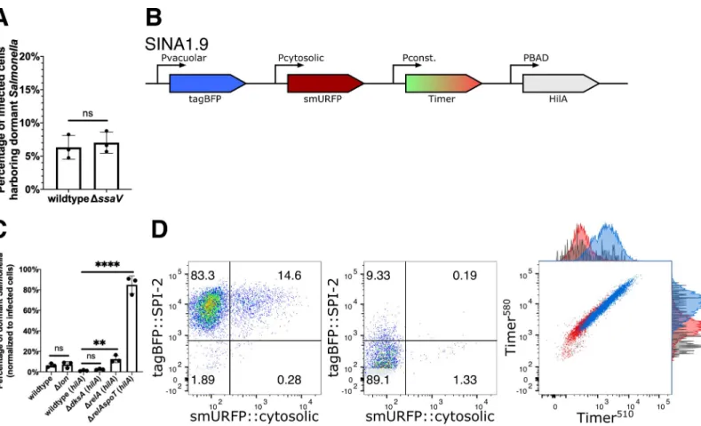
S. Typhimurium dormancy is not a result of the loss of T3SS2 effector
secretion
We then studied whether the lack of T3SS2 effector secretion drivesS. Typhimurium
dor-mancy in epithelial cells. Using our SINA1.1 reporter in theS. Typhimurium SPI-2 secretion
deficient mutantΔssaV [34], we observed no significant difference in the proportion of Vac --Cyt-S. Typhimurium between wild type and the ΔssaV mutant (Fig 3A). Altogether, the for-mation of dormantS. Typhimurium is not a consequence caused by the lack of T3SS2 effector
secretion during the infection of epithelial cells.
S. Typhimurium dormancy is regulated by (p)ppGpp biogenesis
Class II toxin-antitoxin (TA) systems regulate the dormancy formation of non-pathogenicE. coli in laboratory conditions [35]. TA systems are comprised of a toxin and an antitoxin that counter-balances the toxin to regulate bacterial physiology, including growth arrest. A major TA system involves the stringent response mediated by the monofunctional (p)ppGpp
synthases RelA and bifunctional (p)ppGpp synthases SpoT, after which (p)ppGpp binds to DksA to mediate transcription reprogramming for bacterial adaptation. The surge in (p) ppGpp levels also activates the ATP-dependent Lon protease to degrade Type II antitoxins to release the free toxins [36–38]. In recent reports, stringent response has been associated with slow growingS. Typhimurium populations, and TA systems are implicated in S. Typhimurium
dormancy in macrophages [14,16]. Therefore, we assessed the links between the stringent response andS. Typhimurium dormancy in epithelial cells, studying the mutant strains (i) ΔrelA ((p)ppGpp synthase), (ii) ΔrelAΔspoT ((p)ppGpp synthases), (iii) ΔdksA
((p)ppGpp-binding transcription regulator) and (iv)Δlon (protease targeting antitoxin). With the Δlon mutant, we did not observe any difference in the level of dormantS. Typhimurium population
in infected cells (~5–10%), suggesting that Lon protease is dispensable forS. Typhimurium
dormancy in epithelial cells (Fig 3C). AsΔrelAspoT and ΔdksA were reported to suffer reduced invasiveness in epithelial cells due to the reduced SPI-1 expression, we thus constructed SINA1.9 (Fig 3B), a derivative of SINA1.1 with an additional cassette for an inducible expres-sion ofhilA to compensate the reduced invasiveness of the mutants (S12 Fig) following a
Fig 3.S. Typhimurium dormancy is negatively regulated by SpoT. (A) HeLa cells were infected with SINA1.1-harboring S. Typhimurium, the abundance of Vac-Cyt -population in wild type and SPI-2 mutantΔssaV infected cells were quantified with flow cytometry at 6 h pi. (n = 3) (B) Schematic diagram for the construction of SINA derivative, SINA1.9, yielded from the introduction of an arabinose-induciblehilA expression cassette into SINA1.1. SINA1.9 was used to rescue the reduced invasiveness
ofΔdksA, ΔrelA and ΔrelAspoT mutant strains. (C) HeLa cells were infected with SINA1.1 or SINA1.9-harboring S. Typhimurium, the abundance of Vac-Cyt-population
in (p)ppGpp biogenesis and regulon mutants,Δlon, ΔdksA, ΔrelA and ΔrelAspoT were quantified by flow cytometry at 6 h pi (n = 3) (D) Distribution of Vac-Cyt-, Vac+Cyt-and Vac+Cyt+populations inhilA-expressing wild type (Left) and ΔrelAspoT mutant (Middle) infected HeLa cells at 6 h pi quantified by flow cytometry. Overlay
Timerbacprofile (Right) of Vac-Cyt-(red) and Vac+Cyt-(blue) populations of wild type and Vac-Cyt-population ofΔrelAspoT mutant (grey) in infected HeLa cells
quantified by flow cytometry at 6 h pi. (3 independent experiments) At least a total of 1000 events of infected cells were analyzed by flow cytometry in triplicate experiments. Statistics were performed using unpaired t test. ns: not significant (P > 0.05),��P < 0.01,����P < 0.0001.
previously published experimental strategy [39]. With the SINA1.9-complemented mutant strains, we obtained rescued invasiveness as compared to wild typeS. Typhimurium. This
allowed us to address the requirement of (p)ppGpp biogenesis and (p)ppGpp-regulated tran-scription forS. Typhimurium persistence. A significant increase in the Vac
-Cyt-population was observed inΔrelAspoT, whereas the increment was less pronounced in the ΔrelA single mutant and was indifferent inΔdksA mutant, when comparing with the wild type strain (Fig 3C and 3D, Left and Middle panel). As SpoT has been reported to regulate SPI-2 expression [40], we further confirmed that the Vac-Cyt-population ofΔrelAspoT shared a comparable metabolic profile as the one observed in the wild type strain (Fig 3D, Right panel). Together, these results suggested that (p)ppGpp stringent response mediated by SpoT but not RelA is required to restrict dormancy entry ofS. Typhimurium within epithelial cells independent of
the DksA regulon, while SPI-2 effector expression and secretion and Lon protease are dispensable.
Discussion
S. Typhimurium has been reported to survive in different host cells by adopting distinctive
metabolic profiles, subcellular localizations and replication rates, which has also been proposed to account for various clinical complications. Herein, we report a dormant population ofS.
Typhimurium residing in a unique vesicular compartment in epithelial cells of the intestine. These dormant epithelialS. Typhimurium persist within host cells for a prolonged period. The
SINA reporter system was instrumental for the discovery of enterocyte-borne dormantS.
Typhimurium as it allowed the simultaneous depiction of the metabolism, subcellular localiza-tion and replicalocaliza-tion rate of the intracellular bacteria. The compatibility of the SINA system with microscopy and flow cytometry offers the opportunity for multi-omics analysis as well as high-throughput genetic and chemical screenings on genes and compounds that influence the bacterial pathophysiology.
The dormantS. Typhimurium, while remaining viable in the absence of SPI-2 expression,
reside in a unique vesicular compartment distinct from the RAB5, RAB7, RAB11 or LAM-P1-labelled SCV or the LC3-positive autophagosomes. Upon endocytosis, endosomes are either recycled or matured and eventually degraded via fusion with lysosomes. The SCV shares such a fate if T3SS2 effectors are not secreted to hijack the vesicular maturation pathway [34]. Therefore, we propose that dormantS. Typhimurium reside in a vesicular compartment idle
to endocytic trafficking pathways that are independent of T3SS2 effectors. Such a diversion from default endocytic pathways is also observed by other bacterial pathogens, such asShigella,
and despite decades of research, no strong molecular markers have been identified for the short-livedShigella containing vacuole [41]. The subsequent resumption of metabolism and SPI-2 expression potentially serve as a signal to reengage the dormant membrane-enclosedS.
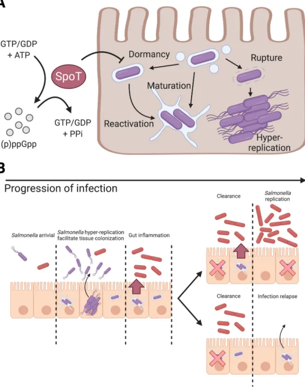
Typhimurium with endocytic trafficking pathways for remodeling the SCV into the conven-tional replicative niche (Fig 4A). The persistence of dormantS. Typhimurium in host cell for
up to at least 7 days in our tested condition is also striking as the bacteria inside this vacuolar compartment are likely to have restricted access to the extracellular nutrients.
S. Typhimurium has been reported previously to enter dormant or persistent states within a
modified SCV from a range of host cell types, including macrophages and fibroblasts [12,13]. Considering the presumably identicalS. Typhimurium dormancy observed across the
differ-ent cell models, there are substantial distinctions among the targeted host cell types in terms of the detection approaches and bacterial physiology. The firstS. Typhimurium
antibiotics-toler-ant persisters were identified in macrophages using a dilution reporter on non-replicatingS.
Fig 4. Schematic illustration of the role of (p)ppGpp alarmone pathway onS. Typhimurium dormancy in enterocytes and the proposed pathophysiological implication ofS. Typhimurium dormancy in enterocytes. (A) Schematic diagram of S.
Typhimurium lifestyles and the regulatory role of SpoT onS. Typhimurium dormancy in human epithelial cells. S. Typhimurium
can opt for three distinct lifestyles: cytosolic, vacuolar and dormant, which exhibits discernible subcellular localization, replication rate and metabolism. The entry of dormant state is negatively regulated by (p)ppGpp synthatase SpoT, while the regulatory mechanism on the dormancy exit remains to be determined. (B) Schematic diagram ofS. Typhimurium infection progression in
the gut epithelium. AsS. Typhimurium reaches the intestinal epithelium, a portion of S. Typhimurium expresses T3SS1 (purple) to
enter host cells and adopts various intracellular lifestyles. DistinctS. Typhimurium lifestyles support rapid tissue colonization and
gut inflammation to increase competitiveness of luminalS. Typhimurium (red). (Top) Reactivation of dormant S. Typhimurium
leads to prolonged gut inflammation that supports the continuous growth ofS. Typhimurium at gut lumen. (Bottom) Dormant S.
Typhimurium reactivates after the eradication of gutS. Typhimurium, which serves as the reservoirs of infection relapse.
The dormancy is regulated by the TA system toxin, TacT that halts protein translation and induces antibiotic persistence, where theS. Typhimurium subsequently exits dormancy and
activates SPI-2 [16,29]. Slow growingS. Typhimurium were also identified in fibroblasts as
early as 2003, where 0.001% of the bacteria survived until the end of the studied time course [12]. Studies in fibroblasts have deciphered major genetic determinants of bacterial persis-tence, however the underlying mechanisms of persistence with regards to bacterial viability, antibiotics, the pathological implication have been characterized in more depth in macro-phages recently [11,42]. In the different infected host cells intracellularS. Typhimurium
acti-vates SPI-2 to remodel the SCV for replication and interacts with endocytic trafficking pathways. In fibroblasts, the SCV subsequently interacts with host aggrephagy, where the majority of theS. Typhimurium residing in the SCV are eradicated whereas the remaining S.
Typhimurium were proposed to persist [11]. In epithelial cells, the reportedS. Typhimurium
dormancy by us is distinct from that in fibroblast and macrophage. They are different in the commence of dormancy, the capacity to replicate and the SCV microenvironment [13]. The distinct niches of dormantS. Typhimurium may reflect cell-type specific vesicular trafficking,
for example SPI-2 expression level and SCV maturation in these different target cells is not identical (S4,S8andS9Figs). It could also be possible that the way of entry impacts the devel-opment of dormantS. Typhimurium. Epithelial dormant S. Typhimurium is independent of
Lon protease and is negatively regulated by SpoT, contrasting to that in macrophage that requires Lon protease, (p)ppGpp synthases RelA and SpoT [14,38]. The substantial difference between theS. Typhimurium dormancy sheds light on their potentially diverge
pathophysio-logical implications as well as the molecular cue and mechanism that signal the establishment and exit of dormancy. The extensive work on TA systems andS. Typhimurium physiology in
fibroblasts would serve as significant groundwork for the further studies of enterocyte-borne dormantS. Typhimurium [42]. It will be interesting to investigate why these regulatory mod-ules are differentially involved in the formation of dormantS. Typhimurium in the different
cell types, and whether the distinct niche impacts their expression and implication.
(p)ppGpp, is a bacterial alarmone that functions as a key regulator of bacterial physiology. The (p)ppGpp-mediated stringent response has been closely associated with antibiotics persis-tence via inhibition of protein synthesis and transcription reprogramming [43–45]. The persis-tentS. Typhimurium in macrophages is dependent on a (p)ppGpp-Lon protease-Class II TA
systems axis, where TacT leads to a halt in protein translation [16]. In non-pathogenicE. coli
andS. Typhimurium models, the loss of Lon and its downstream regulated TA systems leads
to a diminished antibiotics persister formation due to the inactivity of toxins [46]. In our find-ings, the dormant phenotype is negatively regulated by (p)ppGpp synthases SpoT and partially by RelA, but independent of DksA and Lon protease-mediated pathways (Fig 4A). Our finding contrasts the current understanding on the role of stringent response on bacterial persistence, where stringent response is activated by various stress signals and (p)ppGpp synthesis would act on its molecular target to achieve persistence. Therefore, we suggest that bifunctional SpoT is required while monofunctional RelA is dispensable inS. Typhimurium dormancy in
entero-cytes, which echoes the previous report on the requirement of SpoT but not RelA inS.
Typhi-murium invasion and colonization of anin vivo model [39]. The essence of SpoT but not RelA forS. Typhimurium dormancy could suggest that either or both the (p)ppGpp hydrolysis and
synthase function is required, orrelA is not expressed during the course of infection.
Consid-ering thatS. Typhimurium dormancy is independent of DksA and Lon, it implies that
dor-mancy is likely to be mediated by pathways independent of DksA transcription
reprogramming and Lon protease-mediated degradation. As RelA and SpoT function to con-vert GDP and GTP to (p)ppGpp, and SpoT hydrolyzes (p)ppGpp to give GTP/GDP and pyro-phosphate, an imbalance of RelA/SpoT activity upsets the bacterial energy status, which could
potentially act as a cue for dormancy. As dormantS. Typhimurium do not co-exist with S.
Typhimurium of other lifestyles (Fig 2F), the host cell status is also implied to serve a regula-tory role onS. Typhimurium dormancy.
With the traits we uncovered in the dormantS. Typhimurium within epithelial cells, this
population could represent the intestinal persister, given the close association between bacte-rial dormancy and antibiotic persistence. Besides the proposed antibiotic persistence and hori-zontal gene transfer, the physiological features of enterocyte-borne dormantS. Typhimurium
could also provide two plausible benefits toS. Typhimurium colonization of the host gut [47] (Fig 4B): 1) Dormancy and delayed expression of SPI-2 allowS. Typhimurium to evade cellular
immunity during early invasion and to provide a sustained and extended SPI-2 expression at tissue scale, whereS. Typhimurium reactivated from dormancy supports SPI-2 expression as
classic vacuolarS. Typhimurium is eradicated. The sustained SPI-2 expression fuels gut
inflammation to release electron acceptors forS. Typhimurium survival benefits in the gut
lumen [33]. 2) PersistentS. Typhimurium resided within the intestinal tissue serves as the
source of subsequent infection relapse or systemic spread, where the maximum duration of persistence and molecular cues for reactivation remain to be elucidated (Fig 4B).
Materials and methods
Mammalian cell culture
HeLa cervical adenocarcinoma cells, Caco-2 colorectal adenocarcinoma cells, 3T3 mouse fibroblasts and THP-1 acute monocytic leukemia cells were purchased from American Type Culture Collection (ATCC) and used within 20 passages of receipt. HeLa cells and 3T3 cells were cultured in Dulbecco’s Modified Eagle Medium (DMEM, high glucose, GlutaMAX Sup-plement, ThermoFisher) containing 10% (v/v) heat-inactivated fetal bovine serum (FBS, Sigma) and incubated at 37˚C with 5% CO2and 100% humidity. Caco-2 cells were cultured in
DMEM containing 10% FBS, 1% Non-essential amino acids (Gibco), 1% HEPES (Gibco), 1% Penicillin/Streptomycin (Gibco) and incubated at 37˚C with 5% CO2and 100% humidity.
THP-1 cells were cultured in RPMI-1640 medium (ThermoFisher) containing 10% FBS and incubated at 37˚C with 5% CO2and 100% humidity. HeLa and 3T3 cells were seeded in
12-well tissue-culture treated plates (Corning Costar) at a density of 9x10⁴cells/well 48 hours prior to infection. Caco-2 cells were polarized using Corning BioCoat Assay System (Corning) following manufacturer’s protocol. THP-1 cells were seeded in 12-well tissue-culture treated plates at a density of 9x10⁴cells/well 96 hours prior to infection, and differentiated in 50μg/ mL phorbol 12-myristate 13-acetate (PMA, Sigma) for 24 hours, and incubated in RPMI-1640 + 10% for 72 hours. For immunofluorescence staining, HeLa cells were seeded on UV-treated glass coverslips (Marienfeld) in 12-well plates 48 hours prior to infection. For cell sorting experiments, HeLa cells were seeded in 10 cm tissue-culture treated dishes (Corning Costar) at a density of 1.8x10⁶cells/well 48 hours prior to infection.
Bacterial strains
Bacterial strains and plasmids used in this study are listed inS1andS2Tables, respectively. All mutants were constructed using bacteriophageλ red recombinase system from parental stain
S. Typhimurium Typhimurium strain SL1344 using primers listed inS3 Table[48]. HA-tagged T3SS2 effector strains were generated by transducing JL129 with P22 phage lysate (a generous gift from Ste´phane Me´resse, Centre d’Immunologie de Marseille-Luminy, France). Bacteria were cultured in Lysogeny broth (LB) supplemented with appropriate antibiotics, where neces-sary (Ampicillin 100μg/mL; Kanamycin 50 μg/mL).
Plasmid construction
The replication rate module,Timerbacis a generous gift from Dr. Dirk Bumann (University of Basel, Switzerland) [20]. To construct the localization module,tagBFP was amplified from
pHRdSV40-NLS-dCas9-24xGCN4_v4-NLS-P2A-BFP-dWPRE using primers tagBFP_fw and tagBFP_rv, and replaced the GFP in pM973 to yield vacuolar module (pPssaG-tagBFP) [19,49].
For the cytosolic module (pPuhpT-smURFP), smURFP-HO-1 and uhpT promoters were
ampli-fied from pBADsmURFP-HO-1 (smURFP_fw and smURFP_rv) and S. Typhimurium gDNA
(uhpT_fw and uhpT_rv), respectively, and replaced thesfGFP and mxiE promoter in pTSAR1
[50,51]. The vacuolar (Vac_fw and tagBFP_rv) and cytosolic (uhpT_fw and Cyt_rv) modules were amplified and inserted intoEcoRV and SmaI sites, respectively, of pBlueScript II KS (+)
to generate pSINA-int. The localization module on pSINA-int was excised and inserted betweenSalI and SphI sites of pBR322 Timerbac
to yield pSINA1.1. pSINA1.4 was generated by replacingTimerbacwithGFP in pBR322 Timerbacand inserted the vacuolar module at theSalI
andSphI sites. pSINA1.5 was generated by inserting the amplified inducible smURFP cassette
(Ara_fw and Ara_rv) and vacuolar cassette between theSalI and EagI sites of pBR322
Time-rbac. pSINA1.7 was constructed by revertingTimerbactoDsRed by site-directed mutagenesis
using DsRed_fw and DsRed_rv. pBADhilA was generated by inserting the amplified hilA
(hilA_fw1 and hilA_rv1) between theBamHI and PmeI sites of pBAD smURFP-HO-1. The
induciblehilA cassette was amplified using primer hilA_fw2 and hilA_rv2 and inserted into
theEcoRV site of pSINA1.1 to generate pSINA1.9.
Bacterial infections
Bacteria strains were streaked from glycerol stock on LB agar plates with appropriate antibiot-ics 2 days prior to infection. Three bacterial colonies were picked for overnight culture in LB medium supplemented with 0.3 M NaCl with shaking at 37˚C. 150μL overnight culture was subculture in 3 mL LB + 0.3 M NaCl (1:20 dilution) with shaking at 37˚C for 3 h. For strains harboring pSINA1.9, 0.1% L-arabinose was supplemented to the subculture 1 h before harvest. Bacteria were harvested with centrifugation (1 mL, 6000 xg, 1 min, RT), washed once in 1 x
PBS and resuspended in DMEM with no FBS. HeLa cells were infected at a MOI of ~100 for 25 min at 37˚C. Extracellular bacteria were removed and washed with 1 x PBS (5X). Cells were then incubated in DMEM + 10% FBS for 1 h, washed with 1 x PBS (3X), incubated in DMEM + 10% FBS for 2 h, washed with 1 x PBS (3X) and then incubated in DMEM + 10% FBS supple-mented with 10μg/mL gentamicin for the remaining time course of the infection.
Flow cytometry
At designated time points, cells were washed with 1 x PBS (1X) and detached with 0.05% Tryp-sin for 5 min at 37˚C. Detached cells were mixed with equal volume of DMEM + 10% FBS, passed through 40μm strainer and collected by centrifugation (500 x g, 5 min, 4˚C). Cell pel-lets were dislodged and fixed in 4% PFA (15 min, RT). Fixed cells were washed with 1 x PBS (2X) and resuspended in 200μL 1 x PBS for further analysis. For digitonin permeabilization experiment, cells were permeabilized with 45μg/mL digitonin (1 min, RT) or 0.25% saponin (30 min, RT), then washed and stained with anti-S. Typhimurium primary antibody and
Alex-a488-conjugated goat anti-rabbit secondary antibody [21]. The fluorescence intensities of the samples were assayed with LSR Fortessa (BD) (tagBFP Ex: 405 nm Em: 450/50 nm; Timer510 Ex: 488 nm Em: 525/50 nm; Timer580Ex: 562 nm Em: 582/15 nm; smURFP Ex: 633 nm Em: 670/30 nm) and analyzed with FlowJo (v10.0.4). The recorded events were gated according to the strategy described (S2 Fig).
Cell sorting
At designated time points, cells were washed with 1 x PBS (1X) and detached with 0.05% Tryp-sin for 5 min at 37˚C. Detached cells were mixed with equal volume of DMEM + 10% FBS, passed through 40μm strainer and collected by centrifugation (500 x g, 5 min, 4˚C). Cells were washed with 1 x PBS (1X) and resuspended in DMEM + 10% FBS supplemented with 10μg/ mL gentamicin. SYTOX Green (ThermoFisher) was supplemented to differentiate dead cells when necessary. Cells were sorted with Aria III (BD) (tagBFP Ex: 405 nm Em: 450/50 nm; Timer510and SYTOX Green Ex: 488 nm Em: 530/30 nm; Timer580Ex: 561 nm Em: 586/15 nm; smURFP Ex: 633 nm Em: 660/20 nm) to collect uninfected cells, infected cells with dor-mant or SPI-2S. Typhimurium populations. The recorded events were gated according to the
strategy described (S2 Fig).
Immunofluorescence microscopy
Cells seeded on coverslips were washed with 1 x PBS (1X) and fixed in 4% PFA (8 min, RT). After washing with 1 x PBS (3X), cells were permeabilized and blocked in 1 x PBS, 20% FBS, 0.25% saponin (30 min, RT). Coverslips were washed with 1 x PBS (3X) and incubated with anti-RAB5, anti-RAB7, anti-RAB11, anti-LC3 or anti-LAMP1 primary antibodies and phal-loidin-rhodamine diluted in 1 x PBS, 2% FBS (60 min, RT), and then washed with 1 x PBS (3X) and incubated with Cy5-conjugated goat anti-rabbit secondary antibodies diluted in 1 x PBS, 2% FBS (60 min, RT). Stained coverslips were then washed with 1 x PBS (3X) and mounted on SuperFrost Plus microscope sides (Thermo Scientific) with ProLong Gold Anti-fade Mountant without DAPI (Invitrogen). Samples were imaged with Perkin Elmer Ultra-view confocal spinning disk microscope equipped with Volocity software and a 20X/1.3 NA air objective. Images were analyzed with FIJI (NIH) [52] and figures were prepared using Inkscape (v1.0.1).
Colony forming unit plating
Infected HeLa cells were enriched by cell sorting, where 1000 infected cells were sorted for each sample. The cells were then collected by centrifugation at 500 xg for 5 min, and
subse-quently lysed in 0.1% Triton X-100 for 5 min at room temperature. The lysed cells were then serially diluted and plated on LB agar plates with appropriate antibiotics.
Dormant
S. Typhimurium persistence assay
Infected HeLa cells harboring dormantS. Typhimurium were enriched by cell sorting using
the gate Vac-Cyt-, and plated on 12-wells plates in DMEM + 10% FBS + Gen10. The medium was replaced with fresh DMEM + 10% FBS + Gen10to avoid the growth ofS. Typhimurium
being released from dead cells. Cells were harvested at 24 h and 168 h pi for analysis and CFU plating.
Ciprofloxacin survival assay
A final concentration of 10μg/mL of ciprofloxacin (CIP) were supplemented to the cell culture medium of the infected cells at 3 h pi. The cells were harvested at 6 h pi for cell sorting and CFU plating. CIP was administered at 3 h pi, which offered sufficient time for the infected population to differentiate into Vac-Cyt-, Vac+Cyt-and Vac+Cyt+for downstream enrichment of Vac-Cyt-, Vac+Cyt-populations.
Serial sectioning transmission electron microscopy
At 6 h pi, infected HeLa cells harboring Vac-Cyt-S. Typhimurium were harvested by cell
sort-ing. Enriched cells were allowed to adhere on specific dishes (MatTek) pre-coated with 50 mg/ ml fibronectin (Sigma) for 3 hours. For EM sample preparation, the adhered cells were fixed with 4% PFA (EMS), 2.5% glutaraldehyde (Sigma-Aldrich) in 0.2 M HEPES for 1 hour at room temperature. Fixed samples were washed with 1 x PBS for three times, and position of interest were defined by fluorescent light microscopy. For further sample preparation, the fixed cells washed three times by the addition of fresh 0.1 M Caco buffer (pH 7.2) and post-fixed in 1% osmium (EMS) in 0.1 M Caco buffer (pH 7.2) enriched with 1.5% potassium ferro-cyanide (Sigma-Aldrich) for 1 h. After three washes in 0.1M Caco buffer, samples were incu-bated in 0.2% of Tannic acid in water for 30 min at room temperature. Samples are post-fixed for a second time in 1% of osmium for 1 h, washed with water and incubated in 2% uranyl ace-tate dissolved in 25% ethanol for 1 h. Samples were then gradually dehydrated in an ethanol (Sigma-Aldrich) series ranging from 50% to 100%. Samples were embedded in PolyBed812 resin (EMS), followed by polymerization for 48 h at 60˚C. The resin embedded samples were removed from the dishes after gentle heating.
For the correlative microscopy, the region of interest was determined thanks to landmarks printed below embedded samples. Embedded cells were sectioned with an ultramicrotome (Leica, UC7) with 70 nm thickness. Thin serial sections were collected on a single slot grid (Agar scientific). Serial sections were observed with a transmission electron microscope TEM Technai T12 (ThermoFisher) at 120kV.
Statistical analysis
Unless further specified in the figure legend, data were analyzed for statistical significance with a Mann-Whitney test using Prism 8.0 (GraphPad).P value of � 0.05 is considered statistically
significant.�P < 0.05,��P < 0.01,���P < 0.001,����P < 0.0001, ns: not significant/ P � 0.05.
Supporting information
S1 Table.S. Typhimurium strains used in this study.
(DOCX)
S2 Table. Plasmids used in this study.
(DOCX)
S3 Table. Primers used for molecular cloning in this study.
(DOCX)
S4 Table. Antibodies used in this study.
(DOCX)
S1 Fig. Construction strategy of SINA1.1. The vacuolar and cytosolic modules were first
individually tested with GFP (pM973 and puhpT-GFP), and then switched to tagBFP and smURFP, respectively. The vacuolar (PssaG-tagBFP) and cytosolic (PuhpT-smURFP) modules
were subsequently amplified and introduced into pBR322 TimerbacbetweenSphI and SalI sites
to yield SINA1.1. (TIF)
S2 Fig. Gating strategy of SINA1.1 reporter system. Analyzed events were first gated for
“Cells” on SSC-A vs FSC-A plot to remove cell debris. In the “Cells” events, “Uninfected” pop-ulation was gated by double-negative; “Infected” was gated by double-positive on Timer580vs
Timer510plot. To gate for the basal intensity of SINA1.1 at 1 h pi, four quadrants were drawn in the “Infected” events on tagBFP::SPI-2 vs smURFP::cytosolic plot, where the biological interpretations of the four quadrants were denoted in the bottom-right sketch.
(TIF)
S3 Fig. Localization modules indicate subcellular localization ofS. Typhimurium. (A)
Gat-ing strategy for applyGat-ing SINA1.7 for digitonin assay. HeLa cells were infected with SINA1.7--harboring wild typeS. Typhimurium, and harvested at 6 h pi for analysis by flow cytometry.
The events were first gated for “Cells” to remove cell debris and subsequently gated for “unin-fected” and “in“unin-fected” based on DsRed signal. The “in“unin-fected” events were subsequently gated for Vac-Cyt-, Vac+Cyt-and Vac+Cyt+on tagBFP::SPI-2 vs smURFP::cytosolic plot. The fluores-cence profiles FITC::S. Typhimurium (after immunostaining using S. Typhimurium
anti-body) of Vac-Cyt-, Vac+Cyt-and Vac+Cyt+and “uninfected” were plotted as overlay histograms. The gating strategy displays a positive control sample treated with saponin. (B) Schematic diagram for the constructions of the SINA derivative SINA1.7, where Timerbacwas replaced with DsRed as compared to SINA1.1. (C) Digitonin assay on SINA-1.7 harboring wild typeS. Typhimurium-infected HeLa cells at 6 h pi, signal intensities of uninfected (black),
Vac-Cyt-(red), Vac+Cyt-(blue) and Vac+Cyt+(green) populations immunostained against anti-S. Typhimurium. (D) Digitonin assay on SINA-1.7 harboring wild type S. Typhimurium
infected HeLa cells at 6 h pi, signal intensity of Vac-Cyt-population unpermeabilized (black, negative control), permeabilized with digitonin (red) and saponin (maroon, positive control). (E) Digitonin assay on SINA-1.7 harboring wild typeS. Typhimurium infected HeLa cells at 6
h pi, signal intensity of Vac+Cyt-population unpermeabilized (black, negative control), per-meabilized with digitonin (blue) and saponin (navy, positive control). (F) Digitonin assay on SINA-1.7 harboring wild typeS. Typhimurium infected HeLa cells at 6 h pi, signal intensity of
Vac+Cyt+population unpermeabilized (black, negative control), permeabilized with digitonin (green) and saponin (dark Green, positive control).
(TIF)
S4 Fig. SINA1.1 performance in HeLa cells at 2 h, 4 h and 6 h pi. HeLa cells were infected
with wild typeS. Typhimurium harboring SINA1.1. (A) Infected cells were harvested and
ana-lyzed at time intervals of 2 h, 4 h and 6 h pi. (Left) Timerbacprofile of total cells at 2 h (top), 4 h (middle) and 6 h (bottom) pi in HeLa cells. (Right) Fluorescence output of the localization module of infected cells at 2 h (top), 4 h (middle) and 6 h (bottom) pi in HeLa cells. (B-C) Time-lapse microscopic acquisition of theS. Typhimurium intracellular lifestyle.
Representa-tive images of SINA1.1 signal output of vacuolar (B) and cytosolic (C)S. Typhimurium. Scare
bars are 10μm. (TIF)
S5 Fig.S. Typhimurium exhibits distinct replication rates and metabolism in HeLa cells.
HeLa cells were infected with SINA1.1-harboringS. Typhimurium and harvested at 6 h pi for
analysis by flow cytometry. The three infected cell populations, Vac-Cyt-, Vac+Cyt-and Vac +-Cyt+on tagBFP::SPI-2 vs smURFP::cytosolic plot were backgated on Timer580vs Timer510 plot. Timer580and Timer510intensities were extracted from each event. (A) Quantification of Green:red ratio of Vac-Cyt-, Vac+Cyt-and Vac+Cyt+population in Timerbacplot at 6 h pi. Green:red ratios were calculated by dividing Timer510by Timer580values, and plotted against infected cell populations. (B) Quantification of the slope of the best-fitted line of Vac-Cyt-, Vac+Cyt-and Vac+Cyt+population in Timerbacplot at 6 h pi. For each population, a best-fitted line was plotted on the Timer580vs Timer510plot to extract the slopes for each infected cell populations. At least a total of 1000 events of infected cells were analyzed by flow cytometry in
triplicate experiments. The bars represent the mean value, unpaired t-tests were carried out,
�
P < 0.05,����P < 0.0001, ns: not significant.
(TIF)
S6 Fig. DormantS. Typhimurium are observed as early as 2 h pi in HeLa cells. HeLa cells
were infected with SINA1.1-harboringS. Typhimurium, and harvested at 1 h, 2 h and 3 h pi
for analysis by flow cytometry. The infected cells were gated and the fluorescence profiles of vacuolar submodule PssaG-tagBFP at 1 h (black), 2 h (red) and 3 h (blue) pi were plotted as
overlaying histograms. (TIF)
S7 Fig. Performance of SINA1.1 in Caco-2 cells. Polarized Caco-2 monolayers were infected
with SINA1.1-harboringS. Typhimurium and harvested at 1 h and 6 h pi for analysis by flow
cytometry. (Left) Timerbacprofile of Vac-Cyt-(red) and Vac+Cyt-(blue) populations and total cells (black) at 1 h (top) and 6 h (bottom) pi in Caco-2 cells. (Right) Distribution of Vac-Cyt -and Vac+Cyt-populations at 1 h (top) and 6 h (bottom) pi in polarized Caco-2 cells.
(TIF)
S8 Fig. Performance of SINA1.1 in 3T3 cells. 3T3 cells were infected with SINA1.1-harboring
S. Typhimurium and harvested at 1 h, 6 h and 24 h pi for analysis by flow cytometry. (Left)
Timerbacprofile of Vac-Cyt-(red), Vac+Cyt-(blue) and Vac+Cyt+ (green) populations and total cells (black) at 1 h (top), 6 h (middle) and 24 h (bottom) pi in 3T3 cells. (Right) Distribu-tion of Vac-Cyt-, Vac+Cyt+and Vac+Cyt+populations at 1 h (top), 6h (middle) and 24 h (bot-tom) pi in 3T3 cells.
(TIF)
S9 Fig. Performance of SINA1.1 in THP-1 cells. Differentiated THP-1 cells were infected
with SINA1.1-harboringS. Typhimurium and harvested at 1 h, 6 h and 24 h pi for analysis by
flow cytometry. (Left) Timerbacprofile of Vac-Cyt-(red) and Vac+Cyt-(blue) populations and total cells (black) at 1 h (top), 6 h (middle) and 24 h (bottom) pi in THP-1 cells. (Right) Distri-bution of Vac-Cyt-and Vac+Cyt-populations at 1 h (top), 6 h (middle) and 24 h (bottom) pi in differentiated THP-1 cells.
(TIF)
S10 Fig. Serial sectioning TEM determines the subcellular localization of Vac-Cyt-S. Typhimurium. (A) Brightfield and fluorescent microscopy image of region of interest on
Mat-Tek dish. (B) Brightfield and fluorescent microscopy image of cells of interest harboring Vac --Cyt-S. Typhimurium. (C) TEM image of cell of interest in labelled region from (B). (D)
Magnified TEM image of labelled region from (C). (TIF)
S11 Fig. Infected cells harboring dormantS. Typhimurium are viable. HeLa cells were
infected with SINA1.7 harboringS. Typhimurium, harvested at 6 h pi and stained with
SYTOX Green and analyzed by flow cytometry. The infected cells were gated and the fluores-cence profiles of SYTOX Green in uninfected cell (black), Vac-Cyt-(red), Vac+Cyt-(blue) and Vac+Cyt+(green) were plotted as offset histograms.
(TIF)
S12 Fig. Ectopic expression ofhilA rescues the loss of invasiveness. HeLa cells were infected
with variousS. Typhimurium strains and harvested at 6 h pi for flow cytometry analysis. The
losses of invasiveness inΔdksA and ΔrelAspoT mutants are rescued by ectopic expression of
cells were analyzed by flow cytometry in triplicate experiments. The bars represent the mean value, unpaired t-test was carried out,����P < 0.0001.
(TIF)
S1 Movie. Connected toFig 1D: Time-lapse microscopy shows the fluorescence signal out-put from SINA1.1 in Vac+Cyt-intracellularS. Typhimurium population. Brightfield and
fluorescence output of Timerbac, PssaGand PuhpTfrom SINA1.1-harboringS. Typhimurium
exhibiting Vac+Cyt-profile. Images were taken every 15 min starting from 1 h pi. (AVI). (AVI)
S2 Movie. Connected toFig 1D: Time-lapse microscopy shows the fluorescence signal out-put from SINA1.1 in Vac+Cyt-and Vac-Cyt+intracellularS. Typhimurium population.
Brightfield and fluorescence output of Timerbac, PssaGand PuhpTfrom SINA1.1-harboringS.
Typhimurium exhibiting Vac+Cyt-and Vac-Cyt+profiles. Images were taken every 15 min starting from 1 h pi. (AVI).
(AVI)
S3 Movie. Connected toFig 1D: Time-lapse microscopy shows the fluorescence signal out-put from SINA1.1 in Vac-Cyt-intracellularS. Typhimurium population. Brightfield and
fluorescence output of Timerbac, PssaGand PuhpTfrom SINA1.1-harboringS. Typhimurium
exhibiting Vac-Cyt-profile. Images were taken every 15 min starting from 1 h pi. (AVI). (AVI)
Acknowledgments
We thank the members of the Dynamics of Host-Pathogen Interactions Unit for the construc-tive comment and discussion. We are grateful for the generous plasmid gift from D. Bumann, and we acknowledge S. Me´resse for the P22 lysates of tagged PipB2-2HA.
Author Contributions
Conceptualization: Chak Hon Luk, Jost Enninga.
Data curation: Chak Hon Luk, Camila Valenzuela, Magdalena Gil, Le´a Swistak, Yuen-Yan
Chang.
Formal analysis: Chak Hon Luk, Camila Valenzuela, Magdalena Gil, Le´a Swistak, Yuen-Yan
Chang, Jost Enninga.
Funding acquisition: Chak Hon Luk, Jost Enninga.
Investigation: Chak Hon Luk, Camila Valenzuela, Magdalena Gil, Le´a Swistak, Yuen-Yan
Chang, Adeline Mallet, Jost Enninga.
Methodology: Chak Hon Luk, Le´a Swistak, Perrine Bomme, Adeline Mallet. Project administration: Jost Enninga.
Resources: Jost Enninga. Supervision: Jost Enninga.
Validation: Camila Valenzuela, Magdalena Gil, Le´a Swistak.
Visualization: Chak Hon Luk, Perrine Bomme, Adeline Mallet, Jost Enninga. Writing – original draft: Chak Hon Luk, Jost Enninga.