HAL Id: hal-01307758
https://hal-amu.archives-ouvertes.fr/hal-01307758
Submitted on 26 Apr 2016HAL is a multi-disciplinary open access
archive for the deposit and dissemination of sci-entific research documents, whether they are pub-lished or not. The documents may come from teaching and research institutions in France or abroad, or from public or private research centers.
L’archive ouverte pluridisciplinaire HAL, est destinée au dépôt et à la diffusion de documents scientifiques de niveau recherche, publiés ou non, émanant des établissements d’enseignement et de recherche français ou étrangers, des laboratoires publics ou privés.
Distributed under a Creative Commons Attribution - NonCommercial - NoDerivatives| 4.0
Patterns of pain-free response in 497 cases of classic
trigeminal neuralgia treated with Gamma Knife surgery
and followed up for least 1 year.
Constantin Tuleasca, Romain Carron, Noémie Resseguier, Anne Donnet, P
Roussel, Jean Gaudart, Marc Levivier, Jean Régis
To cite this version:
Constantin Tuleasca, Romain Carron, Noémie Resseguier, Anne Donnet, P Roussel, et al.. Patterns of pain-free response in 497 cases of classic trigeminal neuralgia treated with Gamma Knife surgery and followed up for least 1 year.. Journal of Neurosurgery, American Association of Neurological Surgeons, 2012, �10.3171/2012.8.GKS121015)�. �hal-01307758�
L
ars Leksell first introduced the concept of ste-reotactic radiosurgery in 1951, when he treated a patient suffering from classic TN using a proto-type-guiding device linked to a dental x-ray machine.14Patterns of pain-free response in 497 cases of classic
trigeminal neuralgia treated with Gamma Knife surgery
and followed up for least 1 year
Clinical article
Constantin tuleasCa, M.D.,1,4,5 RoMain CaRRon, M.D.,1
noéMie ResseguieR, M.D., M.sC.,2 anne Donnet, M.D.,3 PhiliPPe Roussel, M.D.,1
Jean gauDaRt, M.D., Ph.D.,2 MaRC levivieR, M.D., Ph.D.,4,5anD Jean Régis, M.D.1
1Functional and Stereotactic Neurosurgery Unit, Centre Hospitalier Universitaire La Timone, Assistance Publique-Hopitaux de Marseille, Aix-Marseille University, INSERM, UMR 1106, Marseille; 2Department of Public Health and Medical Information, Centre Hospitalier Universitaire La Timone, Assistance Publique-Hopitaux de Marseille; Aix-Marseille University, UMR 912, SESSTIM; INSERM, UMR 912, SESSTIM; IRD, UMR 912, SESSTIM; Marseille; 3Department of Neurology, Clinical Neuroscience Federation, Centre Hospitalier Universitaire La Timone, Assistance Publique-Hopitaux de Marseille, France; 4Centre Hospitalier Universitaire Vaudois, Neurosurgery Service and Gamma Knife Center, Lausanne; and 5University of Lausanne, Faculty of Biology and Medicine, Lausanne, Switzerland
Object. The goal of this study was to establish whether clear patterns of initial pain freedom could be identified
when treating patients with classic trigeminal neuralgia (TN) by using Gamma Knife surgery (GKS). The authors compared hypesthesia and pain recurrence rates to see if statistically significant differences could be found.
Methods. Between July 1992 and November 2010, 737 patients presenting with TN underwent GKS and
pro-spective evaluation at Timone University Hospital in Marseille, France. In this study the authors analyzed the cases of 497 of these patients, who participated in follow-up longer than 1 year, did not have megadolichobasilar artery– or multiple sclerosis–related TN, and underwent GKS only once; in other words, the focus was on cases of classic TN with a single radiosurgical treatment. Radiosurgery was performed with a Leksell Gamma Knife (model B, C, or Per-fexion) using both MR and CT imaging targeting. A single 4-mm isocenter was positioned in the cisternal portion of the trigeminal nerve at a median distance of 7.8 mm (range 4.5–14 mm) anterior to the emergence of the nerve. A me-dian maximum dose of 85 Gy (range 70–90 Gy) was delivered. Using empirical methods and assisted by a chart with clear cut-off periods of pain free distribution, the authors were able to divide patients who experienced freedom from pain into 3 separate groups: patients who became pain free within the first 48 hours post-GKS; those who became pain free between 48 hours and 30 days post-GKS; and those who became pain free more than 30 days after GKS.
Results. The median age in the 497 patients was 68.3 years (range 28.1–93.2 years). The median follow-up
period was 43.75 months (range 12–174.41 months). Four hundred fifty-four patients (91.34%) were initially pain free within a median time of 10 days (range 1–459 days) after GKS. One hundred sixty-nine patients (37.2%) be-came pain free within the first 48 hours (Group PF≤ 48 hours), 194 patients (42.8%) between posttreatment Day 3 and
Day 30 (Group PF(> 48 hours, ≤ 30 days)), and 91 patients (20%) after 30 days post-GKS (Group PF> 30 days). Differences in
postoperative hypesthesia were found: in Group PF≤ 48 hours 18 patients (13.7%) developed postoperative hypesthesia,
compared with 30 patients (19%) in Group PF(> 48 hours, ≤ 30 days) and 22 patients (30.6%) in Group PF> 30 days (p = 0.014).
One hundred fifty-seven patients (34.4%) who initially became free from pain experienced a recurrence of pain with a median delay of 24 months (range 0.62–150.06 months). There were no statistically significant differences between the patient groups with respect to pain recurrence: 66 patients (39%) in Group PF≤ 48 hours experienced pain recurrence,
compared with 71 patients (36.6%) in Group PF(> 48 hours, ≤ 30 days) and 27 patients (29.7%) in Group PF> 30 days (p = 0.515).
Conclusions. A substantial number of patients (169 cases, 37.2%) became pain free within the first 48 hours.
The rate of hypesthesia was higher in patients who became pain free more than 30 days after GKS, with a statistically significant difference between patient groups (p = 0.014).
(http://thejns.org/doi/abs/10.3171/2012.8.GKS121015)
Key WoRDs • freedom from pain • trigeminal neuralgia • pattern •
Gamma Knife surgery • stereotactic radiosurgery • treatment response
Abbreviations used in this paper: BNI = Barrow Neurological
Institute; GKS = Gamma Knife surgery; Group PF = pain-free group; MS = multiple sclerosis; TN = trigeminal neuralgia.
C. Tuleasca et al.
The use of GKS in the treatment of TN became moreand more frequent starting in the 1990s, and an increas-ing number of articles confirmincreas-ing its safety and efficacy have appeared since 1996,1–4,6,10,13,16–21,24 in what we called
a “revolution” in functional neurosurgery in our recent editorial.22
The mechanisms of action that could explain the ef-fectiveness of GKS for TN remain unclear, even though we now have persuasive evidence that demyelination of trigeminal sensory fibers plays an important role.15
Sev-eral animal studies have also been performed in attempts to clarify the pathophysiological aspects of postoperative GKS effects.12,27
As we pointed out in our editorial,22 the number of
articles about the role of GKS in the armamentarium for treating TN is continually growing. Thus far, few of them discuss the mechanism of action and neuromodulator ef-fect of this radiosurgical technique because in many as-pects the radiobiology remains unclear.
Methods
Patient Population and Selection for Treatment
Between July 1992 and November 2010, 737 patients presenting with intractable TN were treated with GKS and followed up prospectively at the Timone University Hospital in Marseille, France. We accepted for treatment patients fulfilling the criteria of the International Head-ache Society,7 which included long-standing pain
refrac-tory to pharmacological treatment with agents such as carbamazepine, phenytoin, baclofen, gabapentin, and so forth.
Four hundred ninety-seven patients with more than 1 year of follow-up comprised the study group that we finally analyzed. We deliberately excluded from our study patients whose TN was related to a megadolichobasilar artery or MS, as well as those who underwent a second GKS treatment. We focused only on cases of classic TN in which there was only 1 radiosurgical procedure. Radiosurgical Technique
During this 18-year study, various models of the Gamma Knife (Elekta AB) were used: models B, C, 4C, and Perfexion. After a local anesthetic agent had been applied, the Leksell model G stereotactic frame (Elekta) was affixed to the patient head. All 497 patients (100%) underwent stereotactic MR imaging and CT scanning so that we could identify the position of the trigeminal nerve. The MR imaging sequences that we used included T2-weighted constructive interference in steady state without contrast and contrast-enhanced T1-weighted images. The CT routinely supplemented this imaging to correct any distortion errors on the MR images.
A single 4-mm isocenter was used for all 497 patients and was positioned in the cisternal portion of the trigemi-nal nerve, at a median distance of 7.8 mm (range 4.5–14 mm) from the nerve’s emergence from the brainstem.
Patients continued their medication unchanged for 1 month after GKS and then were able to diminish their drug doses depending on the treatment’s efficacy. We
normally saw these patients for a neurological examina-tion, including an assessment of facial sensibility and mo-tility and corneal reflex, at 3 months, 6 months, and 1 year post-GKS, and then yearly thereafter.
A team consisting of a neurosurgeon, radiation on-cologist, and a medical physicist performed dose selec-tion and planning.
Follow-Up and Assessment of Outcome
The director of Marseille-Mediterranean University and the ethical committee of Timone University Hospital approved our study. Follow-up information was obtained by direct clinical evaluation or telephone interview by the first author (C.T.), who was not involved in the selection of cases for treatment.
We evaluated the probability of onset of sensory dis-turbance and pain recurrence in patients who became pain free after GKS. We recorded and analyzed the pre- and post-GKS latency intervals, paying attention to date every event, the use of medication, the need for further surgical procedures, and so forth, as well as the time needed for every patient who became pain free to develop new or recurrent disorders, so that we could assess all available information accurately.
Pain was scored using the BNI pain scale,24 which
uses the following grades: I, no trigeminal pain with no medication; II, occasional pain that does not require medication; IIIa, no pain with continued medication; IIIb, persistent pain controlled by medication; IV, some pain not adequately controlled by medication; and V, severe pain with no relief.
For hypesthesia we used the BNI facial hypesthe-sia scale,24 which uses the following grades: I, no facial
numbness; II, mild facial numbess but not bothersome; III, facial numbness that is somewhat bothersome; and IV, facial numbness that is very bothersome. For patients presenting with facial nerve sensory dysfunction, we in-quired about their quality of life as it related to TN and whether this sensory problem bothered them. We also asked these patients whether they had mastication diffi-culties.
Statistical Analysis
Data were recorded using Microsoft Excel 2000. All statistical analyses were performed using R software (version 2.12.0, R Foundation for Statistical Computing). The survival R package was used for survival analysis.
We first conducted a descriptive analysis of recorded data for all 497 patients. We then compared the character-istics of all 3 groups, Analysis of variance was performed when conditions were verified; otherwise we performed the Kruskal-Wallis test. For qualitative variables, we per-formed the chi-square test when valid or the Fisher test otherwise.
To evaluate outcomes such as hypesthesia and pain recurrence, the time-to-event was estimated using the Kaplan-Meier method. A bivariate analysis was then per-formed to identify predictive factors among the collected variables. For qualitative variables, Kaplan-Meier curves were used to represent time-to-event graphically among
the different groups and were compared using the uni-variate log-rank test. Proportionality of hazards was as-sessed graphically by using log cumulative hazard plots. All tests were 2-sided and p values < 0.05 were judged to be significant.
Evaluating and Deciding Patterns of Pain Free
We tried to establish different patterns of pain-free responses. We used empirical tests to divide patients into different groups depending on the time that passed after GKS before they became pain free. We also made a chart showing the distribution of patients and clear cut-offs of freedom from pain (Fig. 1), with 2 major peaks, one at 2 days (48 hours) and the other at 30 days. Other small or medium peaks could be seen, but the number of patients achieving them was half or less compared with the other 2 peaks. That is why we finally chose 3 patient groups: patients who became pain free within the first 48 hours after GKS (Group PF≤ 48 hours); those who became pain
free more than 48 hours after GKS but before or at 30 days after the procedure (Group PF(> 48 hours, ≤ 30 days)); and
those who became pain free more than 30 days after GKS (Group PF> 30 days). We tested the patients’ clinical
parame-ters and tried to establish whether the rates of hypesthesia and pain recurrence were higher in one group or another. and if these results were statistically significant.
Results
General Data
The median age of the 497 patients was 68.3 years (range 28.1–93.2 years). The median age in Group PF≤ 48 hours was 68.3 years (range 33.1–90.8 years), compared
with 68.5 years (range 28.8–93.2 years) in Group PF(> 48 hours, ≤ 30 days) and 68.3 years (range 43.3–87.8 years) in
Group PF> 30 days (p = 0.991).
The median time between the first appearance of TN and treatment in Group PF≤ 48 hours was 76.5 months
(range 0.8–531 months), compared with 67.5 months (range 1–387.5 months) in Group PF(> 48 hours, ≤ 30 days) and
71 months (range 5–529 months) in Group PF> 30 days (p =
0.554).
The median follow-up period was 43.75 months (range 12–174.4 months).
Four hundred fifty-four patients (91.34%) initially be-came pain free at a median time of 10 days (range 1–459 days) after GKS. Of these, 169 (37.2%) became pain free within the first 48 hours (Group PF≤ 48 hours), compared
with 194 (42.8%) who became pain free between Day 3 and Day 30 (Group PF(> 48 hours, ≤ 30 days)) and 91 patients
(20%) who became pain free after 30 days (Group PF>
30 days). Table 1 shows initial freedom from pain as well
as the development of hypesthesia and pain recurrence within the 3 groups.
We studied the clinical parameters in all 3 subpopu-lations. The pre-GKS assessment showed no statistically significant differences (see Table 2) between the 3 groups regarding the side of the pain (p = 0.73), presence of bi-lateral pain (p = 0.85), number of dermatomes involved in the pain (p = 0.41), presence of atypical (p = 0.805) or
continuous (p = 0.201) pain, neurovascular conflict evi-dent on preoperative MR images (p = 0.743), or the sex of the patients (p = 0.612).
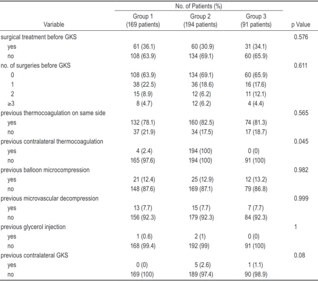
The number and types of previous surgical interven-tions were also assessed. A summary is shown in Table 3. Whether the patient had undergone previous surgical treatment (p = 0.576), the number of preoperative GKS interventions (p = 0.611), and most types of previous in-tervention—thermocoagulation on the same side (p = 0.565), balloon microcompression (p = 0.982), microvas-cular decompression (p = 0.999) and glycerol injection (p = 1)—were not statistically significant. Only previous contralateral thermocoagulation (p = 0.04) was statisti-cally significant for patterns of pain freedom.
Regarding preoperative hypesthesia, we found no statistically significant difference between the 3 pain-free groups (p = 0.874), as seen in Table 4. Only 1 person had preoperative anesthesia dolorosa; that patient was in Group PF≤ 48 hours (p = 0.572).
TABLE 1: Initial freedom from pain in and development of hypesthesia and pain recurrence in the 3 patient groups*
No. of Patients (%) (total no. of patients = 454)
Variable Group 1 Group 2 Group 3 p Value initially pain free 169 (37.2) 194 (42.8) 91 (20)
hypesthesia 0.014 yes 18 (13.7) 30 (19) 22 (30.6) no 113 (86.3) 128 (81) 50 (69.4) pain recurrence 0.515 yes 59 (34.9) 71 (36.6) 27 (29.7) no 110 (65.1) 123 (63.4) 64 (70.3)
* Group 1 = Group PF≤ 48 hours; Group 2 = Group PF(> 48 hours, ≤ 30 days); Group
3 = Group PF> 30 days.
Fig. 1. Graph depicting the distribution of pain-free intervals after
radiosurgery in a population of 497 patients with classic TN and only 1 GKS treatment. (Patients with megadolichobasilar artery– or MS-related TN were not included in the study group.)
C. Tuleasca et al.
Post-GKS Hypesthesia
Among all 497 patients, the hypesthesia rate at 5 years was 20.4%; at 7 years the rate reached 21.1% and remained stable until 14 years post-GKS, with a median delay of onset of 12 months (range 1–65 months).
Statistically significant differences (p = 0.014) be-tween the 3 pain-free groups were identified for post-GKS hypesthesia: postoperative sensory disturbances developed in 18 patients (13.7%) in Group PF≤ 48 hours,
com-pared with 30 patients (19%) in Group PF(> 48 hours, ≤ 30 days)
and 22 patients (30.6%) in Group PF> 30 days. Table 5 shows
these findings. When hypesthesia was classified as mild or severe (discreet or important, respectively), the p value was 0.823, and when it was assessed using the BNI scale, the p value was 0.598.
Until 1 year after GKS, the risk of hypesthesia was similar in the 3 groups, but afterward there was a strong risk of hypesthesia in Group PF> 30 days.
We conclude that the lowest hypesthesia rate was found in patients who became pain free earliest (Group PF≤ 48 hours, 13.7% of patients) and the highest
hypesthe-sia rate was found in patients who became pain free last (Group PF> 30 days, 30.6% of patients).
Figure 2 shows the probability of hypesthesia onset within the 3 subgroups.
Probability of Maintaining Pain Relief and Management of Pain Recurrence
One hundred fifty-seven patients (34.4%) who be-came pain free after GKS experienced a recurrence of TN pain with a median delay of 24 months (range 0.62– 150.06 months).
There were no statistically significant differences in the pain recurrence rates between patient groups: 66 pa-tients (39%) in Group PF≤ 48 hours, 71 patients (36.6%) in
Group PF(> 48 hours, ≤ 30 days), and 27 (29.7%) patients in Group
PF> 30 days experienced at least 1 recurrence of pain (p =
0.515).
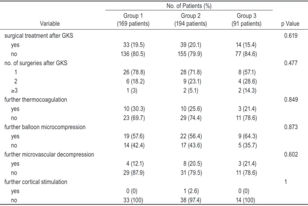
Whether patients underwent additional surgical inter-vention (p = 0.619); if performed, the number of additional surgical procedures (p = 0.477); and the type of additional surgical intervention (p = 0.849 for thermocoagulation; p = 0.873 for balloon microcompression; p = 0.602 for mi-crovascular decompression; and p = 1 for cortical stimula-tion) were not found to be statistically significant. A sum-mary of these findings is shown in Table 6.
Figure 3 shows the probability of maintaining pain relief in the 3 subgroups.
Evaluation of Outcome at the Last Follow-Up
At the last follow-up, our results showed no
statisti-TABLE 2: Preoperative assessment of the 3 patient groups regarding some clinical aspects and the neurovascular conflict seen on preoperative MR images
No. of Patients (%)
Variable Group 1 (169 patients) Group 2 (194 patients) Group 3 (91 patients) p Value
side of pain 0.73 rt 94 (55.6) 102 (52.6) 52 (57.1) lt 75 (44.4) 92 (47.4) 39 (42.9) bilat pain 0.85 yes 7 (4.1) 6 (3.1) 4 (4.4) no 162 (95.9) 188 (96.9) 87 (95.6)
no. of territories of pain 0.41
1 87 (51.5) 116 (59.8) 48 (52.7) 2 71 (42) 63 (32.5) 37 (40.7) 3 11 (6.5) 15 (7.7) 6 (6.6) atypical pain 0.805 yes 36 (21.3) 37 (19.1) 20 (22) no 133 (78.7) 157 (80.9) 71 (78) continuous pain 0.201 yes 42 (24.9) 34 (17.5) 17 (18.7) no 127 (75.1) 160 (82.5) 74 (81.3) previous neurovascular conflict seen on MRI 0.743 yes 92 (54.4) 110 (56.7) 54 (59.3) no 77 (45.6) 84 (43.3) 37 (40.7) sex of patient 0.612 male 79 (46.7) 82 (42.3) 43 (47.3) female 90 (53.3) 112 (57.7) 48 (52.7)
cally significant differences between the 3 subgroups (p = 0.874 for the BNI classification). Table 7 shows these results.
Discussion
Even if the role of GKS in the armamentarium for treating TN is now well established, the radiobiological mechanisms of action associated with the procedure re-main poorly understood.
When we discuss clinical and histopathological chains of events we cannot separate the causes of TN, which can differ greatly from one case to another: com-pression by an overlying artery or vein (thought to ac-count for 80%–90% of cases9) with focal demyelination,
close apposition of demyelinated axons, few residual oligodendrocytes, and no inflammatory cells;8 primary
demyelinating disorders (MS) with demyelination ex-tending along the proximal part of the trigeminal nerve root and, in some cases, to the junction with the periph-eral nerve system, juxtaposed axons, and the presence of variable thinly myelinated fibers; masses in the posterior
fossa, small infarcts or angiomas in the pons or medulla oblongata, prosthetic materials inserted during microvas-cular decompression;5 bone compression of the nerve;23,25
and idiopathic cases or, less frequently, familial TN.11 In
the majority of cases, the cause of TN seems to be demy-elination, especially present at the level of the trigeminal root entry zone.15
Few studies have been conducted to understand the mechanism of action that occurs when GKS is used as a tool for treatment.
TABLE 3: Previous surgical intervention
No. of Patients (%)
Variable (169 patients)Group 1 (194 patients)Group 2 (91 patients)Group 3 p Value
surgical treatment before GKS 0.576
yes 61 (36.1) 60 (30.9) 31 (34.1)
no 108 (63.9) 134 (69.1) 60 (65.9)
no. of surgeries before GKS 0.611
0 108 (63.9) 134 (69.1) 60 (65.9)
1 38 (22.5) 36 (18.6) 16 (17.6)
2 15 (8.9) 12 (6.2) 11 (12.1)
≥3 8 (4.7) 12 (6.2) 4 (4.4)
previous thermocoagulation on same side 0.565
yes 132 (78.1) 160 (82.5) 74 (81.3)
no 37 (21.9) 34 (17.5) 17 (18.7)
previous contralateral thermocoagulation 0.045
yes 4 (2.4) 194 (100) 0 (0)
no 165 (97.6) 194 (100) 91 (100)
previous balloon microcompression 0.982
yes 21 (12.4) 25 (12.9) 12 (13.2)
no 148 (87.6) 169 (87.1) 79 (86.8)
previous microvascular decompression 0.999
yes 13 (7.7) 15 (7.7) 7 (7.7)
no 156 (92.3) 179 (92.3) 84 (92.3)
previous glycerol injection 1
yes 1 (0.6) 2 (1) 0 (0)
no 168 (99.4) 192 (99) 91 (100)
previous contralateral GKS 0.08
yes 0 (0) 5 (2.6) 1 (1.1)
no 169 (100) 189 (97.4) 90 (98.9)
TABLE 4: Preoperative hypesthesia in the 3 patient groups
No. of Patients (%) Preop Hypesthesia Group 1 (169 patients) Group 2 (194 patients) Group 3 (91 patients) Valuep no hypesthesia 131 (77.5) 158 (81.4) 72 (79.1) slight hypesthesia 34 (20.1) 33 (17) 18 (19.8) 0.874 severe hypesthesia 4 (2.4) 3 (1.5) 1 (1.1)
C. Tuleasca et al.
Kondziolka et al.12 irradiated 2 male baboons
(Pap-io cynocephalus anubis) using doses of 80 and 100 Gy. Light and electron microscopic examinations were per-formed to analyze specimens of the trigeminal nerve 6 months after the procedure. The authors discovered acute degenerating axons with some identifiable myelinated ax-ons, small foci of necrosis (including Schwann cell nu-clei necrosis), and normal trigeminal ganglion when they used 80 Gy. At 100-Gy doses they found axon degenera-tion with myelin vacuoladegenera-tion and expansion of the endo-neurial intracellular matrix, which was consistent with edema. In one specimen, almost the entire width of the nerve was necrotic. Axon degeneration was noted outside the necrotic zone, but the histological features normal-ized toward the ganglion. The authors concluded that the histological effects were dose related and consisted of
primary axon injuries. They also observed no evidence of inflammation 6 months after GKS, and the extent of nerve edema was mild. Kondziolka et al. presumed that acute inflammation likely occurs after nerve irradiation (perhaps within the first few days) and may have effects on pathophysiological nerve processes, relieving pain quickly.
Szeifert et al.26 studied the histopathological findings
in a patient who suffered from TN and was treated 2 times by GKS (in the first procedure, the maximum dose was 90 Gy; in the second procedure, performed 10 months later, the maximum dose was 70 Gy). The patient died of a hemorrhagic stroke 26 days following the second inter-vention, and an autopsy was performed. Histopathologi-cal studies demonstrated changes in the trigeminal nerve that differed according to the sites of each treatment dose. Chronic radiation-induced changes were identified 11 months after the first treatment, at the site treated with 90 Gy: there was a well-circumscribed, hypocellular fi-brotic lesion with hyaline-degenerated collagen bundles and scattered fibrocytes. Acute radiation-induced chang-es, indicative of the early consequences of radiosurgery, were identified 26 days after the second treatment, at the site treated with 70 Gy: there was a sharply demarcated necrotic center containing fibrinoid material and tissue debris, which was encircled by surviving nerve bundles.
Conclusions
Our study was rather empirical (but also based on a statistical chart of distribution) in classifying the dis-tribution of pain-free patients into the 3 subtypes. Our data indicate that pathophysiological mechanisms simi-lar to those described in the other cited studies could be involved, but also some new ones—unknown for the moment—that could help explain the different patterns of freedom-from-pain responses after GKS. A substan-tial number of patients, 169 (37.2%), became pain free within the first 48 hours (Group PF≤ 48 hours) after GKS;
this is quite a large population. These patients had the lowest rate of hypesthesia in our series (13.7%),
particu-TABLE 5: Hypesthesia after GKS
No. of Patients (%)
Variable (169 patients)Group 1 (194 patients)Group 2 (91 patients)Group 3 p Value
hypesthesia after GKS 0.014 yes 18 (13.7) 30 (19) 22 (30.5) no 113 (86.3) 128 (81) 50 (69) type of hypesthesia 0.823 discrete 13 (72.2) 21 (70) 14 (63.6) important 5 (27.8) 9 (30) 8 (36.4) BNI type of hypesthesia 0.598
mild facial numbness 15 (83.3) 27 (90) 18 (81.8) facial numbness, somewhat bothersome 2 (11.1) 3 (10) 3 (13.6) facial numbness, very bothersome 1 (5.6) 0 (0) 1 (4.6)
Fig. 2. Post-GKS hypesthesia in the 3 pain-free patient subgroups:
larly compared with patients in the Group PF> 30 days, in
which hypesthesia developed in 22 patients (30.6%) (data statistically significant, p = 0.014). The pain recurrence rate was 39% in Group PF≤ 48 hours, compared with 26.6%
in Group PF(> 48 hours, ≤ 30 days) and 29.7% in Group PF> 30 days,
but these data were not statistically significant (p = 0.515).
Given that targeting a small volume of normal tissue with a high radiation dose probably does not produce a destructive effect, the neuromodulatory mechanisms of GKS still need to be further analyzed and understood.
Disclosure
Professor Régis declares receiving support for non–study related clinical or research effort from Elekta AB. No other potential conflicts have been reported.
Author contributions to the study and manuscript preparation include the following. Conception and design: Tuleasca, Carron, Levivier, Régis. Acquisition of data: Tuleasca. Analysis and inter-pretation of data: Tuleasca, Resseguier, Gaudart. Drafting the article: Tuleasca. Critically revising the article: all authors. Reviewed submitted version of manuscript: all authors. Approved the final ver-sion of the manuscript on behalf of all authors: Tuleasca. Statistical analysis: Resseguier, Gaudart. Administrative/technical/material
TABLE 6: Management of pain recurrence with number and type of further surgical interventions
No. of Patients (%)
Variable (169 patients)Group 1 (194 patients)Group 2 (91 patients)Group 3 p Value
surgical treatment after GKS 0.619
yes 33 (19.5) 39 (20.1) 14 (15.4)
no 136 (80.5) 155 (79.9) 77 (84.6)
no. of surgeries after GKS 0.477
1 26 (78.8) 28 (71.8) 8 (57.1) 2 6 (18.2) 9 (23.1) 4 (28.6) ≥3 1 (3) 2 (5.1) 2 (14.3) further thermocoagulation 0.849 yes 10 (30.3) 10 (25.6) 3 (21.4) no 23 (69.7) 29 (74.4) 11 (78.6)
further balloon microcompression 0.873
yes 19 (57.6) 22 (56.4) 9 (64.3)
no 14 (42.4) 17 (43.6) 5 (35.7)
further microvascular decompression 0.602
yes 4 (12.1) 8 (20.5) 3 (21.4)
no 29 (87.9) 31 (79.5) 11 (78.6)
further cortical stimulation 1
yes 0 (0) 1 (2.6) 0 (0)
no 33 (100) 38 (97.4) 14 (100)
TABLE 7: Outcome at the last follow-up based on BNI classification No. of Patients (%) Variable Group 1 (169 patients) Group 2 (194 patients) Group 3 (91 patients) Valuep BNI classification 0.874 good outcome 165 (97.6) 188 (96.9) 88 (96.7) poor outcome 4 (2.4) 6 (3.1) 3 (3.3)
Fig. 3. Probability of maintaining pain relief in the 3 pain-free pa-
tient subgroups: Group PF≤ 48 hours, Group PF(> 48 hours, ≤ 30 days), and Group PF> 30 days.
C. Tuleasca et al.
support: Tuleasca, Donnet, Roussel, Levivier, Régis. Study supervi-sion: Donnet, Roussel, Gaudart, Levivier, Régis.
References
1. Brisman R: Gamma knife radiosurgery for primary manage-ment for trigeminal neuralgia. J Neurosurg 93 (Suppl 3): 159–161, 2000
2. Brisman R, Mooij R: Gamma knife radiosurgery for trigemi-nal neuralgia: dose-volume histograms of the brainstem and trigeminal nerve. J Neurosurg 93 (Suppl 3):155–158, 2000 3. Flickinger JC, Pollock BE, Kondziolka D, Phuong LK, Foote
RL, Stafford SL, et al: Does increased nerve length within the treatment volume improve trigeminal neuralgia radiosurgery? A prospective double-blind, randomized study. Int J Radiat
Oncol Biol Phys 51:449–454, 2001
4. Fountas KN, Lee GP, Smith JR: Outcome of patients undergo-ing gamma knife stereotactic radiosurgery for medically re-fractory idiopathic trigeminal neuralgia: Medical College of Georgia’s experience. Stereotact Funct Neurosurg 84:88– 96, 2006
5. Fujimaki T, Hoya K, Sasaki T, Kirino T: Recurrent trigemi-nal neuralgia caused by an inserted prosthesis: report of two cases. Acta Neurochir (Wien) 138:1307–1310, 1996 6. Han JH, Kim DG, Chung HT, Paek SH, Kim YH, Kim CY, et
al: Long-term outcome of gamma knife radiosurgery for treat-ment of typical trigeminal neuralgia. Int J Radiat Oncol Biol
Phys 75:822–827, 2009
7. Headache Classification Subcommitee of the International Headache Society: The International Classification of Head-ache Disorders: 2nd edition. Cephalalgia 24 Suppl 1:9–160, 2004
8. Hilton DA, Love S, Gradidge T, Coakham HB: Pathological findings associated with trigeminal neuralgia caused by vas-cular compression. Neurosurgery 35:299–303, 1994 9. Jannetta PJ: Neurovascular compression in cranial nerve and
systemic disease. Ann Surg 192:518–525, 1980
10. Knafo H, Kenny B, Mathieu D: Trigeminal neuralgia: out-comes after gamma knife radiosurgery. Can J Neurol Sci 36: 78–82, 2009
11. Knuckey NW, Gubbay SS: Familial trigeminal and glossopha-ryngeal neuralgia. Clin Exp Neurol 16:315–319, 1979 12. Kondziolka D, Lacomis D, Niranjan A, Mori Y, Maesawa S,
Fellows W, et al: Histological effects of trigeminal nerve ra-diosurgery in a primate model: implications for trigeminal neuralgia radiosurgery. Neurosurgery 46:971–977, 2000 13. Kondziolka D, Zorro O, Lobato-Polo J, Kano H, Flannery TJ,
Flickinger JC, et al: Gamma Knife stereotactic radiosurgery for idiopathic trigeminal neuralgia. Clinical article. J
Neuro-surg 112:758–765, 2010
14. Leksell L: Sterotaxic radiosurgery in trigeminal neuralgia.
Acta Chir Scand 137:311–314, 1971
15. Love S, Coakham HB: Trigeminal neuralgia: pathology and pathogenesis. Brain 124:2347–2360, 2001
16. Lunsford LD, Young RF: Radiosurgery for trigeminal neural-gia. Surg Neurol 54:285–287, 2000
17. Massager N, Lorenzoni J, Devriendt D, Desmedt F, Brotchi J, Levivier M: Gamma knife surgery for idiopathic trigeminal neuralgia performed using a far-anterior cisternal target and a high dose of radiation. J Neurosurg 100:597–605, 2004 18. Massager N, Lorenzoni J, Devriendt D, Levivier M:
Radiosur-gery for trigeminal neuralgia. Prog Neurol Surg 20:235–243, 2007
19. Pollock BE, Phuong LK, Gorman DA, Foote RL, Stafford SL: Stereotactic radiosurgery for idiopathic trigeminal neuralgia.
J Neurosurg 97:347–353, 2002
20. Régis J, Arkha Y, Yomo S, Murata N, Roussel P, Donnet A, et al: [Radiosurgery in trigeminal neuralgia: long-term results and influence of operative nuances.] Neurochirurgie 55:213– 222, 2009 (Fr)
21. Régis J, Metellus P, Hayashi M, Roussel P, Donnet A, Bille-Turc F: Prospective controlled trial of gamma knife surgery for essential trigeminal neuralgia. J Neurosurg 104:913–924, 2006
22. Régis J, Tuleasca C: Fifteen years of Gamma Knife surgery for trigeminal neuralgia in the Journal of Neurosurgery: his-tory of a revolution in functional neurosurgery. Editorial. J
Neurosurg 115 Suppl:2–7, 2011
23. Reilly MM, Valentine AR, Ginsberg L: Trigeminal neuralgia associated with osteogenesis imperfecta. J Neurol
Neuro-surg Psychiatry 58:665, 1995
24. Rogers CL, Shetter AG, Fiedler JA, Smith KA, Han PP, Speiser BL: Gamma knife radiosurgery for trigeminal neuralgia: the initial experience of The Barrow Neurological Institute. Int J
Radiat Oncol Biol Phys 47:1013–1019, 2000
25. Ruelle A, Datti R, Andrioli G: Cerebellopontine angle osteo-ma causing trigeminal neuralgia: case report. Neurosurgery
35:1135–1137, 1994
26. Szeifert GT, Salmon I, Lorenzoni J, Massager N, Levivier M: Pathological findings following trigeminal neuralgia radiosur-gery. Prog Neurol Surg 20:244–248, 2007
27. Zhao ZF, Yang LZ, Jiang CL, Zheng YR, Zhang JW: Gamma Knife irradiation-induced histopathological changes in the trigeminal nerves of rhesus monkeys. Laboratory investiga-tion. J Neurosurg 113:39–44, 2010
Manuscript submitted May 19, 2012. Accepted August 1, 2012.
Portions of this study were presented in abstract form at the 16th International Meeting of the Leksell Gamma Knife Society, Sydney, Australia, March 2012.
Please include this information when citing this paper: DOI: 10.3171/2012.8.GKS121015.
Address correspondence to: Constantin Tuleasca, M.D., Centre
Hos pitalier Universitaire Vaudois, Neurosurgery Service and Gamma Knife Center, Rue de Bugnon 44-46, BH-08, CH-1011 Lausanne, Switzerland. email: constantin.tuleasca@gmail.com.