Memory Th Cell Signature That Is Similar to and
Cross-Reactive with
Streptococcus pneumoniae
Stian Andre´ Engen1*, Ha˚kon Valen Rukke1, Simone Becattini2, David Jarrossay2, Inger Johanne Blix1,3, Fernanda Cristina Petersen1., Federica Sallusto2., Karl Schenck1.
1 Department of Oral Biology, University of Oslo, Oslo, Norway, 2 Institute for Research in Biomedicine, Universita` della Svizzera Italiana, Bellinzona, Switzerland, 3 Department of Periodontology, University of Oslo, Oslo, Norway
Abstract
Background:Carriage of and infection with Streptococcus pneumoniae is known to predominantly induce T helper 17 (Th17) responses in humans, but the types of Th cells showing reactivity towards commensal streptococci with low pathogenic potential, such as the oral commensals S. mitis and S. salivarius, remain uncharacterized.
Methods:Memory CD4+T helper (Th) cell subsets were isolated from healthy human blood donors according to differential expression of chemokine receptors, expanded in vitro using polyclonal stimuli and characterized for reactivity against different streptococcal strains.
Results:Th cells responding to S. mitis, S. salivarius and S. pneumoniae were predominantly in a CCR6+CXCR3+subset and produced IFN-c, and in a CCR6+CCR4+subset and produced IL-17 and IL-22. Frequencies of S. pneumoniae-reactive Th cells were higher than frequencies of S. mitis- and S. salivarius-specific Th cells. S. mitis and S. pneumoniae isogenic capsule knock-out mutants and a S. mitis mutant expressing the serotype 4 capsule of S. pneumoniae showed no different Th cell responses as compared to wild type strains. S. mitis-specific Th17 cells showed cross-reactivity with S. pneumoniae.
Conclusions: As Th17 cells partly control clearance of S. pneumoniae, cross-reactive Th17 cells that may be induced by commensal bacterial species may influence the immune response, independent of capsule expression.
Citation: Engen SA, Valen Rukke H, Becattini S, Jarrossay D, Blix IJ, et al. (2014) The Oral Commensal Streptococcus mitis Shows a Mixed Memory Th Cell Signature That Is Similar to and Cross-Reactive with Streptococcus pneumoniae. PLoS ONE 9(8): e104306. doi:10.1371/journal.pone.0104306
Editor: Bernard Beall, Centers for Disease Control & Prevention, United States of America Received May 20, 2014; Accepted July 7, 2014; Published August 13, 2014
Copyright: ß 2014 Engen et al. This is an open-access article distributed under the terms of the Creative Commons Attribution License, which permits unrestricted use, distribution, and reproduction in any medium, provided the original author and source are credited.
Data Availability: The authors confirm that all data underlying the findings are fully available without restriction. All relevant data are within the paper and its Supporting Information files.
Funding: This work was supported by the European Research Council grant number 323183 PREDICT (to FS). The Institute for Research in Biomedicine, Bellinzona, is supported by the Helmut Horten Foundation (http://www.helmut-horten stiftung.org/en/homepage.html). The funders had no role in study design, data collection and analysis, decision to publish, or preparation of the manuscript.
Competing Interests: The authors have declared that no competing interests exist. * Email: [email protected]
.These authors contributed equally to this work.
Introduction
A reciprocal beneficial relationship has developed between hosts and their symbionts throughout evolution. In the human oral cavity, more than 700 bacterial species can be found [1,2] of which a healthy person can host more than 200 [3]. In order for the commensals to persist in their niches, it is important that adequate host-microbe interplay is established. This comprises immune exclusion by keeping microbes from interacting with host cell by mucus, SIgA and/or antimicrobial peptides, and immune elimination by innate and adaptive responses without the induction of inflammation [4].
S. mitis is a pioneer bacterial species that colonizes the nasopharynx and all sites of the oral cavity from early infancy. Its predominance persists during life and in adultsS. mitis is found in the oral cavity of nearly all persons.S. mitis is closely related to
S. pneumoniae which also resides in the oronasopharynx: the species may share as much as 39% of their genes, including many of the virulence genes [5]. Despite their genetic similarity, S. pneumoniae causes serious infections in about 14.5 million children every year, whereasS. mitis rarely causes disease [6].
After the second year of life, a drastic reduction in carriage and disease rate caused by S. pneumoniae occurs, independent of capsular serotype [7]. This reduction is attributed to the development of serum IgG and secretory antibodies [8], and to antigen-specific T cell responses [9,10]. Oral carriage state ofS. mitis is probably partly regulated by secretion of salivary SIgA [11,12], but the role of Th cells has not been explored.
Naı¨ve T helper (Th) cells develop into different polarized effector Th subsets that are tailored to effectively cope with the type of infection, including Th1 and Th2 that produce IFN-c or IL-4, respectively [13]. More recently, Th17 [14], Th22 [15,16],
and Th9 [17] have been described, which produce IL-17, IL-22 or IL-9, respectively. Th cell subsets with a mixed phenotype have been also identified, including T cells producing IL-17 and IFN-c, or IL-17 and IL-4 [18–20]. In this study, we set out to examine the phenotype and functional property of in vivo-primed memory CD4+Th cells reactive with antigens from the oral commensal speciesS. mitis and Streptococcus salivarius, and compare it to that ofS. pneumoniae.
Utilizing a T cell screening method, we found that memory Th cells reactive againstS. mitis and S. salivarius are predominantly found in the CCR6+Th1 and Th17 subsets, a distribution similar to that obtained for S. pneumoniae. In addition, we observed interspecies cross-reactivity among S. mitis–reactive and S. pneumoniae-reactive T cell clones.
Material and methods Bacterial strains
Bacterial strains included S. mitis (CCUG 31611T, 62644, 62641), S. pneumoniae (TIGR4, Sero 1, D39) and S. salivarius (JIM8777) (Table 1). Isogenic capsule deletion mutants ofS. mitis 31611T (S. mitis Dcps) and S. pneumoniae TIGR4 (S. pneumoniae Dcps) and a capsule switch mutant of S. mitis 31611T expressing the serotype 4 capsule ofS. pneumoniae TIGR4 (S. mitis 31611T TIGR4) were constructed as described before [21]. All strains were grown in Todd Hewitt Broth (THB) (BD Biosciences, Franklin Lakes, NJ). Over night cultures were diluted in THB and grown to OD = 1 at 600 nm. Cells were harvested by centrifuga-tion at 5000 g for 10 min at 4uC, washed in endotoxin free Dulbecco’s-PBS (Sigma-Aldrich, St. Louis, MO) and UV-inacti-vated for 30 min using UVC 500 Crosslinker (GE Healthcare, Fairfield, CT). The UV-treated bacterial suspensions were aliquoted and frozen at 280uC.
Blood samples and cell sorting
Blood from anonymized healthy donors was obtained from the Swiss Blood Donation Centers of Basel and Lugano, and used in compliance with the Swiss Federal Office of Public Health (authorization n. A000197/2 to F.S). No submission to a local
ethics committee was needed because volunteer donors from the national registry sign an informed consent form (Swiss Red Cross, Medical Questionnaire and Informed Consent Form, version 09), stating that their blood could be used for medical research after definitive anonymization. PBMCs were obtained using Ficoll-Paque PLUS (GE Healthcare) gradient centrifugation. CD14+ monocytes and CD4+T cells were isolated by positive selection using magnetic beads (Miltenyi Biotec, Bergisch Gladbach, Germany). CD14+cells were collected and frozen at –80uC for later use. In order to sort distinct Th subsets, CD142CD4+cells were incubated with the following antibodies: anti-CD45RA (Qdot655, Invitrogen, Carlsbad, CA); anti-CXCR3 (APC, BD Biosciences); anti-CCR4 (PE-Cy7, BD Biosciences); anti-CCR6 (Brilliant Violet 605, BioLegend, San Diego, CA); anti-CCR10 (PE, R&D systems, Minneapolis, MN); anti-CD8 (PECy5, Beck-man Coulter, Brea, CA); anti-CD19 (PECy5, BeckBeck-man Coulter); anti-CD25 (PECy5, Beckman Coulter); anti-CD56 (PECy5, Beckman Coulter). CD45RA2CD82CD192CD252CD562 cells were sorted on a FACSAria (BD Biosciences) into the following subsets: i. CXCR3+CCR42CCR62CCR102, ii. CCR6+CXCR3+ CCR42CCR102(both enriched in Th1 and defined thereafter as Th1 and CCR6+Th1, respectively), iii. CCR4+CXCR32CCR62 CCR102 (enriched in Th2); iv. CCR6+CCR4+CXCR32 CCR102 (Th17), and v. CCR6+CCR4+CCR10+CXCR32 (en-riched in Th22). The sorting strategy is summarized in Table S1. Cytokine production by the sorted cell subsets was measured in the 24-hour culture supernatants after activation with immobilized anti-CD3 (clone TR66, 5mg/ml) and anti-CD28 (clone CD28.2; BD Biosciences; 1mgl/ml) antibodies using the cytometric bead array (CBA) (eBiosciences, San Diego, CA), carried out according to the manufacturer’s protocol. The characteristics of the subsets, as assayed by cytokine secretion, is shown in Figure S1.
T cell library construction and screening
T cell libraries were established as described before [22]. Cells were grown in complete media (CM) comprising RPMI-1640 supplemented with 2 mM glutamine, 1% v/v non-essential amino acids, 1% v/v sodium pyruvate, 0.1% v/v 2-mercaptoethanol, penicillin (50 U/mL) and streptomycin (50mg/mL) (Gibco, Table 1. Streptococcal strains used in this study.
Strains Description Source S. mitis
SK575 (62644) Corresponds to CCUG 62644 CCUG* SK579 (62641) Corresponds to CCUG 62641 CCUG CCUG 31611T
Wild type S. mitis biovar 1 type strain; corresponds to NCTC12261 CCUG S. mitis Dcps; MI016 CCUG31611T
cps::kan; KanR
S. mitisTIGR4; MI030 CCUG31611 T
V TIGR4 cps locus; KanR
, ErmR
S. pneumoniae
D39 Corresponds to NCTC 7466 NCTC**
Serotype 1 Corresponds to sequence type 306 Clinical isolate, Norwegian Institute of Public Health TIGR4 Wild type serotype 4, transformable strain, sequenced genome
S. pneumoniae Dcps; SP011 TIGR4 cps::erm; ErmR
S. salivarius JIM8777
*CCUG: Culture collection, University of Go¨teborg. **NCTC: National cultures of Type Cultures. doi:10.1371/journal.pone.0104306.t001
Carlsbad, CA), and 5% v/v human serum (Swiss Blood Center), unless stated otherwise. Memory Th cell subsets obtained by cell sorting as described above, were plated in 96-well U-bottom plates (2000 cells/well) and polyclonally stimulated with 1mg/mL PHA (Sigma-Aldrich), in the presence of irradiated (45 Gy) allogeneic PBMCs and 500 U/mL IL-2. After 7d, the cells were transferred to 24-well plates for further expansion of total 20d. Half of the volume of the medium was changed every other day. For stimulation, the cultures were washed three times in RPMI-1640 with HEPES (Gibco) and 1% v/v FCS, before each well was tested for streptococcal antigen-reactivity by 3d co-culture of 2.56105T cells and 2.06104irradiated (45 Gy) autologous monocytes, pulsed with whole cell UV-inactivated bacteria (MOI: 100:1) for 5 h. At day 3 of the co-culture, [3H]-thymidine was added and proliferation was measured on a beta-counter after 18 h.
Cross reactivity assay
Wells containing Th17 cells reactive to S. mitis and S. pneumoniae were cloned by limiting dilution. First, T cells (2.56105) were stained with CFSE and co-cultured with irradiated
(45 Gy) autologous monocytes pulsed with whole cell UV-inactivated bacteria for 5 h. At day 7 of co-culture, CFSE-low proliferating cells were sorted and plated at 0.5 cells/well, stimulated with 1mg/mL PHA and 0.56104irradiated allogeneic PBMCs/well in CM supplemented with 500 U/mL IL-2 in 384-well plates. During 20 d of expansion, proliferating 384-wells were transferred to 96-well U-bottom plates and further to 24-well plates before re-stimulating with whole-cell UV-inactivated bacteria as described above.
Inhibition assay
Tetanus toxoid (TT)-reactive Th17 memory T cells were sorted and cloned as described above. 2.56104T cells were co-cultured with a 2-fold dilution series of irradiated monocytes, ranging from 46104to 1.256103monocytes/well, and 5mg/mL TT alone or in the presence of whole cells fromS. mitis or S. pneumoniae (MOI: 100:1). After 3 d in culture, [3H]-thymidine was added and proliferation was determined by [3H]-thymidine incorporation after 18 h.
Cytometric bead array (CBA)
To quantify IFN-c, IL-17A and IL-22, supernatants of CCR6+ Th1 and Th17 cells stimulated with S. mitis 31611T and S. pneumoniae TIGR4 were analyzed using the cytometric bead array (CBA) (eBiosciences, San Diego, CA), carried out according to the manufacturer’s protocol.
Statistical analysis
Student t tests were used to assess differences in cytokine secretion. P values of lower than 0.05 were considered to indicate statistically significant.
Results
Circulating T helper memory cells show a heterogeneous signature after stimulation with S. mitis, similar to that obtained with S. pneumoniae
Five CD4+ memory Th cell subsets from PBMCs of healthy donors were isolated according to the expression of chemokine receptors as described before [22]. For each CD4+T cell subset in each donor, 48 cell lines were established and polyclonally expanded for 16–20 days prior to screening. Each T cell line was then screened for reactivity against autologous monocytes
pulsed with three S. mitis strains (62644, 62641, 31611T) and responding T cells were detected by [3H]-thymidine incorpora-tion. Cultures containing proliferating T cells were identified by incorporation of [3H]-thymidine and precursor frequencies were calculated based on the number of negative wells, according to the Poisson distribution and expressed per million cells [23]. A representative example obtained from one of the donors is shown in Figure 1 and the distribution of responding T cells in the different subsets for the 6 donors analyzed is summarized in Figure 2. The raw data and the subset distribution of wells reactive with the streptococcal species for each donor as percentage of the total of all subsets for each strain are shown in Tables S2 and S3, respectively. The frequency of Th17 cells was highest while that of the Th22 cells was lowest for all strains. Intraspecies (S. mitis 62644, 62641, 31611T) signatures were similar, but frequencies of memory T cells reactive to S. mitis strain 62641 were slightly enhanced in all subsets. The frequencies of Th subsets responsive to the commensalS. salivarius were similar to that observed for S. mitis.
The signatures were viewed relative to that ofS. pneumoniae, strains TIGR4, D39, Sero 1, representing serotypes 4, 2, and 1, respectively. The profiles of these three strains were similar in the Th1, Th2 and Th22 subsets but strain D39 displayed a stronger reactivity to the CCR6+Th1 and Th17 subsets (Figure 2). The frequencies of theS. pneumoniae-reactive cells were consistently and markedly higher than those forS. mitis and S. salivarius.
Capsule expression does not significantly affect the pattern of streptococci-reactive Th cell subset frequencies and relative distribution
S. pneumoniae strains are divided into serotypes according to the polysaccharide composition of their capsules.S. mitis strains can also express capsule [21]. Deletion of theS. mitis capsule or replacing it with capsule from S. pneumoniae TIGR4 did not significantly affect reactivities (Figure 1 and 2). InS. pneumoniae TIGR4, capsule deletion had no significant effect on Th cell subset frequencies. Capsule from type 1S. pneumoniae strains, such as strain Sero 1, have zwitterionic characteristics and can activate T cell-dependent immune responses [24]. In the present study, however, no significant difference in frequency of Th reactive with type 1 pneumococcus relative to type 2 (D39) or type 4 (TIGR4) was observed (Figure 2).
S. mitis shows a suppressive effect on T cell responses to unrelated antigen
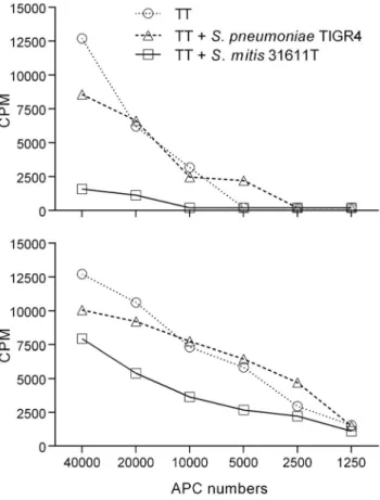
To investigate if the overall lower Th cell responses observed for S. mitis compared to S. pneumoniae could be due to antigen-unspecific T cell inhibitory effects ofS. mitis, tetanus toxoid (TT)-specific Th17 cell clones were co-cultured with autologous monocytes pulsed with TT alone or with TT and S. mitis 31611T orS. pneumoniae TIGR4. Adding S. mitis cells to the co-cultures reduced the ability of the TT-specific T cells to respond to TT, while little effect was observed whenS. pneumoniae cells were added (Figure 3).
S. mitis and S. pneumoniae induce IFN-c, IL-17 and IL-22 secretion by CCR6+Th1 and Th17 cells
As shown in Figures 1 and 2, the highest response to the streptococcal strains was observed within the CCR6+Th1 and Th17 memory cell subsets. For closer characterization, quantities of signature cytokines, i.e. IFN-c, IL-17A and IL-22, were examined by a cytometric bead array (CBA) in supernatants of the T cell lines co-cultured 3 d with monocytes pulsed withS. mitis
orS. pneumoniae wild types. CCR6+Th1 cells released more IFN-c and less IL-17 than the Th17 IFN-cells (Figure 4). To evaluate the differences in cytokine secretion between cultures stimulated with eitherS. mitis or S. pneumoniae, the donors’ cytokine secretion data were averaged within each stimulating strain (3S. mitis and 3 S. pneumoniae strains; Figure 4). The means for the S. mitis and 3 S. pneumoniae stimulations were then compared using Student t tests to reveal differences between the species. No statistically significant differences in cytokine secretion were detected within the CCR6+Th1 cells (P.0.05). The Th17 cells stimulated withS. pneumoniae, however, released more IFN-c, IL-17 and IL-22 than those stimulated withS. mitis (P,0.05; Figure 4).
Th17 clones show cross-reactivity for S. mitis and S. pneumoniae
As similar T cell subset patterns were found forS. mitis and S. pneumoniae and as the library comprised wells that showed reactivity to bothS. mitis and S. pneumoniae strains, we examined whether this could be due to cross-reactive T cells. Cells from
Figure 1. Distribution of antigen-specific memory Th subsets after exposure to streptococcal antigens by autologous monocytes. Raw data of a T cell library of a representative donor screened for reactivity for a panel of ten streptococci. Antigen-specific activity was quantified as a response in increased T cell proliferation determined by thymidine incorporation. Each graph represents the screening of reactivity of one T cell subset to the pane00l of bacteria and each circle represents one well of the respective subset. The dashed line represents lowest counts per minute (CPM) values included in the analysis.
doi:10.1371/journal.pone.0104306.g001
Figure 2. Subset distribution of CD4+ memory T cells in response to oropharyngeal-associated streptococcal bacteria. Distribution of single donor (circles) (N = 3–6) and mean (bars) frequencies of CD4+memory T cells among the CCR62Th1, CCR6+Th1,
Th2, Th17 and Th22 subsets reactive to streptococcal antigens. Data are presented as reactive cells per one million cells in the respective subsets. Open bars: S. mitis; closed bars: S. pneumoniae; hatched bars: S. salivarius. 62644: S. mitis CCUG 62644; 62641: S. mitis CCUG 62641; 31611T: S. mitis CCUG 31611T; 31611T Dcps: S. mitis CCUG 31611T capsule deletion mutant; 31611T TIGR4: S. mitis CCUG 31611T mutant with capsule from S. pneumoniae TIGR4; D39: S. pneumoniae D39; Sero 1: clinical isolate of S. pneumoniae serotype 1; TIGR4: S. pneumoniae TIGR4; TIGR4 Dcps: S. pneumoniae TIGR4 capsule deletion mutant; JIM8777: S. salivarius JIM8777.
doi:10.1371/journal.pone.0104306.g002
Figure 3. Response of TT-specific Th17 memory cell clones to native antigen in combination with bacterial cells. Tetanus toxoid (TT)-specific CD4+Th17 memory T cell clones were co-cultured with autologous monocytes and either TT alone or TT and S. mitis 31611T or S. pneumoniae TIGR4 (MOI: 100:1) for 3 d before proliferation was determined by [3H]-thymidine incorporation (A: clone 1, B: clone 2).
Th17 wells that were reactive with S. mitis 62641 or S. pneumoniae D39 in the initial stimulation were cloned and re-stimulated with the panel of bacterial strains. Clones initially reactive toS. mitis showed both inter- and intraspecies reactivity (Figure 5A), indicating cross-reactivity, while clones initially reactive to S. pneumoniae showed considerable cross-reactivity to allS. mitis strains (Figure 5B).
Discussion
Little is known about the subsets of Th cells responsive to commensal bacteria [25]. Here, we used a high-throughput T cell library method to map the Th cell signature that recognizes antigens fromS. mitis and S. salivarius. CD4+CD45RA2memory Th cells were sorted into subsets based on differential chemokine
receptor expression. This sorting was based on the knowledge of a co-regulation of effector function and migratory properties during T cell differentiation, a mechanism which ensures selective recruitment of different effector T cells to inflamed tissues in response to inflammatory chemoattractants [26]. Compared with in vitro antigen stimulation of unfractionated T cells and subsequent phenotyping, sorting of the cells before stimulation has the advantage of establishing the in vivo subsets’ identity before the cells are brought into culture, ensuring an ‘‘untouched’’ phenotype. We chose to expose the T cell cultures to whole, UV-inactivated bacterial cells to avoid compromising the integrity of surface molecules by unfavorable thermal or chemical conditions. The primary aim of this study was to determine the distribution of Th subsets that recognize antigens from the ubiquitous oral commensalS. mitis in healthy donors. We found that the numbers
Figure 4. Cytokine production by CCR6+Th1 and Th17 cells in response to streptococci. Quantities of IFN-c (A), IL-17A (B) and IL-22 (C) were determined in supernatants of CCR6+Th1 and Th17 CD4+memory T cells co-cultured for 3 d with autologous monocytes and whole cell, UV-inactivated S. mitis 31611T or S. pneumoniae TIGR4 (MOI: 100:1). Bars represent averaged values of different donors (symbols). Th17 cells secreted statistically significantly more IFN-c, IL-17A and IL-22 when stimulated with S. pneumoniae as compared with stimulation with S. mitis (Student t test; P , 0.05; see text).
of S. mitis- and S. salivarius-reactive Th cells were heteroge-neously distributed among the five subsets tested, but with a predominance of CCR6+Th1 and Th17 cells.S. pneumoniae is genetically closely related toS. mitis and was therefore included in the study. As S. pneumoniae has a marked pathogenic potential and S. mitis seldom causes disease, different distributions of response in the Th subset signature might be expected, but the results obtained showed that the Th response reactive with S. pneumoniae is strikingly similar to that of S. mitis. The signature and frequencies of responding cells toS. salivarius also coincided with that of S. mitis. The prominent Th17 response to S. pneumoniae is in accordance with studies on human lymphoid tonsillar tissue [27] and bronchoalveolar lavage [28], which show Th1/Th17- and IL-17A-dependent pneumococcal clearance, respectively.
The CCR6+Th1 subset is a recently described Th subset that has characteristics of both Th1 and Th17 subsets. Th1 and Th17 lymphocytes are characterized by specific transcription factors, surface receptors and cytokine secretion (Th1: T-bet, CXCR3, and IFN-c; Th17: RORC, CCR4/CCR6, and IL-17). Upon
polyclonal stimulation, the intermediate CCR6+Th1 subset can secrete both IL-17 and IFN-c and express RORC and T-bet ([8,18,29,30]. Expression of the Th17 surface marker CD161 and shared TCR clonality indicate that CCR6+Th1 cells are of Th17 ancestry [29]. Furthermore, Th17 cells exposed to Th1-polarizing conditions convert to the intermediate CCR6+Th1 cells (RORC+, T-bet+, IL-17+ IFN-c+), while Th1 cells exposed to Th17-polarizing conditions remain Th1 cells [29,30]. A proportion of Th1 cells are thought to be of Th17 origin due to expression of factors characteristic for Th17 (RORC, CCR6, CD161) [29]. In contrast to polyclonal stimulation, however, exposure of CCR6+ Th1 cells toMycobacterium tuberculosis PPD revealed an antigen-specific response characterized by absence of IL-17 secretion [18]. Our present observation that the CCR6+Th1 cell response to the streptococcal strains tested produced IFN-c but not IL-17 complies with this observation [18].
The CCR6+ Th1 and Th17 subsets presently examined produced a mixture of IFN-c, IL-17 and IL-22 in response to streptococcal stimulation, effector cytokines that can be involved in the clearance of S. pneumoniae in humans. IFN-c and IL-17 support macrophage and neutrophil defenses, respectively [26]. In a human infection model, carriage ofS. pneumoniae increased the prevalence of CD4+IL-17A+TNF+IFN-c+Th cells in lungs and blood, as compared to non-carriers [28]. Another study showed an IFN-c-dependent but IL-17-independent clearance of invadingS. pneumoniae [31]. Finally, significant increases in proportions of IFN-c+CD4+cells were seen in patients with community-acquired pneumonia [32]. IL-22 promotes epithelial proliferation, expres-sion of antimicrobial proteins involved in host defense in the skin, airways and intestine, and production of inflammatory mediators and chemokines from epithelial cells [33]. Knock-out of IL-22 renders mice more susceptible to infection with S. pneumoniae than wild type animals [34]. IL-22 by itself has protective and regenerative functions, but together with IL-17, it supports inflammation [33]. Here, we observed that Th17 cells secreted significantly higher amounts of IFN-c, IL-17 and IL-22 when challenged withS. pneumoniae species as compared with S. mitis. This suggests thatS. pneumoniae can induce a more pronounced inflammatory response as compared withS. mitis.
A consistently lower frequency of Th cells responsive toS. mitis compared to S. pneumoniae was observed. The finding that S. mitis reduced proliferation of T cell clones specific for an unrelated antigen (TT) suggests an inhibitory effect of S. mitis. This inhibition can either be on the APCs directly by preventing activation and/or antigen presentation, or directly on the memory Th cells by interfering with the APC-T cell interaction. TT-induced T cell proliferation was sustained upon addition of S. pneumoniae which indicated that the inhibitory effect is not a shared trait betweenS. mitis and S. pneumoniae, but specific for S. mitis. Another cause for the higher frequencies of S. pneumoniae-specific cells as compared with those for S. mitis can be the occurrence of more species-specific immunogenic antigens in the former species.
Expression of capsule is a hallmark of virulence of S. pneumoniae and the capsules comprise different serotypes that are important for the species to evade immune responses [35]. Capsule antigens comprise multi-epitopic repetitive carbohydrate units and have been considered as T cell-independent antigens, with exception of that fromS. pneumoniae type 1 (Sp1) that has zwitterionic properties, and is capable of activating T cells in an MHC class II-dependent manner [24,36]. Recently,S. mitis also has been shown to express capsule [21]. We tested isogenic capsule deletion mutants ofS. mitis and S. pneumoniae but little difference in Th cell signatures was observed as compared with wild type
Figure 5. Cross-reactivity of CD4+Th17 memory T cell clones in
response to oropharyngeal-associated streptococcal strains. Th17 cells from wells initially reactive to S. mitis 62641 (A) and S. pneumoniae D39 (B) were cloned by limiting dilution and distribution of intra- and interspecies cross-reactivity was determined by thymidine incorporation. Bars represent single re-stimulation of each clone and data are presented as counts per minute (CPM).
strains. Replacing native capsule ofS. mitis 31611T with capsule of S. pneumoniae TIGR4 neither altered the Th cell responses relative to theS. mitis wild type. This indicates that other factors than capsule expression are responsible for the raised frequencies of antigen-specific Th cells toS. pneumoniae as compared with S. mitis. The present human study support previous findings showing that murine splenocytes from animals injected with whole-cell vaccine induce high IL-17 responses, independent of presence or type of capsule [37]. In addition, the lack of differences in Th cell responses to T dependent capsule serotype (Sp1) and T cell-independent capsule serotype (S. pneumoniae TIGR4) supports the notion that capsule is not recognized by CD4+memory T cells in vivo.
Heterologous immunity, the immunity that can develop to one pathogen after a host has been exposed to non-identical pathogens, has been closely studied in viral diseases [38], but less is known in bacterial infections. It can be mediated by specific cross-reactive T cells or antibodies, but can also be less specific [38]. We observed many cultures containing Th17 cells reactive forS. mitis and S. pneumoniae and hypothesized this could be due to cross-reactive cells. Indeed, Th cell clones from cultures responsive toS. mitis showed both intra- and inter-species cross-reactivity. Recently, 12 immunogenic Th17 antigens were isolated from soluble fractions of S. pneumoniae cell extracts [37]. We inspected the genomes of the three S. mitis strains used in this study and this revealed homologues to the 12 prominent T cell antigens from S. pneumoniae (90 to 100% identity) in strains 62644 and 62641 (data not shown). In the S. mitis type strain 31611T, 11 of the antigens were found. This means that the current cross-reactivity findings can be due to antigens common to S. mitis and S. pneumoniae. In the present investigation, it is not possible to identify the antigenic origin of any clones, but the existence of Th cell clones cross-reactive for the commensal S. mitis and the pathogenic S. pneumoniae can mean that immunologic memory induced by exposure to S. mitis or other related commensal streptococci can affect both carriage and
clearance ofS. pneumoniae since it is known that S. pneumoniae– specific Th17 cells play a role in these processes [28].
In conclusion, the similar and cross-reactive T memory cell responses against S. mitis and S. pneumoniae indicate that the species have the potential to influence their mutual colonization. It is possible that carriage distributions of S. mitis strains will be shown to affect the performance of future experimental pneumo-coccal vaccine formulations.
Supporting Information
Figure S1 Cytokine secretion within Th subsets. Th subsets were sorted as described in Material and Methods and cytokine secretion in supernatant was measured by the CBA method after polyclonal stimulation with anti-CD3/anti-CD28 for 24 h. Bars represent averaged values of three samples and flags indicate standard deviation.
(DOCX)
Table S1 Overview of surface markers used for sorting of CD45RA-CD4+Th subsets.
(DOCX)
Table S2 Numbers of T cells reactive with streptococci per 16106T cells per subset per donor.
(DOCX)
Table S3 Wells reactive with streptococci as percentage of total number of wells for all subsets within each strain.
(DOCX)
Author Contributions
Conceived and designed the experiments: SAE HVR IJB FCP FS KS. Performed the experiments: SAE SB DJ. Analyzed the data: SAE HVR FCP FS KS. Contributed reagents/materials/analysis tools: SAE HVR SB FCP FS KS. Contributed to the writing of the manuscript: SAE FCP FS KS.
References
1. Aas JA, Paster BJ, Stokes LN, Olsen I, Dewhirst FE (2005) Defining the normal bacterial flora of the oral cavity. J Clin Microbiol 43: 5721–32.
2. Dewhirst FE, Chen T, Izard J, Paster BJ, Tanner AC, et al. (2010) The human oral microbiome. J Bacteriol 192: 5002–17.
3. Zaura E, Keijser BJ, Huse SM, Crielaard W (2009) Defining the healthy ‘‘core microbiome’’ of oral microbial communities. BMC Microbiol 9: 259. 4. Brandtzaeg P (2001) Inflammatory bowel disease: clinics and pathology. Do
inflammatory bowel disease and periodontal disease have similar immuno-pathogeneses? Acta Odontol Scand 59: 235–43.
5. Donati C, Hiller NL, Tettelin H, Muzzi A, Croucher NJ, et al. (2010) Structure and dynamics of the pan-genome ofStreptococcus pneumoniae and closely related species. Genome Biol 11: R107.
6. O’Brien KL, Wolfson LJ, Watt JP, Henkle E, Deloria-Knoll M, et al. (2009) Burden of disease caused byStreptococcus pneumoniae in children younger than 5 years: global estimates. Lancet 374: 893–902.
7. Granat SM, Ollgren J, Herva E, Mia Z, Auranen K, et al. (2009) Epidemiological evidence for serotype-independent acquired immunity to pneumococcal carriage. J Infect Dis. 200: 99–106.
8. Rapola S, Ja¨ntti V, Haikala R, Syrja¨nen R, Carlone GM, et al. (2000) Natural development of antibodies to pneumococcal surface protein A, pneumococcal surface adhesion A, and pneumolysin in relation to pneumococcal carriage and acute otitis media. J Infect Dis. 182: 1146–52.
9. Lipsitch M, Whitney CG, Zell E, Kaijalainen T, Dagan R, et al. (2005) Are anticapsular antibodies the primary mechanism of protection against invasive pneumococcal disease? PLoS Med. 2: e15.
10. Lundgren A, Bhuiyan TR, Novak D, Kaim J, Reske A, et al. (2012) Characterization of Th17 responses toStreptococcus pneumoniae in humans: Comparisons between adults and children in a developed and a developing country. Vaccine 6; 30: 3897–907.
11. Wirth KA, Bowden GH, Kirchherr JL, Richmond DA, Sheridan MJ, et al. (2008) Humoral immunity to commensal oral bacteria in human infants: evidence thatStreptococcus mitis biovar 1 colonization induces strain-specific salivary immunoglobulin A antibodies. ISME J 2: 728–38.
12. Kirchherr JL, Bowden GH, Richmond DA, Sheridan MJ, Wirth KA, et al. (2005) Clonal diversity and turnover ofStreptococcus mitis bv. 1 on shedding and nonshedding oral surfaces of human infants during the first year of life. Clin Diagn Lab Immunol 12: 1184–90.
13. Mosmann TR, Cherwinski H, Bond MW, Giedlin MA, Coffman RL (1986) Two types of murine helper T cell clone. I. Definition according to profiles of lymphokine activities and secreted proteins. J Immunol 136: 2348–57. 14. Langrish CL, Chen Y, Blumenschein WM, Mattson J, Basham B, et al. (2005)
IL-23 drives a pathogenic T cell population that induces autoimmune inflammation. J Exp Med 201: 233–40.
15. Duhen T, Geiger R, Jarrossay D, Lanzavecchia A, Sallusto F (2009) Production of interleukin 22 but not interleukin 17 by a subset of human skin-homing memory T cells. Nat Immunol 10: 857–63.
16. Trifari S, Kaplan CD, Tran EH, Crellin NK, Spits H (2009) Identification of a human helper T cell population that has abundant production of interleukin 22 and is distinct from T(H)-17, T(H)1 and T(H)2 cells. Nat Immunol 10: 864–71. 17. Veldhoen M, Uyttenhove C, van Snick J, Helmby H, Westendorf A, et al. (2008) Transforming growth factor-beta ‘reprograms’ the differentiation of T helper 2 cells and promotes an interleukin 9-producing subset. Nat Immunol 9: 1341–6. 18. Acosta-Rodriguez EV, Rivino L, Geginat J, Jarrossay D, Gattorno M, et al. (2007) Surface phenotype and antigenic specificity of human interleukin 17-producing T helper memory cells. Nat Immunol 8: 639–46.
19. Cosmi L, De Palma R, Santarlasci V, Maggi L, Capone M, et al. (2008) Human interleukin 17-producing cells originate from a CD161+CD4+ T cell precursor. J Exp Med 205: 1903–16.
20. Cosmi L, Maggi L, Santarlasci V, Capone M, Cardilicchia E, et al. (2010) Identification of a novel subset of human circulating memory CD4(+) T cells that produce both IL-17A and IL-4. J Allergy Clin Immunol 125: 222–30. 21. Rukke HV, Hegna IK, Petersen FC (2012) Identification of a functional capsule
locus inStreptococcus mitis. Mol Oral Microbiol 27: 95–108.
22. Geiger R, Duhen T, Lanzavecchia A, Sallusto F (2009) Human naive and memory CD4+ T cell repertoires specific for naturally processed antigens analyzed using libraries of amplified T cells. J Exp Med 206: 1525–34.
23. Lefkovits I, Waldmann H (1999) Limiting dilution analysis of cells of the immune system. Oxford: Oxford University Press. 285 p.
24. Kalka-Moll WM, Tzianabos AO, Bryant PW, Niemeyer M, Ploegh HL, et al. (2002) Zwitterionic polysaccharides stimulate T cells by MHC class II-dependent interactions. J Immunol 169: 6149–53.
25. Belkaid Y, Bouladoux N, Hand TW (2013) Effector and memory T cell responses to commensal bacteria. Trends Immunol 34: 299–306.
26. Zielinski CE, Corti D, Mele F, Pinto D, Lanzavecchia A, et al. (2011) Dissecting the human immunologic memory for pathogens. Immunol Rev 240: 40–51. 27. Pido-Lopez J, Kwok WW, Mitchell TJ, Heyderman RS, Williams NA (2011)
Acquisition of pneumococci specific effector and regulatory Cd4+ T cells localising within human upper respiratory-tract mucosal lymphoid tissue. PLoS Pathog 7: e1002396.
28. Wright AK, Bangert M, Gritzfeld JF, Ferreira DM, Jambo KC, et al. (2013) Experimental human pneumococcal carriage augments IL-17A-dependent T-cell defence of the lung. PLoS Pathog 9: e1003274.
29. Nistala K, Adams S, Cambrook H, Ursu S, Olivito B, et al. (2010) Th17 plasticity in human autoimmune arthritis is driven by the inflammatory environment. Proc Natl Acad Sci U S A 107: 14751–6.
30. Ramesh R, Kozhaya L, McKevitt K, Djuretic IM, Carlson TJ, et al. (2014) Pro-inflammatory human Th17 cells selectively express P-glycoprotein and are refractory to glucocorticoids. J Exp Med 211: 89–104.
31. Glennie SJ, Banda D, Gould K, Hinds J, Kamngona A, et al. (2013) Defective pneumococcal-specific Th1 responses in HIV-infected adults precedes a loss of control of pneumococcal colonization. Clin Infect Dis 56: 291–9.
32. Paats MS, Bergen IM, Hanselaar WE, van Zoelen EC, Verbrugh HA, et al. (2013) T helper 17 cells are involved in the local and systemic inflammatory response in community-acquired pneumonia. Thorax 68: 468–74.
33. Rutz S, Eidenschenk C, Ouyang W (2013) IL-22, not simply a Th17 cytokine. Immunol Rev 252: 116–32.
34. Ivanov S, Renneson J, Fontaine J, Barthelemy A, Paget C, et al. (2013) Interleukin-22 reduces lung inflammation during influenza A virus infection and protects against secondary bacterial infection. J Virol 87: 6911–24.
35. Pletz MW, Maus U, Krug N, Welte T, Lode H (2008) Pneumococcal vaccines: mechanism of action, impact on epidemiology and adaption of the species. Int J Antimicrob Agents 32: 199–206.
36. Velez CD, Lewis CJ, Kasper DL, Cobb BA (2009) Type I Streptococcus pneumoniae carbohydrate utilizes a nitric oxide and MHC II-dependent pathway for antigen presentation. Immunology 127: 73–82.
37. Moffitt KL, Malley R, Lu YJ (2012) Identification of protective pneumococcal T(H)17 antigens from the soluble fraction of a killed whole cell vaccine. PLoS One 7: e43445.
38. Welsh RM, Che JW, Brehm MA, Selin LK (2010) Heterologous immunity between viruses. Immunol Rev 235: 244–66.