ALKYLATION DAMAGE IN MAMMALIAN CELLS: AN ANALYSIS OF THE MUTAGENIC POTENTIAL OF 04-METHYLTHYMINE ADDUCTS
by
KARA DENISE BEST ALTSHULER
B.S., Texas A&M University, 1987
SUBMITTED IN PARTIAL FULFILLMENT OF THE REQUIREMENTS FOR THE DEGREE OF
DOCTOR OF PHILOSOPHY
at the
MASSACHUSETTS INSTITUTE OF TECHNOLOGY MARCH 1994
o Massachusetts Institute of Technology, 1994 () Signature of Author Division of Toxicology March 15, 1994 Certified by John M. Essigmann Thesis Supervisor Accepted by
MASSACHLUSETS 4YUSTI TUT .... ....- ,,"1 h Y
Steven R. Tannenbaum Chairman, Commitee on Graduate Students
APR
2 0
1994
,YThis doctoral thesis has been examined by a Committee of the Division of Toxicology as follows:
Professor William G. Thilly
Chairman
Professor John M. Essigmanr. , .
-Thesis Supervisor
-~
I
Professor Helmut Zarbl
(
,...
ALKYLATION DAMAGE IN MAMMALIAN CELLS: AN ANALYSIS OF THE MUTAGENIC POTENTIAL OF 04-METHYLTHYMINE ADDUCTS
by
Kara Denise Best Altshuler
Submitted to the Division of Toxicology on March 15, 1994 in partial fulfillment of the requirements for the Degree of Doctor of Philosophy
ABSTRACT
The mutagenic effects of two alkyl-DNA adducts were investigated in
mammalian cells with differing repair phenotypes. A shuttle vector system was used that consisted of a plasmid, pCNheI, which contains a pML2 bacterial origin of replication, an SV40 mammalian origin of replication, the neomycin resistance gene and SV40 polyadenylation sequences. This plasmid can integrate into and replicate within the genome of mammalian cells.
Three different oligonucleotides were positioned within a single NheI site in the pCNheI shuttle vector: the control oligonucleotide, 5'-GCTAGC-3', an
oligonucleotide with the same sequence containing 04MeThy in place of the single thymine, and an oligonucleotide with 06MeGua replacing the second guanine. These modified shuttle vectors were then transfected into two types of Chinese hamster ovary (CHO) cells: one CHO type possessed an endogenous 06-methylguanine
DNA methyltransferase (MGMT) enzyme (mex') and the other was deficient in this repair activity (mex-). The progeny shuttle vectors were amplified from the cellular genomic DNA by using polymerase chain reaction (PCR) technology. The mutation frequencies arising from the replication of the adducts were analyzed by NheI digestion. The mutation frequencies arising from both adducts were high in the repair deficient cells (28-50% for 04MeThy and 7-8.5% for O6MeGua). The
mutation frequencies for O4MeThy were significantly higher than those for O6MeGua
in the same cell line. In methyltransferase proficient cells (mex+), the mutation frequency for O4MeThy remained high (22-42%), whereas the mutation frequency
for O6MeGua dropped to background levels (<1%) in these cells. These results indicated that the 04MeThy adduct was not a substrate for repair by the mammalian methyltransferase. In addition, the high mutation frequency values arising due to replication of the adducted thymine in both mex- and mex+ cells indicated that this
adduct may not be a substrate for repair by any repair system in the CHO cells. Sequence analysis of the PCR products showed the mutations to be exclusively T--C transitions for 04MeThy and G---A transitions for O'MeGua.
Individual G418' clones were selected from CHO cells that had been transfected with O4MeThy and O6MeGua modified shuttle vectors. Mutation
frequency values for the mex- clones arising from both vector types were determined in order to obtain more information into how each cell processed the alkylated bases. Thesis Supervisor: Dr. John M. Essigmann
Acknowledgements
A great number of very special people deserve credit for helping me complete my thesis work and it is a pleasure for me to mention them here.
First, I thank my parents, Jim and Carol, for being the terrific people they are, for supporting me throughout my every endeavor and for giving me such a great foundation which allowed me to reach for my highest goals. I also want to thank
my sister, Kristi, for being such a good friend and confidant and for showing me that sisters are treasures to have.
I owe many thanks to my advisor, John Essigmann, for his support, his
understanding in times of trouble and for letting me go on my extended vacations. Thanks also to the members of my committee, Bill Thilly, Helmut Zarbl and Ed Loechler for their helpful suggestions and their never-ending search for the scientific truth.
I am grateful to Colette Hodes and Manjit Dosanjh not only for the shuttle vectors and adducted oligos that I used in my thesis, but also for their scientific insight and their friendship. Both of them have shown me that you can get what you want out of life; it just may take traveling down a different road.
I owe a big debt of gratitude to Kim, Denise and Laurie for keeping it all together in the lab and for suffering through my endless baby stories. Denise, you'll always be my Garth Brooks informant! I also want to thank my UROP, Cindy Parrish, for her able assistance and cheery personality.
I would like to give a special thanks to Roberta Pastorelli. Roberta has been a dear friend, showing me how to be excited about science even when nothing is working and teaching me how to look for the beautiful things in life. In addition to teaching me Italian (Ciao, bonazza!), she expanded my cultural horizons and shows me still that if you look at it the right way, an ocean's span is really just right next door. I owe a debt of gratitude to a few members of the Stubbe lab for maintaining my mental health while I was completing my thesis. These people are responsible for filling my days with laughter and helping me enjoy many fun nights at the Border. Thanks go to Squire Booker (it work!) for being my "Texas Connection". Special thanks go to Michael Absalon for being such a good friend, helping me with my project, listening to my ideas, and showing me the characteristics of a true scientist. I would like to thank my friend and benefactor, Lisa Naser Bradley, for the
countless things she has done for me during and after her time in the lab. I would also like to thank Susan Verghis for her scientific advice, generosity and discussions about graduate school and family. I also thank Elizabeth Trimmer for her
friendship, her constant willingness to listen, and for being such a nice person and Debbie Kreutzer for lifting my spirits with her bright personality, encouraging me with her supportiveness and for being a great friend.
Extra special thanks go to Dipti Mathur for innumerable reasons. She has proven that a true friend loves you unconditionally. We've laughed together, cried together and with our husbands, solved the world's problems together.
I thank my in-laws, Tom and Suzanne, for being so supportive and giving me a great place to live and to finish my thesis. I thank Chanter, Jesse and Cody for their love, devotion and their understanding when I tried to pack more members into our
family!
I want to thank my precious son, Joshua, for being so accomodating when I dragged him into work with me for months on end. In addition to being the world's happiest baby and biggest flirt, Josh has given my life balance and has shown me the true
meaning of sacrifice and love.
Finally, I want to thank my husband, Tom, for being my best friend and fan. I dedicate this thesis to him, for without his belief in me, I would never have been able to accomplish this work. His constant love, encouragement and understanding
have enabled me to reach for the stars. This is for you, babe.
...
TABLE OF CONTENTS Title Page 1 Committee Page 2 Abstract 3 Acknowledgements 5 Table of Contents 7 List of Figures 9 List of Tables 11 List of Abbreviations 12 I. Introduction 13
II. Literature Survey 16
A. Introduction 17
B. The role of 04AlkThy adducts in carcinogenesis 18
C. Mutagenesis by 04AlkThy adducts 26
D. Repair of 04AlkThy and O6AlkGua adducts 30
E. The role of O6AlkGua in carcinogenesis 48
F. Shuttle vector systems for investigating mutagenesis in
mammalian cells 52
III. Materials and Methods 61
IV. Results 76
A. Use of the shuttle vector system to investigate
04MeThy mutagenesis 77
B. Introduction of a single 04MeThy or O6MeGua into
the NheI site of the shuttle vector pCNheI 82 C. Mutagenesis by 04MeThy and 06MeGua in CHO cells 101 D. Mutational specificity of 04MeThy and O6MeGua in
CHO cells 119
E. Analysis of individual clones that have arisen during
V. Discussion
A. Advantages and limitations of the shuttle vector system
used to determine mutagenesis in mammalian cells 133 B. Types of mutations induced by 04MeThy in O6MeGua-DNA
methyltransferase deficient mammalian cells 136 C. Comparison of the mutation frequencies of 04MeThy and
06MeGua in mammalian cells of differing repair capacities 139 D. Repair of 04MeThy and O6MeGua in CHO cells possessing
O6MeGua-DNA methyltransferase repair protein 143 E. Mutation frequencies of individual clones arising from
mex-and mex÷ cells transfected with 04MeThy and 06MeGua
shuttle vectors 145
F. Probability distribution of mutations in CHO mex- and mex+ cells using shuttle vectors transfected by the
calcium phosphate precipitate technique 150 G. Experimental error in the transfection protocol 175 VI. Concluding remarks and suggestions for future research 178
References 189
Appendix A. Mutation Frequency Analysis by Oligonucleotide
Hybridization 207
Appendix B. Mutation Frequency Analysis by Mismatch Amplification
Mutation Assay (MAMA) 220
Biographical Sketch 252
LIST OF FIGURES
Figure No. Title Page
1. Sites of in vivo and in vitro alkylation in nucleic acids 19
2. The pCNheI shuttle vector system 78
3. Overall transfection scheme 83
4. Sequence of the pCNheI shuttle vector that is amplified by the
polymerase chain reaction 85
5. Scheme for positioning an adducted oligonucleotide into the gapped
heteroduplexes to make the pCNheI shuttle vector 87 6. HPLC chromatograms of hexamers containing 04MeThy or O6MeGua 90
7. Characterization of pCNheI vectors into which the haxamers GCTAGC, GC(O4Me)TAGC or GCTA(O6Me)GC have been ligated 93
8. Repair of 04MeThy- and O6MeGua-adducted shuttle vectors by the
Ada bacterial methyltransferase 99
9. Assay for 06-methylguanine-DNA methyltransferase activity in CHO
mex- and mex÷ cells extracts. 104
10. PhosphorlmagerT M printout of the gel used to measure the mutation
frequencies arising at the NheI site of the samples in Transfection 1. 112 11. PhosphorImagerTM printout of the gel used to measure the mutation
frequencies arising at the NheI site of the samples in Transfection 3. 114 12. Sequencing analysis of the mutations arising from 04MeThy in mex
and mex' CHO cells. 120
13. Sequence analysis of the mutations arising from 06MeGua in mex
and mex÷ CHO cells. 123
14. Grouping of mutation frequencies from individual clones arising in
15. Time frame of the transfection and selection protocol. 152 16. Separation of transfected CHO cells prior to G418 selection. 159 17. The first cell doubling and the distribution of base-pairs in the
replicated DNA. 171
18. The second cell doubling (from two cells to four) and the
distribution of mutations in the replicated DNA. 173 19. Amplification sequence with hybridization oligonucleotides
surrounding adduct site. 217
20. Overall scheme for amplifying mutant DNA using the MAMA
technique. 222
21. pCNheI shuttle vector with MAMA primers and internal
standard primers. 225
22. 04MeThy and O6MeGua sequences amplified by mutant-specific
primers. 227
23. Sequence of internal standard. 229
24. Reconstruction experiment testing the efficacy of the MAMA
technique. 234
25. PhosphorlmagerTM printout of acrylamide gel with MAMA PCR
fragments. 236
26. Plot of the log values of the MAMA amplification products of a
genomic DNA sample. 238
27. MAMA amplification of genomic DNA from cells transfected with
pCNheI and pCNhel-O6MeGua. 246
28. Amplification of wild-type DNA using 06-MeGua mutant-specific
primers. 248
29. MAMA amplification of genomic DNA from cells transfected with
LIST OF TABLES
Table No. Title Page
1. Proportions of alkylated bases present in DNA after alkylating
agent treatment 21
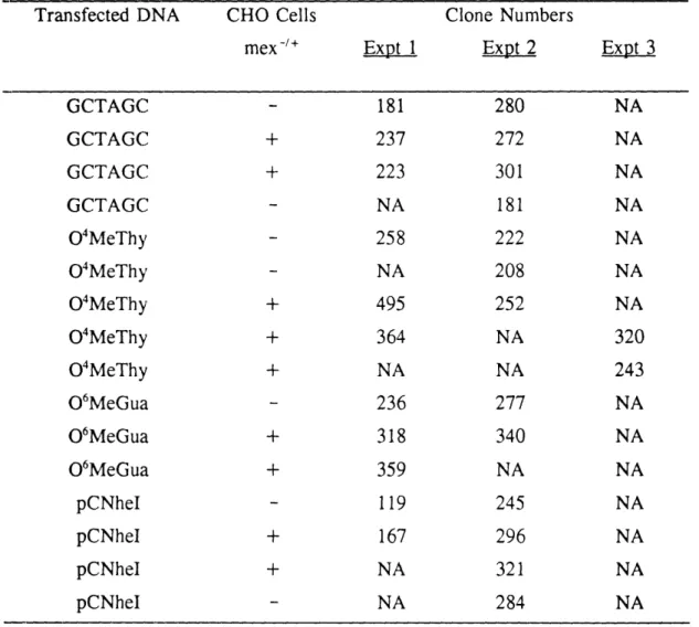
2. Summary of numbers of clones obtained in each transfection 108 3. Mutation frequency data (Transfection 1)-NheI digestion assay 116 4. Mutation frequency data (Transfection 3)-NheI digestion assay 117 5. Mutation frequency values for individual clones
(Transfection 3)--04MeThy and O6MeGua in mex- cells 129
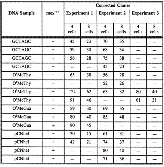
6. Clone numbers from Transfections 1 and 3 corrected for division
of transfected cells into four or eight cells prior to selection 161 7. Numbers of independent clones (corrected values) that would
generate the mutation frequencies observed in Transfection 1 162 8. Numbers of independent clones (corrected values) that would
generate the mutation frequencies observed in Transfection 3 163 9. Mutation frequency data (Transfection 1) using NEB NheI and
Betascope 603 209
10. Hybridization frequency of KHpCXW to a mixed mutant population
of amplified DNA from pCXmnI and pCNheI 213
11. Mutation frequencies (%) for Transfection 1 samples determined by
oligonucleotide hybridization 219
12. Mutation frequencies (%) from Transfection 1 samples--MAMA
analysis for O4MeThy-induced mutations 241
13. Mutation frequencies (%) from Transfection 3 samples--MAMA
LIST OF ABBREVIATIONS 3AlkAde 7AlkGua AT bp C CHO cis-DDP CsCl DBU DEN DMH DMN DMSO DNA EBV EDTA EMS ENU G418r HPLC HPRT kD LTR 3MeAde 7MeGua MNNG MNU neor NDMA NNK 04AlkThy 04MeThy O6AlkGua O6MeGua PCR PNK SDS SSPE TE TLC UV W 3-alkyladenine 7-alkylguanine ataxia telangiectasia base pair
Crick strand of the shuttle vector pCNheI Chinese hamster ovary
cis-diamminedichloroplatinum(II) cesium chloride 1,8-diazobicyclo(5.4.0)-undec-7-ene diethylnitrosamine dimethylhydrazine dimethylnitrosamine dimethylsulfoxide deoxyribonucleic acid Epstein-Barr virus
ethylene diamine tetraacetic acid ethylmethanesulfonate
ethylnitrosourea G418 resistant
high pressure liquid chromatography
hypoxanthine-guanine phosphoribosyl transferase kilodalton
long terminal repeat 3-methyladenine 7-methylguanine N-methyl-N'-nitro-nitrosoguanidine methylnitrosourea neomycin resistance nitrosodimethylamine
4-(N-methyl-N-nitrosamino)- 1-(3-pyridyl)- 1-butanone 04-Alkylthymine
04-Methylthymine 06-Alkylguanine 06-Methylguanine
polymerase chain reaction polynucleotide kinase sodium dodecyl sulfate
Sodium chloride, Sodium pyrophosphate, EDTA 10 mM Tris-HC1 buffer (pH 8), 1 mM EDTA thin layer chromatography
ultraviolet
Over the last few years much progress has been made toward the elucidation of the molecular mechanisms underlying the onset and progression of genetic diseases. One hypothesis is that certain chemicals and radiation can cause lesions or adducts in DNA, and the misreplication or misrepair of these lesions results in a fixed
mutation, permanently changing the genetic makeup of the organism (Miller, 1978). In the specific case of carcinogenesis, the presence of this genetic change affects ordinary cellular functions in such a way as to contribute to the conversion of a normal cell into a tumor cell. Multiple genetic changes may be necessary to force the full transformation to malignancy.
In order to test this model of carcinogenesis, one must first know which chemical lesions within DNA are characteristic of an exposure, how prevalent those lesions are and which mutations, if any, they may cause. Adduct and mutation spectra analyses give information as to the types of adducts that are formed by a chemical in a certain sequence context of a target gene and the resulting mutations arising in those same sequences (Miller, 1983). If mutations are measured before and after exposure, comparison of the adduct spectrum and mutation spectrum after alkylating agent treatment can give information as to the specific adducts that may have been responsible for the resultant mutations. These studies cannot impart information as to the frequency with which a single adduct is misreplicated by the replication machinery of a certain cell. In addition, these studies cannot be
informative as to the importance of individual adducts in the mutagenic process that is widely assumed to precede the neoplastic transformation of a cell induced by alkylating agents or other chemicals that attack DNA. Techniques of introducing a
site-specifically modified vector with a unique chemical modification to the DNA have evolved to accomplish these tasks (Basu and Essigmann, 1990).
Alkylating agents are widely used as chemotherapeutic agents and they are frequently found in the human environment (Bartsch and Montesano, 1984). They form a variety of adducts with DNA bases, primarily at the nitrogen and oxygen atoms (Singer, 1985). Two such adducts are O6MeGua and 04MeThy, both of
which are believed, on the basis of circumstantial evidence, to be precarcinogenic lesions in animal models. O6MeGua is found in much larger amounts in the genome
than the latter adduct and is arguably the most important premutagenic lesion formed by alkylating agents (Day et al., 1987). In selected circumstances, the presence of O4MeThy in the genome correlates well with the onset of carcinogenesis in several animal models (Singer et al., 1981; Swenberg et al., 1984, 1986). The mutagenic potential of this lesion is thus of interest despite its lower incidence. There exists an intriguing controversy over whether the same repair system (in animal models) recognizes these two adducts. Therefore, the investigation of mammalian repair enzymes that might affect these adducts was also of interest.
This work has undertaken a direct comparison of the mutagenic potential of O6alkyl- and O4alkyl-DNA adducts. These studies were undertaken in cells deficient
in or possessing the mammalian methyltransferase (MGMT) in order to assess directly the impact of that repair protein on the ultimate appearance of mutations from the two DNA lesions.
A. Introduction
Mammalian cells provide environmental agents a plethora of biomolecules with which to interact. DNA is one of the most likely targets of reaction owing to its nucleophilic nature. Although there are few data upon which to make a general statement, it seems reasonable to predict that DNA would display varying degrees of chemical accessibility during various phases of the cell cycle and as it carries out its biochemical functions, most notably transcription. The covalent interaction of DNA with chemicals and radiation leads to DNA adducts in which one or more nucleotide atoms are joined to the attacking species (Miller, 1978). If these adducts remain unrepaired prior to replication of the cellular genome, they may lead to mutations through misreplication and misrepair (Singer and Kusmierek, 1982). Alkylating agents are an important class of chemicals noted for their high reactivity with certain atoms in DNA, most notably the electron-rich oxygen and nitrogen atoms of the bases. The high reactivity of alkylating agents with DNA is exploited in their use as chemotherapeutic agents, where the desirable biological endpoint of cytotoxicity is achieved. This characteristic has a negative effect, however, since alkylators are also often potent mutagens and carcinogens (Lawley, 1976; Miller, 1978). It is also noteworthy that a growing literature (Rydberg and Lindahl, 1982; Rebeck and Samson, 1991; Xiao and Samson, 1993) has shown that endogenously generated alkylating agents may contribute significantly to the background of spontaneous mutations known to be experienced by all cells.
Investigations into the biochemical mechanisms underlying alkylating agent-induced mutagenesis and carcinogenesis have provided evidence that certain DNA
adducts are premutagenic (Wood and Essigmann, 1991). It is through mutagenesis that the DNA lesions presumably facilitate neoplastic transformation (Pegg, 1977). One lesion found in significant amounts is 06-methylguanine (O6MeGua). This lesion is likely the most important adduct formed by alkylating agents and it has been widely studied for its mutagenic and cytotoxic potential in many bacterial and
eukaryotic hosts (Pegg, 1984; Topal et al., 1986; Day et al., 1987).
Other alkyl adducts may also play an important role in carcinogenesis. For example, the relevance of 04-alkylthymine (O4AlkThy) adducts in the induction of
cancer has been investigated (Singer, 1984; Dyroff et al., 1986; Brent et al., 1988). The long persistence of the 04AlkThy adducts in the cell after alkylating agent exposure (Richardson et al., 1985), and the extraordinarily high mutagenic potency of the lesion in vitro (Dosanjh et al., 1990) and in vivo (Dosanjh et al., 1991), indicate that this type of lesion might indeed play a role in the initiation of mutagenesis and carcinogenesis.
B. The role of O4AlkThy adducts in carcinogenesis
The interaction of alkylating agents with DNA creates a wide variety of
chemical lesions. Alkylating agents can interact with virtually all of the oxygen and nitrogen atoms in cellular DNA and RNA. A variety of the adducts formed by alkylating agents is shown in Figure 1. The approximate proportions of each adduct formed in DNA as a result of treatment with several alkylating agents are given in Table 1.
Figure 1. Sites of in vivo and in vitro alkylation in nucleic acids. (Adapted from
URIDINE OR TH YMIDINE CYTIDINE GUANOSINE ADENIOSINE BACKBONE
Table 1. Proportions of Alkylated Bases Present in DNA after Alkylating Agent Treatment'
Percentage of Total Alkylation by the following Alkylators DMN MNU DMH 1-Alkyladenine 3-Alkyladenine 7-Alkyladenine 3-Alkylguanine 7-Alkylguanine 06-Alkylguanine 3-Alkylcytosine 02-Alkylcytosine 3-Alkylthymine 02-Alkylthymine 04-Alkylthymine Alkylphosphates MMS 0.7 8 1.5 0.8 68 7.5 DEN ENU 1.2 11 1.9 0.7 83 0.3 0.5 0.1 0.3 0.1 0.1-0.7
' Table adapted from Pegg, 1984
0.3 4 0.4 0.6 12 8 0.2 3 0.8 7 1-4
Studies have shown that carcinogenesis arising in target organs after treatment with a specific alkylating agent correlates with the presence and persistence of a few alkylated bases arising after treatment with the alkylating agent (Loveless, 1969; Goth and Rajewsky, 1974). These studies have primarily focused on the
O6AlkGuas, as these DNA lesions are typically persistent after treatment with alkylating agents. This persistence is believed to increase the chance for
misreplication by the host replication machinery as the adduct remains in the DNA for a long period of time. In addition, the G:C--A:T transition, the mutation known to be caused by O'AlkGua (Loechler et al., 1984; Ellison et al., 1989) is the most common mutation observed in the DNA of tumors resulting from alkylating agent exposure.
Recently, other studies have shown the persistence and predominance of
04AlkThy lesions in the DNA of organs exhibiting carcinogenic endpoints as a result of dosing animals with alkylating agents. Singer et al. (1981) dosed perinatal rats with ethylnitrosourea (ENU) and analyzed the amounts of different alkyl adducts in the liver, the brain (target organ for this carcinogen) and other tissues in the rats at differing time points. By contrast with other adducts, including alkylguanines, the O4EtThy lesion was not appreciably removed from the cellular DNA after 75 hours. This evidence suggests that 04EtThy might possibly be the lesion responsible for
mutagenesis and resulting carcinogenesis. However, the potential roles of different adducts and mechanisms are not ruled out by this study.
In a subsequent investigation, Swenberg and coworkers (1984) treated F-344 male rats with 40 ppm diethylnitrosamine (DEN) ad libidum in their water supply
according to a daily regimen that selectively induces hepatocellular carcinomas. They quantified the O4EtThy and O6EtGua in the hepatocytes and nonparenchymal cells of sacrificed animals using competitive radioimmunoasssay with high-affinity monoclonal antibodies. These two cell types were of interest because the
hepatocytes possessed the 06-methylguanine methyltransferase repair protein in
amounts three to five times higher than the levels contained in nonparenchymal cells. The researchers found that O4EtThy accumulated in hepatocyte DNA during the first 28 days of DEN exposure, approximating a steady-state level of -1 x 10⁻⁵
04EtThy/dT. This steady-state level was maintained from 28 to 77 days of DEN exposure. The authors did not give an hypothesis as to why the 04EtThy levels did not continue to increase throughout the period of DEN exposure. However, they calculated the 04EtThy half-life to be approximately seven days and they suggested that the half-life represented a combination of adduct dilution due to DNA
replication, adduct loss due to cell turnover, and possible enzymatic removal of 04EtThy from DNA. By contrast, the O6EtGua level did not accumulate in the
hepatocytes, but peaked after two days of DEN exposure at a level of -3.7 x 10⁻⁷ O6EtGua/dG. The quick removal of the adducted guanine was presumably due to the methyltransferase repair protein in these cells. The nonparenchymal cells contained only half as much 04EtThy as the hepatocytes, but this level of adducted thymine was 2.5 times the level of O6EtGua in the same cells. This study suggests
that the increased persistence of 04EtThy (and any resulting mutagenesis mediated by the lesion) may be the causative factor contributing to the resultant tumorigenic endpoint in F-344 rats treated with DEN.
A further study by Dyroff and coworkers (1986) provides evidence that the
accumulation of the 04EtThy lesion is linked to the incidence of hepatocellular carcinoma in rats treated with this carcinogen. In this study, the investigators treated male F-344 rats of varying ages with 40 ppm DEN given ad lib. in the drinking water for up to 10 weeks. They found that treatment of eight-week old rats with the carcinogen, while causing significant amounts of 04EtThy adducts to
accumulate within the hepatocellular DNA (up to a steady-state level of
approximately 7-10 x 10-6 mol 04EtThy/mol dT), did not induce any carcinomas of the hepatocytes in these animals. In contrast, a 100% incidence of hepatocellular carcinomas occurred in four-week old rats administered DEN for 6, 8 or 10 weeks and the 04EtThy steady-state concentration was similar to that seen in the eight-week old rats given the same treatment. The researchers surmised that the difference in cancer initiation seen in the two ages of rats indicated the importance of DNA replication and liver cell division in the initiation process. The O6EtGua was
efficiently repaired in the hepatocytes of treated animals and was not believed to be a precarcinogenic lesion in this study.
A more recent study investigated the activation of the K-ras protooncogene in lung tumors in mice dosed with nitrosamines (Belinsky et al., 1989). This study showed that in 25% of the lung tumors resulting in A/J mice treated with 4-(N-methyl-N-nitrosamino)- 1 -(3-pyridyl)-l-butanone (NNK) or nitrosodimethylamine (NDMA), the base change was an A:T--G:C transition in codon 61. This mutation suggested the presence of 04MeThy in the DNA as a result of chemical dosing since this lesion is known to cause T--C transitions both in vitro (Abbott and Saffhill,
1977; Singer et al., 1984, 1986a, b) and in vivo (Preston et al., 1986; Dosanjh et
al., 1991). The remaining 75% of the tumors exhibited G:C-- A:T transitions in
codon 12 of the K-ras gene, indicating that O6MeGua might have been the
premutagenic lesion responsible for oncogene activation. It should be noted that the above data only correlated the presence of 04MeThy and O6MeGua lesions with the
mutations in the tumors in the study by Belinsky and coworkers. Other lesions may have been the premutagenic species.
The bulk of cancer studies done to date has attempted to explain the initiation of cancer in a specific organ with the concentration and persistence of a particular chemical lesion within the DNA of that organ. The logic is that the higher the proportion of adduct present, and the longer it stays within the DNA (this variable is highly dependent upon the cellular repair capacity for the adduct), then the higher the probability that cellular replication of the adduct will cause a mutation. This mutation might then lead to the initiation of cancer through another undetermined mechanism. Many such studies have correlated the persistence of O6AlkGua adducts
with the onset of cancer in an organ containing high amounts of these lesions (Loveless, 1969; Goth and Rajewsky, 1974; Kleihues and Margison, 1976; Swann and Magee, 1968; Nicoll et al., 1975). Following this trend, it is fair to speculate from the literature that 04AlkThy might, in fact, be the premutagenic lesion
C. Mutagenesis by O'AlkThy adducts
Several laboratories have been instrumental in exploring the stability,
conformation and mutagenic properties of O4AlkThy lesions in DNA in both in vitro
and in vivo environments. Abbott and Saffhill were the first researchers to propose that O4MeThy might be mutagenic based on in vitro results (1977). They treated
poly(dA-dT) with dimethylsulphate (DMS) or N-methyl-N-nitrosourea (MNU) and
determined the amounts of each alkylated base formed in the alternating copolymer. They then used the methylated DNA as a template for E. coli DNA polymerase I
and determined the extent of correct and faulty replication. The researchers found that DMS caused no misreplication by the bacterial polymerase, but MNU caused a significant amount of dGMP to be incorporated into the polymerized strand of DNA. By contrast, incorporation of dCMP was not observed. Abbott and Saffhill
hypothesized that the misreplication of the methylated DNA was a result of dGMP pairing with O4MeThy as they observed a virtual one to one ratio between the
amount of incorporated guanine base and the amount of 04MeThy adducts present in the methylated substrate (Abbott and Saffhill, 1977).
These results were corroborated by subsequent studies by Singer and coworkers (Singer et al., 1983; Singer et al., 1984; Singer et al., 1986a, b). These
investigations showed that E. coli polymerase I and the Klenow fragment of pol I could incorporate 04MeThy, O4EtThy and O4IpThy into activated DNA and poly
[d(A:T)] sequences in the place of T, albeit with less efficiency. The kinetics of incorporation of the adducted thymines were such that the smaller the alkyl group, the faster and greater the extent of incorporation (Singer et al., 1986a). Dosanjh et
al. (1990) extended these results in their investigations of the incorporation of certain
nucleotides opposite a specific 04MeThy adduct in a DNA template. The authors annealed a 25-mer oligonucleotide with 04MeThy positioned at nucleotide 21 with a complementary oligonucleotide of 19 bases which ended one base 5' to the adducted thymine. They then added either dATP or dGTP at varying concentrations and
measured the extension of the primer by E. coli Klenow fragment or Drosophila
melanogaster polymerase cx-primase complex. Their studies showed that the pairing
of O4MeThy:G was favored 10-fold over the 04MeThy:A or T:G base pairs. The primers were not readily extended in the presence of dATP. In contrast, when dGTP was added to the mixture, both polymerases extended the primers past the adducted thymine to similar extents, indicating that the 3'-5' exonuclease capability of the Klenow fragment was not efficiently removing the mismatch (Dosanjh et al.,
1990).
More recently, Dosanjh et al. (1993) have investigated the kinetics of incorporation of these same two nucleotide triphosphates opposite 04MeThy and 04EtThy. Using the same 25-mer sequence discussed above as a template, the authors examined the extension of a primer complementary to the template molecule but that terminated two nucleotides 5' proximal to the adducted base. They
measured the incorporation of either dGTP or dATP at varying concentrations opposite the 04MeThy and 04EtThy bases by assaying the amount of full length
double-stranded molecules produced by E. coli Klenow fragment. The researchers found that both the 04MeThy:G and 04EtThy:G base pairs formed at least 10-fold
preference for the methyl adduct. Polymerization was complete beyond both alkylated thymines, indicating that neither adduct was a block to replication and the size of the adduct was not significant in determining the mutagenic potential or mutation type arising from replication of the modified base. In contrast, T:G base pairs were formed very infrequently (frequency of formation = 1.2 x 10⁴ compared
to the formation of T:A base pairs). In addition, dATP was placed opposite the O4AlkThy bases to a very low extent, with a frequency of •2 x
10' . The other
nucleotides, dCTP and dTTP were not observed to incorporate opposite the alkylthymine adducts. The limit of sensitivity of this assay was 10-6-10. (M.
Dosanjh, personal communication), so that if dCTP or dTTP were inserted opposite the alkylthymine lesions at a lower frequency than these low values, this
incorporation would not have been observed. The incorporation studies discussed above show that 04AlkThy adducts can form erroneous base pairs that ultimately lead to T-+C transitions.
The mutagenic potential of 04AlkThy bases has also been studied in vivo. Preston et al. (1986) examined the mutations arising from replication of
site-specifically positioned O4MeThy residues in E. coli spheroplasts. The modified base
was the terminal nucleotide of a primer annealed to the (+) strand of OX174 am3 DNA. The substrate was extended using E. coli DNA pol I with an excess of dNTPs. The resulting double-stranded substrate was transfected and replicated within ada- (repair-deficient) and ada÷ (repair-proficient) E. coli. The adduct caused a mutation frequency in the repair-deficient cells that was 10 times greater than that of the repair-proficient cells. DNA sequenced from 20 independent mutant plaques
exclusively showed a T-+C transition arising from replication of the adduct (Preston
et al., 1986).
Recent studies have also examined the mutagenic potential of 04MeThy in comparison to the O6MeGua adduct (Dosanjh et al., 1991). These studies involved a
site-specifically modified M13mpl9 single-stranded genome with the O4MeThy or O6MeGua lesion positioned within a unique NheI restriction site in the genome. Replication of these adducts within wild-type (repair-proficient) E. coli cells resulted in a 12% mutation frequency arising from 04MeThy and no detectable mutations arising from O6MeGua. When the cells were treated with an MNNG (N-methyl N'-nitro-N-nitrosoguanidine) challenge to remove all methyltransferase repair proteins, the mutation frequency for 04MeThy rose to 24%, whereas the O6MeGua mutation
frequency rose to 1.1%. The mutations arising from 04MeThy were T-WC transitions, confirming earlier reports. O'MeGua induced G->A transitions in this
system (Dosanjh et al., 1991). This study showed that in bacteria, the 04MeThy adduct was significantly more mutagenic than O6MeGua.
The mutagenic potential of 04EtThy adducts has also been investigated in vivo. Klein and coworkers (1990) transfected an extrachromosomally-replicating shuttle vector containing a single site-specific 04EtThy lesion into HeLa cells. The progeny
molecules were removed from the cells 72 hours post-transfection, and unreplicated material was digested with DpnI. This enzyme cleaves GATC sequences only when the A base is methylated (Lacks and Greenberg, 1975) and this modification occurs only in bacterial cells. Therefore, unreplicated duplexes of this sequence will have both strands methylated while replicated sequences will be hemimethylated and hence
will be resistant to cleavage. The digested DNA was transfected into E. coli for determination of mutation frequency. The adduct induced a mutation frequency
ranging from 19-27%, with the average being 23%. The predominant mutations arising from replication of the adduct in the human cells were single T--C transitions at the adduct site. The studies discussed above have shown very clearly that
O4AlkThy adducts are highly mutagenic in bacterial and mammalian cells and are much more mutagenic than O'MeGua lesions in a bacterial environment.
D. Repair of O4AlkThy and O6AlkGua Adducts
Bacterial cells have well documented capabilities for repairing 04AlkThy
adducts. The E. coli Ada repair protein is a 39 kD enzyme encoded by the inducible ada gene (Sedgwick, 1983; Teo et al., 1984). There are two cysteine residues in
the protein (Margison et al., 1985; Sedgwick et al., 1988), one for removing the methyl groups from O6MeGua and 04MeThy adducts (McCarthy et al., 1984;
Takano et al., 1988), and the other for removing methyl groups from
methylphosphotriesters (McCarthy and Lindahl, 1985; Weinfeld et al., 1985). The removal of the methyl groups from damaged DNA is accomplished by an internal cysteine residue in a step that inactivates the protein. Since there seems to be no mechanism for removing the methyl group from the S-methylcysteine, the protein cannot act upon any other methylated base (Lindahl et al., 1982; Demple et al.,
1982) and thus is not a true enzyme. The Ada protein can also remove larger alkyl groups from guanine bases, including ethyl, n-propyl and n-butyl, but at slower rates than it does the methyl groups (Pegg and Dolan, 1985; Pegg et al., 1985).
After repairing methylphosphotriesters, the Ada protein initiates the adaptive response in bacteria (Samson and Cairns, 1977) by activating certain genes whose products are involved in repair of other alkylated adducts. These genes include the
alkA gene (McCarthy et al., 1984; Evenson and Seeberg, 1982), the ada-alkB
operon (Teo et al., 1986; Nakabeppu and Sekiguchi, 1986; Kataoka and Sekiguchi, 1985) and the aidB gene (Volkert et al., 1986). The adaptive response is turned on in response to low levels of alkylating agents, resulting in a several hundred fold increase in alkyltransferase activity that protects the cell from the mutagenic and cytotoxic effects of the alkylated bases.
More specifically, the adaptive response is a highly controlled DNA repair pathway that is distinct from other inducible repair responses in E. coli (such as the SOS response) and it is responsive only to alkylation damage (Lindahl et al., 1988). After exposure of bacteria to a methylating agent, the DNA is alkylated at several positions. The Ada methyltransferase (which is typically found in very low levels in untreated bacteria) removes a methyl group from a methylphosphotriester (S
diastereoisomer only; McCarthy and Lindahl, 1985; Weinfeld et al., 1985; Hamblin
et al., 1985) to the cysteine residue at position 69 in the N-terminal portion of the
protein (Sedgwick et al., 1988). The methylation of the Ada protein at this N-terminal cysteine converts the methyltransferase from a weak transcriptional activator into a strong one (Teo et al., 1986). The methylated Ada protein then binds to a conserved AAANNAAAGCGCA sequence in the promoter regions of the ada and
alkA genes (Teo et al., 1986, Nakabeppu and Sekiguchi, 1986; Nakabeppu et al.,
polymerase-binding site in the ada promoter, and the conserved sequence overlaps with the same polymerase-binding site in the alkA promoter (Nakabeppu and Sekiguchi, 1986; Nakabeppu et al., 1985). The methylated Ada protein that is bound to the conserved sequence may facilitate recognition of the two promoters by RNA polymerase by direct contact with the polymerase or by inducing a change in DNA conformation (Lindahl et al., 1988). Increased transcription of the ada-alkB operon results in a several-fold increase in the levels of the Ada methyltransferase and the AlkB protein, whose function has not yet been identified. However,
experiments indicate that the protein is responsible for DNA repair and is not simply detoxifying the alkylating agent (Lindahl et al., 1988). In addition, alkB mutants are phenotypically similar to alkA or tag mutants (Kataoka and Sekiguchi, 1986; Kondo
et al., 1986). The Tag protein repairs 3-methyladenine only, whereas the AlkA
protein repairs 3-methyladenine, 3-methylguanine and 7-methylguanine, as well as 02-methylthymine and 02-methylcytosine (Riazuddin and Lindahl, 1978; Thomas et
al., 1982; Lindahl, 1976; Laval, 1977; McCarthy et al., 1984). Increased
transcription of the alkA gene induces the levels of the AlkA protein 20-fold. The methylated Ada protein also induces the production of the AidB protein, but the purpose of the latter protein is, as yet, unknown (Volkert and Nguyen, 1984; Volkert et al., 1986). Interestingly, E. coli mutants for the AidB protein are resistant to killing by alkylating agents (Lindahl et al., 1988).
The Ada protein can be cleaved into two peptides, one 20 kD and the other 19 kD in size (Teo et al., 1984; Teo, 1987). The 19 kD fragment has the cysteine
moiety that repairs O6MeGua and O4MeThy lesions and the 20 kD fragment repairs
the methylphosphotriesters.
The Ogt protein is a second methyltransferase repair protein present in bacteria that has been recently isolated (Potter et al., 1987; Rebeck et al., 1988). This 19 kD protein is constitutive in bacteria and can repair O6MeGua and 04MeThy adducts, but not methylphosphotriesters (Rebeck et al., 1988). The protein is present in bacteria that do not contain the ada gene, hence it is encoded by a separate gene (Potter et al., 1987; Rebeck et al., 1988) and it does not seem to be inducible.
Studies have been done investigating the in vitro kinetics of repair of 04MeThy adducts with the different bacterial methyltransferases. Wilkinson and coworkers (1989) incubated modified dodecanucleotides with either purified Ogt protein or the
19 kD fragment of the Ada protein and determined that both enzymes could repair O4MeThy. Interestingly, they found that the Ogt protein repaired the 04MeThy
adducts 84 times faster than the Ada protein. More recently, Sassanfar et al. (1991) have found that a 25mer containing a specific 04MeThy adduct inactivated Ogt with an IC50 value of 3 nM. The same oligonucleotide inactivated the Ada
methyltransferase with an IC50 value of 27.5 nM. These data suggest that the affinity
of Ogt for 04MeThy is approximately nine-fold greater than the affinity of the Ada protein for the same adducted base.
Methyltransferase repair proteins have been isolated from many eukaryotes (Wilkinson et al., 1989; Koike et al., 1990) and the gene sequences have been cloned from human (Tano et al., 1990; Rydberg et al., 1990), rat (Potter et al.,
1991), mouse (Santibanez-Koref et al., 1992; Shiraishi et al., 1992) and yeast (Xiao
et al., 1991). Mammalian methyltransferases transfer methyl groups from the
06-position of guanine to a cysteine residue within the protein in a second-order
stoichiometric reaction. Like the mechanism of the bacterial methyltransferases, this
in situ transfer is a suicide reaction, resulting in inactivation of the mammalian
enzyme. In addition, the mammalian protein can repair larger alkyl moieties, but the rate of repair generally decreases as the moiety size increases (Pegg et al., 1984;
Pegg and Dolan, 1985; Pegg et al., 1985). However, the methyltransferase has a strong preference for the benzyl moiety on the 06-position of guanine (O6BzGua). This lesion is highly preferred by the enzyme; inactivation of the methyltransferase by O6BzGua occurs more rapidly than with O6MeGua (Dolan et al., 1990; 1991). A
2.5 MM concentration of O6BzGua is sufficient to cause a > 90% loss of
methyltransferase activity within 10 minutes, whereas 0.2 mM O6MeGua incubated with the enzyme for 60 minutes was required to bring about the same reduction (Dolan et al., 1990).
The mechanism of action of the human methyltransferase by 06BzGua was recently determined (Pegg et al., 1993). The cysteine residue in the active site of the human methyltransferase binds to the O6BzGua and removes it, forming
S-benzylcysteine and Gua in stoichiometric proportions. The mechanism is in agreement with the increased rate of removal of O6BzGua as compared with
06MeGua, in that a larger benzyl group would be much more likely to enter into a bimolecular displacement reaction than a methyl group (Pegg et al., 1993).
compared to O6MeGua to inactivate the mammalian methyltransferase; the 06-allylguanine residue is not as readily repaired as 06BzGua, however (Pegg et al., 1993). By contrast, 04-benzylthymine was not a substrate for repair by any yeast,
bacterial or human alkyltransferases in the study (Pegg et al., 1993).
A recent study indicated that a 28 amino acid carboxy-terminal tail of the mammalian methyltransferase (found in humans, rats and mice) and not present in the bacterial methyltransferases, indirectly affects binding of O'BzGua by the
methyltransferase (Morgan et al., 1993). Deletion of this 28 amino acids caused the rate of complete inactivation of methyltransferase by O6BzGua to be reduced five-fold over that of the intact methyltransferase. Deletion of the carboxyl-terminal tail also prevents binding of the enzyme to O6MeGua at low temperatures. The study suggests that the tail has a role in the conformation of the enzyme's binding site (Morgan et al., 1993).
Despite the conserved acceptor site within the methyltransferases from differing species, these varied methyltransferases have different substrate specificities. Unlike the bacterial methyltransferases, the mammalian enzyme will not repair
methylphosphotriesters and the majority of evidence suggests that it cannot repair O4AlkThy (Brent et al., 1988), although this is a matter of some controversy (Becker and Montesano, 1985; Koike et al., 1990; Sassanfar et al., 1991). In addition, most mammalian methyltransferase repair proteins do not seem to be part of a general inducible adaptive response to low levels of alkylating agents.
Early studies in whole animals suggested that the MGMT protein could be induced two- to three-fold in the livers of rats chronically fed dimethylnitrosamine
(DMN; Montesano et al., 1979). Montesano and coworkers extended their investigations by establishing the necessary dose and length of pretreatment with DMN to induce the repair response (Montesano et al., 1980). They investigated the
induction of MGMT in male BD IV and female Sprague-Dawley rats to obtain information on sex and strain differences. Their results showed that administering 0.75 mg/kg DMN per day through stomach tubes to female Sprague-Dawley rats for six weeks increased the level of the MGMT protein in the rat livers 2.5-fold over untreated rats. This increase was the same as that found in the livers of the same type of rat given 1 or 2 mg/kg DMN per day for four weeks. In BD IV rats, a maximum induction effect was seen after three weeks of feeding with 2 mg/kg DMN per day as measured by the level of O6MeGua in the liver DNA six hours after administration of 14C-DMN at the same dose level. After the three week DMN pretreatment, the O'MeGua levels dropped four-fold as compared to untreated control animals; by contrast, 7-methylguanine (7MeGua) and 3-methyladenine (3MeAde) levels did not decrease during the course of treatment (Montesano et al.,
1980), suggesting that the repair induction was specific for O6MeGua and not the result of general repair of alkylated bases.
A subsequent study also investigated the induction of MGMT with DMN. This study showed that this repair protein could be induced in rat hepatocytes (isolated from the whole liver prior to MGMT assay) but not in those same cells of C3H or C57BL mice (Lindamood et al., 1984). This study showed that the mice and F-344 rats that were given 10, 30 or 100 ppm DMN in their drinking water for 16 days exhibited very different levels of O6MeGua and 7MeGua in their liver DNA. The
doses obtained through the drinking water were calculated based on the surface area of the animal and the corrected data showed that each animal ingested similar amounts of DMN at each concentration level. The O'MeGua/7MeGua ratio in the hepatocytes of the F-344 rats was consistently low (<.02) at all dosages of the alkylating agent. This ratio of O6MeGua to 7MeGua increased dramatically over background (<0.02) in both mouse strains at the 30 ppm dosage, where it was 0.05 and 0.13 for C57BL and C3H mice, respectively, and 0.07 at the 100 ppm dosage in
C57BL mice. This represents a 4-13-fold increase in these ratios for the mice
hepatocyte DNA at the higher dosages. The 100 ppm dose killed all C3H mice after four days of treatment; this strain of mice has a high incidence of spontaneous
hepatocellular carcinoma and histopathological analyses of the livers of the dead mice showed severe necrosis and "collapse of liver architecture" (Lindamood et al., 1984). The above data suggested that the MGMT protein was being induced and mediating the reduction in alkylated guanine in the rat hepatocytes. That response was, in fact, what Lindamood and coworkers observed. The rat MGMT protein was induced to 150% of the level of control protein values at the 10 ppm dosage and 200% of the level of control values at the 30 and 100 ppm doses. The above studies indicated that chronic administration of DMN to rats induced the levels of the liver MGMT protein 1.5-2.5-fold, but the mouse liver MGMT protein was not induced.
The MGMT protein has been shown to be non-inducible in other rodent species. Bamborschke and associates pretreated the Mongolian gerbil with 0.75 mg DMN/kg/day for 35 days. The researchers observed that the pretreatment did not increase the ability of the rodent liver to remove 14C-06MeGua from DNA after a
single intra peritoneal injection of '4C-DMN at a dose of 2 mg/kg as determined by HPLC analysis of unmethylated guanine and radiolabeled O6MeGua (Bamborschke et
al., 1983).
Samson and Schwartz showed that MNNG pretreatment of CHO cells and a human skin fibroblast cell line (GM637) resulted in the increased resistance of the cell strains to the cytotoxic and genotoxic effects of MNNG and MNU (Samson and Schwartz, 1980). The researchers incubated the cells in media with 0.01 Jg/mL MNNG every six hours for 48 hours. Pretreated cells that were subsequently challenged with 0.1-0.2 CIg/mL MNNG experienced a two-fold increase in survival over control cells. Both cell strains that had been pretreated with MNNG for at least 48 hours were resistant to the induction of sister chromatid exchanges by exposure to MNNG (0-0.05 jig/mL), MNU (0-2.5 /ig/mL), and ENU (0-25 pg/mL). The cells were not resistant to the induction of sister chromatid exchanges (SCEs) by UV light, however. The study did not specify whether the CHO cells used were mex or mex÷, nor did the researchers assay for an increase in MGMT level. Therefore,
despite the argument by the authors that the response observed appeared to be specific for alkylating agents, it was not necessarily due to an increase in MGMT levels or an extension of the half-life of the protein.
Waldstein and coworkers (1982) induced a three-fold increase in the amount of MGMT protein contained in extracts from HeLa CCL2 cells as assayed by the removal of methyl groups from radiolabeled alkylated DNA. They accomplished this induction by treating the cells with 25 ng/mL MNNG, four separate times in 24 hours. The maximum amount of induction was seen eight hours after the last
treatment of cells with the chemical, and the MGMT level decreased to one-half the maximal level within the next 24 hours. Interestingly, treatment of the cells with MNNG at concentrations higher than a cumulative dose of 100 ng/mL induced a response significantly lower than the 100 ng/mL dosing regimen (Waldstein et al., 1982).
Similar results were obtained when a rat hepatoma cell line (H4) was incubated in the presence of MNNG, methylmethanesulphonate (MMS) and
ethylmethanesulphonate (EMS; Frosina and Laval, 1987). This study is unique, however, because the induction occurred after a single administration of the alkylating agent. H4 cells incubated in a 10 jiM concentration of MNNG first suffered a depletion of their cellular levels of MGMT. The levels of repair protein per cell then increased after six hours to reach a maximum level (approximately three times the level in untreated cells) 48 hours after chemical treatment. The repair protein levels decreased rapidly after this time point. EMS and MMS also induced MGMT levels in the H4 cells, but MMS did so without an initial decrease in the levels of the repair protein and EMS did so with an initial decrease in MGMT of <50%. A 1 mM dose of MMS induced the MGMT level five-fold over control levels and a 10 mM EMS treatment induced a four-fold increase in the levels of the protein.
The possibility that this latter induction may be part of a larger regulated response must be considered because EMS and MMS produce far fewer O'AlkGua residues in DNA (Pegg, 1984) in relation to other alkylated bases, such as
is upregulated in response to an activated form of the mammalian methylpurine glycosylase. The slight induction seen after treatment with MNNG was the same level (three-fold) observed in other cells strains and organs discussed above.
Fritz and coworkers performed an extensive study of the induction of the MGMT protein in several human and rodent cell lines treated with MNNG (Fritz et
al., 1991). The authors assayed the increase in MGMT mRNA transcripts from the
cell lines as compared with untreated controls. They found that varying
concentrations of MNNG (1 nM-15 /tM) added to cells for four or eight hours in the media were able to induce mRNA production for the MGMT three- to five-fold in the H4 and FTO-2B cell lines (both rat hepatomas). By contrast, MNNG treatment was unable to induce mRNA transcripts for the repair protein in the following mex cell lines despite the presence of the MGMT gene within these cells: CHO-9, V79 and HeLa MR. In addition, MNNG treatment failed to increase MGMT mRNA levels in those mex+ cells tested that showed normal mRNA levels that paralleled their MGMT activity: NIH 3T3, HeLa S3 and HepG2. More importantly, the rat hepatoma MGMT mRNAs were also induced by MNU, MMS, N-hydroxyethyl-N-chloroethylnitrosourea, UV light and X-rays. This latter induction by non-alkylating DNA damaging agents suggested that the induction of MGMT was a response to non-specific DNA damage (Fritz et al., 1991).
The extensive study discussed above corroborated data obtained by other researchers that showed the rat liver MGMT was inducible by acetylaminofluorene pretreatment (Cooper et al., 1982), single and multiple doses of aflatoxin (Chu et
tetrachloride, 1,2-dimethylhydrazine (1,2-DMH), DEN (diethylnitrosamine) and DMN (Pegg and Perry, 1981). Pegg and Perry also showed that partial hepatectomy in the rat increased the levels of MGMT protein.
The increase in MGMT levels in response to non-specific DNA damage is not limited to rat liver, however. Murine fibroblasts (C3H 10T1/2 strain) exposed to X-rays (1.5 Gy) were more resistant to the cytotoxic and mutagenic effects of
subsequent MNNG treatments (0.2, 0.6 and 1.0 pg/mL treatment) than non-irradiated cells (van Hofe and Kennedy, 1991). The above data suggested that it may have been cell regeneration or hormonal changes caused by the toxic agents used in the treatments that caused the increased levels of methyltransferase repair protein. However, the studies by Frosina and Laval (1987) and Fritz and coworkers
(1991) indicated that the induction of MGMT protein as a result of treatment with alkylating agents or other DNA damaging agents may be part of a system of coordinated regulation, not unlike that in E. coli.
Recent evidence from the laboratory of Dr. F. Laval (1991) extends that
hypothesis. Dr. Laval treated rat hepatoma cells (H4) in culture with cis-diammine-dichloroplatinum II (cis-DDP, 5 /tM), N-methyl-9-hydroxyellipticinium (2.5 j/g/mL) and y-rays (3 Gy). mRNA levels for the MGMT and 3-methyladenine glycosylase were determined by probing with the rat cDNAs for these proteins. Each DNA damaging agent induced the levels of each repair protein to a similar extent. The MGMT mRNA was increased two- to five-fold and the 3MeAde glycosylase mRNA was increased two- to three-fold. The increases for both transcripts showed a time dependence; 24 hours were required to observe a significant response and the high
level of protein reached at that time point was maintained for 48 hours. mRNA levels had returned to control levels by 96 hours post-treatment (Laval, 1991). DNA amplification was not observed in the cells, which suggested that increased
trasncription was triggered by the DNA damage. The above studies indicated that the adaptive response in mammalian cells is limited mostly to rat hepatoma cell lines and hepatocytes from whole organs, with few non-rat cell lines exhibiting a similar response. In addition, the response seems to be triggered as a result of general DNA damage rather than being specific for alkylation damage.
As mentioned above, many investigators have studied the repair of 04AlkThy adducts with the mammalian methyltransferase repair proteins. Much of this work has been summarized in a concensus view presented by several researchers in the field of chemical carcinogenesis (Brent er al., 1988) and it is their opinion that 04AlkThy adducts are repaired by a mechanism different than that in bacterial cells. A few of the studies contributing to this viewpoint are discussed in the following paragraphs.
Many investigators have incubated mammalian cell extracts with poly(dA-dT) polymers that were treated with 3H-MNU. Brent (1985) incubated this substrate with methyltransferase purified from human cultured lymphoblasts (CCRF-CEM line) and found that the enzyme was not depleted by the substrate, but it was depleted by an MNU-treated poly(dG-dC). Yarosh and coworkers (1985) treated poly(dT) with 'H-MNU, then annealed the copolymer to poly(dA). The resultant double-stranded substrate was then incubated with human liver cell extracts, but no removal of the methyl groups from the alkylated thymines was seen. Positive
controls performed simultaneously showed the removal of methyl groups from O6MeGua adducts formed in MNU-treated DNA by the same liver cell extracts. In
addition, methyl groups from O6MeGua and 04MeThy in the substrates were
removed by an E. coli BS21 extract (ada', repair proficient cell line). A 3H-MNU
treated poly(dT) substrate annealed to poly(dA) was also used by Dolan et al. (1984) and Domaradzki et al. (1984) to investigate 04MeThy repair. The researchers found no significant repair of the methylated pyrimidines after incubating the substrates with partially purified rat liver extracts or crude extracts prepared from transformed human fibroblasts (XP12BE). Studies involving the use of poly(dA:dT) to
investigate the repair of alkylated thymines by mammalian proteins may not be optimal since this DNA copolymer most likely does not mimic or accurately
represent cellular DNA. Thus, poly(dA:dT) may not be a recognizable substrate for any repair system capable of recognizing and removing modified thymines.
Hall and Karran (1986) used a variety of substrate polynucleotides treated with 'H-MNU to study the activity of methyltransferase towards O4MeThy adducts. They
found that an excess of partially purified calf thymus methyltransferase repair protein did not recognize the 04MeThy adducts present in the substrate, whereas the same amount of E. coli methyltransferase removed all methyl groups from the substrate.
Pegg, Dolan and coworkers (Scicchitano et al., 1986; Dolan et al., 1988a, b) have investigated the repair of alkylated adducts using oligomers of defined
sequence. Self-complementary dodecamers containing a single O6MeGua or
O4MeThy lesion were incubated with rat liver and human tumor cell extracts. The O6MeGua-containing oligomers were demethylated by the extracts (Scicchitano et
al., 1986; Dolan et al., 1988a), but not the 04MeThy-containing oligomers (Dolan et
al., 1988b). Studies show that the ethylated pyrimidine is also not a good substrate
for repair by the mammalian methyltransferase. Dolan and coworkers (1984) treated
14C-ENU-treated DNA, containing 2.5% 04EtThy and 7.8% O6EtGua (fractions of
the total ethylation), with purified rat liver methyltransferase for eight hours. In this time, all the O6EtGua was removed, but no loss of 04EtThy was detected.
Despite the apparent inability of mammalian methyltransferases to repair 04AlkThy lesions in vivo or in vitro, these lesions are actively removed from the genomes of animals treated with alkylating agents, albeit very slowly. Recent
animal studies have shown that the half-life (t,,2) of 04MeThy in liver DNA of rats is
20 hours (Richardson et al., 1985), while the t112 of 04EtThy ranges from two and a
half days in the brain tissue of 10 day old BD IX rats (Singer et al., 1981) to eleven days in the hepatocytes of rats (Richardson et al., 1985).
Studies using cultured mammalian cells also show evidence for an active repair system for the adducts. 04MeThy molecules incorporated into the DNA of cultured
V79 cells by the uptake of precursor nucleosides, were removed rapidly with a tl 2
2-3 hours (Saffhill and Fox, 1980). Bodell and coworkers (1979) treated cultured human fibroblasts with ENU and performed HPLC analysis of the DNA. They found that the 04EtThy adducts were removed with a half-life of 40-60 hours. Wani
and D'Ambrosio (1987) also found that ENU-treated human skin fibroblasts and kidney epithelial cells showed a gradual biphasic removal of the alkylated thymine as a function of post-treatment time in culture. Using sensitive immunoassays to