HAL Id: hal-03008428
https://hal.archives-ouvertes.fr/hal-03008428
Submitted on 16 Nov 2020
HAL is a multi-disciplinary open access
archive for the deposit and dissemination of
sci-entific research documents, whether they are
pub-lished or not. The documents may come from
teaching and research institutions in France or
abroad, or from public or private research centers.
L’archive ouverte pluridisciplinaire HAL, est
destinée au dépôt et à la diffusion de documents
scientifiques de niveau recherche, publiés ou non,
émanant des établissements d’enseignement et de
recherche français ou étrangers, des laboratoires
publics ou privés.
Distributed under a Creative Commons Attribution| 4.0 International License
Flora Reverchon, Vidian De Concini, Vanessa Larrigaldie, Sulayman
Benmerzoug, Sylvain Briault, Dieudonnée Togbe, Bernhard Ryffel, Valérie F J
Quesniaux, Arnaud Menuet
To cite this version:
Flora Reverchon, Vidian De Concini, Vanessa Larrigaldie, Sulayman Benmerzoug, Sylvain Briault, et
al.. Hippocampal interleukin-33 mediates neuroinflammation-induced cognitive impairments. Journal
of Neuroinflammation, BioMed Central, 2020, 17 (1), �10.1186/s12974-020-01939-6�. �hal-03008428�
R E S E A R C H
Open Access
Hippocampal interleukin-33 mediates
neuroinflammation-induced cognitive
impairments
Flora Reverchon
1,2, Vidian de Concini
1, Vanessa Larrigaldie
1, Sulayman Benmerzoug
1,3, Sylvain Briault
1,4,
Dieudonnée Togbé
5, Bernhard Ryffel
1, Valérie F. J. Quesniaux
1and Arnaud Menuet
1*Abstract
Background: Interleukin (IL)-33 is expressed in a healthy brain and plays a pivotal role in several neuropathologies, as protective or contributing to the development of cerebral diseases associated with cognitive impairments. However, the role of IL-33 in the brain is poorly understood, raising the question of its involvement in immunoregulatory mechanisms.
Methods: We administered recombinant 33 (rm33) by intra-hippocampal injection to C57BL/6 J (WT) and IL-1αβ deficient mice. Chronic minocycline administration was performed and cognitive functions were examined trough spatial habituation test. Hippocampal inflammatory responses were investigated by RT-qPCR. The microglia activation was assessed using immunohistological staining and fluorescence-activated cell sorting (FACS).
Results: We showed that IL-33 administration in mice led to a spatial memory performance defect associated with an increase of inflammatory markers in the hippocampus while minocycline administration limited the
inflammatory response. Quantitative assessment of glial cell activation in situ demonstrated an increase of proximal intersections per radius in each part of the hippocampus. Moreover, rmIL-33 significantly promoted the outgrowth of microglial processes. Fluorescence-activated cell sorting analysis on isolated microglia, revealed overexpression of IL-1β, 48 h post-rmIL-33 administration. This microglial reactivity was closely related to the onset of cognitive disturbance. Finally, we demonstrated that IL-1αβ deficient mice were resistant to cognitive disorders after intra-hippocampal IL-33 injection.
Conclusion: Thus, hippocampal IL-33 induced an inflammatory state, including IL-1β overexpression by microglia cells, being causative of the cognitive impairment. These results highlight the pathological role for IL-33 in the central nervous system, independently of a specific neuropathological model.
Keywords: Interleukin-33, Interleukin-1, Microglia, Memory
© The Author(s). 2020 Open Access This article is licensed under a Creative Commons Attribution 4.0 International License, which permits use, sharing, adaptation, distribution and reproduction in any medium or format, as long as you give appropriate credit to the original author(s) and the source, provide a link to the Creative Commons licence, and indicate if changes were made. The images or other third party material in this article are included in the article's Creative Commons licence, unless indicated otherwise in a credit line to the material. If material is not included in the article's Creative Commons licence and your intended use is not permitted by statutory regulation or exceeds the permitted use, you will need to obtain permission directly from the copyright holder. To view a copy of this licence, visithttp://creativecommons.org/licenses/by/4.0/. The Creative Commons Public Domain Dedication waiver (http://creativecommons.org/publicdomain/zero/1.0/) applies to the data made available in this article, unless otherwise stated in a credit line to the data.
* Correspondence:[email protected]
Flora Reverchon and Vidian de Concini are co-first authors.
1UMR7355, Experimental and Molecular Immunology and Neurogenetics,
CNRS and University of Orléans, 3B rue de la Ferollerie, 45071 Orléans, France Full list of author information is available at the end of the article
Introduction
IL-33 is a member of the interleukin-1 (IL-1) cytokine family that plays important roles in various disorders including allergy, autoimmune, or cardiovascular dis-eases through its receptor ST2 and co-receptor IL-1 accessory protein (IL-1RAcP) [1]. Recently, IL-33 has also been involved in the pathogenesis of central ner-vous system (CNS) diseases such as neurodegenerative diseases, stroke, or infectious diseases. Broadly and highly expressed in the CNS in physiological condi-tions, IL-33 is described as a key regulator of neuro-inflammation [2–4].
In experimental autoimmune encephalomyelitis (EAE), a model of multiple sclerosis disease (MS), a systemic administration of recombinant IL-33, from the day of immunization until day 18, induces a protective effect [5]. However, the intraperitoneal administration of anti-IL-33 neutralizing antibodies also delayed the onset and the severity of EAE [6]. These apparently opposite findings highlight the dual function of IL-33. Moreover, this dual function of IL-33 has also been observed in Alzheimer’s disease (AD). IL-33 is highly expressed in the vicinity of amyloid plaques and in glial cells in brain sections from AD patients suggesting that a prolonged IL-33 production may induce inflammatory molecule release and contribute to the AD pathogenesis with neuronal damage [7]. However, more recently, Saresella et al. [8] demonstrated a decrease of IL-33 in the serum of AD patients as compared with healthy controls. These clinical data highlight a complex pro- and anti-inflammatory properties of IL-33 in AD patients acting both at the central and systemic level. IL-33 dual func-tions have also been observed in CNS infectious diseases. We previously reported the essential role of the IL-33 receptor ST2 in the pathogenesis of experimental cerebral malaria (ECM) caused by Plasmodium berghei Anka (PbA)-infection in mice. We showed that ST2-deficient mice were resistant to PbA-induced neuropath-ology [9] and demonstrated a deleterious role of CNS endogenous IL-33 in the neuropathogenesis associated with cognitive disorders [10]. Surprisingly, IL-33 defi-cient mice were not resistant to ECM [11] and IL-33 systemic administration improved antimalarial drug treatment of ECM via Treg cells [12, 13]. Thus, IL-33 has dual effects on infection, inflammation, and diseases of the CNS [1] raising the question of the cellular and immunomodulators involved.
Immunohistological analyses and IL-33/citrine reporter mice showed that astrocytes [14,15] and oligodendrocytes [10,16] are the main cellular sources of IL-33 within the CNS. Moreover, ST2 receptor is overexpressed by astro-cytes and microglial cells under pathophysiological condi-tions [14]. Microglia could be the first glial cells to respond to IL-33 stimulation through the ST2/IL-1RAcP
receptor complex [15]. We previously showed a deleteri-ous effect of CNS endogendeleteri-ous IL-33 through the activation of microglia leading to IL-1β release in ECM [10]. IL-33 is not only involved [15, 16] but essential for the microglial activation [17]. Given the importance of microglia in the neurotoxic or neuroprotective inflamma-tory responses, CNS IL-33 may be a key factor in the neuroinflammatory processes and associated with cogni-tive impairments.
In this study, we show that recombinant mouse IL-33 administration in the hippocampus led to microglial cell activation and increased IL-1 production associated with cognitive disturbance.
Materials and methods
Mice and ethics statement
C57BL/6 J (wild-type; WT) male mice under specific pathogen-free (SPF) condition at 8 weeks of age were purchased from Janvier Labs (Le Genest Saint Isle, France). Mice deficient for both IL-1α and IL-1β were bred in the Transgenose Institute animal facility (CNRS UPS44, Orleans, France). They were issued from an intercross between IL-1α ΚΟ and IL-1β ΚΟ mice [18]. As they were backcrossed 10-fold on C57BL/6 J back-ground, C57BL/6 J control was used. Mice were housed at four per propylene cage with woodchip bedding, and kept under controlled conditions of temperature (20– 22 °C), humidity (50%), and bright cycle (12/12-h dark/ light), with free access to chow pellets and water. The animals were previously habituated to our animal facility at 4 weeks and used in experimental settings at 8 weeks of age. All animal experimental protocols complied with the French ethical and animal experiments regulations (see Charte Nationale, Code Rural R 214–122, 214–124 and European Union Directive 86/609/EEC) and were approved by the “Ethics Committee for Animal Experi-mentation of CNRS Campus Orleans” (CCO), registered (N°3) by the French National Committee of Ethical Re-flexion for Animal Experimentation, under N° CLE CCO 2015-1084 and by the French “Ministère de l’enseigne-ment supérieur, de la recherche et de l’innovation”, under number APAFIS #19264.
Intrahippocampal microinjection
Mice divided into 4 groups, received intrahippocampal injections of either vehicle PBS containing 0.1% BSA as a carrier (PBS-BSA) or recombinant mouse (rm) IL-33 protein (R&D Systems, Abingdon, UK; 200 ng/μl in PBS-BSA), in the absence or in the presence of minocycline hydrochloride (MP Biomedicals, Illkirch, France) was ad-ministered daily (i.p, 50 mg/kg in NaCl 0.9%) during 10 days, including 7 days before surgery and 3 days post-surgery. Before intrahippocampal injections, mice anes-thetized with ketamine/xylazine (100μL/10 g i.p. of 29.4
mg/mL ketamine plus 3.05 mg/mL xylazine) were se-cured in the stereotaxic apparatus (KOPF instruments, Lidingö, Sweden). Burr holes were drilled bilaterally in the skull above the hippocampus at 2.0 mm posterior to bregma, and ±1.8 mm lateral to bregma. Then, mice re-ceived bilateral intrahippocampal injection of rmIL-33 protein at 400 ng in a total volume of 2μL of PBS-BSA by side. Control animals received PBS-BSA vehicle. A 10-μL Hamilton syringe (Hamilton, Reno, NV, USA) controlled by a Stereotaxic Injector (KD Scientific, Holliston, USA) was used to inject the solution at a rate of 0.25μL/min in the hippocampus at −1.80 mm to Bregma. After the surgery and to facilitate recovery, each mouse was placed alone per cage until the end of the experiments. Groups of sham animals were subjected to a similar hippocampal surgery, without PBS-BSA or rmIL-33 injection with or without minocycline pretreatment.
Spatial habituation test
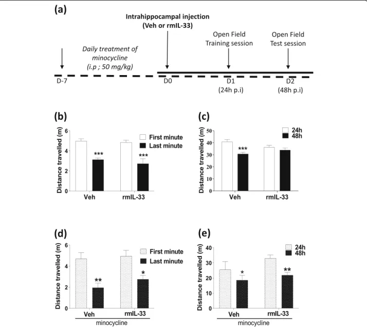
Spatial habituation to a novel environment is commonly used for the exploration of non-associative learning and memory processes linked to hippocampal structures [19–21]. As previously described [22], to explore the learning component, 1 day after the surgical interven-tion, the animal was allowed to explore an open field (OF) (40 cm × 40 cm) for 10 min (trial session). After 24 h, the mouse was re-exposed for 10 min to the same OF (test session). During each session, the exploratory measures were quantified using the Ethovision tracking system (version 10, Noldus Technology, Wageningen, Netherlands). Locomotor activity was indexed by the distance traveled in the entire open-field arena. To explore intrasession habituation during the trial session, the distance traveled between the first and the last minute was compared. The intersession habituation was assessed by comparing the full distance traveled during both sessions. All sessions were performed at 10 lux to
limit the anxiogenic component of the novel
environment.
Real-time quantitative polymerase chain reaction (RT-qPCR)
At the indicated time, total RNA from the hippocampus was isolated using TRI-Reagent (Sigma-Aldrich, Saint-Quentin Fallavier, France) as previously described [10] and reverse transcripted (Superscript III reverse tran-scriptase, Invitrogen, Carlsbad, CA). Quantitative real-time PCR reactions were performed using GoTaq qPCR-Master Mix (Promega, Charbonnières-les-Bains, France) and primers for Nos2, Il1b, Tnfa, Ifng, Arg1, Chil3, Il10, and Igf1(Qiagen, Hilden, Germany). After normalization using 18S-RNA expression as a housekeeping gene, raw data were analyzed by the 2ΔΔCtmethod [23].
Tissue preparation and immunofluorescence
For immunostaining, mice were deeply anesthetized and transcardially perfused with ice-cold PBS followed by 4% paraformaldehyde (PFA). The brains were collected, post-fixed for 48 h in 4% PFA, and cryo-protected in a 30% sucrose solution for 1 week. Then, 14μm brain cryo-sections mounted onto glass slides were incubated in citrate buffer (pH = 6) at 80 °C for 30 min, followed by incubation with blocking solution (TBS 1X; 1% BSA; 10% FCS; 0.3% Triton; 1% NaN3) during 45 min in a wet chamber at room temperature. After incubation overnight at 4 °C with anti-Iba-1 antibody (Abcam, Cam-bridge, England, ab5076; 1:500), the sections were washed in TBS and incubated with Alexa 488 secondary antibody (Abcam, ab150129, 1:1000) for 1 h. The slides were rinsed, then counter-stained with DAPI for 10 min,
mounted with Fluoromount-G (SouthernBiotech,
Birmingham, England), and dried before observation using ZEISS AXIOVERT 200 M/Apotome microscope (Zeiss, Oberkochen, Germany). Serial sections were collected at ×20 magnification to reconstruct each whole-hippocampal image software (ZEN2.1, Zeiss). The images were collected as Z-series of 18 optical slices to obtain a sufficient resolution to perform the morpho-logical analysis of microglial cells. For each mouse, 3 representative stacks of images of the hippocampus were recorded. Positive cells for Iba-1 were counted (50–100 cells) and their morphology analyzed in each area e.g. the cornu ammonis (CA)1/CA2, CA3 and the dentate gyrus (DG). Image analysis and processing were per-formed with the software Image J -Fiji [24] using the “concentric circles” plugin. For the Sholl analysis, the intersection number per radian was defined each 5μm from the center of each cell (n = 3 mice per treatment with 50-100 microglia analyzed per mouse). This ana-lysis was performed by a blinded experimenter.
Fluorescence-activated cell sorting
The hippocampus of 3 mice perfused with phosphate-buffer saline (PBS) was pooled and the cellular suspen-sions were prepared using the Neural Tissue Dissociation Kit (Miltenyi Biotec, Paris France), according to the manufacturer’s instructions. Cells were stained with extra-cellular conjugated antibodies: Fixable Viability Dye (eBiosciences™, 65-0865-14, 1/800), anti-CD45 V450 (BD Horizon™, 560501, 1:100), anti-CD11b PerCP/Cy5 (BD Pharmingen™, 560993, 1:100) and blocked with non-conjugated anti-CD16/32 (BD Pharmingen™, 553142, 1: 100) for 20 min at 4 °C. Then, the cells were washed be-fore fixation. Intracellular IL-1β pro-form stained with PE-conjugated specific antibody (eBioscience™, 12-7114-80, 1:20) was visualized after cell permeabilization for 20 min at 4 °C with Cytofix/Cytoperm Plus Kit (BD Biosci-ences, Paris, France). This antibody recognizes only the
pro-form of mouse IL-1β and does not detect the cleaved and secreted mature IL-1β form. Cells were then washed and re-suspended in lysing solution (BD FACS™ Lysing Solution) before the acquisition. Data were acquired with a flow cytometer (BD FACSCanto II) and analyzed with FlowJo v7.6.5 software (Tree Star, Ashland, OR). Very low SSC and very low FSC were excluded to strictly define the populations of interest. IL-1β pro-form staining was mea-sured using geometric mean fluorescence intensity (GMFI). For the analysis, live single cells were pre-gated. Then, CD11b+/CD45low cells were gated as microglia, while CD11b+/CD45high cells or CD11b−/CD45high cells were gated as infiltrating macrophage or lymphocyte cells, respectively. FMO controls were also included to define populations of Fixable Viability Dye cells and CD45, CD11b, and IL-1β-expressing cells.
Statistical
Statistical significance was determined with GraphPad Prism v6 (GraphPad Software, La Jolla, CA). Standard errors of the mean are reported as SEM. To analyze non-parametric data, Mann-Whitney test for 2 series was used or Kruskal-Wallis followed by Dunn’s multiple comparison for more series. P values≤ 0.05 were consid-ered statistically significant.
Results
Local hippocampal rmIL-33 injection impairs long-term memory
We previously proposed a role for IL-33/ST2 signaling pathway in the hippocampus in the cognitive impair-ments after PbA-infection, especially on the memory process [10]. In this respect, we asked whether CNS IL-33 overexpression, mimicked here by an exogenous rmIL-33 administration locally in the hippocampus, could influence cognitive functions. After bilateral intrahippocampal injections of rmIL-33 or vehicle, non-associative learning and memory retrieval processes were explored by spatial habituation test in an open-field apparatus as described in Fig. 1a. The time spent in the central square was similar in all tested groups 24 h post-surgery (Addition file 1), suggesting an absence of specific anxiogenic response. The total distance trav-eled 24 h post-surgery decreased from 1–10 min during the first session in a novel environment for both ve-hicle- and rmIL-33-treated mice (Fig. 1b), correspond-ing to appropriate habituation to spatial novelty. The total distance traveled during the test session at 48 h was significantly reduced in the vehicle group, as compared with the training session at 24 h, indicating a normal ability to retrieve the previous exploratory information from memory processes, e.g., a proper long-term habituation process (Fig. 1c). In contrast, rmIL-33-treated mice showed no reduction of distance
traveled at 48 h, as compared with the 24h training ses-sion, indicative of a disturbed long-term habituation process (Fig. 1c). These findings showed that rmIL-33 hippocampal administration impaired spatial memory retrieval processes.
Minocycline prevents the long-term memory impairment induced by rmIL-33
IL-33 is considered as an immunomodulator of various neuropathologies [2]. To investigate the impact of the immune response on rmIL-33-induced cognitive impair-ment, minocycline which is an inflammatory anti-biotic able to cross the blood-brain barrier [25] was used. Minocycline pre-treatment was administrated daily, starting 7 days prior to vehicle and rmIL-33 intra-hippocampal administration. In our experimental condi-tions (Fig. 1a), minocycline treatment did not affect anxiogenic response to a novel environment (Addition file 1). In addition, habituation to spatial novelty in vehicle or rmIL-33-treated animals during the trial session was also conserved (Fig. 1d). In contrast, the impairment of long-term habituation previously observed after rmIL-33 administration (Fig. 1c) was absent in minocycline-treated animals (Fig. 1e). In-deed, mice receiving minocycline treatment showed a decrease in distance traveled at 48 h compared with the distance traveled at 24 h even after rm-IL-33 ad-ministration. These data thus suggest that minocy-cline treatment prevents the deleterious effect of rmIL-33 administration on spatial memory retrieval. These data demonstrate that minocycline should pre-vent the deleterious effect of rmIL-33 administration on spatial memory retrieval.
IL-33 drives inflammatory response in the hippocampus
We next asked whether the effect of minocycline on re-storing rmIL-33-induced cognitive impairment may be associated with its reduction of a neuroinflammatory response [25]. We evaluated the time course of neuroin-flammatory processes in the hippocampus 24 h and 48 h after a single injection of rmIL-33 alone or in the pres-ence of minocycline pre-treatment in the hippocampus (Fig.2). The slight increase in pro-inflammatory markers expression seen at 24 h post-injection in terms of Nos2, Il1b, Tnfa, Ifng (Fig. 2a to d), as well as anti-inflammatory markers Arg1, Chil3, Il10 and Igf1 (Fig.2e to h) was observed both in vehicle- and rmIL-33-treated mice, as compared with the sham group, suggesting an inflammatory response to the microinjection itself. This inflammatory response was resolved at 48 h in vehicle-treated control animals, returning to the level of the sham group. However, at 48 h, a time point correspond-ing to the cognitive impairment, rmIL-33-treated mice showed high levels of hippocampal expression of
inflammatory markers, as compared with the vehicle group. Thus, rmIL-33 administration delayed the reso-lution of inflammation. Interestingly, minocycline treat-ment reduced the expression of Il1b and Ifng observed 48 h after the rmIL-33 administration (Fig. 2b, d) while it had no effect on the other parameters studied. Thus, minocycline treatment partially reduces the deleterious effects of rmIL-33 on the resolution of inflammation by limiting the overexpression of Il1b and Ifng. We must
notice that Il1a expression analysis showed a similar re-sponse to Il1b but fold inductions were widely reduced (Addition file 2).
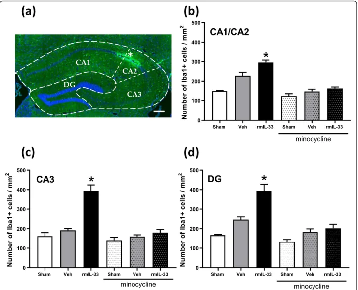
Intrahippocampal administration of rmIL-33 exacerbates microglial activation
To further analyze IL-33 implication in the inflammatory processes, we next investigated the effect of rmIL-33 on microglia 48 h after intrahippocampal administration. Fig. 1 Effect of hippocampal exogenous IL-33 associated with minocycline pre-treatment on spatial memory and habituation. a Experimental design. Mice were intrahippocampally injected with rmIL-33 or vehicle solution (PBS-BSA 0.1%). The cognitive behavior was tested on day 1 by a first open-field session, followed by a second session on day 2. The short term habituation was analyzed during the first session on day 1. b and d The distance traveled between the first and the last minute in the open-field were compared. c and e The long-term habituation was accessed by comparing the distance traveled between the first session (24 h) and the last session (48 h). Two cohorts have been studied: (b and c) vehicle or rmIL-33 treated without minocycline, (d and e) vehicle or rmIL-33 treated with minocycline. Under our experimental conditions, IL-33 impaired the intersession habituation (c). This effect on spatial memory retrieval was prevented by minocycline administration (e). Values are mean ± SEM, n = 8–15 per group corresponding to a pool of 2 independent experiments. Two-way ANOVA followed by a Sidak post hoc test was used to analyze the distance traveled among groups. *P≤ 0.05, **P ≤ 0.01, ***P ≤ 0.001
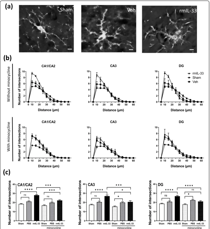
Immunochemistry experiments were performed to quan-tify the number of Iba-1+ microglial cells in the different hippocampal areas (Fig.3a). After vehicle injection, there was no significant difference in the number of Iba-1+cells in the cornu ammonis (CA), CA1/CA2 (Fig. 3b), CA3 (Fig. 3c), and the dentate gyrus (DG) (Fig. 3d), as com-pared to sham groups. However, the number of Iba-1+ glial cells was increased after rmIL-33 administration in the three hippocampal areas. Interestingly, minocycline treatment prevented rmIL-33-induced increase of micro-glial cell numbers in all areas of the hippocampus. To go further, microglia activation was investigated at 48 h post-injection. Microglial cells exhibited a typical activated morphology 48 h after vehicle administration, as com-pared with sham controls and this activated phenotype was more prominent after rmIL-33 administration (Fig.
4a). The Sholl analysis was used to provide a quantitative assessment of glial cell activation in situ (Fig. 4b). We demonstrated an increase of proximal intersections per ra-dius in the CA1/CA2, CA3, and the DG 48 h post-injection of vehicle, which was more pronounced after rmIL-33 treatment (Fig.4b). Although hippocampal injec-tion itself slightly modified microglia morphology, rmIL-33 significantly promoted the outgrowth of microglial pro-cesses, in agreement with an activated state. This effect of rmIL-33 administration was prevented by minocycline pre-treatment in the three hippocampal areas analyzed (Fig.4b, c). These findings show that rmIL-33 administra-tion induced microglial activaadministra-tion and proliferaadministra-tion/re- proliferation/re-cruitment in the hippocampus, and this effect was sensitive to the anti-inflammatory effect of minocycline.
Hippocampal exogenous rmIL-33 induces an increase of microglial cells expressing pro-IL-1β
To dissect the effect of rmIL-33 on microglia functions, we performed flow cytometry on dissociated cells from hippocampal tissues, 48 h after vehicle or rmIL-33 treat-ment. We determined the frequency of microglia, mac-rophages, and lymphocytes in hippocampal samples from sham, vehicle- and rmIL-33-treated groups. The gating strategy of live cell analysis is shown (Fig. 5a). Group comparison showed an increasing trend of
CD11blow/CD45high cells defined as lymphocytes and CD11b+/CD45high cells defined as macrophages (Fig. 5b, c) after vehicle or rmIL-33 administration. However, there was an increase in terms of CD11b+/CD45lowcells defined as microglial cells in rmIL-33-treated mice, as compared with vehicle control group (Fig. 5d), in agree-ment with the immunohistochemical data (Fig.3). More-over, intracellular staining using a pro-IL-1β specific antibody demonstrated overexpression of pro-IL-1β by hippocampal microglial cells exposed to rmIL-33 (Fig.
5e, f). Altogether, these data suggest that rmIL-33 induced an increase of hippocampal microglial cells expressing pro-IL-1β.
Exogenous rmIL-33-induced cognitive impairments require IL-1 signaling
As rmIL-33 administration induced microglia prolifera-tion/recruitment with IL-1β overexpression, we next questioned whether IL-1 contributes to the maintenance of inflammation and the cognitive disorders induced by exogenous rmIL-33. To address this question, we injected rmIL-33 in the hippocampus of mice deficient for IL-1α and IL-1β (IL-1αβ-/-) and evaluated their responses in spatial memory tasks at 24 h and 48 h post-administration. The decrease of distance traveled exhibited by vehicle-treated-IL-1αβ-/-mice (Fig. 6a) was similar to WT mice at 24 h after rmIL-33 administration (Fig.1b), indicating that IL-1αβ-/- mice displayed as WT mice normal intra-session habituation. However, at 48 h post-rmIL-33 in-jection, the decrease in traveled distance indicating that IL-1αβ-/- mice retained spatial memory retrieval (Fig.
6b), in contradiction with the rmIL-33-treated wild type mice, previously observed (Fig. 1c). Moreover, we showed that in the absence of IL-1αβ, rmIL-33 treat-ment induced an increase in the expression of key inflammatory mediators in the hippocampus (Nos2, Tnfa, Ifng, Arg1, Chil3 and Il10) of IL-1αβ-/- mice, 48 h post-injection (Fig. 6c–j) which is similar in WT mice (Fig.2). These results indicate that hippocampal IL-1 ex-pression, and most likely IL-1β, is required for rmIL-33-induced cognitive impairment independently of upstream inflammatory mediators.
(See figure on previous page.)
Fig. 2 mRNA expression of inflammatory markers in hippocampi of IL-33-injected mice. mRNA expression of pro-inflammatory markers (Nos2 in a, IL1b in b, Tnfa in c and Ifng in d) or anti-inflammatory markers (Arg1 in e, Chil3 in f, IL10 in g, and Igf1 in h) were quantified in hippocampi at 24 h and 48 h post-surgery by RT-qPCR normalized against 18S RNA. Relative fold change in vehicle group (grey bar) and in rmIL-33 group (black bar) were quantified versus sham group (S; white bar). Minocycline-treated mice were also analyzed at 48 h post-surgery (dotted bar). IL-33 injection delayed the resolution of inflammation highlighted by an increase of inflammatory markers at 48 h administration. A partial reduction of this effect was observed under minocycline exposure, especially for IL-1b and Ifng mRNA. Data are represented as mean ± SEM (n = 4–6). Statistical comparisons were made using Kruskal-Wallis followed by Dunn’s multiple comparison test for each group vs. Sham. *P ≤ 0.05, **P ≤ 0.01, ***P ≤ 0.001. In addition, a comparison between rmIL-33 (48 h) and rmIL-33 + mino (48 h) groups was performed using the Mann-Whitney test. #P≤ 0.05, ##P≤ 0.01
Discussion
The implications of IL-33 has been described in many neuropathologies [1], not only as protective [14, 16] but also as disruptor [15,17] of neuronal homeostasis. IL-33 exerts pleiotropic effects on the immune system, both on type 2 and type 1 immune responses, in the periphery but also at the CNS level. Despite the presence of IL-33 in a healthy brain [4] and in CNS pathologies [1], the multifold functions of IL-33 in CNS remain unclear. To elucidate the role of endogenous IL-33 in the CNS, the
present study explored the consequences of
intrahippocampal injection of recombinant IL-33 on cognitive function and neuroinflammatory processes.
Using spatial habituation tasks in an open field, allow-ing to address hippocampal non-associative learnallow-ing and memory processes [19–21,26], we show that the habitu-ation to a novel environment was intact in IL-33 hippo-campal treated mice 1-day post-surgery. These results indicate that neither the micro-lesion induced by the injection nor the IL-33 treatment had a neurological impact on learning at this stage. However, 48 h after intrahippocampal injection, IL-33-treated mice displayed Fig. 3 Effects of 33 treatment on the number of microglial cells in the hippocampal areas. Mice were intrahippocampally injected with rmIL-33 or vehicle solution (PBS-BSA 0.1%) with or without minocycline pretreatment as in Fig.1a and brain sections analyzed at 48 h. a Representative immunohistochemical staining (Dapi in blue and Iba1 in green) in the hippocampal section from vehicle mouse indicating the injection site (*). Scale bar = 200μm. The dotted lines indicate the different areas of the hippocampal formation (CA1, CA2, CA3, and DG=Dentate Gyrus). b, c, d Histograms showing the number of Iba1+cells by mm2quantified in each area (CA1/CA2 in b, CA3 in c, and DG in d). Quantification of Iba1+ cells was analyzed from 3–5 sections per mouse (n = 3–4 for each group). In rmIL-33-injected mice, the number of microglia was exacerbated in all hippocampal areas. Interestingly, under minocycline treatment, these increases were drastically limited. Data are represented as mean ± SEM. Statistical comparisons were analyzed using Kruskal-Wallis and uncorrected Dunn’s test to generate P values for each paired comparison (each group vs. Sham without minocycline). *P≤ 0.05
Fig. 4 Effects of rmIL-33 treatment on microglial morphology in hippocampal areas. a Representative high magnification of Iba1+microglia in
sham, vehicle- or rmIL-33-treated mice (Scale bar = 10μm). b, c Sholl analysis was performed after Iba1 immunohistochemistry on the hippocampal section from 48 h post-surgery mice. b To evaluate the ramification complexity of microglial cells, the number of process intersections per radius was reported graphically in the curve. c Bar graphs show the cumulated number of intersections at distances up to 25μm from the soma. The analyses were performed for each hippocampal area and for each group without or with minocycline treatment. In all hippocampal areas, Sholl analysis showed that microglia maintained hyper-ramified state following IL-33 injection, although vehicle treatment induced an intermediate-ramified state, as compared to sham. The minocycline pre-treatment impaired IL-33 effect on microglial morphology. Data are represented as mean ± SEM. Statistical comparisons were made using One-way ANOVA followed by Tukey’s multiple comparison test. *P ≤ 0.05, ***P ≤ 0.001, ****P ≤ 0.0001
a complete impairment of spatial memory retrieval. Unlike control mice, they were not able to recognize the previously explored environment, indicating that long-term habituation was significantly affected after rmIL-33 administration. These results suggest that a massive IL-33 release might disturb neuronal function and affect the memory retrieval process. IL-33 has been previously involved in cognitive defects observed in neuropatho-logical conditions such as reflected in Alzheimer’s disease, multiple sclerosis, and experimental cerebral malaria [1,10, 22]. Our data further show that injecting recombinant IL-33 directly in the hippocampus could mimic an acute exposure of IL-33 and its effects on cognitive processes.
To explore the link between the cognitive defect induced by IL-33 and the neuroinflammatory response, mice were pre-treated with minocycline. This antibiotic is able to cross the blood-brain barrier and exhibits anti-inflammatory properties preventing memory deficits in several neuropathologies [25]. In the present study, chronic administration of minocycline alone before intrahippocampal injections in control mice did not affect learning and spatial memory processes. However, our data also reveal that pre-treatment with minocycline seems to prevent the spatial memory retrieval impair-ment induced by IL-33 administration. This rescue of the IL-33-induced phenotype suggests that the cognitive impairments induced by IL-33 involved a neuroinflam-matory process.
Previous studies demonstrated that IL-33/ST2 pathway modulated the production of cytokines and chemokines in neuropathological conditions [1, 3, 17]. We assessed the direct effects of IL-33 role on the inflammatory context by gene expression analysis. We quantified mRNA expression levels in the hippocampus of molecu-lar markers usually used to define pro-inflammatory or regulatory immune response [27]. Nos2, Il1b, Tnfa, and Ifng are mediators of pro-inflammatory responses whereas Arg1, Chil3, Il10, and Igf1 are associated with immunoregulatory mechanisms. In control mice, we ob-served a transient inflammatory response induced by the injection at 24 h and resolving at 48 h post-injection.
This transient response to a slight trauma is correlated with the ability of the organism to return to a homeosta-sis state without adverse effects on behavior [28]. However, at 48 h, the intrahippocampal IL-33 injection induced a neuroinflammatory environment with overex-pression of pro-inflammatory and immunoregulatory markers mRNA. Minocycline administration reduced this inflammatory context in terms of Il1b and Ifng ex-pression at 48 h, contributing to the resolution of in-flammation. These results suggest that exogenous IL-33 induces a neuroinflammatory phenotype associated with long-term habituation disturbance.
To explore the cellular process involved in IL-33-induced immune response, we focused on microglia, the first active immune barrier in the CNS strongly express-ing IL-33 receptor ST2 [14]. We investigated the hippo-campal microglia reaction by immunochemistry using Iba-1 staining. Indeed, in response to a neuroinflamma-tory context induced by LPS administration, resident microglia alter their shape in a specific way as compared with infiltrated peripheral cells with a rounder morph-ology [29,30]. Sholl analysis on Iba1 immunofluorescent staining revealed a significant increase of proximal inter-sections per radius in the CA1/CA2, CA3, and DG regions 48 h after IL-33 treatment. This reactive morph-ology associated with an increase of microglial cell number demonstrated maintenance of their activated state. Minocycline administration through its anti-inflammatory activity attenuated the microglia activation of IL-33 treated mice, in line with previous reports in cognitive disorders [31,32]. This result suggests that the deleterious function of IL-33 pathway on spatial memory retrieval processes requires microglia activation, espe-cially in the hippocampal formation. Indeed, in healthy conditions, microglia regulate neuronal activity, synaptic plasticity, and adult neurogenesis required for learning and memory. In many neuropathologies, the microglia reactivity state has been characterized based on morpho-logical modifications and the release of cytokines, chemokines, and growth factors, modulating neuronal and synaptic functions. This activated phenotype should be beneficial and associated with inflammatory changes (See figure on previous page.)
Fig. 5 Increased IL-1β expressing microglia in the hippocampus of IL-33-treated mice. Flow cytometry revealing an increased proportion of microglia associated with a IL-1β pro-form overexpression. a Gating strategies to analyze whole hippocampus cell suspensions. A gate was created on non-debris population excluding very low SSC/FSC events. Then, cells were gated on single cells and selected on their live dead staining. This population was then gated according to CD11b and CD45 status as populations of CD11blow/CD45highlymphocytes (b), CD11bhigh/ CD45highmacrophages (c) and CD11bhigh/CD45lowmicroglia (d). The percentage of lymphocytes, macrophages, and microglia obtained from two experiments (n = 4–5 by the group) are graphically reported. e Representative geometric mean fluorescent intensity (GMFI) curve of IL-1β proform expression in microglial gated cells. f Microglial cells MFI for pro-IL-1β immunostaining expressed as ratio control relative to sham. Our flow cytometry analysis shows that compared with sham controls, rmIL-33 treatment induces a higher proportion of microglial cells correlated with a higher expression of IL-1β pro-form. Data are shown as mean ± SEM. Statistical analyses were made by Kruskal-Wallis test, followed by corrected Dunn’s multiple comparison test. *P ≤ 0.05, **P ≤ 0.01
to combat the injury and return to a homeostatic state. However, these defense processes could be over-stimulated and cause significant damage to behavior [33], as we demonstrated hereafter intrahippocampal IL-33 injection.
To confirm the IL-33-induced microglial reactivity, cells from the hippocampi of IL-33-treated mice were analyzed by flow cytometry at 48 h post-surgery. Although neither macrophage nor lymphocyte recruit-ment was observed, microglia number was significantly increased in IL-33-treated mice, confirming our immu-nohistological data. This analysis revealed also an over-expression of the IL-1β immature form in hippocampal microglia 48 h after IL-33 injection. Thus, in vivo, IL-33 treatment promotes IL-1β microglia production, as pre-viously demonstrated in vitro [10], indicating that micro-glia contribute to the pro-inflammatory response. We then hypothesized that the cognitive impairment in-duced by exogenous IL-33 may be mediated in part by microglia derived IL-1. To test this point, we performed IL-33 intrahippocampal microinjections in IL-1αβ defi-cient mice. The absence of IL-1 cytokines prevented spatial memory retrieval impairment induced by IL-33 administration even if neuroinflammatory markers, ex-cept IL-1β and IL-1α, were upregulated. Thus, IL-1β-producing microglia are required for IL-33 neurotoxic effects on cognitive impairment. In our experimental conditions, IL-1 contribution to these cognitive defects impairment could involve its non-immunological activ-ities. Indeed, this cytokine has been described as critical for learning and memory in a dose-dependent manner [34]. Here, we verified that IL-1αβ deficient mice behave
as WT mice in terms of habituation to spatial novelty in our experimental conditions. Prolonged up-regulation of pro-inflammatory cytokines, especially IL-1β, has been associated with a decrease in synaptic plasticity, as well as a deficit in spatial learning [35,36]. This IL-1β disrup-tive effect on cognidisrup-tive functions could involve the in-hibition of long-term potentiation generation at the neuronal level and/or defect of neurotrophic factors pro-duction [34]. All these parameters should be further in-vestigated in our futures studies.
Conclusion
In conclusion, we showed that IL-33 intrahippocampal administration is a valuable tool to mimic a local acute exposition. We provide the evidence that CNS IL-33 directly orchestrates neuroinflammatory mechanisms through microglia activation and overproduction of IL-1-inducing spatial memory disorders. Thus, we suggest that in neuropathological conditions IL-33 released by astrocytes and/or oligodendrocytes may activate micro-glia and induce IL-1-dependent cognitive defects. These results highlight the need to dissociate the CNS versus systemic IL-33 effects, in particular in the context of cerebral diseases.
Supplementary information
Supplementary information accompanies this paper athttps://doi.org/10. 1186/s12974-020-01939-6.
Additional file 1. The emotional state of mice was tested 24H after the intrahippocampal microinjection. Anxiety-like behavior, representing by the time spent in center of the open field, was not different between: (a) mice intrahippocampal injected with vehicle solution (Veh) versus 33, (b) mice intrahippocampal injected with vehicle solution versus rmIL-33 treated with minocycline, (c) IL-1αβ KO mice intrahippocampal injected with vehicle solution versus rmIL-33. Values are mean ± SEM, n = 8-15 per group, Mann-Whitney test was used to compare the time spent in the center between 2 groups.
Additional file 2. Expression of IL-1a mRNA in hippocampi at 24 h and 48 h post-surgery, in Sham, vehicle or IL-33 intra-hippocampal treated mice with or without minocycline pretreatment. mRNA expression of the pro-inflammatory marker IL-1a was quantified in hippocampi by RT-qPCR normalized against 18S RNA. Relative fold change in vehicle group (grey bar) and in IL-33 group (black bar) were quantified versus sham group (S ; white bar). Minocycline treated mice were also analyzed at 48 h post-surgery (dotted bar). IL-33 injection delayed the resolution of inflamma-tion highlighted by an increase of this markers at 48 h administrainflamma-tion. A partial reduction of this effect was observed under minocycline exposure. Data are represented as mean ± SEM (n = 4-6). Statistical comparisons were made using Kruskal-Wallis followed by Dunn’s multiple comparison test for each group vs. Sham.**p≤ 0.01, ***p ≤ 0.001. In addition, com-parison between rmIL-33 (48 h) and rmIL-33 + mino (48 h) groups was performed using the Mann-Whithney test.
Acknowledgements
The authors thank Dr. Marc Le Bert for expert advice in mouse genetics, Mr. David Gosset, responsible of the Cytometry and cellular imaging P@CYFIC platform, and Dr. Jean-Charles Bizot, Key-Obs company director for stereo-taxic equipment.
(See figure on previous page.)
Fig. 6 Effects of hippocampal exogenous IL-33 on spatial memory and habituation and hippocampi mRNA expression in IL-1αβ KO mice. IL-1αβ KO mice were intrahippocampally injected with rmIL-33 or vehicle solution (PBS-BSA 0.1%). Habituation in a novel environment was analyzed in intrasession (a) and in intersession 24 h and 48 h post-surgery (b). mRNA expression of pro-inflammatory markers (Nos2 in c, Il1b in d, Tnfa in e, and Ifng in f) or anti-inflammatory markers (Arg1 in g, Chil3 in h, Il10 in i, and Igf1 in j) was quantified by RT-qPCR normalized against 18S RNA at 48 h post-administration in WT/sham (WT-S), IL-1αβ KO/sham, IL-1αβ KO/vehicle and IL-1αβ KO/rmIL-33-treated groups (n = 4–6). Relative fold change in groups was quantified versus WT/sham group (S; white bar). Data are shown as mean ± SEM. The habituation analysis (a, b), n = 8–10 per group corresponding to a pool of 2 independent experiments. Two-way ANOVA followed by a Sidak post hoc test was used to analyze the distance traveled among the two groups (IL-1αβ KO injected with vehicle or rmIL-33). For qPCR analysis, statistical comparisons were made using Kruskal-Wallis followed by Dunn’s multiple comparison test for each group vs. WT/Sham. *P ≤ 0.05, **P ≤ 0.01, ***P ≤ 0.001
Authors’ contributions
FR, VdC, and VL performed the experiments; FR, Vd.C, DT, and AM conceived the experiments and analyzed the data; FR, VdC, VQ, and AM discussed the results and prepared the paper. SB, BR, VQ provided funding, and AM overall supervision of this study. All authors read and approved the final manuscript.
Funding
This work was supported by CNRS, the University of Orleans, and European funding in the Region Centre-Val de Loire (FEDER N° 2016-00110366 and EX005756).
Availability of data and materials
The datasets used and/or analyzed during the current study are available from the corresponding author on reasonable request.
Ethics approval and consent to participate
All animal experimental protocols complied with the French ethical and animal experiments regulations and were approved under number APAFIS #19264
Consent for publication Not applicable.
Competing interests
The authors declare that they have no competing interests Author details
1
UMR7355, Experimental and Molecular Immunology and Neurogenetics, CNRS and University of Orléans, 3B rue de la Ferollerie, 45071 Orléans, France.2Current address: Center for Molecular Biophysics, CNRS UPR4301,
45071 Orléans, France.3Current address:Department of Urology, Urology
Research Unit, CHUV, Lausanne, Switzerland.4Department of Genetics, Regional Hospital, Orléans, France.5Artimmune SAS, 13 Avenue Buffon,
45071 Orléans-Cedex 2, France.
Received: 6 April 2020 Accepted: 24 August 2020
References
1. Liew FY, Girard JP, Turnquist HR. Interleukin-33 in health and disease. Nat Rev Immunol. 2016;16(11):676–89.
2. Abd Rachman Isnadi MF, Chin VK, Abd Majid R, Lee TY, Atmadini Abdullah M, Bello Omenesa R, et al. Critical Roles of IL-33/ST2 Pathway in neurological disorders. Mediators Inflamm. 2018;2018:5346413. 3. Du L, Hu X, Yang W, Yasheng H, Liu S, Zhang W, et al. Spinal IL-33/ST2
signaling mediates chronic itch in mice through the astrocytic JAK2-STAT3 cascade. Glia. 2019;67(9):1680–93.
4. Hudson CA, Christophi GP, Gruber RC, Wilmore JR, Lawrence DA, Massa PT. Induction of IL-33 expression and activity in central nervous system glia. J Leukoc Biol. 2008;84(3):631–43.
5. Li M, Li Y, Liu X, Gao X, Wang Y. IL-33 blockade suppresses the development of experimental autoimmune encephalomyelitis in C57BL/6 mice. J Neuroimmunol. 2012;247(1-2):25–31.
6. Allan D, Fairlie-Clarke KJ, Elliott C, Schuh C, Barnett SC, Lassmann H, et al. Role of IL-33 and ST2 signalling pathway in multiple sclerosis: expression by oligodendrocytes and inhibition of myelination in central nervous system. Acta Neuropathol Commun. 2016;4(1):75.
7. Xiong Z, Thangavel R, Kempuraj D, Yang E, Zaheer S, Zaheer A. Alzheimer's disease: evidence for the expression of interleukin-33 and its receptor ST2 in the brain. J Alzheimers Dis. 2014;40(2):297–308.
8. Saresella M, Marventano I, Piancone F, La Rosa F, Galimberti D, Fenoglio C, et al. IL-33 and its decoy sST2 in patients with Alzheimer's disease and mild cognitive impairment. J Neuroinflammation. 2020;17(1):174.
9. Palomo J, Reverchon F, Piotet J, Besnard AG, Couturier-Maillard A, Maillet I, et al. Critical role of IL-33 receptor ST2 in experimental cerebral malaria development. Eur J Immunol. 2015;45(5):1354–65.
10. Reverchon F, Mortaud S, Sivoyon M, Maillet I, Laugeray A, Palomo J, et al. IL-33 receptor ST2 regulates the cognitive impairments associated with experimental cerebral malaria. PLoS Pathog. 2017;13(4):e1006322.
11. Shibui A, Takamori A, Tolba MEM, Nambu A, Shimura E, Yamaguchi S, et al. IL-25, IL-33 and TSLP receptor are not critical for development of experimental murine malaria. Biochem Biophys Rep. 2016;5:191–5. 12. Strangward P, Haley MJ, Albornoz MG, Barrington J, Shaw T, Dookie R, et al.
Targeting the IL33-NLRP3 axis improves therapy for experimental cerebral malaria. Proc Natl Acad Sci U S A. 2018;115(28):7404–9.
13. Besnard AG, Guabiraba R, Niedbala W, Palomo J, Reverchon F, Shaw TN, et al. IL-33-mediated protection against experimental cerebral malaria is linked to induction of type 2 innate lymphoid cells, M2 macrophages and regulatory T cells. PLoS Pathog. 2015;11(2):e1004607.
14. Yang Y, Liu H, Zhang H, Ye Q, Wang J, Yang B, et al. ST2/IL-33-dependent microglial response limits acute ischemic brain injury. J Neurosci. 2017; 37(18):4692–704.
15. Yasuoka S, Kawanokuchi J, Parajuli B, Jin S, Doi Y, Noda M, et al. Production and functions of IL-33 in the central nervous system. Brain Res. 2011;1385:8–17.
16. Gadani SP, Walsh JT, Smirnov I, Zheng J, Kipnis J. The glia-derived alarmin IL-33 orchestrates the immune response and promotes recovery following CNS injury. Neuron. 2015;85(4):703–9.
17. Cao K, Liao X, Lu J, Yao S, Wu F, Zhu X, et al. IL-33/ST2 plays a critical role in endothelial cell activation and microglia-mediated neuroinflammation modulation. J Neuroinflammation. 2018;15(1):136.
18. Yamada H, Mizumo S, Horai R, Iwakura Y, Sugawara I. Protective role of interleukin-1 in mycobacterial infection in IL-1 alpha/beta double-knockout mice. Lab Invest. 2000;80(5):759–67.
19. Leussis MP, Bolivar VJ. Habituation in rodents: a review of behavior, neurobiology, and genetics. Neurosci Biobehav Rev. 2006;30(7):1045–64. 20. Vianna MR, Alonso M, Viola H, Quevedo J, de Paris F, Furman M, et al. Role
of hippocampal signaling pathways in long-term memory formation of a nonassociative learning task in the rat. Learn Mem. 2000;7(5):333–40. 21. Bolivar VJ. Intrasession and intersession habituation in mice: from
inbred strain variability to linkage analysis. Neurobiol Learn Mem. 2009; 92(2):206–14.
22. Fu AK, Hung KW, Yuen MY, Zhou X, Mak DS, Chan IC, et al. IL-33 ameliorates Alzheimer's disease-like pathology and cognitive decline. Proc Natl Acad Sci U S A. 2016;113(19):E2705–13.
23. Bustin SA, Benes V, Garson JA, Hellemans J, Huggett J, Kubista M, et al. The MIQE guidelines: minimum information for publication of quantitative real-time PCR experiments. Clin Chem. 2009;55(4):611–22.
24. Schindelin J, Arganda-Carreras I, Frise E, Kaynig V, Longair M, Pietzsch T, et al. Fiji: an open-source platform for biological-image analysis. Nat Methods. 2012;9(7):676–82.
25. Moller T, Bard F, Bhattacharya A, Biber K, Campbell B, Dale E, et al. Critical data-based re-evaluation of minocycline as a putative specific microglia inhibitor. Glia. 2016;64(10):1788–94.
26. Pavkovic Z, Milanovic D, Ruzdijic S, Kanazir S, Pesic V. The influence of propofol anesthesia exposure on nonaversive memory retrieval and expression of molecules involved in memory process in the dorsal hippocampus in peripubertal rats. Paediatr Anaesth. 2018;28(6):537–46. 27. Orihuela R, McPherson CA, Harry GJ. Microglial M1/M2 polarization and
metabolic states. Br J Pharmacol. 2016;173(4):649–65.
28. Buckley CD, Gilroy DW, Serhan CN, Stockinger B, Tak PP. The resolution of inflammation. Nat Rev Immunol. 2013;13(1):59–66.
29. David S, Kroner A. Repertoire of microglial and macrophage responses after spinal cord injury. Nat Rev Neurosci. 2011;12(7):388–99.
30. Herber DL, Maloney JL, Roth LM, Freeman MJ, Morgan D, Gordon MN. Diverse microglial responses after intrahippocampal administration of lipopolysaccharide. Glia. 2006;53(4):382–91.
31. Kobayashi K, Imagama S, Ohgomori T, Hirano K, Uchimura K, Sakamoto K, et al. Minocycline selectively inhibits M1 polarization of microglia. Cell Death Dis. 2013;4:e525.
32. Seabrook TJ, Jiang L, Maier M, Lemere CA. Minocycline affects microglia activation, Abeta deposition, and behavior in APP-tg mice. Glia. 2006;53(7): 776–82.
33. Szepesi Z, Manouchehrian O, Bachiller S, Deierborg T. Bidirectional Microglia-Neuron Communication in Health and Disease. Front Cell Neurosci. 2018;12:323.
34. Liu X, Quan N. Microglia and CNS Interleukin-1: Beyond Immunological Concepts. Front Neurol. 2018;9:8.
35. Oitzl MS, van Oers H, Schobitz B, de Kloet ER. Interleukin-1 beta, but not interleukin-6, impairs spatial navigation learning. Brain Res. 1993;613(1):160–3.
36. Hein AM, Stasko MR, Matousek SB, Scott-McKean JJ, Maier SF, Olschowka JA, et al. Sustained hippocampal IL-1beta overexpression impairs contextual and spatial memory in transgenic mice. Brain Behav Immun. 2010;24(2):243–53.
Publisher’s Note
Springer Nature remains neutral with regard to jurisdictional claims in published maps and institutional affiliations.