Activation of Protein Kinase B Induced by H
2O
2and Heat Shock
through Distinct Mechanisms Dependent and Independent of
Phosphatidylinositol 3-Kinase
1Hiroaki Konishi,' Toshihide Fujiyoshi,* Yasuhisa Fukui,f Hidenori Matsuzaki,'4
Toshiyoshi Yamamoto,* Yoshitaka Ono,J Mirjana Andjelkovic,1 Brian A. Hemmings,1 and Ushio Kikkawa*2
'Biosignal Research Center, Kobe University, Nada-ku, Hyogo, Kobe 657-8501; ^Department of Applied Biological Chemistry, Graduate School of Agriculture and Life Science, The University of Tokyo, Bunkyo-ku, Tokyo 113-8657; ^Department of Biology, Faculty of Science, Kobe University, Nada-ku, Hyogo, Kobe 657-8501; and ^Friedrich Mieacher Institute, CH-4002 Basel, Switzerland
Received September 22, 1999; accepted September 30, 1999
Protein kinase B (PKB) is a downstream target of phosphatidylinositol (PI) 3-kinase in the signaling pathway of growth factors, and is activated by cellular stress such as H2O2 and heat shock. To study the mechanism of the stress-induced activation of PKB, PI 3-kinase products were measured in stress-stimulated cells. Both PI 3,4-bisphosphate and PI 3,4,5-trisphosphate increased in H2O2 -treated cells, and the elevation of these phospholipids and activation of PKB were concurrently blocked by wortmannin, a potent inhibitor of PI 3-kinase. In heat-shocked cells, the level of PI 3,4-bisphosphate did not change while that of PI 3,4,5-trisphosphate increased slightly, and an association between PKB molecules was observed. Two active PKB fractions, presumably monomeric and oligomeric forms, were resolved from heat-shocked cells by gel filtration column chromatography. Activation of the former was suppressed by pretreatment with wortmannin, whereas the generation and activation of the latter were not blocked by the PI 3-kinase inhibitor. Only the monomeric form, but not the oligomeric form, was recovered from H2O2-treated cells, and its activation was prevented by wortmannin. These results indicate that PKB is activated by two distinct mechanisms that are dependent and independent of PI 3-kinase in stress-stimulated cells. Key words: heat shock, hydrogen peroxide (H2O2), phosphatidylinositol 3-kinase, pleck-strin homology domain, protein kinase B.
Protein kinase B (PKB, also named Akt and RAC-protein phosphatidylinositol (PI) 3-kinase in the growth factor
kinase) is a serine/threonine protein kinase with a pleck- signaling pathway {3-6). Namely, PKB was first reported strin homology (PH) domain in its arnino-tenninal region to be activated by direct interaction of PI 3,4-bisphosphate and a catalytic domain in its carboxyl-terminal region (1, (PI 3,4-P2) with its PH domain (7-9), and then the
phos-2). This protein kinase was identified as an enzyme with a phorylation of PKB was shown to be indispensable for its catalytic domain closely related to both cAMP-dependent activation (10-12). In PKBa, Thr308 in the activation loop
protein kinase and protein kinase C, and also as a cellular of its catalytic domain and Ser473 in the carboxyl-terminal
counterpart of a rodent viral oncogene v-akt. Thus far, end region were identified as the phosphorylation sites three mammalian PKB genes, a, fi, and y, have been (12). Later, PDK1 (3-phosphoinositide-dependent protein isolated. Studies on physiological roles of PKB have reveal- kinase) and related enzymes were isolated that catalyze the ed that this protein kinase is a downstream target of phosphorylation of Thr308 of PKB a- in the presence of PI 3,
4,5-trisphosphate (PI 3,4,5-P3) and PI 3,4-P2 (13-16).
'This study was supported in part by research grants from the Thus, the direct association of the PI 3 -kinase products with Scientific Research Funds of the Ministry of Education, Science, the PH domain and phosphorylation by upstream protein Sports and Culture of Japan, the Suntory Institute for Bioorganic ^ ^ ^ m a y b e necessary for full activation of PKB upon
Research, and the Charitable Trust Osaka Cancer Researcher-Fund. ,. , ,. , ,, „ , ,, , , 'To whom correspondence should be addressed. Phone: +81-78-803- stimulation of the cells by growth factors^
5964, Fax: +81-78-803-5972, E-mail: ukikkawa(gkobe-u.ac.jp On the other hand, we have found that PKB is activated Abbreviations: PKB, protein kinase B; PH domain, pleckstrin by heat shock in COS-7 and NIH 3T3 cells in a manner homology domain; PI, phosphatidylinositol; PI 3,4-Pj, phosphatidyl- insensitive to wortmannin, a potent PI 3-kinase inhibitor inositol 3,4-bisphosphate; PI 3,4,5-P,, phosphatidylinositol 3,4,5- (17). The heat shock-induced activation of PKB has been trisphosphate; HA, hemagglultinin; HPLC, high-performance liquid confirmed using different cell lines (18 19) The mecha-chromatography; TLC thin.layer mecha-chromatography; PI 4 5-P, phos- ^ f h e a f c s h o c k.m d u c e d a c tiv a ti o n of the enzyme,
phatadyknositol 4,5-bisphosphate: PI 3-P, phosphatidylinositol 3- , , ,._. ,, ,. ,. , , Phosphate; PI 4-P, phosphatidylinositol 4-phosphate. however, appears to differ among the cell lines employed.
The heat shock-induced activation of PKB is not abrogated © 1999 by The Japanese Biochemical Society. by wortmannin in COS-7 and 3T3-L1 cells (17, 19), but is
sensitive to the PI 3-kinase inhibitor in Swiss 3T3 and 293 cells (18). Different results are reported in NIH 3T3 cells: the heat shock-induced activation of PKB is not prevented by wortmannin (17), whereas heat shock activates PI 3-kinase (20), and the heat shock-induced activation of PKB is prevented by the PI 3-kinase inhibitor (18). Further-more, we have shown that PKB is activated by stimulation with H2O2 in COS-7 cells (21). H2O2- and reactive oxygen
species-induced activation of PKB has been indicated in 3T3-L1, Swiss 3T3, 293, and vascular smooth muscle cells, which are sensitive to wortmannin (18, 19, 22).
In the present study, the PI 3-kinase products (PI 3,4-P2/PI 3,4,5-P3) were measured in cultured mammalian
cells to determine whether stress-induced PKB activation is dependent on or independent of PI 3-kinase. It was found that heat shock generates a slight increase in PI 3,4,5-P3,
but does not produce PI 3,4-P2, whereas H2O2 induces the
formation of both of PI 3,4-P2 and PI 3,4,5-P3 in COS-7 and
293T ceDs. Therefore, the activation mechanism of PKB was analyzed by using various PKB a- mutants and monitor-ing the protein kinase activity of the enzyme recovered from stress-stimulated cells.
MATERIALS AND METHODS
Expression Plasmids—The expression plasmids of
FLAG- and HA-epitope tagged rat PKBa, designated FLAG-PKB and HA-PKB, respectively, were constructed as described previously (17, 23). The cDNA fragments corresponding to amino acids 1-113 and 149-480 of rat PKBo-, which include the PH domain and the protein kinase catalytic domain, respectively, were cloned into pECE vector to make FLAG-epitope tag sequences at their amino-termini. The resulting plasmids were designated FLAG-PH and FLAG-KD, respectively. The expression plasmid of FLAG-epitope tagged rat PKBa in which Arg25
is replaced by Cys was constructed by polymerase chain reaction and designated R25C. The cDNAs of human PKB a and its mutated molecules in which the phosphorylation sites Thr308 and Ser473 are replaced by Ala (12) were
introduced into pECE vector. These FLAG-epitope tagged constructs were designated hPKB, T308A, and S473A, respectively. The primary amino acid sequences of rat and human PKBo- are 98% identical, and conserve these phosphorylation sites (24, 25). The DNA sequences were confirmed by the dideoxy chain-termination method using a DNA Sequencer model 373A (Applied Biosystems).
Cells and Transfection—COS-7 and 293T cells were
maintained in Dulbecco's modified Eagle's medium sup-plemented with 10% fetal calf serum at 37'C in a 5% CO2
incubator. CHO cells were maintained under the conditions described above in the presence of 35//g/ml proline. COS-7 and CHO cells were transfected with each expres-sion vector by electroporation using a Gene Pulser (Bio-Rad), and 293T cells were transfected by a polyamine transfection reagent Trans IT (PanVera). The transfected cells were cultured for 48 h, and treated as described in each experiment. Where indicated, cells were treated with 200 nM wortmannin for 10 min before stimulation.
Immunoprecipitation and Immunoblot—The expressed
proteins were immunoprecipitated at 0-4'C essentially as described (17). Briefly, cells were washed with phosphate-buffered saline, and lysed in 20 mM Tris-HCl, pH7.5,
containing 1 mM EDTA, 1 mM EGTA, 10 mM 2-mercapto-ethanol, 1% Triton X-100, 150 mM NaCl, lOmMNaF, 1 mM Na3VO4, and 50 //g/ml phenylmethylsulfonyl fluoride.
The lysate was centrifuged for 10 min at 18,000 x g, and the supernatant (500-600 //g of protein) was incubated for 1 h with either an anti-FLAG (Sigma) or an anti-HA (12CA5, Boehringer) monoclonal antibody. Then, protein A-Sepharose (Pharmacia) was added and the mixture was incubated for 30 min. The immunoprecipitates were col-lected by centrifugation and washed with 20 mM Tris-HCl, pH 7.5, containing 150 mM NaCl and 1% Triton X-100. The immunoprecipitates were boiled in SDS sample buffer, and proteins were separated by SDS-PAGE and transferred onto an 1mm obi Inn P membrane (Millipore). Monoclonal antibody against either the FLAG- or HA-epitope was used as the first antibody, and alkaline phosphatase-conjugated anti-mouse antibody (Promega) was employed as the second antibody. The color reaction was carried out using 5-bromo-4-chloro-3-indolyl-phosphate and nitro blue tetrazolium as substrates.
Protein Kinase Assay—The enzyme activity of
immuno-precipitated PKB was assayed by measuring the incorpora-tion of radioactivity from [y-32P] ATP into the core histone
fraction prepared from calf thymus (17). Before assay, the immunoprecipitates collected were washed at 0-4"C with 20 mM Tris-HCl, pH 7.5, containing 1 mM EDTA, 1 mM EGTA, 10 mM 2-mercaptoethanol, 150 mM NaCl, and 50 //g/ml phenylmethylsulfonyl fluoride to remove Triton X-100, NaF, and Na3V04. The reaction mixture (25 //I) in
20 mM Tris-HCl, pH 7.5, 10 mM MgCl2) 20 //M ATP,
15-50kBq of [y-32P]ATP, and 200//g/ml core histone, and
the immunoprecipitates were incubated for 30 min at 30°C. After boiling in SDS sample buffer, the phosphorylated proteins were separated by SDS-PAGE, and the radioactiv-ity of the histone band was analyzed with a Bio-imaging Analyzer BAS2000 (Fuji).
Gel Filtration of PKB—Gel nitration analysis was
car-ried out as described by Suzuki et al. (26) with modifica-tions. COS-7 and CHO cells transfected with FLAG-PKB were disrupted with a Dounce homogenizer in 20 mM Tris-HCl, pH 7.5, containing 150 mM NaCl, 10 mM MgCl2,
2 mM EGTA, 10 mM 2-mercaptoethanol, 10 mM NaF, 1 mM Na3V0<, and 50 //g/ml phenylmethylsulfonyl fluoride.
The homogenate was centrifuged at 100,000 X g for 30 min, and a 50-//1 aliquot of the supernatant (100-150//g of protein) was subjected to gel filtration chromatography on a prepacked Superdex-200 column (PC3.2/30) equipped with a SMART System (Pharmacia); the column was equilibrated and eluted with homogenization buffer. The chromatographic procedure was repeated three times, and the corresponding column fractions were combined and subjected to immunoprecipitation by the FLAG anti-body. The procedures above were carried out at 0-4'C. The protein kinase activity in the immunoprecipitates was assayed by measuring the autophosphorylation of the enzyme under the conditions described above in the ab-sence of exogenously added phosphate acceptor protein.
Analysis of Phosphoinositides—COS-7 and 293T cells
were labeled with [32P]orthophosphate (37 MBq/ml) for 4
h in serum- and phosphate-free Dulbecco's modified Eagle's medium. After stimulation, the medium was removed and the reaction was stopped with MeOH: 1 N HC1 (1:1). The lipid was extracted with CHC13, deacylated with
methylamine, and the resulting sample was analyzed by HPLC using a Partisphere 5-SAX anion-exchange chro-matography column (27). The radioactivity in the glycero-phosphoinositide peaks was quantitated by scintillation counting and normalized to the radioactivity in the total lipids. For in vitro analysis, the cells stimulated with H2O2
were suspended in buffer containing 10 mM Tris-HCl, pH 7.5, and 10 mM NaCl for 5 min at 4°C, and then disrupted with a Dounce homogenizer. After removing the cell debris by centrifugation at 500 X g for 5 min, the sample was further centrifuged at 10,000 X g for 45 min and the membrane fraction was suspended in 20 mM Hepes-NaOH, pH 7.5, containing 100 mM NaCl. An aliquot of the membrane fraction was incubated at 25°C with 10 /*M
[y-32P]ATP (370 kBq) and 10 mM MgCl2. The reaction was
stopped by the addition of chloroform :methanol:l N HC1 (10:20:1). The organic phase separated by centrifugation was applied to TLC on a Silica Grel plate (Merck) pretreated with 1% oxalate and developed in chloroform:acetone: methanol:acetic acid:water (40:15:13:12:7). The radioac-tivity on the dried plate was visualized using a Bio-imaging Analyzer. When the lipid sample was analyzed by HPLC, the extracted lipid was deacylated with methylamine as described above.
RESULTS
PKB Activation and 3-Phosphoinositide Production in Stress-Stimulated Cells—The activity of PKB and the
changes in 3-phosphoinositides were measured in stress-stimulated cells (Fig. 1). In transfected COS-7 cells, the PKB activity was elevated by heat shock, and this activa-tion was nearly insensitive to pretreatment of the cells with wortmannin as described previously (17) (Fig. 1A). In these cells, PI 3,4-P2 levels did not change, whereas PI
3,4,5-Pj levels increased slightly and this rise was blocked by the PI 3-kinase inhibitor (Fig. IB). H2O2 treatment
induced PKB activation, consistent with previous reports
(18, 19, 21, 22), and elevated the levels of both PI 3,4-P2
and PI 3,4,5-P3 (Fig. 1, A and B). In contrast to heat shock,
the H2O2-induced activation of PKB was prevented by
wortmannin, and the increases in the levels of PI 3,4-P2 and
PI 3,4,5-P3 were also abrogated completely by wortmannin.
The H2O2-induced generation of 3-phosphoinositides was
further studied using 293T cells (Fig. 1C). PI 3,4,5-P3
levels were elevated transiently by stimulation with either 1 or 10mM H2O2. In cells treated with 10 mM H2O2, PI
3,4-P2 levels increased dramatically after a transient
B
20- 15-10-81 5H
CO%
H
.£ 20 n o 15-10-
5-1
c
I—I hs i—11—•mm
•
H2O2 i—11—iWT 0 Histone «-PKB 1 mM H2O2 Pl 3,4-P2 [ ^ 1 1 ^ li\
PI 3,4,5-P3 WT hs H2O2 Concentration of H2O2 treatment for 10 min0 1 5 7.510 [mM]
Time of 10mMH2O2 treatment
0 2.5 5 7.510 15 [min]
Fig. 1. PKB activity and 3-phosphoinosltide production in
stress-treated cells. (A) PKB activity in COS-7 cells. Cells
transfect-ed with FLAG-PKB were treattransfect-ed either at 45'C for 20 min (hs) or with 10 mM H,Oi for 10 min (H,0i). Untreated control cells are indicated as (C). Cells pretreated with and without wortmannin (WT) are indicated as ( + ) and ( —), respectively. FLAG-PKB was immunopre-cipitated with anti-FLAG antibody. Protein kinase activity was measured using core histone as a substrate (upper). Immunoblot analysis was carried out using anti-FLAG antibody (lower). (B) Changes in the amounts of 3-phosphoinositides in COS-7 cells. Cells were metabolically labeled with ["P]orthophosphate, and treated as described in (A). The fold elevations of PI 3,4-P, and PI 3,4,5-P, were
10 15
[min]
calculated from the normalized radioactivity of each glycerophospho-inositide separated by HPLC. The results are shown as means ± SD for three independent experiments. (C) Changes in the amounts of inositol phospholipids in 293T cells. Cells were metabolically labeled with ["P]orthophosphate, and treated with either 1 mM H,O, (upper) or 10 mM H,0, (lower). The relative levels of PI 3,4-P,, PI 3,4,5-P,, and PI 4,5-P, calculated from the normalized radioactivity of each glycerophosphoinositide separated by HPLC are shown. (D) PKB activity in 293T cells. Cells transfected with FLAG-PKB were serum starved for 16 h and treated with various concentrations of H,0, for 10 min (upper) or with 10 mM H,0, for various times (lower). Protein kinase activity was measured using core histone as a substrate.
increase in PI 3,4,5-P3, but did not change in cells treated
with 1 mM H2O2. In addition, a substantial decrease in the
PI 4,5-bisphosphate (PI 4,5-P2) level was seen in cells
treated with the higher concentration of H2O2, suggesting
that a considerable amount of PI 4,5-P2 was used in the PI
3-kinase pathway. It seems that the PI 3,4,5-P3 generated
from PI 4,5-P2 in cells treated with 10 mM H2O2 is
convert-ed to PI 3,4-P2. PKB activiation was found in 293T cells
stimulated with comparable concentrations of H2O2 (Fig.
ID, upper panel) and could be detected 5 min after treat-ment with 10 mM H2O2 (Fig. ID, lower panel). Therefore,
the onset of PKB activation occurred mostly in parallel with the increase in PI 3,4,5-P3 levels in H2O2-stimulated cells,
and significant PKB activation and increase in PI 3,4-P2
were concomitantly observed in the cells treated with higher concentrations of H2O2.
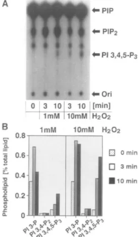
Generation of 3-Phosphoinositides in Cell Membranes—
The results above suggest that PI-3 kinase may be activated intensely by treatment of the ceDs with H2O2. To confirm
this, the generation of 3-phosphoinositides was analyzed using the membrane fraction obtained from 293T cells treated with H2O2. After incubation of the membrane
fraction with [y-32P]ATP, the production of PI 3,4,5-P3
was observed as judged by TLC (Fig. 2A). The increase in PI 3,4,5-P3 was dependent on the concentration of H2O2
employed for the stimulation of the cells. HPLC analysis was performed to examine whether other 3-phosphoino-sitides were produced in the membrane fraction (Fig. 2B). PI 3,4,5-P3 was found as by TLC analysis, and PI
3-phos-phate (PI 3-P) was constantly produced even in membrane fractions prepared from cells not treated with H2O2. PI
3,4-P2, however, was not detected in the membrane
frac-tion. PI-3 kinase can use PI, PI 4-phosphate (PI 4-P), or PI 4,5-P2 as a substrate with comparable affinities in vitro (28). In the membrane, the levels of PI 4-P and PI 4,5-P2
may be comparable and much lower than the PI level. Despite the contents of these phospholipids, PI 3,4,5-P3
was preferentially generated in the membrane fraction obtained from cells treated with H2O2. Therefore, it is
reasonable to assume that PI 4,5-P2 is recognized
specifi-cally by the lipid kinase in the membrane. Presumably, the PI 3,4,5-P3 phosphatase activity, which dephosphorylates
PI 3,4,5-P3 to produce PI 3,4-P2 in vivo (29), may not be
available in the membrane. These results confirm the possibility described above that the accumulation of PI 3,4-P2 in H2O2-treated cells is due to the dephosphorylation of
PI 3,4,5-P3 rather than to the direct phosphorylation of PI
4-P by PI 3-kinase.
Distinct Mechanisms of PKB Activation—Both the
direct association of PI 3,4-P2 with the PH domain (7-9)
and the phosphorylation on Thr308 and Ser473 of PKB* (12)
are important for the regulation of PKB activity by growth factors. Therefore, the roles of the PH domain and site-spe-cific phosphorylation in stress-induced PKB activation were examined using the mutant proteins (Fig. 3). Human PKBo- employed as a control was activated in a manner
<-PI3,4,5-P3
B
3i.
'Q. o a oFig. 2. Generation of 3-phosphoinosltldes in the membrane
fraction of 293T cells. (A) TLC of 3-phosphoinositides. After
stimulation with HiO2 for the indicated time, the membrane fraction
was incubated with [y-'*P]ATP and MgCl2, and the lipids were
extracted and separated by TLC. PIP and PIPi indicate Pl-monophos-phate and Pl-bisphosPl-monophos-phate, respectively. (B) HPLC of 3-phosphoino-sitides. The lipid samples from (A) were deacylated and separated by HPLC. The normalized radioactivities of each glycerophosphoino-si tide are shown.
hs H2O2
WT Histone
hPKB T3O8A S473A R25C
Fig. 3. The activity of PKB mutants in COS-7 cells. Cells were transfected with the FLAG-epitope tagged PKB construct of hPKB, T308A, S473A, or R25C, and treated either at 45 C for 20 min (hs) or with 10 mM H,O, for 10 min (H,0,). Untreated control cells are
indicated as (C). Cells pretreated with and without wortmannin antibody (lower).
(WT) are indicated as ( + ) and ( - ) , respectively. FLAG-epitope tagged molecules were immunoprecipitated with anti-FLAG antibody. Protein kinase activity was measured using core histone as a substrate (upper). Immunoblot analysis was carried out using anti-FLAG
identical to rat PKBa. The R25C mutant, in which Arg26 in
the PH domain is replaced by Cys, and which does not bind inositol phospholipids (3), was not activated in cells stimu-lated with H2O2. The replacement of Thr308 with Ala
eliminated its basal activity as previously described (12), and the T308A mutant showed no protein kinase activity in H2O2-treated cells. Therefore, both the direct association
of PI 3-kinase products with the PH domain and the phosphorylation of Thr308 seem to be necessary for the
H2O2-induced activation of PKB, as in the case of the
regulation of PKB activity by growth factors. The S473A mutant was activated in cells stimulated with H2O2 and in
heat-shocked cells, suggesting that the phosphorylation of Ser473 may not be indispensable for the stress-induced
activation of PKB a.
In heat-shocked cells, however, the R25C mutant was efficiently activated, and wortmannin did not block the activation of this mutant. These results suggest that the R25C mutant is activated without the direct binding of PI 3-kinase products to its PH domain. Furthermore, trace but significant enzyme activity was found in the immunopre-cipitate of the T308A mutant, although the T308A mutant itself has no catalytic activity (12). It has been reported that PKB forms a protein complex by protein-protein interactions between the PH domains, and that PI 3,4-P2
promotes the dimerization of PKB in vitro (8, 30). Thus,
the possibility of the association of the T308A mutant and PKB expressed endogenously in host cells was examined. To analyze the PKB protein complex, HA-PKB was intro-duced into COS-7 cells with either FLAG-PH, FLAG-KD, or FLAG-PKB (Fig. 4, A and B). HA-PKB was co-immuno-precipitated constitutively with FLAG-PH (Fig. 4A, left panel), consistent with the previous report (30). The association of these two full length molecules was observed in heat-shocked cells, but not in control or H2O2-treated
cells (Fig. 4A, right panel). HA-PKB did not associate with FLAG-KD (Fig. 4A, middle panel). Therefore, heat shock induces the association of PKB molecules through their PH domains.
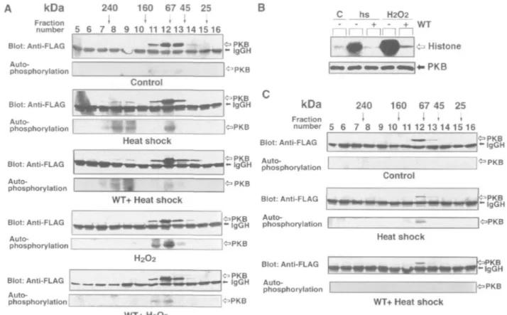
Protein Complex Formation—FLAG •'PKB was expressed
in COS-7 cells, and the supernatant fraction of the cells was subjected to gel filtration column chromatography to ana-lyze the formation of the PKB protein complex. FLAG-PKB was immunoprecipitated from each column fraction, and detected by immunoblot analysis and by monitoring its autophosphorylating activity (Fig. 5A). The recombinant protein from unstimulated control cells was recovered as a single peak in column fractions 11-13 and showed practi-cally no protein kinase activity. When the cells were stimulated by H2O2, active PKB was recovered in column
fractions 11-13, and the activation is completely blocked by pretreating the cells with wortmannin. Although the
appar-HA-PKB + FLAG-PH HA-PKB + FLAG-KD hs H2O2 HA-PKB + FLAG-PKB Blot: Anti-HA Blot: Anti-FLAG c a - (gGH IgGH FLAG-KD hs i—i r~
MM
H2O2 1 1 1 1 1WT +• HA-PKB — IgGH ^FLAG-PKB • I g G H IP: Anti-FLAG B HA-PKB PH domainFig. 4. Oligomerizatlon of PKB in heat-shocked cells. (A) Interaction of PKB and deletion mutants in COS-7 cells. Cells were co-transfected with HA-PKB and either FLAG-PH (left), FLAG-KD (center), or FLAG-PKB (right). Cells were treated either at 45"C for 20 min (hs) or with 10 mM H,O, for 10 min (H,O,). Untreated control cells are indicated as (C). Cells pretreated with and without wortmannin (WT) are indicated as ( + ) and ( —), respectively.
Kmase domain
FLAG-epitope tagged molecules were immunoprecipitated with anti-FLAG antibody. Immunoblot analysis was carried out using the anti-HA antibody (upper) or the anti-FLAG antibody (lower). The positions of IgG [heavy (H) and light (L) chains] are indicated. (B) Schematic representation of the epitope-tagged PKB molecules used in (A).
kDa 240 160 67 45 25 Fraction * I < < , number 5 6 7 8 9 10 11 1 2 1 3 1 4 1 5 1 6 B Blot: Anti-FLAG Auto- I phosphorylation : O P K B - igGH I^PKB c I I
b
I—
hs inr H2O2 lrnrn• d
——I
Control Blot: Anti-FLAG Auto-phosphorylation Blot: Anti-FLAG Auto-phosphorylation kDa 240 160 67 45 25 Fraction < » ' ' ' number 5 6 7 8 9 10 1112 13 14 15 16Heat shock Blot: Anti-FLAG
i r-u Auto-' 9U M phosphorylation PKB - I g G H IOPKB Control WT+ Heat shock Blot: Anti-FLAG Auto-phosphorylation [ H2O2 Blot: Anti-FLAG LM — i Auto- r phosphorylation L ^ P K B - IgGH JOPKB W T + H 2 O 2
Fig. 5. Gel filtration analysis of PKB in stress-treated cells. (A) PKB recovered from COS-7 cells. Cells transfected with FLAG-PKB were treated either at 45'C for 20 min (Heat shock) or with 10 mM H20i for 10 min (H,02). Untreated control cells are indicated as
(Control). Cells pretreated with wortmannin before stimulation are indicated as (WT). The supernatant fractions of cells were applied to a gel filtration column, and FLAG-PKB in the column fractions was immunoprecipitated with anti-FLAG antibody. The immunoprecipi-tated samples were subjected to the immunoblot analysis using anti-FLAG antibody (upper) and the autophosphorylating activity of
Blot: Anti-FLAG PKB - I g G H > P K B . , . ** IgGH phosphorylation Blot: Anti-FLAG Auto-phosphorylation Heat shock «=>PKB - I g G H WT+ Heat shock
PKB was measured (lower). The positions of the IgG heavy chain (H) are indicated. The fraction numbers from column chromatography and the positions of the molecular weight markers are indicated at the top of the control panels. (B) PKB activity in CHO cells. Cells transfected with FLAG-PKB were treated and protein kinase assay and immuno-blot analysis were carried out as described in the legend to Fig. 1A. (C) PKB recovered from CHO cells. Cells transfected with FLAG-PKB were treated at 45'C for 20 min with or without pretreatment with wortmannin, and the supernatant fractions were subjected to gel nitration analysis as described in (A).
ent size of the enzyme recovered in fractions 11-13 is 70 kDa, slightly larger than the calculated molecular mass of 57 kDa of the recombinant protein (24), the enzyme is presumably a monomeric form as it is found in H2O2-treated cells. In contrast, two peaks of protein kinase
activity were recovered from heat-shocked cells in column fractions 7-9 and 11-13. The PKB activity that appeared in column fractions 7-9 remained even in cells pretreated with wortmannin. Therefore, the PKB activity obtained in these higher molecular weight fractions seems to be an oligomeric form corresponding to the PKB protein complex observed in Fig. 4A. The enzyme recovered in these higher molecular weight fractions shows an apparent molecular mass of 200 kDa. The activity in heat-shocked cells that eluted in fractions 11-13 disappeared when the cells were pretreated with wortmannin. As heat shock induces a faint increase in the PI 3,4,5-P3 level as shown in Fig. IB, it is
reasonable to assume that the generation of active PKB activity recovered in fractions 11-13 is regulated by PI 3-kinase. In heat- shocked COS-7 cells, therefore, PKB is activated by two distinct mechanisms: one dependent on PI 3-kinase without the formation of the protein complex, and the other independent of PI 3-kinase accompanying its oligomerization.
In transfected CHO cells, PKB was activated both by H2O2 and by heat shock as in the case of COS-7 cells, but
wortmannin efficiently prevented the activation of PKB induced not only by H2O2 but also by heat shock (Fig. 5B).
On gel filtration column chromatography, FLAG-PKB expressed in CHO cells was obtained in a monomeric form from both control and heat-shocked cells, and the PKB protein complex was not found in heat-shocked cells (Fig. 5C). Wortmannin blocked the heat shock-induced activa-tion of the monomeric form of the enzyme as in COS-7 cells. The PI 3-kinase inhibitor also prevented the activa-tion of the monomeric form of PKB in CHO cells treated with H2O2 (data not shown). These results indicate that the
generation of active PKB that accompanies the formation of the protein complex is not a universal mechanism, but depends on the cell type.
DISCUSSION
PI 3-kinase was shown to be activated by cellular stresses such as H2O2 and heat shock. The levels of both PI 3,4-P2
and PI 3,4,5-P3 were elevated in cells treated with H2O2 as
in cells stimulated with growth factors (28). H2O2 is
a role in the signaling pathway (31), and thus it is attractive to assume that H2O2 takes part in the activation process of
PI 3-kinase induced by growth factors. The mechanism for PI 3-kinase activation by H2O2 remains unknown at
pres-ent, but it seems possible that H2O2 added to the culture
medium activates PKB through PI 3-kinase by mimicking the mechanism of growth factor stimulation. Heat shock also activated PI 3-kinase in COS-7 cells as reported in NIH 3T3 cells (20), and a trace increase in the amount of PI 3,4,5-P3 was observed. Consistent with the increase in the
PI 3-kinase product, a small PKB fraction was activated by heat shock in COS-7 cells in a wortmannin-sensitive manner. In CHO cells, however, the heat-shock induced activation of PKB was completely blocked by the PI 3-kinase inhibitor. Thus, this enzyme may be activated by heat shock through PI 3-kinase in Swiss 3T3 and 293 cells, where the heat shock-induced activation of PKB is sup-pressed by PI 3-kinase inhibitors (18). As no apparent increase in the amount of PI 3,4-P2, which interacts with
the PH domain of PKB (7-9), was observed in the heat-shocked cells, it is necessary to analyze the role of PI 3,4,5-P3 in the regulation of PKB activity through the PH
domain.
In addition to Thr308 in the catalytic domain, Ser473 in the
carboxyl-terminal region of PKBa is phosphorylated in cells stimulated by growth factors (12). Recently, integrin-linked kinase has been shown to phosphorylate Ser'73,
which is stimulated in a PI 3-kinase-dependent manner
(32). On the other hand, it has been reported that PDK1,
which phosphorylates Thr308, interacts with other proteins
and thus shows a different substrate specificity to recognize Ser473 (33). Even though the S473A mutant was activated
in stress-stimulated cells, Ser473 was phosphorylated in
H2O2-treated and heat-shocked cells (data not shown). It
seems possible that Ser473 is phosphorylated by these PI
3-kinase-dependent protein kinases in stress-stimulated cells, because PI 3-kinase is activated by cellular stress.
PKB was activated not only in a PI 3-kinase-dependent manner, but also in a manner independent of lipid kinase in heat-shocked COS-7 cells. The PI 3-kinase-independent activation of PKB is not a universal mechanism, but may occur in the cellular responses of certain cell types. PKB has also been reported to be activated in a PI 3-kinase-independent manner in rat epididymal fat cells stimulated by /S-adrenergic agonists (34) and in 293 EBNA and COS-7 cells treated with cAMP-elevating agents (35). Although the molecular mechanism is not clear, phosphorylation at a site(s) distinct from Thr308 or Ser473 in PKBa has been
suggested to be important for its cAMP-induced activation
(35). In this study, a PKB fraction was detected as an
oligomeric form of PKB protein complex in heat-shocked cells. It has been reported that PI 3,4-P2 promotes the
dimerization of PKB (8, 30), but the PKB protein complex was formed in heat-shocked cells in which PI 3,4-P2 was not
generated. Furthermore, the apparent size of the PKB protein complex was 200 kDa, which is larger than the estimated molecular mass of the PKB dimer. The mole-cular composition of the PKB protein complex is not clear at present, and needs to be analyzed. We have previously identified a small heat shock protein, Hsp27, that associates specifically with PKB in stress-stimulated cells (21). Hsp27 associates with certain proteins as a chaperone (36) and may play a role in the complex formation and activation
of PKB. PKB has been shown to play a critical role in protection against apoptosis (2). A preliminary experiment indicated that H2O2-induced apoptosis is suppressed in a
CHO cell line that stably overexpresses PKB a. As PKB is activated by H2O2-treatment, it is suggested that PKB may
take part in the protection of cells from damage caused by cellular stress. Further studies are required to clarify the precise mechanism of PKB activation and the role of the enzyme in the cellular response to stress stimulation.
We thank Dr. Yasutomi Nishizuka for discussion and Ms. Yukiko Kimura for secretarial assistance.
REFERENCES
1. Hemmings, B.A. (1997) Akt signaling: linking membrane events to life and death decisions. Science 275, 628-630
2. Coffer, P.J., Jin, J., and Woodgett, J.R. (1998) Protein kinase B (c-Akt): a multifunctional mediator of phosphatidylinositol 3-kinase activation. Biochem. J. 335, 1-13
3. Franke, T.F., Yang, S.I., Chan, T.O., Datta, K., Kazlauskaus, A., Morison, D.K., Kaplan, D.R., and Tsichlis, P.N. (1995) The protein kinase encoded by the Akt proto-oncogene is a target of the PDGF-activated phosphatidylinositol 3-kinase. Ceil 81, 727-736
4. Burgering, B.M.T. and Coffer, P.J. (1995) Protein kinase B (c-Akt) in pho8phatidylinositol-3-OH kinase signal transduction. Nature 376, 599-602
5. Cross, D.A.E., Alessi, D.R., Cohen, P., Andjelkovic, M., and Hemmings, B.A. (1995) Inhibition of glycogen synthase kinase-3 by insulin mediated by protein kinase B. Nature 378, 785-789 6. Kohn, A.D., Kovacina, K.S., and Roth, R.A. (1995) Insulin
stimulates the kinase activity of RAC-PK, a pleckstrin homology domain containing Ser/Thr kinase. EMBO J. 14, 4288-4295 7. Klippel, A., Kavanaugh, W.M., Pot, D., and Williams, L.T.
(1997) A specific product of phosphatidylinositol 3-kinase direct-ly activates the protein kinase Akt through its pleckstrin ho-mology domain. Mol. Cell. Biol. 17, 338-344
8. Franke, T.F., Kaplan, D.R., Cantley, L.C., and Toker, A. (1997) Direct regulation of the Akt proto-oncogene product by phos-phatidylinositol-3,4-bisphosphate. Science 275, 665-668 9. Freeh, M., Andjelkovic, M., Ingley, E., Reddy, K.K., Falck, J.R.,
and Hemmings, B.A. (1997) High affinity binding of inositol phosphates and phosphoinositides to the pleckstrin homology domain of RAC/protein kinase B and their influence on kinase activity. J. Biol. Chem. 272, 8474-8481
10. Kohn, A.D., Takeuchi, F., and Roth, R.A. (1996) Akt, a pleck-strin homology domain containing kinase, is activated primarily by phosphorylation. J. Biol Chem. 271, 21920-21926 11. Andjelkovic, M., Jakubowicz, T., Cron, P., Ming, X.F., Han,
J.W., and Hemmings, B.A. (1997) Activation and phosphoryla-tion of a pleckstrin homology domain containing protein kinase (RAC-PK/PKB) promoted by serum and protein phosphatase inhibitors. Proc Natl. Acad. ScL USA 93, 5699-5704
12. Alessi, D.R., Andjelkovic, M., Caudwell, B., Cron, P., Morrice, N., Cohen, P., and Hemmings, B.A. (1996) Mechanism of activation of protein kinase B by insulin and IGF-1. EMBO J. 15, 6541-6551
13. Alessi, D.R., James, S.R., Downes, C.P., Holmes, A.B., Gaffney, P.R.J., Reese, C.B., and Cohen, P. (1997) Characterization of a 3-phosphoinositide-dependent protein kinase which phosphor-ylates and activates protein kinase Ba. Curr. Biol. 7, 261-269 14. Stokoe, D., Stephens, L.R., Copeland, T., Gaffney, P.R., Reese,
C.B., Painter, G.F., Holmes, A.B., McCormick, F., and Hawkins, P.T. (1997) Dual role of phosphatidylinositol-3,4,5-trisphos-phate in the activation of protein kinase B. Science 277, 567-570 15. Alessi, D.R., Deak, M., Casamayor, A., Caudwell, F.B., Morrice, N., Norman, D.G., Gaffney, P., Reese, C.B., MacDougall, C.N., Harbison, D., Ashworth, A., and Bownes, M. (1997) 3-Phospho-inositide-dependent protein kinase-1 (PDK1): structural and functional homology with the Drosophila DSTPK61 kinase. Curr.
Biol. 7, 776-789
16. Stephens, L., Anderson, K., Stokoe, D., Erdjument-Bromage, H., Painter, G.F., Holmes, A.B., Gaffney, P.R., Reese, C.B., McCormick, F., Tempst, P., Coadwell, J., and Hawkins, P.T. (1997) Protein kinase B kinases that mediate phosphatidylino-sitol 3,4,5-trisphosphate-dependent activation of protein kinase B. Science 279, 710-714
17. Konishi, H., Mateuzaki, H., Tanaka, M., Ono, Y., Tokunaga, C, Kuroda, S., and Kikkawa, U. (1996) Activation of RAC-protein kinase by heat shock and hyperosmolarity stress through a pathway independent of phosphatidylinositol 3-kinase. Proc. NatL Acad. Sci. USA 93, 7639-7643
18. Shaw, M., Cohen, P., and Alessi, D.R. (1998) The activation of protein kinase B by HiOj or heat shock is mediated by phosphoinositide 3kinase and not by mitogenactivated protein kinase -activated protein kinase-2. Biochem. J. 336, 241-246
19. Tirosh, A., Potaahnik, R., Bashan, N., and Rudich, A. (1999) Oxidative stress disrupts insulin-induced cellular redistribution of insulin receptor substrate-1 and phosphatidylinositol 3-kinase in 3T3-L1 adipocytes. A putative cellular mechanism for im-paired protein kinase B activation and GLUT4 translocation. J. Biol. Chem. 274, 10595-10602
20. Lin, R.Z., Hu, Z.W., Chin, J.H., and Hoffman, B.B. (1997) Heat shock activates c-Src tyrosine kinases and phosphatidylinositol 3-kinase in NIH3T3 fibroblasts. J. Biol. Chem. 272, 31196-31202
21. Konishi, H., Matsuzaki, H., Tanaka, M., Takemura, Y., Kuroda, S., Ono, Y., and Kikkawa, U. (1997) Activation of protein kinase B (Akt/RAC-protein kinase) by cellular stress and its association with heat shock protein Hsp27. FEBS Lett. 410, 493-498 22. Ushio-Fukai, M., Alexander, R.W., Akers, M., Yin, Q., Fujio, Y.,
Walsh, K., and Griendling, K.K. (1999) Reactive oxygen species mediate the activation of Akt/protein kinase B by angiotensin II in vascular smooth muscle cells. J. Biol. Chem. 274, 22699-22704
23. Konishi, H., Tanaka, M., Takemura, Y., Matsuzaki, H., Ono, Y., Kikkawa, U., and Nishizuka, Y. (1997) Activation of protein kinase C by tyrosine phosphorylation in response to H2O2. Proc.
Natl. Acad. Sci. USA 94, 11233-11237
24. Konishi, H., Shinomura, T., Kuroda, S., Ono, Y., and Kikkawa, U. (1994) Molecular cloning of rat RAC protein kinase a and fi and their association with protein kinase C £. Biochem. Biophys. Res. Commun. 205, 817-825
25. Jones, P.F., Jakubowicz, T., Pitossi, F.J., Maurer, F., and Hemmings, B.A. (1991) Molecular cloning and identification of a serine/threonine protein kinase of the second-messenger sub-family. Proc. Natl. Acad. Sci. USA 88, 4171-4175
26. Suzuki, A., Sugiyama, Y., Hayashi, Y., Nyu-i, N., Yoshida, M.,
Nonaka, I., Ishiura, S., Arahata, K., and Ohno, S. (1998) MKBP, a novel member of the small heat shock protein family, binds and activates the myotonic dystrophy protein kinase. J. Cell Biol.
140, 1113-1124
27. Fukui, Y., Kornbluth, S., Jong, S.-M., Wang, L.-H., and Hanafusa, H. (1989) Phosphatidylinositol kinase type I activity associates with various oncogene products. Oncogene Res. 4, 283-292
28. Toker, A. and Cantley, L.C. (1997) Signalling through the lipid products of phosphoinositide-3-OH kinase. Nature 387, 673-676 29. Kavanaugh, W.M., Pot, D.A., Deuter-Reinhard, M., Jefferson, A.B., Norris, F.A., Masiarz, F.R., Cousens, L.S., Majerus, P.W., and Williams, L.T. (1996) Multiple forms of an inositol poly-phosphate 5-phosphatase form signaling complexes with She and Grb2. Curr. Biol. 6, 438-445
30. Datta, K., Franke, T.F., Chan, T.O., Makris, A., Yang, S.I., Kaplan, D.R., Morrison, D.K., Golemis, E.A., and Tsichlis, P.N. (1995) AH/PH domain-mediated interaction between Akt molecules and its potential role in Akt regulation. Mol. Cell. BioL 15, 2304-2310
31. Sundaresan, M., Yu, Z.-X., Ferrans, V.J., Irani, K., and Finkel, T. (1995) Requirement for generation of H2O2 for
platelet-derived growth factor signal transduction. Science 270, 296-299 32. Delcommenne, M., Tan, C, Gray, V., Rue, L., Woodgett, J., and
Dedhar, S. (1998) Phosphoinositide-3-OH kinase-dependent regulation of glycogen synthase kinase 3 and protein kinase B/ AKT by the integrin-linked kinase. Proc. Natl. Acad. Sci. USA 95,11211-11216
33. Balendran, A., Casamayor, A., Deak, M., Paterson, A., Gaffney, P., Currie, R., Downes, C.P., and Alessi, D.R. (1999) PDK1 acquires PDK2 activity in the presence of a synthetic peptide derived from the carboxyl terminus of PRK2. Curr. Biol. 9, 393-404
34. Moule, S.K., Welsh, G.I., Edgell, N.J., Foulstone, E.J., Proud, C.G., andDenton, R.M. (1997) Regulation of protein kinase Band glycogen synthase kinase-3 by insulin and /5-adrenergic agonists in rat epididymal fat cells. Activation of protein kinase B by wortmannin-sensitive and -insensitive mechanisms. J. Biol. Chem. 272, 7713-7719
35. Filippa, N., Sable, C.L., Filloux, C, Hemmings, B., and Van Obberghen, E. (1999) Mechanism of protein kinase B activation by cyclic AMP-dependent protein kinase. Mol. Cell. Biol. 19, 4989-5000
36. Kato, K., Hasegawa, K., Goto, S., and Inaguma, Y. (1994) Dissociation as a result of phosphorylation of an aggregated form of the small stress protein, hsp27. J. BioL Chem. 269, 11274-11278