Interactive Image Guided Surgical Navigation
-A First Step Towards Minimally Invasive Orthopaedic Surgery
C. Scheer, E. Arm, O. Schwarzenbach*, B. Jost, L.-P. Nolte, H. Visarius, U. Rohrer
M.E. Müller Institute for Biomechanics and *Department of Orthopaedic Surgery, University of Bern, 3010 Bern, Switzerland INTRODUCTION
In the field of minimally invasive surgery an major obstacle is the limited access to the anatomy of interest. Interactive image guided surgical navigation (IIGSN) has recently proven to be an effective means for improving surgical accuracy and safety in conventional open procedures [1]. The goal of the present work was to apply IIGSN to "keyhole"-type Orthopaedic surgeries by introducing novel registration and referencing techniques. CLINICAL ASPECTS
The current stage in open surgery stabilisation includes pedicle screw insertion in the thoracolumbar and cervical spine and during scoliosis surgery.
Applied minimally invasive techniques like laparo/thoracoscopic surgery (trans-, retroperitoneal, transthoracic) have the following short comings:
time-consummg
several specialists are involved expensive equipment
high-risk learning curve
A*. ..·.
m^wm
Figure J: Scheme of subcutaneous spinal siabilisation
Our research focus is towards minimally invasive spine stabilisation that offers potential benefits, such äs:
• no muscle damage
• decrease of intraoperative radiation exposure • time saving
• smaller amount of anaesthesia • precise pedicle screw insertion
As a first application the percutanous delivery of so-called Schanz-screws for external spinal stabilisation was
chosen. A registration probe has been developed, which was carrying 6 CT-compatible markers.
External äs well äs subcutaneous internal fixation is supported by this System.
REALIZED SYSTEM
The challenge was the visualisation of operated Segments (see Figure 2) and the development of a fiducial marker carrier (see Figure 3) for the dedicated Implantation site.
^'^^^^^^^c^^i^fa^
Surgical objcct
Any hone fragmenl
Virtual object: Any mediccd iniage
. . .
T\
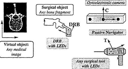
· · · · ' · · · ·-Figure 2: Involved co-ordinate spaces
Based on the concept of rigid body mechanics applied by the motion analysis System OPTOTRAK® 3020 (Northern Digital, Waterloo, Canada) several rigid bodies have to be tracked in space (see Figure 2). Each one is equipped with infrared LEDs tracked by the OPTOTRAK®. At least three LEDs are needed to establish a local co-ordinate System (COS).
Differently from framebased stereotactic approaches [2], we are using a so-called dynamic reference base (COSDRB). All tooFs co-ordinates are later on expressed
in this local COS. Right before the surgery, this base is rigidly attached to a newly developed clamp.
Tests on cadaver spines with different fiducial marker carriers led to the clamp's final small design.
The clamp consists of radiolucent POM (see Figure 3) and titanium spheres. These spheres are embedded at defined positions. The clamp is attached to the spinous process percutaneously through a small incision under local anaesthesia prior to image acquisition (CT or MRI scan). It resides in this place, until the following surgery has been performed.
The transformation between the "real" world and the patient's digital equivalent (image) is called matching.
Figure 3: Special clamp carryingßducial markers and iheir counterparts on the spinous process, visible in an
MRI scan
Preoperatively, the titanium spheres are registered in the CT image. In the OR the surgeon points towards six small indents on the clamp that are in a known position relative to the centres of the spheres. By knowledge of these paired points in COS07 and COSDRB the Workstation (Sun
j Microsystems, Eschenbach, Switzerland) is able to calculale the transformation MDRB~*CT (see Figure 2).
l
' The surgeon is now able to verify the matching accuracy
by real-time visualisation of a pointer in the CT image. Also the fluoroscope is used to assure this.
Figure 4: Graphical user Interface with interactive real-time tool visualisation in a CT scan
In the next Step the insertion of a Kirschner wire is interactively guided (see Figure 4) by a marker carrier equipped drill guide (see Figure 5). This wire is used for the drilling process with a hollow drill.
Later, the use of fluoroscopy validated these actions and proved the accuracy of this Intervention.
A preoperative planning of the screw paths (trajectories) which enables defmed guidance, äs well äs real-time display of tools without planning, is possible.
Custom made Software allows the interactive image guided percutanous delivery of the Schanz-screws by displaying 2D views of saggital, lateral, and horizontal cuts through the CT images.
Figure 6: OR scene including drill guide and dynamic reference base
After being validated on several cadavers, the new approach was applied during three surgeries, one open stabilisation and two external fixators.
DISCUSSION
The proposed ncw way of referencing has already proven its practical application in three clinical cases.
Together with dedicated 2D views of the anatomy to be operated on, our System enables real-time guidance of a pneumatic drill during percutanous Schanz-screw delivery with minimum radiation exposure when compared to conventional fluoroscope guided insertions.
Nevertheless, the preoperatively embedded clamp could be driven to smaller sizes, which would come closer to the goal of more "minimally" invasive surgery.
In future approaches the referencing/matching procedures could be solved by intraoperative contour matching with fluoroscopy or ultrasound. Another promising Option lies in the combination of endoscopic/microscopic surgery with existing concepts of Computer assisted orthopaedic surgery (CAOS).
REFERENCES
[1] L.-P. Nolte, L. Zamorano, H. Visarius, U. Berlemann, F. Langlotz, E. Arm, O. Schwarzenbach: Clinical evaluation of a System for precision enhancement in spine surgery. Clin. Biomech. 10:293-303 (1995) [2] L. Zamorano, C. Chavantes, M. Dujovny, G. Malik, J.
Ausman: Stereotactic endoscopic interventions in cystic and intraventricular brain lesions. Acta Neurochirurgica, Suppl. 54:69-76 (1992)