UV Filters with Antagonistic Action at Androgen Receptors in the
MDA-kb2 Cell Transcriptional-Activation Assay
Risheng Ma, Bea Cotton, Walter Lichtensteiger, and Margret Schlumpf
1 Institute of Pharmacology and Toxicology, University of Zurich,CH-8057, Zurich, SwitzerlandReceived November 23, 2003; accepted March 24, 2003
The fact that certain ultraviolet (UV) filters used in cosmetics display estrogenic activity prompted us to study potential actions on androgen receptors (AR) in the human breast carcinoma cell line MDA-kb2, which expresses functional endogenous AR and glucocorticoid receptors (GR) and is stably transfected with a luciferase reporter plasmid. Dihydrotestosterone (DHT), methyl-trienolone (R1881), methyltestosterone, danazol, and andro-stenedione increased luciferase activity, with EC50values between
0.11 nM (R1881), 0.14 nM (DHT), and 73.5 nM (androstenedione). DHT-induced luciferase gene expression was inhibited by nonste-roidal antiandrogens, hydroxyflutamide, flutamide, bicalutamide, and vinclozolin. In contrast, the steroidal AR agonist/antagonist cyproterone actetate showed agonistic activity in the absence and presence of DHT, which was not blocked by hydroxyflutamide and thus seems not to be mediated by AR. GR-mediated activation of luciferase by dexamethasone was 100 times less potent than DHT and was not antagonized by hydroxyflutamide. The cell line was used for screening of UV filters, benzophenone–3 (Bp-3), benzo-phenone– 4, 3-benzylidene camphor, 4-methylbenzylidene cam-phor, butyl-methoxy-dibenzoylmethane, homosalate (HMS), octyl-dimethyl-PABA, and octyl-methoxycinnamate. Two of these, Bp-3 and HMS, antagonized DHT-induced AR activation below cyto-toxic concentrations, with IC50of 5.57 10
– 6
M (HMS) and 4.98 10– 6
M (Bp-3). None of the eight UV filters displayed agonistic activity when tested alone, but high concentrations of Bp-3 induced an increase of luciferase activity in the presence of dexamethasone, which was not blocked by hydroxyflutamide or the estrogen an-tagonist, ICI 182,780. These data indicate that the UV filters Bp-3 and HMS possess antiandrogenic activity in vitro in addition to estrogenic activity.
Key Words: MDA-kb2 cells; androgen receptor; androgen;
anti-androgen; endocrine disruptor; pesticide; UV filter.
The presence in the environment of increasing amounts of
synthetic chemicals is causing concern about unknown
long-term toxicities, especially the possibility of interference with
endocrine systems (IPCS, 2002; Kelce et al., 1998). We
pre-viously identified estrogenic activity in several ultraviolet (UV)
filters in vitro and in vivo (Schlumpf et al., 2001). This
prompted us to initiate a study on their potential interaction
with androgen receptors. UV filters are added to sunscreen
products in continuously higher amounts, since sun protection
factors (SPF) increased from SPF 1–2 in 1950 to SPF 40 – 60
today (SPF
⫽ ratio of UV light dose producing minimal
erythema in the protected skin versus the unprotected skin). In
addition, UV filters are also included in cosmetics for product
protection and stability. These persistent chemicals have been
detected in fish (Nagtegaal et al., 1997) and in human milk (Hany
and Nagel, 1997). Interactions of xenobiotics with androgens may
cause developmental abnormalities, as shown following the
ex-posure to the antiandrogenic pesticides vinclozolin, procymidone,
or linuron (Kelce et al., 1994, 1997; Lambright et al., 2000; Ostby
et al., 1999). As there is 100% sequence homology between rat
and human AR ligand binding domains, analogous effects can be
expected to be mediated by human AR (Kelce et al., 1998).
In this study, we validated the human AR expressing cell
line MDA-kb2 with known androgens and antiandrogens and
used the system to test UV filters for androgenic or
antiandro-genic activity in vitro. Eight of the most frequently used
compounds were chosen among the total of 30 UV filters
admitted for use in Switzerland. The human breast carcinoma
cell line, MDA-kb2, expresses high levels of functional
endog-enous androgen receptor (AR) and also glucocorticoid receptor
(GR), while estrogen receptor alpha and progesterone receptor
are not detectable at the RNA level, and estrogen receptor beta
is expressed only at low levels (Hall et al., 1992). The cells are
stably transfected with a luciferase transporter plasmid driven
by the mouse mammary tumor virus promoter (MMTV) that
can be activated through both AR and GR (Wilson et al.,
2002). Compounds acting through AR or GR can therefore
induce luciferase expression. It was shown earlier that known
AR antagonists like hydroxyflutamide, the vinclozolin
metab-olites M1 and M2, p,p
⬘DDE, and linuron-inhibited
dihydrotes-tosterone (DHT)-induced gene expression in this cell line
(Wil-son et al., 2002).
MATERIALS AND METHODS
Chemicals. Methyltrienolone (R1881, also called metribolone) (CAS no. 965-93-5) was purchased from NEN™(NEN™ Life Science Products, Inc., 1To whom correspondence should be addressed at Institute of
Pharmacol-ogy and ToxicolPharmacol-ogy, University of Zurich, Winterthurerstrasse 190, CH-8057, Zurich, Switzerland. Fax: ⫹41 (1) 635-6857. E-mail: schlumpm@ pharma.unizh.ch.
Copyright © 2003 by the Society of Toxicology
Boston, MA 02118). 5␣-dihydrotestosterone (DHT) (CAS no. 521-18-6, pu-rity ⱖ 99.0%), methyltestosterone (Met-T) (CAS no. 58-18-4, purity ⱖ 97.0%), androstenedione (CAS no. 63-05-8, purityⱖ 98.0%), and dexameth-asone (DEX) (CAS no. 50-02-2, purityⱖ 97.0%) were obtained from Fluka (Fluka Chemie Gmbh CH-9471 Buchs, Switzerland), danazol (CAS no. 17230-88-5, purityⱖ 98.0%), flutamide (CAS no. 13311-84-7, purity ⱖ 99%), and cyproterone acetate (CAS no. 427-51-0, purityⱖ 98.0%) from Sigma (Sigma-Aldrich Chemie GmbH, Schnelldorf, Germany), vinclozolin (CAS no. 50471-44-8, purityⱖ 99.4%) from Riedel de Haen (Rdh Laborchemikalien GmbH & Co. KG D-30918 Seelze). Bicalutamide (Casodex, ICI176.334) was a gift from Dr. W. Ko¨rner (Bayerisches Landesamt fu¨r Umweltschutz, D-86179
Augs-burg, Germany). Hydroxyflutamide (OHF) was obtained from Schering-Plough Research Inst., Kenilworth, NJ 07033). The UV screens 3-(4-methyl-benzylidene) camphor (4-MBC, Eusolex 6300) (CAS no. 36861-47-9 ⬎ 99.7%), 2-ethylhexyl-4-dimethylamino-benzoate (OD-PABA, Eusolex 6007 ⬎ 98.5%) (CAS no. 21245-02-3), 4-tert-butyl-4⬘-methoxy-dibenzoylmethane (B-MDM, Eusolex 9020) (CAS no. 70356-09-1 ⬎ 98%), 2-ethylhexyl-4-methoxycinnamate (OMC, Eusolex 2292) (CAS no. 5466-77-3⬎ 98%), 2-hy-droxy-4-methoxybenzophenone (Benzophenone-3, Bp-3, Eusolex 4360) (CAS no. 131-57-7 ⬎ 99%), 3,3,5-trimethylcyclohexyl salicylate (Homosalate, HMS, Eusolex HMS) (CAS no. 118-56-9⬎ 98%), and 3-benzylidencamphor (3-BC) (CAS no. 15087-24-8⬎ 99.9%) were purchased from Merck (Di-etikon, Switzerland), 2-benzoyl-5-methoxy-1-phenol-4-sulfonic acid (Benzo-phenone 4, Bp-4) (CAS no. 4065-45-6, purityⱖ 97.0) from Fluka (Fluka Chemie Gmbh, CH-9471 Buchs, Switzerland).
Stock solutions of the compounds were prepared in absolute ethanol at a concentration of 10–2
M, stored at –20°C, and diluted to desired concentrations in L-15 (LEIBOVITZ) medium (Gibco, Cat No. 11415– 049, Lot No. 3041839). The final ethanol concentrations in the medium did not exceed 1% (v/v). This concentration did not affect cell proliferation (Schlumpf et al., 2001).
Cell line and cell culture conditions. The MDA-kb2 cell line was kindly provided by K. Bobseine and L. E. Gray (Endocrinology Branch, U.S. EPA, Research Triangle Park, NC). For routine maintenance, cells were grown in 25-cm2
canted neck tissue culture plastic flasks (Falcon, Oxnard, CA) in Leibovitz’s L-15 medium at 37°C in a humidified incubator under regular atmospheric conditions (no CO2). The medium was supplemented with 10%
heat-inactivated (56°C, 30 min) fetal bovine serum (FBS, Lot No. 1077868, Cat No. 16000 – 044, Life & Technologies, GIBCo, Grand Island, NY), and 1% (v/v) (final concentration) antibiotic-antimycotic agent (Gibcobrl, Cat No. 15240 – 062, Lot No. 1078238).
AR-mediated gene-reporter activation assay in MDA-kb2 cells. Tests were carried out according to the protocol of Wilson et al. (2002) with several modifications. MDA-kb2 cells were trypsinized and seeded into 96-well plates (Costar NY, USA) at a density of about 1⫻ 104 cells/well with 100 l
media/well using a multichannel pipettor. After the cells had attached, medium was removed and replaced by dosing medium. For every experiment, wells in column no. 6 were filled with medium as negative control, wells in column no. 7 with 1% ethanol in medium as solvent control, wells in column no. 12 with 10 nM DHT as a positive control (0.1 or 0.5 nM for testing AR antagonists), and wells in column no. 1 with 1M flutamide or bicalutamide alone (for comparison with the test chemical alone). Wells in columns no. 2–5 and 8 –11 were filled with different concentrations of the test chemical, rows 1– 4
FIG. 1. Androgen receptor (AR)-mediated reporter gene activation
in-duced by androgen agonists in MDA-kb2 cells. Mean⫾ SEM of five inde-pendent experiments. Ethanol concentrations (v/v): from 1:1010
to 1:103
. Filled circle, DHT; open circle, R1881; filled diamond, Met-T; filled upward-pointing triangle, danazol; filled downward-pointing triangle, androstenedione.
TABLE 1
Potency of Androgen Receptor and Glucocorticoid Receptor Agonists in the MDA-Kb2 Cell Transcriptional Activation Assay
Chemicals EC50(M) a Agonists alone Agonists⫹ 1M flutamide Agonists⫹
1M hydroxyflutamide Relative androgenic potencya,b
(%)M DHT 1.36⫾ 0.17 ⫻ 10–10 4.22⫾ 0.32 ⫻ 10–10 * 5.11⫾ 1.33 ⫻ 10–9 ** 100 R1881 1.11⫾ 0.09 ⫻ 10–10 3.89⫾ 0.32 ⫻ 10–10 * 128.99⫾ 22.53 Met-T 1.25⫾ 0.32 ⫻ 10–9 3.09⫾ 1.20 ⫻ 10–9 1.25⫾ 0.11 ⫻ 10–8 ** 11.28⫾ 1.30 Danazol 5.55⫾ 0.52 ⫻ 10–9 1.66⫾ 0.19 ⫻ 10–8 * 7.48⫾ 0.43 ⫻ 10–8 ** 2.41⫾ 0.26 Androstenedione 7.35⫾ 1.33 ⫻ 10–8 1.69⫾ 0.24 ⫻ 10–7 * 2.35⫾ 0.63 ⫻ 10–6 ** 0.21⫾ 0.03 Dexamethasone 1.26⫾ 0.14 ⫻ 10–8 1.23⫾ 0.18 ⫻ 10–8 1.29⫾ 0.23 ⫻ 10–8 a
Mean⫾ SEM of five independent experiments.
bRAP⫽ EC50(DHT)
EC50(test compound)⫻ 100.
* and **, different from agonists alone for p⬍ 0.05 and p ⬍ 0.01, respectively.
together with DHT and rows 5– 8 without DHT. Cells were incubated over-night at 37°C.
For measuring luciferase activity, medium was removed. Cells were washed gently two times with Dulbecco’s phosphate-buffered saline (PBS) at room temperature. Lysis Buffer (Promega, Cat No. E1351, Lot No. 119684) was added (25l/well), and cells were left at room temperature for 30 min. The contents of the wells (25l/well) were transferred onto a white Dynatech microtiter plate (DYNEX Technologies, Inc., Chantilly, VA). The plate was read in a luminometer ML 1000 [Dynatech Laboratories, Chantilly, VA], with injection set to deliver 25l 1 mM D-luciferin and 25 l reaction buffer (25 mM glycylglycine, 15 mM MgCl2, 5 mM ATP, 0.1 mg/ml BSA, pH 7.8) using
a flash-detection program.
MTT reduction assay. Cytotoxicity was estimated with the MTT [3-(4,5-dimethylthiazol-2-yl)-2,5-diphenyltetrazolium bromide] dye reduction assay as described by Mosmann (1983), with some modifications. Briefly, cells were seeded at 10,000 cells/100l into 96-well flat-bottom culture plates, grown for 24 h, and then treated with the chemicals at the indicated concentrations and time intervals. After incubation with chemicals, 0.1 mg (20l/well of 5 mg/ml in PBS) MTT was added. Plates were further incubated for 4 h at 37°C. The medium was then discarded, 100 l of DMSO was added to each well to
FIG. 2. Effects of androgen receptor
antagonists in MDA-kb2 cells. Data are expressed as mean⫾ SEM. Open circle, antagonist alone; filled square, antagonist in presence of 0.5 nM DHT; filled circle, antagonist in presence of 0.1 nM DHT; open upward-pointing triangle, 0.5 nM DHT; open downward-pointing triangle, 0.1 nM DHT. * and ** significant differ-ence (p⬍ 0.05 and p ⬍ 0.01) as com-pared to activation by 0.1 or 0.5 nM DHT, respectively. Ethanol concentra-tions (v/v): from 1:108
to 1:103
. TABLE 2
Antiandrogenic Potency of Antiandrogens and UV Filters in MDA-kb2 Cells Chemicalsa IC50 (in 0.5 nM DHT) (M)b IC50 (in 0.1 nM DHT) (M)2 Hydroxyflutamide 2.54⫾ 0.09 ⫻ 10–7(4) 3.45⫾ 0.20 ⫻ 10–8(4) Flutamide 3.62⫾ 0.19 ⫻ 10–6(4) 7.88⫾ 1.05 ⫻ 10–7(6) Bicalutamide 8.30⫾ 0.46 ⫻ 10–8(4) 3.84⫾ 1.44 ⫻ 10–7(5) Vinclozolin 7.92⫾ 2.13 ⫻ 10–7(5) 1.09⫾ 0.14 ⫻ 10–7(5) Bp-3 2.85⫾ 1.18 ⫻ 10–5(6) 4.98⫾ 0.64 ⫻ 10–6(6) HMS 1.31⫾ 0.51 ⫻ 10–5(6) 5.57⫾ 0.54 ⫻ 10–6(6)
Note. Inactive UV filters (Fig. 5): Bp-4, 3-BC, 4-MBC, B-MDM,
OD-PABA, OMC.
aChemicals: Bp-3: benzophenone-3; HMS: homosalate; Bp-4:
benzophe-none 4; 3-BC: 3-benzylidenecamphor; 4-MBC: 3-(4-methylbenzylidene) cam-phor; B-MDM: 4-tert-butyl-4⬘-methoxy-dibenzoylmethane; OD-PABA: 2-eth-ylhexyl-4-dimethylamino-benzoate; OMC: 2-ethylhexyl-4-methoxycinnamate.
dissolve the formazan crystals formed, and the plate was agitated for 1 min. Absorption at 540 nm (reference filter 620 nm) was quantified with a micro-plate reader (Anthos labtec reader). MTT-reduction for each treatment was expressed as a percentage of control values.
Data analysis. Results were expressed as mean fold induction compared to negative control (medium)⫾ SE of the mean (SEM). Luciferase activities of negative control and solvent control were identical. Data were analysed by two-way ANOVA followed by Bonferroni pairwise comparisons (SYSTAT
5.01 software). Differences between groups were considered statistically sig-nificant at p⬍ 0.05.
Nonlinear regression and calculation of EC50 and IC50 values were
per-formed with GraphPad Prism 2.01 (GraphPad Software, Inc., San Diego, CA 92121). Mean EC50or IC50values of compounds were calculated from the EC50
or IC50values of individual experiments.
EC50is the concentration of the agonist producing 50% maximal induction
of luciferase activity in MDA-kb2 cells, whereas, IC50is the concentration of
the antagonist producing 50% inhibition of agonist-induced luciferase activity in the cells.
RESULTS
Validation of the MDA-kb2 Cell Assay: Androgen Receptor
Agonists
MDA-kb2 cells were exposed to known androgenic
chemi-cals, 5
␣-dihydrotestosterone (DHT), methyltrienolone (R1881,
metribolone), methyltestosterone (Met-T), danazol, and
andro-stenedione. The five androgens increased luciferase activity of
MDA-kb2 cells in a concentration-dependent manner (Fig. 1).
DHT and R1881 induced luciferase expression significantly
from 0.06 nM and 0.03 nM, respectively (compared with
negative control p
⬍ 0.05). The rank order of EC
50and relative
androgenic potency values corresponded to the known
poten-cies of the chemicals (Table 1) (Foster and Cunha, 1999; Wiita
et al., 1995).
Effect of Androgen Antagonists on DHT-Induced AR
Activation
Nonsteroidal AR antagonists.
When administered alone,
flutamide, bicalutamide, and the pesticide vinclozolin did not
show a detectable effect on luciferase activity in MDA-kb2
cells (Fig. 2). Hydroxyflutamide induced luciferase activity at
the highest concentration tested (10
M) in the absence or
presence of DHT. All four compounds antagonized luciferase
activation by 0.1 or 0.5 nM DHT in a concentration-dependent
FIG. 3. (A) Effect of cyproterone
acetate (CPA) on luciferase activity of MDA-kb2 cells in the presence or ab-sence of 0.1 nM DHT. Open circle, CPA in presence of 0.1 nM DHT; filled circle, CPA alone; open downward-pointing tri-angle, 0.1 nM DHT. Mean ⫾ SEM of four independent experiments. **Signif-icant difference from negative control (p ⬍ 0.01). ⫹⫹Significant difference (p⬍ 0.01) as compared to activation by 0.1 nM DHT alone. Ethanol concentra-tions (v/v): from 1 : 107
to 1 : 103
. (B) Effect of hydroxyflutamide on luciferase activity of MDA-kb2 cells in the pres-ence of 1M CPA. Mean ⫾ SEM of three independent experiments. Open cir-cle, 1M CPA; filled circle, hydroxyflu-tamide in presence of 1M CPA. Ethanol concentration (v/v): from 1:107
to 1:103
.
FIG. 4. Effect of dexamethasone on luciferase activity in MDA-kb2 cells
in the absence and presence of 1M flutamide and 1 M hydroxyflutamide. Mean⫾ SEM of five independent experiments. Filled circle, dexamethasone; open circle, dexamethasone in presence of 1 M flutamide; open triangle, dexamethasone in presence of 1 M hydroxyflutamide. **Dexamethasone without flutamide and hydroxyflutamide, significant difference from negative control (p⬍ 0.01). Ethanol concentrations (v/v): from 1:109to 1:104.
manner. IC
50values are shown in Table 2. The effect of
hydroxyflutamide was significant at and above concentrations
of 0.01
M or 0.06 M (Fig. 2A).
Cyproterone acetate.
In contrast to the nonsteroidal AR
antagonists, cyproterone acetate (CPA) increased luciferase
activity dose-dependently in MDA-kb2 cells when given alone
(Fig. 3A). The effect of DHT (0.1 nM) on luciferase activity
seemed almost unchanged by low concentrations of CPA; the
slight reduction at around 100 nM CPA is statistically not
significant. At concentrations above 1
M, CPA increased
luciferase activity to the same extent as in the absence of DHT
(Fig. 3A). The agonistic effect of 1
M CPA was not inhibited
by hydroxyflutamide (Fig. 3B).
Glucocorticoid Receptor (GR) Agonist-Induced Luciferase
Activation
Since MDA-kb2 cells contain both endogenous AR and GR,
compounds that act through either receptor could activate the
luciferase reporter. Dexamethasone (DEX) increased luciferase
expression in MDA-kb2 cells in a concentration-dependent
manner (Fig. 4), with a significant effect at 5 nM and above
(compared with negative control p
⬍ 0.01). The EC
50of
FIG. 5. Antagonism by the UV
fil-ters benzophenone-3 (Bp-3) (A) and ho-mosalate (HMS) (B) of DHT (0.1 nM or 0.5 nM)-induced androgen receptor (AR) activation in MDA-kb2 cells and absence of antagonism by other six UV filters (C and D). Mean⫾ SEM of six independent experiments. ** significant difference vs. 0.1 nM or 0.5 nM DHT (p ⬍ 0.01). Ethanol concentrations (v/v): from 1:106
to 1:102
dexamethasone, 12.6
⫾ 1.38 nM, was about 100 times higher
than that of DHT.
Differentiation between AR and GR agonists.
When AR
and GR agonists were tested in the presence of 1
M flutamide
or 1
M hydroxyflutamide, both compounds antagonized the
increase in luciferase activity induced by androgens, but
showed no antagonism against dexamethasone (Table 1, Fig. 4).
Screening of UV Filters for Androgenic or Antiandrogenic
Activity
In order to assess possible agonistic or antagonistic actions
of UV filters on AR in MDA-kb2 cells, the cells were exposed
to eight UV filters in the absence or presence of 0.1 or 0.5 nM
DHT. The list of test chemicals included 4-MBC, 3-BC,
OD-PABA, B-MDM, OMC, Bp-3, Bp-4, and HMS. None of the
compounds displayed agonistic activity (Fig. 5). However, two
UV-filters, Bp-3 and HMS, reduced DHT-induced AR
activa-tion in MDA-kb2 cells in a concentraactiva-tion-dependent manner
(Figs. 5A and 5B; Table 2). As indicated by the MTT reduction
assay, this effect was not due to cytotoxicity, which was seen
only at and above 200
M (Fig. 6). 4-MBC, 3-BC, OD-PABA,
B-MDM, Bp-4, and OMC did not inhibit AR activation by
DHT across a wide concentration range (Figs. 5C and 5D).
Bp-3 and HMS were also tested with respect to possible
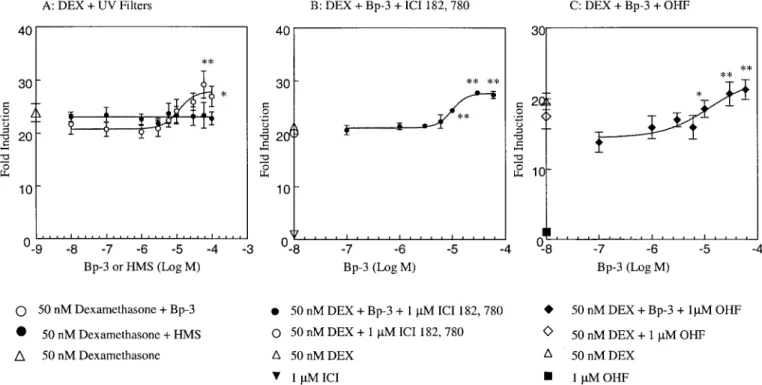
interactions with GR activation (Fig. 7). HMS did not change
the effect of 50 nM dexamethasone on luciferase activity. High
concentrations of Bp-3 induced a small increase in luciferase
activity beyond the level produced by dexamethasone alone.
FIG. 6. Concentration-response curves for effects of Bp-3 and HMS on
MTT reduction in MDA-kb2 cells. Mean⫾ SEM of four independent exper-iments. * and **, significantly lower than control (p⬍ 0.05 and p ⬍ 0.01).
FIG. 7. (A) Effect of Bp-3 and HMS on luciferase activity in MDA-kb2 cells in the presence of 50 mM dexamethasone. Mean⫾ SEM of five independent
experiments. Ethanol concentrations (v/v): from 1 : 106 to 1 : 102. (B) Effect of Bp-3 on luciferase activity in MDA-kb2 cells in the presence of 50 nM
dexamethasone and 1M ICI 182, 780. Mean ⫾ SEM of five independent experiments. **Significant difference vs. 50 nM dexamethasone and 1 M ICI 182, 780 (p⬍ 0.01). Ethanol concentrations (v/v): from 1:106 to 1:102. (C) Effect of Bp-3 on luciferase activity in MDA-kb2 cells in the presence of 50 nM
dexamethasone⫹ 1M hydroxyflutamide. Mean ⫾ SEM of five independent experiments. * and **: significant difference vs. 50 nM dexamethasone ⫹ 1 M hydroxyflutamide (p⬍ 0.05 and p ⬍ 0.01). Ethanol concentrations (v/v): from 1:106to 1:102.
This effect was not prevented either by the estrogen antagonist
ICI 182,780 nor by hydroxyflutamide (Fig. 7B,C).
DISCUSSION
MDA-kb2 cells represent a cell line that expresses both
endogenous human AR and GR and has been stably transfected
by Kathy Bobseine with the MMTV-luciferase gene (Wilson et
al., 2002). In our validation experiments, AR agonists [5
␣-dihydrotestosterone (DHT), methyltrienolone (R1881,
metri-bolone), methyltestosterone, androstenedione, and danazol]
displayed EC
50values and relative androgenic potencies
cor-responding to the potency range reported in earlier studies
(Foster and Cunha, 1999; Wiita et al., 1995). A distinction
between AR and GR agonists can be achieved with androgen
antagonists such as hydroxyflutamide, which antagonized the
effect of DHT on luciferase expression without concomitant
antagonism of dexamethasone. These results are comparable to
findings of Wilson et al. (2002). The data indicate that the cell
line represents a sensitive tool for the screening of AR
ago-nists.
The assay likewise proved to be suitable and sensitive for the
detection of AR antagonistic effects of nonsteroidal
antiandro-gens such as hydroxyflutamide, flutamide, bicalutamide, and
vincolozolin (Bauer et al., 1998; Gray et al., 1994; Kelce et al.,
1994; Waller et al., 1996; Wong et al., 1995). Vinclozolin is
converted in vivo into two metabolites, M1 and M2, with
higher antiandrogenic activity than the parent compound
(Kelce et al., 1994). The IC
50value of the parent compound
observed in the present study is in the range of the activity
reported by Wilson and coworkers (2002).
A complex situation was encountered with cyproterone
actetate (CPA). CPA was characterized by marked agonistic
activity across a wide concentration range when given alone, a
slight, nonsignificant tendency of DHT antagonism at low CPA
concentrations (0.1
M), and an identical dose-response
rela-tionship at high CPA concentrations in the presence or absence
of DHT. CPA is known to possess significant partial AR
agonist activity (Labrie et al., 1987). However, the failure by
hydroxyflutamide to reduce its agonistic effect speaks against
a major role of AR for the agonistic CPA action in the present
cell system. The effect might result from an action on the GR
expressed by MDA-kb2 cells; CPA has been reported to
ex-hibit glucocorticoid activity (Lamberts et al., 1988; Poulin et
al., 1991). A predominance of agonistic effects of CPA was
also observed in MCF7-AR1 cells expressing the human AR
(A-SCREEN assay) (Ma, 2002). Thus, antiandrogens with
mixed activities may present problems for analysis in both
assay systems.
As mentioned in the introduction, the use of UV filters has
greatly increased over the last decades. Since they are
li-pophilic, they may bioaccumulate in the food chain. Evidence
for this has been presented for fish (perch and roach in the
Meerfelder Maar lake, Germany; Nagtegaal et al., 1997), and
for human milk (Hany and Nagel, 1997). The presence in
human milk has recently been confirmed for one UV filter in
this laboratory (unpublished observations). When analyzed for
potential endocrine activity, 4-MBC, OD-PABA, OMC, Bp-3,
and HMS exhibited estrogenic activity in vitro on MCF-7 cells
(increased proliferation, pS
2protein induction). 4-MBC, OMC,
and Bp-3 also increased uterine weight in immature rats
(Schlumpf et al., 2001). Two of these compounds,
benzophe-none- 3 (Bp-3) and homosalate (HMS), also showed significant
androgen (DHT) antagonism in vitro in MDA-kb2 cells. Both
chemicals were completely devoid of agonistic actions when
tested alone. The IC
50values of the two UV filters were in the
low micromolar range, which is the effective range of other
environmental antiandrogenic chemicals such as vinclozolin
and linuron (Vinggaard et al., 1999; Wilson et al., 2002).
While in this cell line, HMS appeared only to antagonize
androgen actions and did not interfere with glucocorticoid
effects, high concentrations of Bp-3 elicited an unexpected
increase of luciferase activity in the presence of
dexametha-sone, beyond the level induced by the glucocorticoid. Since
hydroxyflutamide and the estrogen antagonist ICI 182,780
were unable to block this effect, its nature remains uncertain.
In conclusion, our investigation identified two out of eight
UV filters tested, Bp-3 and HMS, as antiandrogens in the in
vitro MDA-kb2 cell transcription assay. Both compounds also
act as estrogen agonists on MCF-7 cells in vitro, and Bp-3 is
estrogenic in the in vivo uterotrophic assay (Schlumpf et al.,
2001). In vitro estrogenic potencies (ED
503.73
M for Bp-3
and 1.56
M for HMS) and antiandrogenic potencies (Table 2)
are in a similar range, but such comparisons should be done
with caution, considering the different endpoints of the two
assays (proliferation versus reporter gene). Our data support
the notion that some endocrine disrupting chemicals may
in-teract with endocrine regulation by more than one mechanism
(Sohoni et al., 1998). Whether Bp-3 and HMS are also AR
antagonists in vivo remains to be clarified.
ACKNOWLEDGMENTS
The authors would like to thank Kathy Bobseine and L. Earl Gray (U.S. EPA) for the generous gift of MDA-kb2 cells. The investigation was supported by the Swiss Environmental Protection Agency (Bundesamt fu¨r Umwelt, Wald und Landschaft, BUWAL) and Swiss National Research Programme (NRP) 50 (40-66583).
REFERENCES
Bauer, E. R., Meyer, H. H., Stahlschmidt-Allner, P., and Sauerwein, H. (1998). Application of an androgen receptor assay for the characterization of an-drogenic or antianan-drogenic activity of various phenylurea herbicides and their derivatives. Analyst 123, 2485–2487.
Foster, B. A, and Cunha, G. R. (1999). Efficacy of various natural and synthetic androgens to induce ductal branching morphogenesis in the de-veloping anterior rat prostate. Endocrinology 140, 318 –328.
environ-mental antiandrogen: The fungicide vinclozolin alters sex differen-tiation of the male rats. Toxicol. Appl. Pharmacol. 129, 46 –52.
Hall, R. E., Tilley, W. D., McPhaul, M. J., and Sutherland, R. L. (1992). Regulation of androgen receptor gene expression by steroids and retinoic acid in human breast-cancer cells. Int. J. Cancer 52, 778 –784.
Hany, J., and Nagel, R. (1997). Nachweis von UV-Filtersubstanzen in Mut-termilch. Deutsche Lebensmittel-Rundschau. 91, 341–345.
IPCS (International Programme on Chemical Safety) Report. (2002). Global assessment of the state of the science of endocrine disruptors (T. Damstra, S. Barlow, A. Bergman, R. Kavlock, and G. Van Der Kraak, Eds.). IPCS/ WHO, Geneva, Switzerland.
Kelce, W. R., Gray, L. E., and Wilson, E. M. (1998). Antiandrogens as environmental endocrine disruptors. Reprod. Fertil. Dev. 10, 105–111. Kelce, W. R., Lambright, C. R., Gray, L. E., and Roberts, K. (1997).
Vinclo-zolin and p,p⬘-DDE alter androgen-dependent gene expression in vivo con-firmation of an androgen receptor-mediated mechanism. Toxicol. Appl.
Pharmacol. 142, 192–200.
Kelce, W. R., Monosson, E., Gamcsik, M. P., Laws, S. C., and Gray, L. E., Jr. (1994). Environmental hormone disruptors: Evidence that vinclozolin de-velopmental toxicity is mediated by antiandrogenic metabolites. Toxicol.
Appl. Pharmacol. 126, 275–285.
Labrie, C., Cusan, L., Plante, M., Lapointe, S., and Labrie, F. (1987). Analysis of the androgenic activity of synthetic “progestins” currently used for the treatment of prostate cancer. J. Steroid Biochem. 28, 379 –384.
Lamberts, S. W., Uitterlinden, P., and de Jong, F. H. (1988). Rat prostatic weight regression in reaction to ketoconazole, cyproterone acetate, and RU23908 as adjuncts to a depot formulation of gonadotropin-releasing hormone analogue. Cancer Res. 48, 6063– 6068.
Lambright, C., Ostby, J., Bobseine, K., Wilson, V., Hotchkiss, A., Mann, P. C., and Gray, L. E., Jr. (2000). Cellular and molecular mechanisms of action of linuron: An antiandrogenic herbicide that produces reproductive malforma-tions in male rats. Toxicol. Sci. 56, 389 –399.
Ma, R. (2002). Analysis of androgen receptor agonists and antagonists using MCF7-AR1 and MDA-MB-453-KB2 cells. Doctoral Thesis, University of Zurich, Switzerland.
Mosmann, T. (1983). Rapid colorimetric assay for cellular growth and
sur-vival: Application to proliferation and cytotoxicity assays. J. Immunol.
Methods 65, 55– 63.
Nagtegaal, M., Ternes, T. A., Baumann, W., and Nagel, R. (1997). UV-Filtersubstanzen in Wasser und Fischen. UWSF-Z. Umweltchem. O¨ kotox. 9, 79 – 86.
Ostby, J., Kelce, W. R., Lambright, C., Wolf, C. J., Mann, P., and Gray, L. E., Jr. (1999). The fungicide procymidone alters sexual differentiation in the male rat by acting as an androgen-receptor antagonist in vivo and in vitro.
Toxicol. Ind. Health 15, 80 –93.
Poulin, R., Baker, D., Poirier, D., and Labrie, F. (1991). Multiple actions of synthetic “progestins” on the growth of ZR-75–1 human breast cancer cells: An in vitro model for the simultaneous assay of androgen, progestin, estrogen, and glucocorticoid agonistic and antagonistic activities of steroids.
Breast Cancer Res. Treat. 17, 197–210.
Schlumpf, M., Cotton, B., Conscience, M., Haller, V., Steinmann, B., and Lichtensteiger, W. (2001). In vitro and in vivo estrogenicity of UV screens.
Environ. Health Perspect. 109, 239 –244.
Sohoni, P., and Sumpter, J. P. (1998). Several environmental oestrogens are also anti-androgens. J. Endocrinology 158, 327–339.
Vinggaard, A. M., Bonefeld Joergensen, E. C., and Larsen, J. C. (1999). Rapid and sensitive reporter gene assays for detection of antiandrogenic and estrogenic effects of environmental chemicals. Toxicol. Appl. Pharmacol.
155, 150 –160.
Waller, C., Juma, B. W., Gray, L. E., and Kelce, W. R. (1996). Three-dimensional quantitative structure-activity relationships for androgen recep-tor ligands, Toxicol Appl Pharmacol. 137, 219 –227.
Wiita, B., Artis, A., Ackerman, D. M., and Longcope, C. (1995). Binding of 17-alpha-ethyltestosterone in vitro to human sex hormone binding globulin and rat ventral prostate androgen receptors. Ther. Drug. Monit. 17, 377–380. Wilson, V. S., Bobseine, K., Lambright, C. R., and Gray, L. E., Jr. (2002). A novel cell line, MDA-kb2, that stably expresses an androgen- and glucocor-ticoid-responsive reporter for the detection of hormone receptor agonists and antagonists. Toxicol Sci. 66, 69 – 81.
Wong, Choi-iok, Kelce, W. R., Sar, M., and Wilson, E. M. (1995). Androgen receptor antagonist versus agonist activities of fungicide vinclozolin relative to hydroxyflutamide. J. Biol. Chem. 270, 19998 –20003.