HAL Id: hal-02370595
https://hal.archives-ouvertes.fr/hal-02370595
Submitted on 17 Dec 2020HAL is a multi-disciplinary open access archive for the deposit and dissemination of sci-entific research documents, whether they are pub-lished or not. The documents may come from teaching and research institutions in France or abroad, or from public or private research centers.
L’archive ouverte pluridisciplinaire HAL, est destinée au dépôt et à la diffusion de documents scientifiques de niveau recherche, publiés ou non, émanant des établissements d’enseignement et de recherche français ou étrangers, des laboratoires publics ou privés.
Awake Mouse
Ombeline Hoa, Chrystel Lafont, Pierre Fontanaud, Anne Duvoid-Guillou,
Yasmine Kemkem, Rhonda Kineman, Raul Luque, Tatiana Fiordelisio Coll,
Paul Le Tissier, Patrice Mollard
To cite this version:
Ombeline Hoa, Chrystel Lafont, Pierre Fontanaud, Anne Duvoid-Guillou, Yasmine Kemkem, et al.. Imaging and Manipulating Pituitary Function in the Awake Mouse. Endocrinology, Endocrine Society, 2019, 160 (10), pp.2271-2281. �10.1210/en.2019-00297�. �hal-02370595�
Imaging and manipulating pituitary function in the awake mouse
1
Authors: Ombeline Hoa1,#, Chrystel Lafont1, Pierre Fontanaud1, Anne Guillou1, Yasmine Kemkem1,
2
Rhonda D. Kineman2,3, Raul M. Luque4,5,6, Tatiana Fiordelisio Coll7, Paul Le Tissier8, Patrice Mollard1,*
3
4
Affiliations:; 1IGF, CNRS, INSERM, Univ. Montpellier, F-34094 Montpellier, France, 2Research and
5
Development Division, Jesse Brown Veterans Affairs Medical Center, University of Illinois at Chicago,
6
Chicago, Illinois, USA, 3Department of Medicine, Section of Endocrinology, Diabetes, and Metabolism,
7
University of Illinois at Chicago, Chicago, Illinois, USA, 4Maimonides Institute for Biomedical
8
Research of Cordoba (IMIBIC), Reina Sofia University Hospital, Córdoba, Spain, 5Department of Cell
9
Biology, Physiology and Immunology, University of Córdoba, Córdoba, Spain, 6CIBER Fisiopatología
10
de la Obesidad y Nutrición (CIBERobn); Córdoba, Spain, 7Laboratorio de Neuroendocrinología
11
Comparada, Departamento de Ecología y Recursos Naturales, Biología, Facultad de Ciencias,
12
Universidad Nacional Autónoma de México, Ciudad Universitaria, 04510 México, DF, México,
13
8University of Edinburgh, Centre for Discovery Brain Sciences, Edinburgh, EH8 9XD, UK
14
#New address: Center for Interdisciplinary Research in Biology (CIRB), Collège de France, CNRS,
15
INSERM, PSL Research University, Paris, France
16
*Correspondence to:
17
Patrice Mollard, Institut de Génomique Fonctionnelle, 141 rue de la Cardonille, F-34000 Montpellier,
18
France, tel. : +33 4334359270, email : [email protected]
19
20
Running title: Pituitary initiative
21
22
Key words: In vivo imaging, optogenetic tools, viral infection, endocrine manipulation, hormone
23
rhythms
24
Extensive efforts have been made to explore how the activities of multiple brain cells combine to
26
alter physiology through imaging and cell-specific manipulation in different animal models.
27
However, the temporal regulation of peripheral organs by the neuroendocrine factors released by
28
the brain is poorly understood. We have established a suite of adaptable methodologies to
29
interrogate in vivo the relationship of hypothalamic regulation with the secretory output of the
30
pituitary gland, which has complex functional networks of multiple cell types intermingled with
31
the vasculature. These allow imaging and optogenetic manipulation of cell activities in the
32
pituitary gland in awake mouse models, in which both neuronal regulatory activity and hormonal
33
output are preserved. This methodology is now readily applicable for longitudinal studies of
short-34
lived events (e.g. calcium signals controlling hormone exocytosis) and slowly-evolving processes
35
such as tissue remodelling in health and disease over a period of days to weeks.
Introduction
37
In the past decade, there has been an exponential increase in the technical development of novel tools
38
allowing interrogation of the functional interactions of the complex architecture of the mammalian brain
39
in health and disease. These have principally been developed in mouse models, where both organisation
40
and function of the brain largely recapitulates that of higher mammals including humans (1). The
41
availability of a wide-range of genetically-modified mice, combined with novel virus-based approaches
42
to infect specific mouse brain regions, has allowed identification of specific cell-types, manipulation of
43
neuronal circuits with optogenetic techniques and in vivo monitoring of cell activity. Combining these
44
with recently developed optical techniques, such as the use of a gradient-index (GRIN) lens for imaging
45
deep brain regions (2), has resulted in rapid mapping of the activity and connectivity of neuronal
46
networks (3). Although the mammalian brain is exceptionally complex, the increasing prevalence of
47
neurological and neuropsychiatric defects has recently inspired large-scale research programmes, such
48
as the NIH Brain Research through Advancing Innovative Neuro-technologies (BRAIN) Initiative (4,
49
5), to meet this challenge.
50
The brain does not simply work as an isolated unit but forms a functional continuum with other
51
physiological processes (6), especially with the endocrine systems that control basic body functions (7,
52
8). These endocrine systems share complex functional features with the brain, such as hierarchal
multi-53
cellular organization (e.g. presence of “hub” cells which control neighbours (9, 10)), adaptive plasticity
54
(11) and long-term memory (9), suggesting that studies of their function would benefit from application
55
of the novel tools and techniques developed for neuroscience. This is exemplified by the pituitary gland,
56
which acts as an intermediate between the brain and periphery, with endocrine and neural lobes (nerve
57
terminals emanating from hypothalamic vasopressin and oxytocin neurons) connected to the brain by
58
the pituitary stalk and surrounded by brain meninges (see Fig. 1). Interest in monitoring the in vivo
59
function of this gland has recently been increased by large-scale ex vivo imaging, which has revealed
60
3D cell networks that are structurally and functionally organised within the endocrine anterior pituitary
61
(also called the pars distalis); this cell network connectivity is essential for normal gland development
62
(12), coordination of gene expression (13) and pulsatile release of hormones to the periphery (8). To
date, in vivo studies have been limited by the location of the pituitary on the ventral side of the brain,
64
with extensive microsurgery required to expose the gland through the palate bone in
terminally-65
anaesthetised mice to record and manipulate cell function (14). These surgical procedures preclude both
66
longitudinal studies and functional investigation in awake mice.
67
Here, we describe a toolkit for imaging and manipulating pituitary cells in vivo over periods of days to
68
weeks in awake mouse models. We have used these tools to: image the dynamics of pituitary
69
microvascular function and cell signalling (calcium events); locally express exogenous proteins through
70
injection of viral constructs within the parenchyma; and, optogenetically manipulate specific cell
71
networks while monitoring their secretory outputs into the bloodstream. This range of techniques allows
72
analysis of the pituitary gland in awake mammalian models in unparalleled detail, complementing
large-73
scale studies of the brain to further understand neural control of complex physiological systems via
74
endocrine signals.
75
Materials and Methods
76
Animals
77
78
Tg(Gh1-cre)bKnmn (called GH-Cre) (R Kineman, Jesse Brown Veterans Administration Medical
79
Center, Chicago, USA) (15), ROSA26-fl/fl-ChR2-dtTomato and wild-type C57BL/6 mice (6–12 wk
80
old) as indicated in figure legends, were housed in a 12-h light/12-h dark cycle (lights on at 0800 hours
81
and off at 2000 hours) with food and water available ad libitum. All animal procedures were approved
82
by the local ethical committee under agreement CEEA-LR-12185 according to EU Directive
83
2010/63/EU. Since this study included only one experimental group of animals, no randomization or
84
blinding were required.
85
86
Stereotaxic injections of AAV
87
Adult GH-Cre mice and wild-type C57BL/6 were anesthetized with Ketamine/Xylazine (0.1/0.02 mg/g),
88
placed in a stereotaxic apparatus, and given bilateral 1μL injections of
AAV5-CAG-dflox-GCaMP6s-89
WPRE-SV40 (2.52 × 10^13 GC/mL; Penn Vector Core), AAV5-CAG-GCaMP6s-WPRE-SV40 (2.23
× 10^13 GC/mL; Penn Vector Core), AAV2-CAG-GFP (gift from Margarita Arango, IGF, Montpellier),
91
rAAV5/sspEMBOL-CBA-GFP (8 x 10^12 GC/mL; UNC Vector Core),
rAAV8/sspEMBOL-CAG-92
GFP (8 x 10^12 GC/mL; UNC Vector Core) or rAAV9/sspEMBOL-CAG-GFP (9.2 x 10^12 GC/mL;
93
UNC Vector Core) into the pituitary gland at a rate of 100 nL/min. Coordinates were -2.5mm
antero-94
posterior, ±0.4mm lateral to midline, pointed as zero at the superior sagittal sinus. Two dorso-ventral
95
positions were used for injection, 50µm and 400µm over the sella turca -6.15/5.75 mm for ventral
96
injection and -5.6/5.3 mm for dorsal injection. Experiments were conducted from 4 weeks on after
97
injection.
98
99
Optical imaging through a GRIN lens in awake head-fixed mice
100
Adult mice were anesthetized with Ketamine/Xylazine (0.1/0.02 mg/g) and placed in a stereotaxic
101
apparatus to implant a GRIN lens (0.6 mm diameter, 1.5 pitch, 7.5mm length and 150µm working
102
distance, GRINTECH Germany) immediately above the pituitary gland. After a large part of skull was
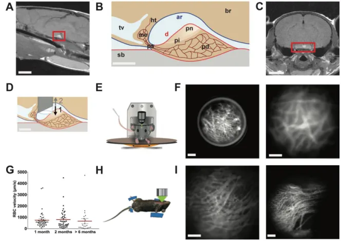
103
exposed, the GRIN lens was placed in 20G1/2 Gauge needle (Ultra-Thin wall, Terumo, USA), with
104
movement restricted by placing a metal rod above it. The needle was inserted at the coordinates -2.5mm
105
antero-posterior, ±0.4mm lateral to midline pointed as zero at the superior sagittal sinus
106
-5.5/5.1 mm dorso-ventral. Then, the needle was removed with the metal rod kept in place so that the
107
GRIN lens stayed in place at the dorsal side of the pituitary. Finally, the metal rod was removed. The
108
GRIN lens and a head-plate were fixed with UV-retractable cement. Prior to and starting from two
109
weeks after surgery, mice were habituated to the wheel and the head-plate fixation system under the
110
microscope every two to three days. Four weeks after surgery, mice were placed on the wheel, the
head-111
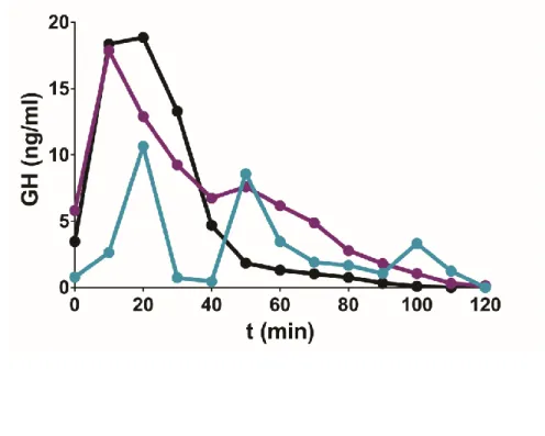
plate fixed, and fluorescence imaging was performed using a stereomicroscope (Zeiss Discovery V.12,
112
Germany), which was fitted with a fluorescence lamp (Lambda LS, Sutter Instrument company, USA),
113
a shutter (Lambda 10-B Smart Shutter, Sutter Instrument Company) and a CMOS ORCA Flash 4.0
114
camera (C11440 Hamamatsu, Japan), all controlled with MetaMorph 7.8.9 software (Molecular
115
Devices, USA).
116
117
In vivo imaging in terminally-anesthetized mice
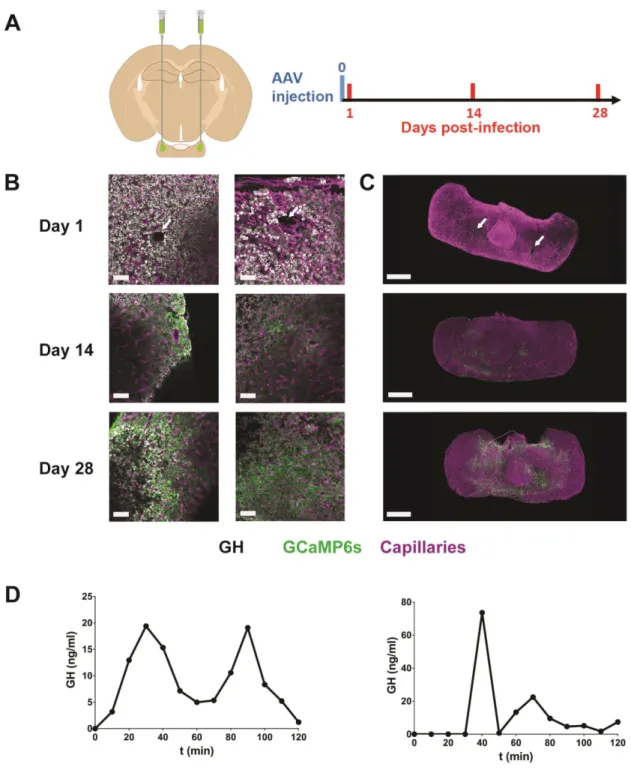
118
Details of the methods can be found in Lafont et al. (2010) (14). In brief, male, 2- to 4-month-old
119
transgenic GH-Cre mice and GH-ROSA26-fl/fl-ChR2-dtTomato mice on a C57Bl6 background were
120
anesthetized by inhalation of isoflurane (1.5% in O2). After dividing the mandibular symphysis, the
121
mucosa overlying the hard palate was parted by blunt dissection under a stereomicroscope to expose an
122
area of palatal periosteal bone. This was thinned with a felt polisher (drill; World Precision Instruments,
123
USA) and then removed with a hook and forceps. The exposed surface of the pituitary gland, visible
124
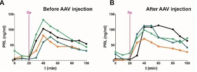
through the hole in the bone, was continuously superfused with a physiological solution.
125
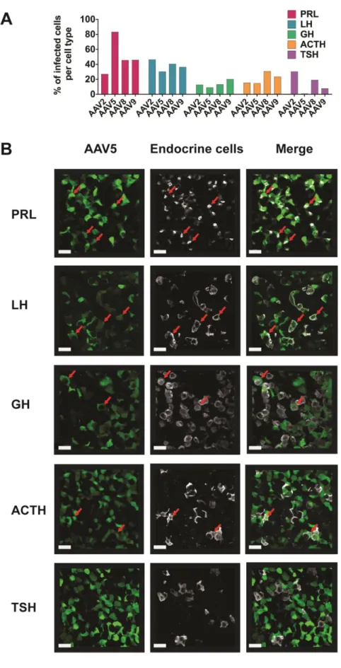
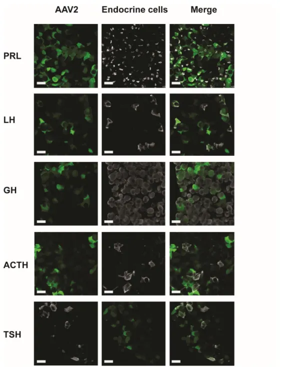
126
In vivo monitoring of blood flow and calcium signals
127
Mice underwent surgery (see above) to visualize either the ventral (terminally-anesthetized animals) or
128
the dorsal side (awake animals) of the pituitary gland. Using the ventral approach 100µl of
129
tetramethylrhodamine isocyonate 150kDa dextran (Sigma Aldrich, USA) was injected into the jugular
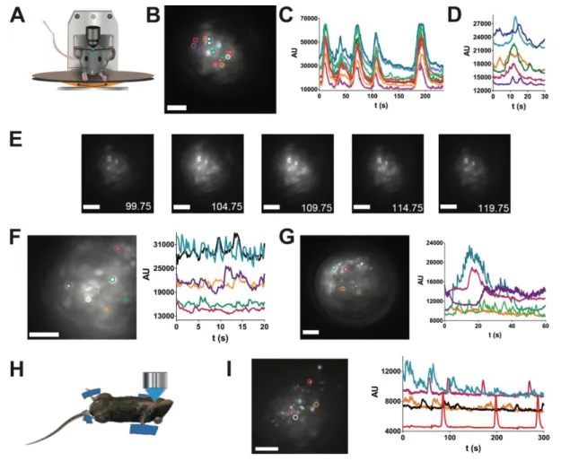
130
vein or in the retro-orbital sinus for GRIN lens approach. Imaging of blood flow was performed at 150
131
to 200 frames/sec using 545nm excitation and 570nm emission filters. When calcium signals were
132
recorded in vivo, experiments were performed as described as above four weeks after stereotaxic
133
injection of GCAMP6s-expressing AAV5. Multi-cellular calcium imaging was typically performed at
134
2-4 frames/sec, using 480nm excitation and 520nm emission filters.
135
136
Optogenetic photostimulation in awake mice
137
GH Cre x ROSA26-fl/fl-ChR2-dtTomato mice were anesthetized with Ketamine/Xylazine (0.1/0.02
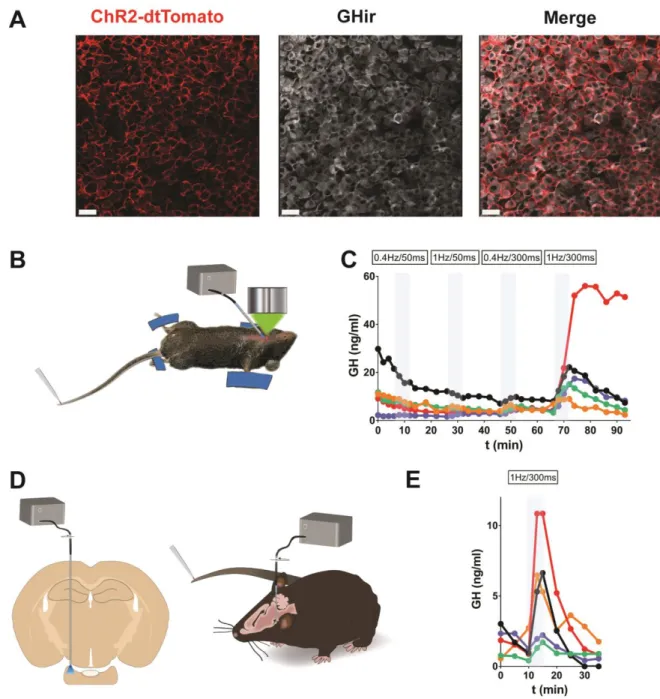
138
mg/g) and placed in a stereotaxic apparatus to implant an optical fiber (diameter: 200μm, Doric Lenses,
139
Canada) immediately above the pituitary gland (stereotaxic coordinates described above). The optical
140
fiber was fixed using UV-retractable cement. Two weeks later, an optical fiber was connected to the one
141
previously implanted, and laser stimulation (488nm) was delivered at 10mW and using various patterns
142
(frequency: 1Hz, exposure time: 300ms) while blood samples were collected as described below.
143
144
GH pulse profiling in mice and GH ELISA
145
A tail-tip blood collection procedure was used to sample blood from C57BL/6 adult mice or transgenic
146
GH-Cre mice; 3μl blood samples were analyzed for GH content by ELISA (16).
147
148
iDISCO+
149
Pituitary glands were removed and fixed by overnight immersion in 4% paraformaldehyde. For the
150
immunoflurescence labelling and clearing, an iDISCO+ clearing protocol was used as described in detail
151
elsewhere(17). Primary antibodies were rat anti-Meca32 (1:100, BD Biosciences Cat# 550563,
152
RRID:AB_393754)(18), guinea pig anti-GH (1:2500, NIDDK-NHPP Cat# AFP12121390,
153
RRID:AB_2756840)(19), rabbit anti-GFP (1:250, Molecular Probes Cat# A-6455,
154
RRID:AB_221570)(20) and secondary antibodies were anti-rat Alexa 647 (Jackson ImmunoResearch
155
Labs Cat# 712-606-150, RRID:AB_2340695)(21), anti-guinea pig Alexa 510 (Jackson
156
ImmunoResearch Labs Cat# 706-166-148, RRID:AB_2340461)(22) and anti-rabbit Alexa 488
157
(Molecular Probes Cat# A-21206, RRID:AB_141708)(23) (dilution: 1:2000). After clearing, transparent
158
pituitary glands were mounted in well glass slides (065230, Dominique Dutscher) in DiBenzyl Ether
159
(Sigma Aldrich). Coverslips were sealed with nail varnish.
160
161
Immunofluorescence staining in fixed pituitary slices
162
Pituitary glands were collected from terminally-anesthetized mice and fixed by overnight immersion in
163
4% paraformaldehyde at 4°C, serial cuts were done at 40µm-thick tissue sections using a vibratome
164
(Leica, Germany). Combinations of the following antibodies were used: guinea pig anti- GH
(NIDDK-165
NHPP Cat# AFP12121390, RRID:AB_2756840)(19), LH (NIDDK-NHPP Cat# rLHb, also
166
AFP571292393, RRID:AB_2665511)(24), PRL (NIDDK-NHPP Cat# AFP65191,
167
RRID:AB_2756841)(25), TSH (NIDDK-NHPP Cat# AFP9370793, RRID:AB_2756856)(26) or ACTH
168
(NIDDK-NHPP Cat# AFP71111591, RRID:AB_2756855)(27) (dilution: 1:2500), rabbit anti-GFP
169
(1:250, Molecular Probes Cat# A-6455, RRID:AB_221570)(20) and rabbit anti-RFP (1:500, Rockland
170
Cat# 600-401-379, RRID:AB_2209751)(28). Primary antibody incubation was performed in PBS, 0.1%
Triton X-100, 2% BSA at 4 °C for 48 h. Sections were then incubated with secondary antibodies for 2h
172
at room temperature. Secondary antibodies were anti-rabbit Alexa 488 (Molecular Probes Cat#
A-173
21206, RRID:AB_141708)(23), anti-guinea pig Alexa 510 (Jackson ImmunoResearch Labs Cat#
706-174
166-148, RRID:AB_2340461)(22), Anti-Rat Alexa 647 (Jackson ImmunoResearch Labs Cat#
712-606-175
150, RRID:AB_2340695)(21), Anti-Guinea Pig Alexa 488 (Jackson ImmunoResearch Labs Cat#
706-176
545-148, RRID:AB_2340472)(29) and anti-rabbit 510 (Jackson ImmunoResearch Labs Cat#
711-166-177
152, RRID:AB_2313568)(30) (1:2000 in PBS, 0.1% Triton X-100, 2%BSA).
178
179
Confocal imaging
180
Fluorescence images of both sliced pituitaries and whole clarified pituitaries were acquired on a Zeiss
181
LSM 780 confocal microscope with 20x, 40x, 63x objectives. Images were analyzed using Imaris
182
(Bitplane, UK).
183
184
MRI image acquisition from mouse brain
185
Animals were scanned on a 9.4T Agilent Varian MRI scanner. A volumic RF43 antenna (Rapid
186
Biomedical) was used. For image acquisition, mice were anesthetized with isoflurane and their heads
187
secured with bite and ear bars. Respiration rate and heart rate were monitored. Animals were scanned
188
using a spin echo sequence with the following parameters: Repetition time 500ms, echo time 10ms, 1
189
echo, averaging 16 times, matrix of 256 × 256 pixels in a FOV of 30x30mm, slices thickness 0.5mm.
190
Total imaging time was 34 min.
191
192
Analysis
193
Blood flow changes were estimated from red blood cell velocities as previously described (14) and
194
analysed using a two-tailed variance ratio test followed by a Mann–Whitney U test for any differences
195
directly attributable to treatment application. Estimation of decay time (τ = 5sec) from calcium signals
196
(27 single calcium transients) recorded in vivo was used to generate simulated calcium rises due to trains
197
of calcium spikes firing at frequencies of either 0.4 or 1Hz. Spike frequencies high enough (1Hz) to
generate robust plateau rises in cytosolic calcium (Figure supplement 6)(31) then guided selection of
199
appropriate frequencies of laser light pulses during optogenetic experiments.
200
Results
201
Longitudinal optical monitoring of pituitary blood flow in awake mice
202
Unravelling the intricacies of pituitary function with cellular in vivo imaging studies lasting days to
203
weeks requires optical access to the gland whilst maintaining both its integrity and that of surrounding
204
tissue. The location of the pituitary (Fig. 1A-C, sagittal and coronal MRI sections of mouse heads and
205
relative schemas, respectively), suggested that the least invasive strategy would be insertion of a GRIN
206
lens though the cortex towards the dorsal side of the pituitary using a stereotaxic frame in anesthetized
207
animals. To overcome the major challenge of crossing the meninges covering the ventral brain without
208
damaging the nearby pituitary tissue (Fig. 1B), the GRIN lens was inserted into the lumen of a needle
209
which was then retracted once the GRIN lens was located correctly (Fig. 1D). The GRIN lens was then
210
fixed to the cranium with UV-retractable cement and a titanium bar with a central opening for the lens
211
was attached to the skull. After at least 3-4 weeks of mouse habituation to being head-fixed under a
212
stereomicroscope fitted with a x20 objective, with the body and limbs being able to move on a treadmill
213
(Fig. 1E), pituitary blood flow was imaged for 0.5 to 2 hours in animals pre-injected in the retro-orbital
214
sinus with fluorescent 150kDa dextran (Fig. 1F, video 1)(31). These in vivo imaging sessions were
215
repeatable every 3-4 days and up to several months after GRIN lens implantation with no alteration in
216
blood flow, assessed by measurements of red blood cell velocities (Fig. 1G). Imaging pituitary blood
217
flow in awake mice using a GRIN lens with a numerical aperture of 0.5 provided image resolution
218
similar to that obtained in terminally-anesthetized animals with ventral surgery and imaged with a
long-219
range (2 cm working distance, N.A. 0.5) objective (Fig. 1H, I) (14). All imaging sessions were
220
performed between one and six months after GRIN lens implantation without noticeable changes of
221
pituitary function, based on preservation of endogenous hormone rhythms (Figure supplement 1)(31).
222
Thus implantation of thin GRIN lenses through two layers of meninges, one at the level of the cortex
and the other covering the ventral side of the brain, allowed long-lasting in vivo imaging of the dorsal
224
side of the pituitary whilst preserving characteristic features of pituitary function.
225
Selective viral delivery and fluorescent protein expression in the pituitary parenchyma
226
Local stereotaxic delivery for expression of specific genes, for example by viral transduction (2), has
227
been an important tool for monitoring the activities of cells in selective brain regions. Whilst this
228
approach has been applied to very large pituitary tumors by trans-auricular injection (32, 33), it has not
229
been described in the pituitary of healthy mice. We developed stereotaxic delivery of viral particles that
230
could easily be combined with in vivo imaging using GRIN lenses with minimal pituitary damage. We
231
first inserted vertically the AAV-containing needle via the cortex and then positioned the needle tip to
232
touch the palate bone. After waiting 5 min, the needle was retracted by 50µm and 400µm to target the
233
ventral and dorsal regions of the pituitary, respectively (Fig. 2A). AAV particles were then injected
234
using a controlled pneumatic pump to transduce cells with an expression cassette encoding the calcium
235
sensor GCAMP6s (34) or GFP under the control of the strong ubiquitous CAG promoter. Virus was
236
routinely injected in both pituitary “wings” (lateral regions are 500-700 µm thick). Pituitaries were then
237
dissected and fixed 1, 14 and 28 days after viral injection (Fig. 2B-C). Although a small region of tissue
238
damage was apparent one day after AAV injection using a needle with an outer diameter of 210µm
239
diameter, this was markedly reduced or absent 2 weeks post-injection and apparently fully repaired after
240
4 weeks. Pituitary tissues were immunostained for fenestrated vessel markers (MECA32), pituitary
241
hormones (e.g. GH) and GCAMP6s in thick pituitary sections (Fig. 2B, top left panels and tissue
242
clarified with the iDISCO+ protocol (Fig. 2C, top right panel) (17). This showed that expression of
243
AAV-CAG-expressed GCAMP6s could be detected 2 weeks post-infection (Fig. 2B-C, middle panels)
244
but was increased and more extensive after 4 weeks (Fig. 2B-C, bottom panels). Consistent with the
245
apparently complete tissue recovery one month after AAV infection (Fig. 2B-C), endogenous (Fig. 2D)
246
and hormonal responses to hypothalamic agonists (Figure supplement 2)(31) were unaltered following
247
stereotaxic injection of AAV.
As the pituitary gland contains five endocrine cell types secreting specific hormones (PRL, LH/FSH,
249
GH, ACTH and TSH), we tested the efficiency of viral transduction in each of these by a range of AAV
250
serotypes expressing CAG promoter driven GFP. All pituitary hormonal cell types were transduced with
251
variable efficiency depending of AAV serotype (Fig. 3, Figures supplement 3-5)(31). For all AAV
252
serotypes, expression of GFP could readily be detected by immunostaining from constructs utilizing a
253
CAG promoter but not those with a CMV promoter (data not shown).
254
255
Pituitary calcium signals in awake mice
256
Having successfully transduced pituitary cells with constructs expressing GCAMP6s by stereotaxic
257
injection of AAV5-CAG-GCAMP6s, we then explored whether this could be used to monitor
258
multicellular calcium signals in awake mice following AAV injection. GRIN lenses were implanted
259
above the dorsal pituitary at the site where AAVs had previously been injected stereotaxically. One
260
month after GRIN lens implantation, it was possible to monitor a wide range of profiles of pituitary
261
calcium transients in awake mice (Fig. 4A-G, video 2)(31), with evidence of cell-cell coordination (Fig.
262
4B-4E, video 3)(31) similar to that previously reported in ex vivo studies on pituitary slice preparations
263
(9, 11, 35). Of note, similar calcium activity was detected in vivo using a ventral imaging approach in
264
anaesthetized mice (14) which had been injected with AAV5-CAG-GCAMP6s (Fig. 4H-I), suggesting
265
that the GRIN lens implantation does not affect calcium signaling.
266
267
Optogenetic manipulation of pituitary hormone pulsatility in awake mice
268
The ability to implant lenses and optical devices into the pituitary of awake mice also enables control of
269
the secretory activity of pituitary cells. For this, we used a Cre-lox strategy by crossing GH-Cre and
270
R26-fl-fl-ChR2-dtTomato mice, resulting in expression of ChR2 specifically in somatotrophs
(GH-271
ChR2; Fig. 5A). To determine which blue laser illumination pattern was efficient at triggering hormone
272
output from somatotrophs, we used the ventral imaging approach in anesthetized mice to stimulate the
273
pituitary cells with a 400 µm diameter fiber optic positioned close to the pituitary surface (Fig. 5B) and
measured GH in blood samples collected from the tail (Fig. 5C). The requirement for a 1Hz stimulation
275
for 300ms to elicit a robust output of GH agrees with simulations of the generation of sustained trains
276
of calcium spikes based on from in vivo calcium spike kinetics (Figure supplement 6)(31). Application
277
of this pattern of laser light triggered GH pulses in awake GH-ChR2 mice chronically-implanted with
278
an optical fiber which was located above the dorsal side of the pituitary (Fig. 5 D-E).
279
280
Discussion
281
By adapting approaches using stereotaxic to access the ventral side of the brain, we have successfully
282
applied a wide range of tools and techniques for imaging and manipulating specific cell activities in the
283
pituitary gland of awake mice. These technical developments now allow the study of the function of this
284
gland and its intimate relationship with the brain in health and disease at a level hitherto not achievable
285
in awake animal models. Analysis of dynamic pituitary function over periods of days to months in
286
animals with intact interactions between multiple organs will provide important insight into a range of
287
conditions with dysregulated physiological function which may occur at different level within an axis.
288
For example, it is unclear to what extent altered pituitary, hypothalamic or ovarian function contributes
289
to the dysregulated LH secretion which is a hallmark of the polycystic ovarian syndrome, the most
290
common endocrine pathology in the reproductive age female (prevalence 7-15% of pre-menopausal
291
women (36).
292
Live imaging with multi-cellular resolution in awake GRIN lens implanted mice is well suited to
real-293
time studies of cell signals, as illustrated here with calcium signals that are essential for hormone
294
exocytosis (8), and can be used to monitor cell-cell communication within the variety of intermingled
295
cell networks wiring the gland (12). As multi-cellular signal events can be directly combined with
296
frequent blood microsamples and high-sensitive hormone ELISA (16), on-line monitoring of
‘stimulus-297
secretion’ coupling (37, 38) is now achievable at the organ (pituitary) level in awake animals, avoiding
298
the well-described blunting of hypothalamic inputs by anesthetics (14). In addition, these studies will
299
be augmented by combining laser light-control of cell functions with monitoring cell activity within the
same field of view of the GRIN lens, which is now possible given the efficiency of optogenetic tools for
301
the control of pituitary cell networks.
302
On-line monitoring and manipulation of in vivo stimulus-secretion coupling is now readily applicable
303
to answer long-standing questions concerning pituitary gland integration of both brain and peripheral
304
signals for the generation of pulsatile hormonal output. For example, it is now clear that dynamic pulses
305
of corticotroph ACTH output is generated by both a combination of both hypothalamic (CRF and
306
vasopressin) inputs and negative cortisol feedback (39). Future use of miniature imaging systems in
307
GRIN lens-implanted animals (3) would allow monitoring and manipulation of corticotroph cell activity
308
regulating the stress axis, with simultaneous modification of environmental conditions in freely-moving
309
mouse models and study of behavioral effects. To date, such interrogation of the role of pituitary
310
corticotrophs in the stress axis has been restricted to simpler animal models, such as larval zebrafish
311
(40), which lack delivery of hypophysiotropic input via a portal blood system and thus may differ in
312
important aspects to humans (8). An ability to manipulate pituitary cell output via optogenetic
313
stimulation and/or inhibition will also allow dissection of the role of specific patterns of pituitary
314
hormone output, for example the sexually dimorphic GH-dependent regulation liver gene expression
315
(41). Male and female GH secretion patterns can now be optogenetically triggered irrespective of sex
316
animal.
317
A remarkable feature of this suite of tools is their capacity to allow long-term pituitary imaging and
318
manipulation in awake animals. With the restriction of studying adult animals, both short-lived cell
319
events (as discussed above) and slowly-evolving remodelling of the tissue, such as angiogenesis and
320
expansion/shrinkage of a cell population can now be examined over weeks to months in individual
321
animals, which act as their own controls (42). This will notably be relevant for visualizing and studying
322
on-line potential repopulation of the pituitary with stem cells/progenitors (43-45) (e.g. fluorescent cells
323
locally injected in immune-suppressed mice), which have the potential to restore cell populations in the
324
hypoplastic pituitary. It will also be possible to explore the function of either sick or healthy tissue zones
325
within one pituitary by local injection of, for example, tumor cells or a virus encoding CRISPR-driven
326
gene mutation in Cas9-expressing mice (46, 47).
In summary, the ability to image at multiple time scales and manipulate the pituitary gland enables the
328
interrogation of pituitary gland function in awake mammalian models and study of how it delivers
329
highly-ordered hormone pulses essential for controlling body functions such as reproduction, growth,
330
stress and metabolism. Since endocrine cells can be photo-painted in situ (10), longitudinal in vivo
331
studies would give access to the history of cells (48) and how they interact with neighbours in their
332
native environment (9, 49). Single-cell multiomics which include transcriptomics, epigenomics and
333
proteomics (50) would then be applicable to individual pituitary cells which have been monitored for
334
days to months in awake mouse models. Together with these newly-developed single-cell level
335
techniques, application of our cellular in vivo imaging and manipulation toolkit to longitudinal studies
336
of awake animal models will provide a unique ability to explore the origin and development of pituitary
337
hormone defects.338
339
340
Acknowledgements341
We thank Margarita Arango (IGF, Montpellier, France) for helpful comments and suggestions about
342
AAV experiments, Jerome Lecoq (Allen Inst., USA) for advices about the use of GRIN lenses, Danielle
343
Carmignac (NIMR-MRC, London UK) for helpful suggestions about AAV injections, Yan Chastagnier
344
(IGF, Montpellier, France) for help and advice about image analysis, and Muriel Asari for her schematic
345
rendition of technical set-ups. Antibodies and Recombinant mouse Growth hormone and Prolactin were
346
supplied by Dr. A.F. Parlow and NIDDK-National Hormone and Pituitary Program (NHPP,
347
TORRANCE, CA). Authors were supported by grants from the Biotechnology and Biological Sciences
348
Research Council, UK (BB/N007026/1) (P.L.T.), U.S. Department of Veterans Affairs, Office of
349
Research and Development Merit Award BX001114; and National Institutes of Health grant
350
R01DK088133 (R.D.K), Junta de Andalucía (CTS-1406, BIO-0139), ISCIII-FIS (PI16/00264)
351
(R.M.L.), ANR-CONACyT 273513, Estancia Sabática apoyada con el Programa PASPA-DGAPA
352
UNAM (T.F.C) , the Agence Nationale de la Recherche (ANR 12 BSV1 0032-01,
ANR-15-CE14-0012-353
01), France-Bioimaging (INBS10-GaL/AR-11/12), Institut National de la Santé et de la Recherche
Médicale, Centre National de la Recherche Scientifique, Université de Montpellier, and Fondation pour
355
la Recherche Médicale (DEQ20150331732) (P.M.). OH was supported by a PhD fellowship from
356
Fondation pour la Recherche Médicale (FDT20160435494). We would also like to thank all members
357
of the Montpellier core facilities IPAM and BioNanoNMRI for unconditional support and thoughtful
358
comments during the course of this work.
359
References
361
362
1. Buzsaki G, Logothetis N, Singer W 2013 Scaling brain size, keeping timing: evolutionary
363
preservation of brain rhythms. Neuron 80:751-764
364
2. Deisseroth K, Schnitzer MJ 2013 Engineering approaches to illuminating brain structure and
365
dynamics. Neuron 80:568-577
366
3. Li Y, Mathis A, Grewe BF, Osterhout JA, Ahanonu B, Schnitzer MJ, Murthy VN, Dulac C 2017
367
Neuronal Representation of Social Information in the Medial Amygdala of Awake Behaving
368
Mice. Cell 171:1176-1190 e1117
369
4. Ecker JR, Geschwind DH, Kriegstein AR, Ngai J, Osten P, Polioudakis D, Regev A, Sestan N,
370
Wickersham IR, Zeng H 2017 The BRAIN Initiative Cell Census Consortium: Lessons Learned
371
toward Generating a Comprehensive Brain Cell Atlas. Neuron 96:542-557
372
5. Jorgenson LA, Newsome WT, Anderson DJ, Bargmann CI, Brown EN, Deisseroth K, Donoghue
373
JP, Hudson KL, Ling GS, MacLeish PR, Marder E, Normann RA, Sanes JR, Schnitzer MJ,
374
Sejnowski TJ, Tank DW, Tsien RY, Ugurbil K, Wingfield JC 2015 The BRAIN Initiative:
375
developing technology to catalyse neuroscience discovery. Philosophical transactions of the
376
Royal Society of London Series B, Biological sciences 370
377
6. Sudhof TC 2017 Molecular Neuroscience in the 21st Century: A Personal Perspective. Neuron
378
96:536-541
379
7. Herbison AE 2016 Control of puberty onset and fertility by gonadotropin-releasing hormone
380
neurons. Nature reviews Endocrinology 12:452-466
381
8. Le Tissier P, Campos P, Lafont C, Romano N, Hodson DJ, Mollard P 2017 An updated view of
382
hypothalamic-vascular-pituitary unit function and plasticity. Nature reviews Endocrinology
383
13:257-267
384
9. Hodson DJ, Schaeffer M, Romano N, Fontanaud P, Lafont C, Birkenstock J, Molino F, Christian
385
H, Lockey J, Carmignac D, Fernandez-Fuente M, Le Tissier P, Mollard P 2012 Existence of
386
long-lasting experience-dependent plasticity in endocrine cell networks. Nature
387
communications 3:605
388
10. Johnston NR, Mitchell RK, Haythorne E, Pessoa MP, Semplici F, Ferrer J, Piemonti L, Marchetti
389
P, Bugliani M, Bosco D, Berishvili E, Duncanson P, Watkinson M, Broichhagen J, Trauner D,
390
Rutter GA, Hodson DJ 2016 Beta Cell Hubs Dictate Pancreatic Islet Responses to Glucose. Cell
391
Metab 24:389-401
392
11. Sanchez-Cardenas C, Fontanaud P, He Z, Lafont C, Meunier AC, Schaeffer M, Carmignac D,
393
Molino F, Coutry N, Bonnefont X, Gouty-Colomer LA, Gavois E, Hodson DJ, Le Tissier P,
394
Robinson IC, Mollard P 2010 Pituitary growth hormone network responses are sexually
395
dimorphic and regulated by gonadal steroids in adulthood. Proceedings of the National
396
Academy of Sciences of the United States of America 107:21878-21883
397
12. Budry L, Lafont C, El Yandouzi T, Chauvet N, Conejero G, Drouin J, Mollard P 2011 Related
398
pituitary cell lineages develop into interdigitated 3D cell networks. Proceedings of the
399
National Academy of Sciences of the United States of America 108:12515-12520
400
13. Featherstone K, Hey K, Momiji H, McNamara AV, Patist AL, Woodburn J, Spiller DG, Christian
401
HC, McNeilly AS, Mullins JJ, Finkenstadt BF, Rand DA, White MR, Davis JR 2016 Spatially
402
coordinated dynamic gene transcription in living pituitary tissue. Elife 5:e08494
403
14. Lafont C, Desarmenien MG, Cassou M, Molino F, Lecoq J, Hodson D, Lacampagne A,
404
Mennessier G, El Yandouzi T, Carmignac D, Fontanaud P, Christian H, Coutry N,
Fernandez-405
Fuente M, Charpak S, Le Tissier P, Robinson IC, Mollard P 2010 Cellular in vivo imaging
406
reveals coordinated regulation of pituitary microcirculation and GH cell network function.
407
Proceedings of the National Academy of Sciences of the United States of America
107:4465-408
4470
15. Luque RM, Amargo G, Ishii S, Lobe C, Franks R, Kiyokawa H, Kineman RD 2007 Reporter
410
expression, induced by a growth hormone promoter-driven Cre recombinase (rGHp-Cre)
411
transgene, questions the developmental relationship between somatotropes and lactotropes
412
in the adult mouse pituitary gland. Endocrinology 148:1946-1953
413
16. Steyn FJ, Huang L, Ngo ST, Leong JW, Tan HY, Xie TY, Parlow AF, Veldhuis JD, Waters MJ,
414
Chen C 2011 Development of a method for the determination of pulsatile growth hormone
415
secretion in mice. Endocrinology 152:3165-3171
416
17. Renier N, Adams EL, Kirst C, Wu Z, Azevedo R, Kohl J, Autry AE, Kadiri L, Umadevi Venkataraju
417
K, Zhou Y, Wang VX, Tang CY, Olsen O, Dulac C, Osten P, Tessier-Lavigne M 2016 Mapping of
418
Brain Activity by Automated Volume Analysis of Immediate Early Genes. Cell 165:1789-1802
419
18. RRID:AB_393754, https://scicrunch.org/resolver/AB_393754420
19. RRID:AB_2756840, https://scicrunch.org/resolver/AB_2756840421
20. RRID:AB_221570, https://scicrunch.org/resolver/AB_221570422
21. RRID:AB_2340695, https://scicrunch.org/resolver/AB_2340695423
22. RRID:AB_2340461, https://scicrunch.org/resolver/AB_2340461424
23. RRID:AB_141708, https://scicrunch.org/resolver/AB_141708425
24. RRID:AB_2665511, https://scicrunch.org/resolver/AB_2665511426
25. RRID:AB_2756841, https://scicrunch.org/resolver/AB_2756841427
26. RRID:AB_2756856, https://scicrunch.org/resolver/AB_2756856428
27. RRID:AB_2756855, https://scicrunch.org/resolver/AB_2756855429
28. RRID:AB_2209751, https://scicrunch.org/resolver/AB_2209751430
29. RRID:AB_2340472, https://scicrunch.org/resolver/AB_2340472431
30. RRID:AB_2313568, https://scicrunch.org/resolver/AB_2313568432
31. Materials to be uploaded to Dryad.
433
32. Riley DJ, Nikitin AY, Lee WH 1996 Adenovirus-mediated retinoblastoma gene therapy
434
suppresses spontaneous pituitary melanotroph tumors in Rb+/- mice. Nat Med 2:1316-1321
435
33. Walls GV, Lemos MC, Javid M, Bazan-Peregrino M, Jeyabalan J, Reed AA, Harding B, Tyler DJ,
436
Stuckey DJ, Piret S, Christie PT, Ansorge O, Clarke K, Seymour L, Thakker RV 2012 MEN1 gene
437
replacement therapy reduces proliferation rates in a mouse model of pituitary adenomas.
438
Cancer Res 72:5060-5068
439
34. Chen TW, Wardill TJ, Sun Y, Pulver SR, Renninger SL, Baohan A, Schreiter ER, Kerr RA, Orger
440
MB, Jayaraman V, Looger LL, Svoboda K, Kim DS 2013 Ultrasensitive fluorescent proteins for
441
imaging neuronal activity. Nature 499:295-300
442
35. Bonnefont X, Lacampagne A, Sanchez-Hormigo A, Fino E, Creff A, Mathieu MN, Smallwood S,
443
Carmignac D, Fontanaud P, Travo P, Alonso G, Courtois-Coutry N, Pincus SM, Robinson IC,
444
Mollard P 2005 Revealing the large-scale network organization of growth hormone-secreting
445
cells. Proceedings of the National Academy of Sciences of the United States of America
446
102:16880-16885
447
36. Hayes MG, Urbanek M, Ehrmann DA, Armstrong LL, Lee JY, Sisk R, Karaderi T, Barber TM,
448
McCarthy MI, Franks S, Lindgren CM, Welt CK, Diamanti-Kandarakis E, Panidis D, Goodarzi
449
MO, Azziz R, Zhang Y, James RG, Olivier M, Kissebah AH, Reproductive Medicine N,
Stener-450
Victorin E, Legro RS, Dunaif A 2015 Genome-wide association of polycystic ovary syndrome
451
implicates alterations in gonadotropin secretion in European ancestry populations. Nature
452
communications 6:7502
453
37. Neher E, Marty A 1982 Discrete changes of cell membrane capacitance observed under
454
conditions of enhanced secretion in bovine adrenal chromaffin cells. Proceedings of the
455
National Academy of Sciences of the United States of America 79:6712-6716
456
38. Thomas P, Surprenant A, Almers W 1990 Cytosolic Ca2+, exocytosis, and endocytosis in single
457
melanotrophs of the rat pituitary. Neuron 5:723-733
458
39. Walker JJ, Spiga F, Waite E, Zhao Z, Kershaw Y, Terry JR, Lightman SL 2012 The origin of
459
glucocorticoid hormone oscillations. PLoS biology 10:e1001341
40. De Marco RJ, Thiemann T, Groneberg AH, Herget U, Ryu S 2016 Optogenetically enhanced
461
pituitary corticotroph cell activity post-stress onset causes rapid organizing effects on
462
behaviour. Nature communications 7:12620
463
41. Waxman DJ, O'Connor C 2006 Growth hormone regulation of sex-dependent liver gene
464
expression. Mol Endocrinol 20:2613-2629
465
42. Pilz GA, Bottes S, Betizeau M, Jorg DJ, Carta S, Simons BD, Helmchen F, Jessberger S 2018 Live
466
imaging of neurogenesis in the adult mouse hippocampus. Science 359:658-662
467
43. Andoniadou CL, Matsushima D, Mousavy Gharavy SN, Signore M, Mackintosh AI, Schaeffer
468
M, Gaston-Massuet C, Mollard P, Jacques TS, Le Tissier P, Dattani MT, Pevny LH,
Martinez-469
Barbera JP 2013 Sox2(+) stem/progenitor cells in the adult mouse pituitary support organ
470
homeostasis and have tumor-inducing potential. Cell Stem Cell 13:433-445
471
44. Perez Millan MI, Brinkmeier ML, Mortensen AH, Camper SA 2016 PROP1 triggers
epithelial-472
mesenchymal transition-like process in pituitary stem cells. Elife 5
473
45. Rizzoti K, Akiyama H, Lovell-Badge R 2013 Mobilized adult pituitary stem cells contribute to
474
endocrine regeneration in response to physiological demand. Cell Stem Cell 13:419-432
475
46. Swiech L, Heidenreich M, Banerjee A, Habib N, Li Y, Trombetta J, Sur M, Zhang F 2015 In vivo
476
interrogation of gene function in the mammalian brain using CRISPR-Cas9. Nat Biotechnol
477
33:102-106
478
47. VanDusen NJ, Guo Y, Gu W, Pu WT 2017 CASAAV: A CRISPR-Based Platform for Rapid
479
Dissection of Gene Function In Vivo. Curr Protoc Mol Biol 120:31 11 31-31 11 14
480
48. Singh SP, Janjuha S, Hartmann T, Kayisoglu O, Konantz J, Birke S, Murawala P, Alfar EA,
481
Murata K, Eugster A, Tsuji N, Morrissey ER, Brand M, Ninov N 2017 Different developmental
482
histories of beta-cells generate functional and proliferative heterogeneity during islet
483
growth. Nature communications 8:664
484
49. van der Meulen T, Mawla AM, DiGruccio MR, Adams MW, Nies V, Dolleman S, Liu S,
485
Ackermann AM, Caceres E, Hunter AE, Kaestner KH, Donaldson CJ, Huising MO 2017 Virgin
486
Beta Cells Persist throughout Life at a Neogenic Niche within Pancreatic Islets. Cell Metab
487
25:911-926 e916
488
50. Macaulay IC, Ponting CP, Voet T 2017 Single-Cell Multiomics: Multiple Measurements from
489
Single Cells. Trends Genet 33:155-168
490
491
493
Figure 1. In vivo imaging of pituitary blood flow in the awake mouse. (A) Sagittal MRI view of the
494
mid-brain from a female mouse. Red rectangle indicates pituitary location below the ventral side of the
495
brain. Scale bar, 3mm. (B) Drawing of a sagittal view of the hypothalamus-pituitary system. Ht,
496
hypothalamus; tv, third ventrical; me, median eminence; ps, pituitary stalk; d, dura mater (in red); ar,
497
arachnoid mater (in blue); pn, pars nervosa; pi, pars intermedia; pd, pars distalis; sb, sphenoidal bone.
498
Scale bar, 300µm. (C) Coronal MRI view of the brain of a female mouse. Red rectangle indicates
499
pituitary location. Scale bar, 3mm. (D) Schema showing the GRIN lens implantation in the arachnoid
500
matter region above the dorsal side of the pituitary. Downward (1) and upward (2) arrows indicate the
501
sequential needle movements when the GRIN lens is positioned above the pituitary. Scale bar, 300µm.
502
(E) Head-fixed in vivo imaging of an awake mouse implanted with a GRIN lens which provides an
503
optical relay between the microscope and pituitary gland. (F) Head-fixed in vivo imaging of pituitary
504
capillaries at low (left panel) and high magnification (right panel). Scale bar, 100µm; representative
505
image of n = 5 female mice. (G) Example of longitudinal monitoring of red blood cell velocities in the
506
same pituitary field viewed from one to six months after GRIN lens implantation; n = 21 to 43 vessels
analyzed per animal, n= 4 female mice. (H) Schematic arrangement of the ventral in vivo imaging
508
approach in terminally-anesthetized mice (14). (I) Ventral in vivo imaging of pituitary capillaries at the
509
level of the pituitary parenchyma (left panel) and entrance (right panel) of different male mice. Scale
510
bar, 100µm. See also Figure supplement 1(31).
511
513
Figure 2. AAV injection into the pituitary. (A) Following bi-lateral AAV injection in anesthetized mice,
514
pituitaries were dissected from terminally anesthetized animals from 1 to 28 days after
GCAMP6s-515
expressing AAV5 injection, fixed and subjected to immunostaining and imaging. (B) and (C) pituitary
516
sections and whole gland (iDISCO+ protocol), respectively. Immunostaining for GH (cells
pseudo-517
coloured in white), GCAMP6s (green) and MECA32 (a marker of fenestrated capillaries, magenta);
518
representative images of n = 2-4 male mice per condition. White arrows indicate presumed needle tissue
damage. Scale bars, 50µm (left panels) and 300µm (right panels), respectively. (D) Endogenous GH
520
pulses prior to and one month after AAV5 injection in the same animal. 3µl blood samples were
521
collected every 5 min at the tail-tip and GH content was then measured using a high-sensitive Elisa
522
assay. See also Figure supplement 2(31).
523
525
Figure 3. Percentage of infected cells per pituitary cell type. (A) Infection efficiency by different AAV
526
serotypes (2, 5, 8 and 9) of endocrine cell types (6 tissue sections/pituitary, n = 3 female mice). The
percentage of infected cells was counted under microscopic observation within each field of infected
528
cells. (B) Examples of co-labelling of endocrine pituitary cells infected by AAV5-CAG-GFP particles
529
(fixed pituitary sections followed by dual immunostaining against hormones and GFP). Scale bars,
530
20µm. See also Figures supplement 3-5(31).
531
533
Figure 4. In vivo calcium imaging in pituitary cells in the awake mouse. (A) Schematic arrangement of
534
calcium imaging in head-fixed animals injected with AAV5-CAG-GCAMP6s particles into the
535
pituitary. (B) Field of GCAMP6s cells viewed from the dorsal pituitary side with the selection of cells
536
as ROIs shown in colored circles in A. Scale bar, 40µm; representative image of n=3 female mice. (C)
537
Coordinated calcium spikes recorded in cells shown in B. (D) Calcium spikes recorded at 10 frames/sec
538
in cells shown in B. (E) Mosaic of GCAMP6s images (bottom right, recording time in sec) show a
539
coordinated increase in calcium spike firing. Scale bar, 50µm (F) and (G). Two examples of calcium
540
recordings in other female animals injected with AAV5-CAG-GCAMP6s particles. Scale bar, 100µm.
541
(H) and (I) Calcium signals in pituitary cells (I) imaged from the ventral side in a terminally-anesthetized
542
animal (H); representative image and trace of n = 2 male mice.
543
545
Figure 5. Optogenetic stimulation of GH pulses in vivo. (A) Co-labelling of dtTomato and GH in the
546
pituitary from a GH-Cre mouse injected with Cre-activated AAV5-GAG-ChR2-dtTomato particles. (B)
547
Laser light illumination of the ventral pituitary side in terminally-anesthetized mice subjected to tail-tip
548
blood sampling (3µl every 3 min). (C) In experimental conditions as in B, trains of blue laser light pulses
549
(300 msec pulses at 1Hz) were able to trigger GH pulses (n = 5 male mice). (D) Laser light illumination
550
of the pituitary in the awake mouse in which tail-tip blood sampling was carried out. (E) GH pulses
551
triggered by a train of laser light pulses (300 msec pulses at 1Hz) in GH-Cre mice injected with
Cre-552
selective AAV5-GAG-ChR2-dtTomato particles (n = 5 male mice). See also Figure supplement 6(31).
554
Supplementary information555
556
Supplementary information557
Imaging and manipulating pituitary function in the awake mouse.
558
559
Hoa O, Lafont C, Fontanaud P, Guillou A, Kemkem Y, Kineman RD, Luque RM, Fiordelisio Coll T,
560
Le Tissier P, Mollard P
561
Endocrinology. 2019 Jul 22. pii: en.2019-00297. doi:10.1210/en.2019-00297.
562
563
564
565
566
567
568
569
Figure supplement 1. Endogenous GH pulses in 3 male mice implanted with a GRIN-lens. Related to
570
Figure 1. In awake animals, 3µl blood aliquots were tail-tip collected every 10min.
571
573
574
575
576
577
Figure supplement 2. Prolactin secretion response in response to the D2 receptor antagonist
578
domperidone (Dp) in mice. Related to Figure 2. In awake animals prior to (A) or one month after
579
pituitary infection (B) with AAV5-CAG-GFP virus particles (about 80% of lactotrophs were infected),
580
domperidone (20 mg/kg (Abcam Biochemicals) was injected i.p. and tail-tip blood samples were then
581
processed using a high-sensitive mPRL Elisa; n = 4 female mice.
582
584
Figure supplement 3. Representative examples of co-labelling of endocrine pituitary cells infected by
585
AAV2-CAG-GFP particles (fixed pituitary sections) in 3 tissue sections /pituitary of 3 female mice.
586
Related to Figure 3. Scale bars, 20µm.
587
589
Figure supplement 4. Representative examples of co-labelling of endocrine pituitary cells infected by
590
AAV8-CAG-GFP particles (fixed pituitary sections) in 3 tissue sections /pituitary of 3 female mice.
591
Related to Figure 3. Scale bars, 20µm.
592
594
Figure supplement 5. Representative examples of co-labelling of endocrine pituitary cells infected by
595
AAV9-CAG-GFP particles (fixed pituitary sections) in 3 tissue sections /pituitary of 3 female mice.
596
Related to Figure 3. Scale bars, 20µm.
597
599
Figure supplement 6. Representation of simulated trains of in vivo calcium spikes from pituitary cells.
600
Based on simulated calcium spikes with a 5sec decay time (see Materials and Methods for details),
601
stimulation at frequencies of 1Hz (A), but not 0.4 Hz (B) was efficient at eliciting a robust calcium
602
plateau rise. Related to Figure 5.