Publisher’s version / Version de l'éditeur:
Vous avez des questions? Nous pouvons vous aider. Pour communiquer directement avec un auteur, consultez la première page de la revue dans laquelle son article a été publié afin de trouver ses coordonnées. Si vous n’arrivez pas à les repérer, communiquez avec nous à PublicationsArchive-ArchivesPublications@nrc-cnrc.gc.ca.
Questions? Contact the NRC Publications Archive team at
PublicationsArchive-ArchivesPublications@nrc-cnrc.gc.ca. If you wish to email the authors directly, please see the first page of the publication for their contact information.
https://publications-cnrc.canada.ca/fra/droits
L’accès à ce site Web et l’utilisation de son contenu sont assujettis aux conditions présentées dans le site LISEZ CES CONDITIONS ATTENTIVEMENT AVANT D’UTILISER CE SITE WEB.
PLoS ONE, 13, 12, 2018-12-12
READ THESE TERMS AND CONDITIONS CAREFULLY BEFORE USING THIS WEBSITE.
https://nrc-publications.canada.ca/eng/copyright
NRC Publications Archive Record / Notice des Archives des publications du CNRC : https://nrc-publications.canada.ca/eng/view/object/?id=50ef9cd4-1ae2-4c18-bdb3-75146630b0af https://publications-cnrc.canada.ca/fra/voir/objet/?id=50ef9cd4-1ae2-4c18-bdb3-75146630b0af
Archives des publications du CNRC
This publication could be one of several versions: author’s original, accepted manuscript or the publisher’s version. / La version de cette publication peut être l’une des suivantes : la version prépublication de l’auteur, la version acceptée du manuscrit ou la version de l’éditeur.
For the publisher’s version, please access the DOI link below./ Pour consulter la version de l’éditeur, utilisez le lien DOI ci-dessous.
https://doi.org/10.1371/journal.pone.0208978
Access and use of this website and the material on it are subject to the Terms and Conditions set forth at
Neutralization of Clostridium difficile toxin B with VHH-Fc fusions
targeting the delivery and CROPs domains
Hussack, Greg; Ryan, Shannon; Van Faassen, Henk; Rossotti, Martin;
Mackenzie, C. Roger; Tanha, Jamshid
Neutralization of Clostridium difficile toxin B
with V
H
H-Fc fusions targeting the delivery and
CROPs domains
Greg HussackID1*, Shannon Ryan1, Henk van Faassen1, Martin Rossotti1, C.
Roger MacKenzie1, Jamshid Tanha1,2,3
1Human Health Therapeutics Research Centre, National Research Council Canada, Ottawa, Ontario, Canada, 2 Department of Biochemistry, Microbiology and Immunology, University of Ottawa, Ottawa, Ontario, Canada, 3 School of Environmental Sciences, University of Guelph, Guelph, Ontario, Canada *Greg.Hussack@nrc-cnrc.gc.ca
Abstract
An increasing number of antibody-based therapies are being considered for controlling bacte-rial infections, including Clostridium difficile by targeting toxins A and B. In an effort to develop novel C. difficile immunotherapeutics, we previously isolated several single-domain antibodies (VHHs) capable of toxin A neutralization through recognition of the extreme C-terminal
com-bined repetitive oligopeptides (CROPs) domain, but failed at identifying neutralizing VHHs that
bound a similar region on toxin B. Here we report the isolation of a panel of 29 VHHs targeting
at least seven unique epitopes on a toxin B immunogen composed of a portion of the central delivery domain and the entire CROPs domain. Despite monovalent affinities as high as KD=
70 pM, none of the VHHs tested were capable of toxin B neutralization; however, modest toxin
B inhibition was observed with VHH-VHH dimers and to a much greater extent with VHH-Fc
fusions, reaching the neutralizing potency of the recently approved anti-toxin B monoclonal antibody bezlotoxumab in in vitro assays. Epitope binning revealed that several VHH-Fcs
bound toxin B at sites distinct from the region recognized by bezlotoxumab, while other VH
H-Fcs partially competed with bezlotoxumab for toxin binding. Therefore, the VHHs described
here are effective at toxin B neutralization when formatted as bivalent VHH-Fc fusions by
tar-geting toxin B at regions both similar and distinct from the bezlotoxumab binding site.
Introduction
Clostridium difficile is a Gram-positive spore-forming bacterium that continues to be a prob-lematic nosocomial pathogen. The symptoms of gastrointestinal C. difficile infections can range from mild diarrhea to pseudomembrane colitis and death. The spore-forming nature of the pathogen coupled with an ability to rapidly colonize patients on broad-spectrum antibiot-ics presents a significant challenge to infection control in healthcare settings. Healthcare asso-ciated costs of managing C. difficile infection were estimated to exceed a staggering $4.5 billion annually in the US alone [1]. Despite the introduction of therapies that include new antibiotic modalities, fecal transplantation and antibody-based immunotherapy, efficacy limitations remain which necessitate the continuous search for more potent therapeutic agents [2,3,4].
a1111111111 a1111111111 a1111111111 a1111111111 a1111111111 OPEN ACCESS
Citation: Hussack G, Ryan S, van Faassen H,
Rossotti M, MacKenzie CR, Tanha J (2018) Neutralization of Clostridium difficile toxin B with VHH-Fc fusions targeting the delivery and CROPs
domains. PLoS ONE 13(12): e0208978.https://doi. org/10.1371/journal.pone.0208978
Editor: Yung-Fu Chang, Cornell University, UNITED
STATES
Received: September 23, 2018 Accepted: November 28, 2018 Published: December 12, 2018
Copyright:© 2018 Hussack et al. This is an open
access article distributed under the terms of the
Creative Commons Attribution License, which permits unrestricted use, distribution, and reproduction in any medium, provided the original author and source are credited.
Data Availability Statement: All relevant data are
within the manuscript and its Supporting Information files.
Funding: This study was funded by the National
Research Council Canada. The funder had no role in study design, data collection and analysis, decision to publish, or preparation of the manuscript.
Competing interests: The authors have declared
C. difficile secretes two large toxins (TcdA and TcdB) that act upon the epithelial cells lining the gastrointestinal tract by first internalizing and then inactivating Rho/Ras proteins, which leads to cell-cytoskeleton disruption and ultimately a loss of epithelial barrier function, severe inflammation and apoptosis [5,6]. TcdA and TcdB are each four-domain proteins composed of an N-terminal glucosyltransferase domain, a central autoprotease cutting domain, a neigh-boring central delivery/translocation domain and a C-terminal region referred to as the com-bined repetitive oligopeptides (CROPs) domain. These two toxins, along with transferase toxin (CDT), are considered the primary virulence factors of C. difficile and have been targeted by toxin-binding polymers, vaccines and antibodies as strategies to control C. difficile-associ-ated disease [5]. With respect to antibodies a number of anti-toxin formats have been explored, including intravenous immunoglobulin therapy, IgG, IgA, IgY, single-chain fragment variable (scFv) and single-domain antibodies (sdAbs) [2,7]. Recently, the TcdB monoclonal anti-body (mAb) bezlotoxumab was FDA-approved for the treatment of recurrent C. difficile infec-tion [8,9].
In an effort to develop novel antibody-based therapeutics for C. difficile infection, our group and others have explored the use of sdAbs as potent anti-toxin neutralizing agents [10,11,12,13,14,15,16,17,18,19]. Camelid-sourced sdAbs (VHHs or Nanobodies) are
recombi-nant antibody fragments that offer the benefits of full-sized mAbs–high target affinity and specificity–with unique properties, most notably their amenability to tandem formatting in various geometries such that multi-specificities can be attained within a single molecule [20,21,22]. We previously isolated several moderate-affinity llama VHHs that targeted the
C-terminal CROPs domain (also referred to as the receptor binding domain or RBD) of TcdA (aa 2304–2710) that were neutralizers of TcdA as single VHHs or in combination [10]. At the
same time, immunization with a small C-terminal fragment of the TcdB CROPs domain (aa 2286–2366) failed to produce neutralizing VHHs.
Here, we immunized a llama with a recombinant TcdB fragment (aa 1751–2366) that encompasses a portion of the central delivery/translocation domain (aa 1751–1833) and the entire CROPs domain (aa 1834–2366). We hypothesized that extending our previous immu-nogen design to include a portion of the delivery/translocation domain and the full-length CROPs domain may lead to neutralizing antibodies. This is supported by recent reports show-ing TcdB bindshow-ing the frizzled (FZD2) family of Wnt receptors [23,24] and the poliovirus receptor-like 3 (PVRL3) receptor [25] through the central delivery/translocation domain, and TcdB binding the chondroitin sulfate proteoglycan 4 (CSPG4) receptor [26] through a region adjacent to the CROPs domain. In addition, several earlier studies showed the isolation of neu-tralizing antibodies to the CROPs region of TcdB [15,27,28]. Despite isolating numerous VHHs with affinities as high as KD= 70 pM, none of the monomeric antibodies were capable
of preventing TcdB-induced cytotoxicity in cell-based assays. However, when reformatted as Fc-fusions, several VHH-Fcs were transformed into potent TcdB neutralizers on par with the
neutralizing efficacy of the recently approved anti-TcdB mAb bezlotoxumab. Thus, TcdB-binding VHHs targeting a fragment of the delivery domain and the complete CROPs domain
are effective neutralizers when presented in the context of larger bivalent VHH-Fc fusions,
pre-sumably due to steric and/or avidity effects not afforded to monomeric VHHs.
Materials and methods
Llama immunization, serum fractionation and serology
Recombinant TcdB (aa 1751–2366; hereafter referred to as TcdB1751-2366) was a generous gift
from Dr. Kenneth Ng (University of Calgary, Calgary, AB, Canada). A llama was immunized with 100 μg of TcdB1751-2366per injection, following a similar immunization schedule and
adjuvanted as previously reported [29]. Serum from blood drawn 42 days post immunization was tested for binding to TcdB1751-2366by ELISA essentially as described [10]. Serum was
frac-tionated according to established protocols with protein G and protein A affinity columns [30] and conventional and heavy-chain IgG fractions tested for TcdB1751-2366binding by ELISA
[10]. The ability of polyclonal fractions to neutralize TcdB VPI 10463 (List Biological Labora-tories) was examined by Vero cell toxin inhibition assays, essentially as described for TcdA [19] with minor modifications. Vero cell monolayers were incubated with a final TcdB con-centration of 10 pM (2.7 ng/mL) or 30 pM (8.1 pg/mL) and 1 μM of day 42 fractionated serum for 72 h at 37˚C and 5% CO2before addition of WST-1 cytotoxicity reagent (Roche,
Missis-sauga, ON, Canada) for 30 min and subsequent absorbance measurement at 450 nm.
Research involving animals
All procedures involving llamas and their care were approved by the National Research Coun-cil Canada Animal Care Committee and by the Animal Care Committee of Cedarlane Labora-tories who is licensed by the Ontario Ministry of Agriculture, Food and Rural Affairs.
Library construction, V
HH isolation and expression
Lymphocytes obtained from serum drawn 42 days post immunization served as a starting point for phagemid library construction. RNA extraction, cDNA synthesis, two rounds of PCR, restriction digestion, ligation into the pMED1 phagemid vector, transformation of elec-trocompetent E. coli and preparation of library phage were all performed as described [10,30]. VHHs were then selected by two approaches. In the first approach, TcdB1751-2366was coated
directly onto microtiter plate wells, plates were blocked with 5% non-fat skimmed milk in PBS-T (PBS + 0.5% (v/v) Tween 20), and library phage applied, washed and eluted with 0.1 M triethylamine, all essentially as reported [10], for three rounds. In the second approach, TcdB1751-2366was first biotinylated (TcdB1751-2366-Biotin) using a commercial EZ-LinkTM
Sulfo-NHS-Biotinylation Kit (ThermoFisher, Ottawa, ON, Canada), according to the manu-facturer’s instructions, and confirmed by Western blotting by probing with streptavidin (SA) conjugated with AP (ThermoFisher). Next, library phage were incubated with TcdB1751-2366
-Biotin (5 nM) in a 1.5 mL Eppendorf tube for 10 min before addition of non-biotinylated TcdB1751-2366competitor (2.5 μM) for 10 min. The mixture was then applied to streptavidin
coated microtiter plates (ThermoFisher) for 5 min before a series of washes with PBS and PBS-T and elution with 0.1 M triethylamine. In each subsequent round the concentration of TcdB1751-2366-Biotin target was reduced and the incubation time with TcdB1751-2366competitor
increased, except for the fourth round in which the incubation time was held at 60 min. Eluted phage clones displaying VHHs from both isolation methods were tested for binding to
immo-bilized TcdB1751-2366in monoclonal phage ELISA as described [10] and those clones producing
the highest absorbance (450 nm) were sequenced, subcloned into the pSJF2H expression vec-tor [10], and expressed and purified from E. coli by IMAC [10,30]. Purified VHHs were probed
by Western blotting using anα-His tag specific IgG conjugated with AP (ThemoFisher).
V
HH characterization
The aggregation state of VHHs was assessed by size-exclusion chromatography (SEC) using a
SuperdexTM75 Increase column (GE Healthcare, Mississauga, ON, Canada) as described [10,31]. VHH thermal unfolding and refolding were examined by circular dichroism
spectros-copy, essentially as reported [31], with the exception of data collection every 0.2˚C and the VHHs were allowed to cool at 25˚C for 3 h before the second thermal melt was performed. The
resonance (SPR) instrument (GE Healthcare) and the Biotin CAPture Kit (GE Healthcare). Approximately 900 resonance units (RUs) of TcdB1751-2366-Biotin were captured before flowing
a two-fold dilution series of SEC-purified VHHs (concentrations ranging from as low as 0.13–2
nM to as high as 31.3–500 nM) over the TcdB1751-2366-Biotin surface using single-cycle kinetic
analysis. All experiments were performed in HBS-EP buffer (10 mM HEPES, pH 7.4, 150 mM NaCl, 3 mM EDTA, 0.005% (v/v) Surfactant P20; GE Healthcare) at 25˚C at a flow rate of 30 μL/min, with a contact time of 2 min and dissociation time of 10 min. Surfaces were regener-ated according to the Biotin CAPture Kit instructions (Regeneration stock 1 and 2, at 10 μL/ min). Reference flow cell subtracted sensorgrams were fit to a 1:1 binding model using the BIAevaluation 4.1 software (GE Healthcare). VHH epitope binning was also performed by SPR
on TcdB1751-2366-Biotin captured surfaces by injection of the first VHH at 10× KDconcentration
for 2 min followed immediately by injection of a mixture of the first VHH + a second VHH at
10× KDconcentration for 2 min, all at a flow rate of 30 μL/min. Binning experiments were also
performed in the reverse format (i.e., VHH2 followed by VHH2 + VHH1). The ability of VHHs
to neutralize TcdB VPI 10463 was examined by Vero cell toxin inhibition assays, essentially as described above for fractionated serum. Vero cell monolayers were incubated with a final TcdB concentration of 1 pM (270 pg/mL) and 1 μM of VHH for 72 h at 37˚C and 5% CO2before
addi-tion of WST-1 cytotoxicity reagent (Roche) for 30 min and subsequent absorbance measure-ment at 450 nm. Before performing TcdB inhibition assays that involved VHHs, dimers and
Fc-fusions, dose-response experiments were always conducted to identify a working TcdB concen-tration that provided ~90% toxicity to account for batch-to-batch variability in TcdB potency.
Generation and characterization of V
HH-V
HH dimers
Using the three highest affinity VHHs targeting unique epitopes (B39, B74 and B167) all
VHH-VHH dimer formats were constructed by splice overlap extension PCR, essentially as
described [31], before ligation into pSJF2H expression vector. Each dimer was separated by a 25 amino acid linker (Gly4Ser)5and contained a C-terminal His6tag for purification. Dimers were
expressed in 1 L E. coli cultures, extracted from the periplasm by osmotic shock, purified by IMAC and assessed for purity and aggregation by SDS-PAGE, Western blot and SEC with a Superdex 200 Increase column (GE Healthcare), according to standard methods. When VHH-VHHs showed higher order aggregates or signs of degradation by SEC, the
non-aggregat-ing monomer peaks were collected and used for subsequent assays. The thermal unfoldnon-aggregat-ing tem-perature of each dimer was determined as described above for monomer VHHs. The dissociation
rate of each VHH-VHH dimer was determined by SPR using conditions described above for
binding to TcdB1751-2366-Biotin captured surfaces. Briefly, dimers and control monomers were
injected at 1 nM for 5 min before 30 min of dissociation, all at 25˚C with a flow rate of 30 μL/ min and in HBS-EP buffer (GE Healthcare). Dissociation phases were fit to a 1:1 dissociation model using the BIAevaluation 4.1 software (GE Healthcare) in order to calculate kds (s-1). The
ability of VHH-VHH dimers to neutralize TcdB (List Biological Laboratories) was examined by
Vero cell toxin inhibition assays as described above for fractionated serum and VHH monomers.
A final TcdB concentration of 3 pM (810 pg/mL), based on dose-response experiments, and a dimer concentration of 1 μM were used. All other assay conditions remained the same. An irrele-vant dimer T5/TC9 [32] was used at 1 μM as a negative control while the TcdB-binding mAb bezlotoxumab (MDX-1388) was used at 250 nM as a positive control.
Generation and characterization of V
HH-Fc fusions
VHHs fused to the N-termini of human IgG1 Fcs were synthesized and subcloned into the
before protein A purification, as described [33,34]. Each VHH was separated from the IgG1 Fc
region by either the human IgG1 hinge or a 35-residue camel/llamaγ2a hinge [35]. The aggre-gation profiles of VHH-Fcs were assessed by SEC using a Superdex 200 column (GE
Health-care). SPR was used to determine the dissociation rates of VHH-Fcs by flowing 1 nM of
SEC-purified antibody over TcdB1751-2366-Biotin surfaces for 2 min, followed by a dissociation time
of 30 min, all at 25˚C with a 30 μL/min flow rate in HBS-EP buffer (GE Healthcare). SPR-based binning experiments between VHH-Fcs and MDX-1388 [36] were performed essentially
as described above for VHH monomers. The ability of VHH-Fcs to neutralize TcdB (List
Bio-logical Laboratories) was examined by Vero cell toxin inhibition assays as described above for VHH monomers and dimers. A final TcdB concentration of 500 fM (135 pg/mL), based on
dose-response experiments, and a VHH-Fc fusion concentration of 250 nM were used. All
other assay conditions remained the same. When two VHH-Fc fusions were used in
combina-tion for TcdB inhibicombina-tion, 125 nM of each VHH-Fc was added. TcdB and VHH-Fcs were not
pre-incubated before the addition to Vero cells.
Results
Llama immunization, serum fractionation and serology
In an attempt to generate TcdB neutralizing VHHs, we started by immunizing a llama with
recombinant TcdB1751-2366which consists of a portion of the central delivery/translocation
domain and entire CROPs domain (Fig 1A and 1B). Serum drawn 35 days and 42 days post immunization showed a clear response to TcdB1751-2366by ELISA (Fig 1C). Serum drawn on
day 42 was separated by fractionation into conventional IgG (cIgG; G2 fraction) and heavy-chain IgG (hcIgG; G1 fraction–a long hinge isotype based on SDS-PAGE, and A1 and A2 frac-tions–a short hinge isotype(s) based on SDS-PAGE,Fig 1D) and showed a typical reactivity pattern by TcdB1751-2366binding in ELISA (Fig 1E). The G2 fraction (cIgG) showed the
stron-gest reactivity towards TcdB1751-2366with an EC50~ 0.1 μg/mL compared to the G1 fraction
(hcIgG) with an EC50~ 2 μg/mL. A1 and A2 fractions (hcIgG) possessed considerably lower
TcdB binding titers. We next examined if the fractionated polyclonal sera could neutralize TcdB in Vero cell cytotoxicity assays. TcdB (10 or 30 pM, final) was added to Vero cells alone or in combination with fractionated serum (1 μM, final) for 72 h before addition of WST-1 reagent (Fig 1F). At 30 pM of TcdB the G2 fraction fully inhibited TcdB, while approximately 30% was inhibited by the A2 fraction, 10% by the G1 fraction and no inhibition with A1. A similar pattern was seen when 3-fold less TcdB was used: near complete inhibition with G2 and A2, approximately 30% by G1 and less than 10% by A1. The inhibition pattern largely matches the TcdB binding data (Fig 1E), although the A2 heavy-chain fraction showed greater inhibition than G1, presumably due to the presence of minor IgM contaminants (Fig 1D) that would efficiently neutralize TcdB due to size and valency. We next proceeded with phage dis-play library construction and created a phage disdis-play library with a size of ~ 3 x 10⁷ indepen-dent transformants.
V
HH isolation and expression
For the isolation of VHHs, two different selection strategies were used: solution panning
(off-rate based selection) and solid-phase panning (Fig 1G). In off-rate based selections 5 nM of biotinylated TcdB1751-2366(Fig 1A) was incubated with the phage-displayed VHH library
fol-lowed by addition of a 500-fold molar excess of non-biotinylated TcdB1751-2366(2.5 μM) for 10
min before capture on streptavidin coated wells, washing and elution in round 1 (Fig 1G). In each subsequent round the amount of biotinylated TcdB1751-2366was decreased and the
selection, TcdB1751-2366was coated directly on standard microtiter plate wells, incubated with
library phage and eluted with high pH. In each round of panning the amount of TcdB1751-2366
coated was reduced. Monoclonal phage ELISA was performed on clones derived from both methods after three or four rounds of panning and those producing the highest ELISA signals were sequenced and sub-cloned for expression in E. coli (Fig 1H). A total of 29 unique VHH
sequences (all differing in CDR3;S1 Table) were expressed and purified by immobilized metal-ion affinity chromatography (IMAC) with purification yields ranging from 3.1–41.3 mg/L before extensive biophysical characterization.
Biophysical characterization of V
HHs
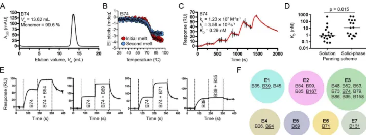
Size-exclusion chromatography (SEC) profiles of the VHHs showed predominantly single
monodispersed peaks devoid of higher order aggregates as expected (Table 1,Fig 2AandS1 Fig). Circular dichroism spectroscopy was used to determine VHH melting temperatures
(Tms) and to assess VHH refolding (Table 1,Fig 2BandS2 Fig). The Tms ranged from 57.6 to
87.2˚C (median Tm= 73.4˚C) and most VHHs could refold, although the completeness of
refolding was sequence dependent. VHH binding affinities and kinetics were determined by
SPR single-cycle kinetic analysis (Table 1,Fig 2CandS3 Fig). VHH affinities (KDs) ranged
Fig 1. Isolation of high-affinity TcdB-binding VHHs. (A, top) Schematic of TcdB. GTD, glucosyltransferase domain; APD, autoprotease domain,
Delivery, delivery/receptor binding domain, CROPs, combined repetitive oligopeptides domain. (A, bottom) A 71.6 kDa fragment of C. difficile toxin B (TcdB1751-2366) was purified, biotinylated and analyzed by non-reducing (NR) SDS-PAGE and Western blot (WB) probed with streptavidin-AP
(SA-AP). Approximately 1 μg of protein was loaded per lane. M, protein molecular weight marker; -B, unlabeled TcdB1751-2366; +B, biotinylated
TcdB1751-2366. (B) TcdB1751-2366was used as an antigen (Ag) for llama immunization with the schedule shown. FCA, Freund’s complete adjuvant; FIA,
Freund’s incomplete adjuvant. (C) ELISA showing the binding response from pre-immune and immune llama sera collected on days 35 and 42 post immunization to coated TcdB1751-2366. (D) Serum from day 42 post immunization was fractionated using protein G and protein A affinity columns
and analyzed by SDS-PAGE under reducing (R) conditions. G1, A1 and A2 fractions contain heavy-chain IgG (hcIgG,�) while the G2 fraction
contains conventional IgG (cIgG,� �). The arrows denote IgM heavy and light chains in the A2 fraction. (E) ELISA showing the binding response of
fractionated day 42 serum to coated TcdB1751-2366. (F) Vero cell cytotoxicity assay demonstrating the effect of fractionated day 42 serum on TcdB
inhibition, 72 h post addition of TcdB and polyclonal antibodies to Vero cell monolayers. TcdB was used at 10 and 30 pM and polyclonal fractionated sera at 1 μM. (G, top) Summary of solution-phase library panning titers using off-rate selection. In each round, the amount of target (biotinylated TcdB1751-2366) was reduced, the amount of competitor (“Comp”; unlabeled TcdB1751-2366) was held constant, and the incubation time of phage
+ target + competitor was increased. After incubation, the complex of phage and biotinylated TcdB1751-2366was captured on streptavidin coated
microtiter plates, washed and eluted. (G, bottom) Summary of solid-phase library panning titers using coated TcdB1751-2366. (H) Phage ELISA
showing binding of phage displayed VHHs to coated TcdB1751-2366. VHHs on phage producing the highest ELISA signals were expressed, purified and
characterized. The Western blot (WB) shows detection of purified VHHs with anα-His6-AP secondary antibody.
from 70 pM to 576 nM and, when classified by selection method (Fig 2D), the 18 VHHs
iso-lated from solution panning were of statistically higher affinity than the 15 VHHs obtained by
solid-phase panning (median KDs of 0.79 nM and 12.3 nM, respectively, and note that the four
VHHs found by both selection methods were counted in each). Next, VHHs with KDs of ~50
nM and stronger were subjected to epitope binning by SPR-based co-injection (VHH 1
fol-lowed by VHH 1 + VHH 2) experiments (Table 1,Fig 2EandS4 Fig). From the 21 VHHs
binned a total of seven non-overlapping TcdB epitopes were found (Fig 2F). The nine VHHs
residing in epitope bin E3 were clonally related (Fig 2F,S1 Table) while the other six bins con-tained predominantly unique, unrelated VHHs. Finally, using VHHs with the highest affinities
and/or slowest off-rates (kds) in each epitope bin (B39, B69, B71, B74, B94, B131 and B167), we
examined the TcdB neutralization capacity in Vero cell assays. At the highest VHH
concentra-tion tested (1 μM) none of the antibodies were capable of inhibiting the cytotoxic effects of TcdB on cells at 1 pM.
Table 1. Biophysical properties of anti-TcdB VHHs.
VHH Panning source Ve(mL) SEC (%)a Tm(˚C)b ka(M-1s-1) kd(s-1) KD(nM) Rmax(RU)c Epitope bin
B26 solution 12.97 99.8 68.3±0.3 5.25×10⁶ 3.45×10⁻² 6.3 149 4 B35 solution 14.07 99.8 76.2±0.4 1.16×10⁶ 1.05×10⁻³ 0.9 50 1 B39 solution 16.05 99.8 87.2±0.2 3.44×10⁷ 2.56×10⁻³ 0.07 43 1 B45 solution 14.84 99.4 79.5±0.4 3.52×10⁶ 3.84×10⁻⁴ 0.1 55 1 B46 solid phase 13.18 99.3 61.0±1.4 1.53×10⁵ 9.43×10⁻³ 62 51 n.d. B48 both 13.07 99.7 82.5±0.3 4.11×10⁶ 5.71×10⁻³ 1.4 73 3 B52 both 12.77 99.9 65.3±0.7 1.29×10⁷ 1.01×10⁻² 0.8 63 3 B53 solid phase 12.97 99.6 77.1±0.0 4.38×10⁶ 7.98×10⁻³ 1.8 72 3 B54 solution 12.85 95.3 81.7±0.3 3.86×10⁷ 1.47×10⁻² 0.4 65 2 B55 solid phase 13.35 99.8 66.6±1.1 3.34×10⁵ 4.93×10⁻² 147 142 n.d. B56 solid phase 14.00 86.7 57.6±0.3 5.46×10⁴ 3.15×10⁻² 576 18 n.d. B65 solid phase 13.26 96.3 84.4±0.2 1.32×10⁴ 2.33×10⁻³ 176 17 n.d. B69 solid phase 13.11 82.1 80.9±0.5 1.17×10⁷ 1.50×10⁻¹ 12.8 102 5 B71 solid phase 12.88 85.9 63.3±0.3 7.76×10⁶ 9.18×10⁻² 11.8 61 6 B73 both 13.41 99.7 80.2±0.0 1.27×10⁷ 9.19×10⁻³ 0.7 63 3 B74 both 13.62 99.6 75.0±0.2 1.23×10⁷ 3.58×10⁻³ 0.3 66 3 B76 solid phase 12.48 99.4 84.2±1.1 1.38×10⁶ 2.17×10⁻¹ 157 74 n.d. B79 solid phase 13.48 99.1 66.9±0.1 5.59×10⁶ 9.72×10⁻³ 1.7 75 3 B85 solution 12.94 99.4 69.7±0.1 1.93×10⁷ 1.14×10⁻² 0.6 73 2 B86 solution 13.03 99.6 72.9±0.1 8.25×10⁶ 3.98×10⁻³ 0.5 71 3 B92 solid phase 12.85 98.9 78.0±0.1 1.51×10⁶ 8.28×10⁻¹ 548 72 n.d. B94 solution 12.58 96.7 69.7±0.4 6.19×10⁵ 8.77×10⁻³ 14.2 385 4 B95 solid phase 13.97 95.1 80.8±0.6 1.12×10⁷ 7.05×10⁻³ 0.6 72 3 B96 solution 13.17 99.7 70.5±0.2 1.37×10⁶ 7.33×10⁻² 53.6 104 n.d. B99 solution 13.05 98.7 69.8±2.2 6.33×10⁷ 1.04×10⁻¹ 1.6 76 2 B131 solution 13.01 100 69.3±2.3 5.47×10⁷ 1.40×10⁻¹ 2.6 73 7 B149 solution 13.45 97.4 83.9±0.0 4.12×10⁴ 2.20×10⁻² 534 133 n.d. B158 solution 13.44 99.4 73.4±0.8 3.44×10⁴ 1.74×10⁻³ 50.5 67 3 B167 solution 13.14 99.6 72.1±0.4 1.53×10⁷ 1.10×10⁻³ 0.07 76 2
a% monomer determined by area under the curve
b(mean ± SD) c
Observed Rmaxwas obtained from a TcdB surface with a theoretical Rmaxof 200 RUs; n.d., not determined.
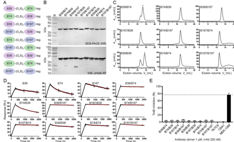
Reformatting V
HHs as dimeric molecules
We next generated VHH-VHH dimers using the three highest affinity VHHs that targeted
unique epitopes (B39, B74 and B167) to determine if biparatopic designs could impart a mea-sureable level of TcdB neutralization not seen with VHH monomers. All possible combinations
of the three antibodies were created including homodimers (Fig 3A), with a standard 25 amino acid linker separating each VHH. Dimers were expressed in E. coli, purified by IMAC
with yields ranging from 2.0 to 12.0 mg/L (Fig 3B) and assessed by SEC (Fig 3C) which revealed a predominantly single monodispersed species with the exception of B39/B74 and B39/B39 dimers that showed higher order aggregates. VHH-VHH dimer Tms ranged from 64.4
to 73.1˚C. SPR off-rate analysis demonstrated nearly irreversible bivalent binding to TcdB 1751-2366surfaces for many of the dimers, approaching the instrument limit of detection (Table 2,
Fig 3D). Vero cell neutralization assays using the dimers showed minor TcdB inhibition with
all nine formats tested ranging from 3.2% (B39/B39) to 7.5% (B167/B39) maximum inhibition
(Table 2,Fig 3E). The negative control T5/TC9 dimer did not inhibit TcdB and the
bench-mark control mAb MDX-1388 reached a maximum inhibition of 76.6%.
Reformatting V
HHs as Fc fusions
Given the modest level of TcdB inhibition seen with the VHH-VHH dimers, we next explored
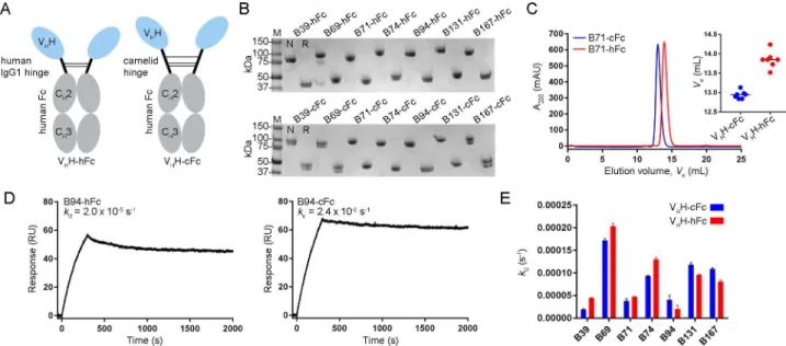
if construction of larger molecules may lead to greater TcdB neutralizing potency. VHH-Fc
fusions (Fig 4A) were constructed using each of the seven high-affinity VHHs from distinct
epitope bins (B39, B69, B71, B74, B94, B131 and B167). The designs consisted of a set of VHH-Fcs with a 15-residue human IgG1 hinge (EPKSCDKTHTCPPCP) and another set of
complementary molecules with a 35 residue camel/llamaγ2a hinge (EPKIPQPQPKPQPQ PQPQPKPQPKPEPECTCPKCP), to explore the possible advantages a longer, more flexible hinge may have on TcdB inhibition. The VHH-Fcs, denoted “VHH-hFc” for molecules
con-taining the human hinge and “VHH-cFc” for molecules containing the camel hinge, were
Fig 2. Biophysical characterization of VHHs. Representative SEC profile (A) and thermal unfolding curve from initial and refolded thermal melts (B).
(C) Representative SPR single-cycle kinetics sensorgram showed high-affinity binding of VHHs to biotinylated TcdB1751-2366immobilized on a CAP
sensor chip. (D) Plot comparing KDs of VHHs isolated from solution and solid-phase panning schemes. The four VHHs isolated from both panning
schemes were included in the analyses. Bars represent the median KDand a P-value < 0.05 was considered significant (Mann Whitney two-tailed
unpaired t-test). (E) Representative sensorgrams demonstrating SPR-based epitope binning. All VHHs were injected at 10x KDconcentrations. (F)
Summary of the TcdB1751-2366epitope bins identified in this study by the pool of VHHs tested. The VHHs in each epitope (E) bin are noted and the
underlined VHHs represent the highest affinity and/or slowest dissociating antibodies in each bin.
expressed in mammalian cells and purified by protein A with yields ranging from 2.5 to 21.6 mg/100 mL of culture (Table 3,Fig 4B). SEC analyses of VHH-Fcs revealed single,
monodis-persed peaks and consistently showed VHHs with camel hinges eluting earlier, most likely due
to their larger hydrodynamic radius (Table 3,Fig 4CandS5A Fig). SPR-determined off-rates demonstrated nearly irreversible bivalent binding to TcdB1751-2366surfaces (Table 3,Fig 4D
andS5B Fig). Comparison of the off-rates (kds) of VHH-Fcs with human or camel hinges did
not reveal significant differences in their ability to bind TcdB1751-2366(Fig 4E).
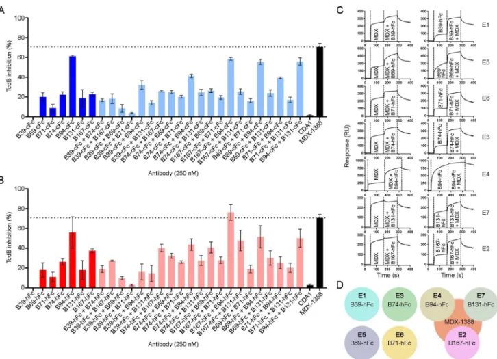
TcdB neutralization with V
HH-Fcs
The neutralization potencies of VHH-Fcs were compared to the benchmark mAb
bezlotoxu-mab (MDX-1388) in TcdB inhibition assays using 500 fM TcdB and 250 nM antibody
(Table 3,Fig 5A and 5B). MDX-1388 showed a maximum TcdB inhibition of 70.7%
com-pared to VHH-Fcs that ranged from 8.9% (B71-cFc) to 61.2% (B94-cFc). There were essentially
no differences between the neutralizing capacities of VHH-Fcs containing the short IgG1
hinge or the longer camel hinge, mirroring the near identical off-rates determined by SPR. Neither format of B39-Fc neutralized TcdB. CDA1 (an anti-TcdA mAb) [36] was included as a negative control and did not neutralize TcdB as expected. To examine possible synergistic Fig 3. Generation and characterization of VHH-VHH dimers. (A) Cartoon diagram of VHH-VHH dimers separated by a 25 amino acid linker (G4S)5and
containing a C-terminal His6“tag”. (B) Dimers were expressed, purified and analyzed by non-reducing (NR) SDS-PAGE and probed byα-His6-AP in
Western blot (WB). M, protein molecular mass marker. (C) SEC profiles of dimers. Asterisks denote the peaks that were selected for SPR analysis when aggregates were present. (D) SPR sensorgrams showing dissociation phases of VHH-VHHs and parent monomers. (E) TcdB neutralization with VHH-VHH
dimers. The final TcdB concentration of 3–10 pM and VHH-VHH concentration of 1 μM was co-incubated with Vero cell monolayers for 72 h before
addition of WST-1 cytotoxicity reagent. MDX-1388 was used at 250 nM. Neutralization values are presented as mean ± SD from n = 3 independent experiments.
effects from combinations of VHH-Fcs on TcdB inhibition, pairs of VHH-Fcs (125 nM + 125
nM) were examined in neutralization assays (S2 Table,Fig 5A and 5B). The VHH-Fc pair of
B94-hFc + B167-hFc achieved a maximum TcdB inhibition of 76.2%, slightly exceeding the neutralization potency of each VHH-Fc alone and that of MDX-1388. In pairs containing
B39-Fc, the non-neutralizing antibody, overall neutralization was reduced by approximately 50% of the maximum inhibiting potency of the second antibody partner, reflecting the fact that 50% less inhibitory antibody was present (Fig 5A and 5B).
Table 2. Biophysical properties of anti-TcdB VHH-VHH dimers.
VHH- VHH Yield (mg/ L)a Ve(mL)b Tm(˚C)c kd(s-1) Maximum TcdB neutralization (%)c,d
B39/B74 9.2 13.2 71.6 ± 0.2 2.2 × 10⁻⁵ 4.4 ± 0.4 B74/B39 3.6 12.2 70.9 ± 1.6 5.9 × 10⁻⁴ 5.3 ± 0.1 B39/B167 9.0 13.0 73.1 ± 1.7 2.4 × 10⁻⁵ 4.9 ± 0.3 B167/B39 2.0 11.9 67.7 ± 1.4 1.7 × 10⁻⁵ 7.5 ± 0.1 B74/B167 5.8 12.0 64.4 ± 2.1 1.4 × 10⁻⁵ 4.2 ± 0.2 B167/B74 2.4 11.7 66.5 ± 0.6 2.4 × 10⁻⁵ 4.1 ± 0.2 B39/B39 12.0 14.2 79.5 ± 3.4 9.7 × 10⁻⁵ 3.2 ± 0.3 B74/B74 2.2 12.0 70.4 ± 3.3 6.3 × 10⁻⁵ 5.4 ± 0.3 B167/B167 6.8 11.6 67.5 ± 2.3 6.0 × 10⁻⁵ 5.1 ± 0.2 MDX-1388 - n.d. n.d. n.d. 76.6 ± 4.4e
aPurification yield from 1 L E. coli culture bV
efrom Superdex 200 c(mean ± SD) d1 μM or
e250 nM antibody + 3 pM TcdB vero cell cytotoxicity (72 h); n.d., not determined.
https://doi.org/10.1371/journal.pone.0208978.t002
Fig 4. Generation and characterization of VHH-Fcs with human or camelid hinges. (A) Cartoon diagram illustrating the two VHH-Fc formats used
with varying hinge length and composition. (B) Purified VHH-Fcs were analyzed by SDS-PAGE under non-reducing (N) and reducing (R) conditions.
(C) Representative VHH-Fc SEC elution profile from a Superdex 200 column. Inset, plot of SEC elution volumes for all VHH-cFcs and VHH-hFcs. (D)
Representative SPR sensorgrams showing 30 min (1800 s) dissociations of VHH-Fc fusions flowing over immobilized TcdB1751-2366surfaces. (E)
Dissociation rate comparison of VHH-Fcs with human or camelid hinges.
V
HH-Fc competition with bezlotoxumab
To determine if our panel of neutralizing VHH-Fcs recognized similar or unique TcdB
epi-topes from that of bezlotoxumab we performed SPR co-injection experiments (Fig 5C and 5D). B39-hFc, B69-hFc, B71-hFc and B74-hFc did not compete with MDX-1388 in either injection sequence, indicating that the four VHH-Fcs bind sites on TcdB completely
indepen-dent of the MDX-1388 binding site. This is unsurprising for B39-hFc given the inability of this antibody to neutralize TcdB. The other three VHHs were previously shown to bind unique
epi-topes as monomers (Fig 2F) suggesting B69-hFc, B71-hFc and B74-hFc recognize three novel TcdB epitopes that support neutralizing antibodies. The results of B94-hFc, B131-hFc and B167-hFc binning with MDX-1388 revealed significant overlap in TcdB binding patterns. When MDX-1388 was injected first, B94-hFc was partially blocked by pre-bound MDX-1388 and B131-hFc and B167-hFc were completely blocked by pre-bound MDX-1388. In the oppo-site orientation, MDX-1388 binding was completed blocked by pre-bound B94-hFc, and par-tially blocked by pre-bound B131-hFc or B167-hFc. Collectively this data suggests the most potent TcdB neutralizing VHH-Fcs (B94-hFc, B167-cFc) bind TcdB at sites that partially
over-lap with the MDX-1388 binding site at the N-terminal end of the CROPs domain [27].
Discussion
In this work we set out to identify high-affinity VHHs capable of neutralizing C. difficile TcdB.
We previously failed to identify monomeric VHH neutralizers when immunizing with a small
C-terminal fragment of the CROPs domain [10]. Here our expanded immunogen design con-taining a portion of the central delivery domain and the entire CROPs domain yielded a num-ber of VHHs that were capable of TcdB inhibition when formatted as dimers and more so as Fc
fusions. It should be noted that our previous TcdB-binding VHHs [10] were not tested as
VHH-Fc fusions and may have been capable of TcdB inhibition, although their affinities were
Table 3. Biophysical properties of anti-TcdB VHH-Fc fusions.
VHH-Fc/mAb Hinge Yield (mg/ 100 mL)a Ve(mL)b kd(s-1) Maximum TcdB neutralization (%)c,d
B39-hFc human 5.7 17.64 4.5 × 10⁻⁵ 0.0 B69-hFc human 13 14.24 2.0 × 10⁻⁴ 18.0 ± 10.2 B71-hFc human 6 13.91 4.7 × 10⁻⁵ 11.0 ± 7.0 B74-hFc human 4.4 13.82 1.3 × 10⁻⁴ 26.3 ± 4.5 B94-hFc human 12.2 13.52 2.0 × 10⁻⁵ 55.9 ± 22.4 B131-hFc human 14.6 13.70 9.6 × 10⁻⁵ 18.0 ± 13.4 B167-hFc human 2.5 13.85 8.1 × 10⁻⁵ 37.5 ± 2.7 B39-cFc camelid 7.9 15.07 2.0 × 10⁻⁵ 0.1 ± 0.1 B69-cFc camelid 9.1 13.13 2.0 × 10⁻⁴ 20.2 ± 5.7 B71-cFc camelid 11.3 12.98 4.7 × 10⁻⁵ 8.9 ± 5.5 B74-cFc camelid 6.8 12.95 9.4 × 10⁻⁵ 22.3 ± 4.2 B94-cFc camelid 11.8 12.84 2.4 × 10⁻⁵ 61.2 ± 1.3 B131-cFc camelid 21.6 12.84 9.6 × 10⁻⁵ 18.7 ± 12.3 B167-cFc camelid 9.2 12.90 8.1 × 10⁻⁵ 22.8 ± 2.9 MDX-1388 human - n.d. n.d. 70.7 ± 13.9
aPurification yield from 100 mL HEK293 culture bV
efrom Superdex 200 c
(mean ± SD)
d250 nM antibody + 500 fM TcdB vero cell cytotoxicity (72 h); n.d., not determined.
considerably weaker than the VHHs isolated in this work. Once again the monomeric VHHs
did not inhibit TcdB, suggesting a steric element that is required for TcdB inhibition when tar-geting the CROPs domain. Consistent with our findings, Yang et al isolated several TcdB-binding VHHs and found five high-affinity VHHs targeting the C-terminal CROPs domain
that failed to inhibit TcdB cytotoxicity while those targeting the N-terminal GTD domain were potent neutralizers [13].
Our work is not the first to report TcdB-inhibiting VHHs binding the CROPs domain.
Andersen et al isolated several inhibitory monomeric VHHs binding this domain, indicating
that TcdB neutralization is possible with monomeric antibodies [15]. It is not clear how they successfully identified VHHs that recognize critical epitope(s) for TcdB function/cell binding,
that were not identified here, in our early work [10] or by [13], while employing a similar immunization strategy. Interestingly two of their non-neutralizing CROPs-binding VHHs
were converted into inhibitory antibodies when displayed on the surfaces of lactobacilli [15], again pointing to a large steric element in imparting TcdB inhibition with antibodies binding this region of the toxin.
Fig 5. TcdB neutralization assays with VHH-Fcs. (A) TcdB neutralization with VHH-Fcs containing a camelid hinge. (B) TcdB neutralization with
VHH-Fcs containing a human hinge. In (A) and (B), the final TcdB concentration of 500 fM and final antibody concentration of 250 nM (single antibody)
or 125 + 125 nM (pairs) were co-incubated with Vero cell monolayers for 72 h before addition of WST-1 cytotoxicity reagent. Neutralization values are presented as mean ± SD from n = 4 independent experiments. (C) Sensorgrams showing SPR-based epitope binning of MDX-1388 mAb with each VH
H-hFc, in both orientations, with antibodies injected at 25–50 × KDconcentrations over TcdB1751-2366surfaces. Epitope bins corresponding to each VH
H-hFc are noted. (D) Summary of VHH-Fc reactivity to TcdB1751-2366illustrating that several VHH-Fcs bind TcdB at sites distinct from MDX-1388, while
others bind TcdB at regions that partially overlap with MDX-1388.
Beyond VHHs there are several potent TcdB-neutralizing mAbs that have been well studied.
The most advanced anti-TcdB mAb is bezlotoxumab which received FDA approval for recur-rent C. difficile infection in 2016. This antibody was originally isolated in a study reported by [36] and demonstrated to bind the C-terminal receptor binding domain (CROPs domain). Structural studies performed by [27] revealed the precise binding site of the mAb lies within the first two of four CROP repeat domains, with each CROP domain consisting of three short repeating units (SRs) followed by one long repeating unit (LR) and two more SRs. SPR binding data showed bezlotoxumab bound two distinct epitopes which was later supported by X-ray crystal structures and homology modeling showing two Fab fragments binding adjacent to each other in the N-terminal half of the CROPs domain (aa 1834–2101). Given bezlotoxumab neutralizes TcdB by preventing binding to mammalian cells [27,37], one can assume that some of our most potent VHH-Fc fusions (B94-Fc, B131-Fc and B167-Fc) neutralize TcdB in a
simi-lar manner given that they partially overlap with bezlotoxumab for TcdB binding. Whether the exact mechanism of inhibition is due to blocking putative carbohydrate binding site interac-tions with a host-cell receptor, or by steric effects precluding the central/delivery domain from making contacts with FZD2, PVRL3 or CSPG4 receptors is unknown. It is interesting to note that the observed Rmaxof B94 in SPR experiments was considerably higher than other
antibod-ies as both a VHH monomer (Table 1) and as a VHH-Fc in binning experiments (Fig 5C),
sug-gesting that B94 may bind to repeating TcdB CROPs domain epitopes. Supporting this idea is the fact that B94-Fc was the only VHH-Fc to completely block MDX-1388 binding when
B94-Fc was bound to TcdB first. If a repeating epitope is bound by B94-Fc, it would suggest a similar mechanism of inhibition to that of MDX-1388 and that affinity improvements may lead to greater neutralizing potency since the monovalent affinity of B94 is relatively weak (KD
= 14 nM) compared to a Fab fragment from MDX-1388 (KD= 19 pM or 370 pM; depending
on the TcdB epitope) [27].
For the other three neutralizing VHH-Fcs described here (B69-Fc, B71-Fc and B74-Fc) their
location for toxin binding and subsequently their mechanism for toxin inhibition also remain unknown. B39, which failed to neutralize TcdB as an Fc fusion and did not overlap with bezlo-toxumab as expected, was previously [12] co-crystalized with a C-terminal fragment of the TcdB CROPs domain (aa 2248–2367 of TcdB from strain 10463) and definitively showed only recognition of a single, non-repeating TcdB epitope. This may suggest that antibodies binding at a distance from the central delivery/translocation domain have minimal effects on TcdB inhibition compared to antibodies binding nearby in the N-terminal half of the CROPs domain. Elsewhere, [28] have successfully identified four inhibitory mAbs targeted to the CROPs domain of TcdB that were neutralizers alone and to a greater extent when combined in pairs. Additionally, TcdB-neutralizing mAbs recognizing the C-terminal GT domain of TcdB have proven to be both potent inhibitors and effective in in vivo protection assays [38,39,40].
There were several other interesting observations of note from this work. The conventional IgG (cIgG) fraction obtained from llama serum post immunization with TcdB1751-2366was
capable of inhibiting TcdB in cytotoxicity assays more efficiently than the three heavy-chain IgG (hcIgG) fractions. While the cIgG binding titer for TcdB was higher than the best hcIgG fraction by approximately 10-fold, which is a typical binding pattern we have observed in sev-eral other immunization campaigns, there were dramatic differences in the inhibition pattern seen between G1 (hcIgG) and G2 (cIgG) fractions. It is possible that a greater agglutination mechanism with camelid cIgGs compared to camelid hcIgGs is at play here and could be due to the physical distance separating the binding arms of each antibody format. We hypothesize that the more compact hcIgG footprint may bias the polyclonal pool toward intra-toxin bind-ing events and that the greater distance afforded to cIgGs promotes both intra-toxin and
inter-toxin binding events, leading to increased agglutination and ultimately greater TcdB inhibi-tion. Supporting this is the fact the A2 (hcIgG) fraction, which showed a much weaker TcdB binding titer than the G1 fraction, possessed considerably greater neutralizing potency likely driven by IgM antibodies found as contaminants in the fractionated serum.
We performed panning experiments in solution using off-rate based selection and this method produced statistically higher affinity VHHs compared to panning on immobilized
antigen. This method of selection was also the source of the VHH-Fcs with the highest
neutral-izing potency, even though two of the three most potent TcdB inhibitors did not possess the highest overall monovalent VHH affinities. It is probable that the solution panning scheme
pre-sented the randomly biotinylated TcdB in a more native-like conformation and made more areas of the protein available for VHH binding compared to selection of VHHs on TcdB-coated
wells. This may explain the higher average binding affinity and greater neutralization potency of VHHs isolated by the solution panning approach. We also examined the use of a longer
camel hinge in place of the human IgG1 hinge to present the VHH in a more natural context
when tethered to the Fc domain. While SEC profiles clearly showed that VHH-Fcs with camel
hinges eluted earlier and thereby suggest a larger hydrodynamic volume, similar dissociation rate constants and TcdB neutralizing potency demonstrated there was no clear benefit to using the longer hinge. We do not completely understand why this was the case but speculate that the camel hinge only moderately expands the binding distance between VHH arms, still less
than the footprint of a cIgG, and that neutralization is dependent on combination of factors including the location, geometry and accessibility of TcdB epitopes.
In summary, the VHHs described here were potent TcdB neutralizing antibodies on par
with bezlotoxumab when formatted as Fc fusions. We envision creating even more effective TcdB-neutralizing agents through optimization of affinities and binding geometries, such as through structure-guided biparatopic designs. Combining anti-TcdB biparatopic VHH-VHH
designs with TcdA-neutralizing VHHs onto a human Fc scaffold would allow for the generation
of ultra-potent toxin inhibitors in a single antibody format, similar to approaches described pre-viously [13,17], while maintaining the steric requirements for TcdB neutralization and long serum half-life. The in vivo efficacy previously demonstrated with multivalent designs that include linking anti-TcdA and anti-TcdB VHHs suggests the inclusion of GTD-targeting
anti-TcdB antibodies is critical [13,17,18]. Our results make a case for targeting the central delivery and CROPs domains with VHHs. Whether including CROPs-targeting VHHs in these designs
will be as potent as those targeting the GTD region remains to be seen. In addition, finer epitope mapping of the VHH-Fcs that bound TcdB at regions distinct from bezlotoxumab will reveal if
these antibodies recognize the central delivery domain or the CROPs domain and the nature of these unique inhibitory epitopes. Finally, the VHHs described here will serve as useful additions
to the reagent toolkit for further refinement of the mechanism of TcdB-host cell interactions.
Supporting information
S1 Fig. SEC chromatograms of VHH monomers. VHHs were passed over a Superdex 75
col-umn at a flow rate of 0.5 mL/min in HBS-EP buffer. Molecular mass standards are shown. The percent monomer was calculated from the peak area under the curve. Ve, elution volume.
(TIF)
S2 Fig. Thermal unfolding of VHH monomers by circular dichroism spectroscopy. (A)
Thermal unfolding of VHHs (50 μg/mL, 3.2 μM) was performed in phosphate buffer and
mea-sured in a 5 mm cuvette at 215 nm. VHHs were allowed to cool at 25˚C for 3 h before the
sec-ond thermal unfolding (refolded melting curve) was performed. (B) Voltage comparing two VHHs (B54 and B99) that aggregate as a consequence of unfolding versus one VHH that does
not (B53). (TIF)
S3 Fig. Single-cycle kinetics sensorgrams showing monomeric VHHs binding to
TcdB1751-2366surfaces. Black lines represent raw data and red lines represent 1:1 binding
model fits. Rate constants and affinities are shown for each antibody. The irrelevant VHH
served as a negative control. For experimental conditions seeMaterials and methods. (TIF)
S4 Fig. Representative sensorgrams from SPR-based epitope binning of VHH monomers
on TcdB1751-2366surfaces. The first VHH was injected at 10× KDconcentration followed
immediately by injection of a mixture of the first VHH + second VHH at 10× KD
concentra-tion. For experimental conditions seeMaterials and methods. (TIF)
S5 Fig. Characterization of VHH-Fc fusions. (A) SEC chromatograms of VHH-Fcs on a
Superdex 200 column at a flow rate of 0.5 mL/min in HBS-EP buffer. (B) SPR sensorgrams illustrating the dissociation of 1 nM VHH-Fcs from TcdB1751-2366surfaces. For experimental
conditions seeMaterials and methods. (TIF)
S1 Table. Clonal relatedness of TcdB-specific VHHs isolated in this study.
(PDF)
S2 Table. Select pairs of VHH-Fcs with the highest TcdB neutralization.
(PDF)
Acknowledgments
We thank Kenneth Ng (University of Calgary) for providing the recombinant TcdB fragment, Shreya Jain (NRC) for assisting in protein expression and Yves Durocher and Denis L’Abbe´ (NRC) for producing the CDA1 and MDX-1388 control mAbs.
Author Contributions
Conceptualization: Greg Hussack, C. Roger MacKenzie, Jamshid Tanha.
Data curation: Greg Hussack, Shannon Ryan, Henk van Faassen, Martin Rossotti.
Formal analysis: Greg Hussack, Shannon Ryan, Henk van Faassen, Martin Rossotti, C. Roger
MacKenzie, Jamshid Tanha.
Methodology: Martin Rossotti.
Resources: C. Roger MacKenzie, Jamshid Tanha.
Supervision: Greg Hussack, C. Roger MacKenzie, Jamshid Tanha. Writing – original draft: Greg Hussack.
Writing – review & editing: Greg Hussack, Shannon Ryan, Henk van Faassen, Martin
Ros-sotti, C. Roger MacKenzie, Jamshid Tanha.
References
1. Dubberke ER, Olsen MA (2012) Burden of Clostridium difficile on the healthcare system. Clin Infect Dis 55 Suppl 2: S88–92.
2. Hussack G, Tanha J (2016) An update on antibody-based immunotherapies for Clostridium difficile infection. Clin Exp Gastroenterol 9: 209–224.https://doi.org/10.2147/CEG.S84017PMID:27536153
3. Napolitano LM, Edmiston CE Jr. (2017) Clostridium difficile disease: Diagnosis, pathogenesis, and treatment update. Surgery 162: 325–348.https://doi.org/10.1016/j.surg.2017.01.018PMID:28267992
4. Hopkins RJ, Wilson RB (2018) Treatment of recurrent Clostridium difficile colitis: a narrative review. Gastroenterol Rep 6: 21–28.
5. Aktories K, Schwan C, Jank T (2017) Clostridium difficile toxin biology. Ann Rev Microbiol 71: 281–307.
6. Chandrasekaran R, Lacy DB (2017) The role of toxins in Clostridium difficile infection. FEMS Microbiol Rev 41: 723–750.https://doi.org/10.1093/femsre/fux048PMID:29048477
7. Hussack G, Tanha J (2010) Toxin-specific antibodies for the treatment of Clostridium difficile: current status and future perspectives. Toxins 2: 998–1018.https://doi.org/10.3390/toxins2050998PMID:
22069622
8. Mullard A (2016) FDA approves antitoxin antibody. Nat Rev Drug Discov 15: 811.
9. Navalkele BD, Chopra T (2018) Bezlotoxumab: an emerging monoclonal antibody therapy for preven-tion of recurrent Clostridium difficile infecpreven-tion. Biologics 12: 11–21.https://doi.org/10.2147/BTT. S127099PMID:29403263
10. Hussack G, Arbabi-Ghahroudi M, van Faassen H, Songer JG, Ng KK, MacKenzie R, et al. (2011) Neu-tralization of Clostridium difficile toxin A with single-domain antibodies targeting the cell receptor binding domain. J Biol Chem 286: 8961–8976.https://doi.org/10.1074/jbc.M110.198754PMID:21216961
11. Hussack G, Keklikian A, Alsughayyir J, Hanifi-Moghaddam P, Arbabi-Ghahroudi M, van Faassen H, et al. (2012) A V(L) single-domain antibody library shows a high-propensity to yield non-aggregating binders. Protein Eng Des Sel 25: 313–318.https://doi.org/10.1093/protein/gzs014PMID:22490957
12. Murase T, Eugenio L, Schorr M, Hussack G, Tanha J, Kitova EN, et al. (2013) Structural basis for anti-body Recognition in the receptor-binding domains of toxins A and B from Clostridium difficile. J Biol Chem 289: 2331–2343.https://doi.org/10.1074/jbc.M113.505917PMID:24311789
13. Yang Z, Schmidt D, Liu W, Li S, Shi L, Sheng J, et al. (2014) A novel multivalent, single-domain antibody targeting TcdA and TcdB prevents fulminant Clostridium difficile infection in mice. J Infect Dis 210: 964–972.https://doi.org/10.1093/infdis/jiu196PMID:24683195
14. Shkoporov AN, Khokhlova EV, Savochkin KA, Kafarskaia LI, Efimov BA (2015) Production of biologi-cally active scFv and VHH antibody fragments in Bifidobacterium longum. FEMS Microbiol Lett 362: fnv083.
15. Andersen KK, Strokappe NM, Hultberg A, Truusalu K, Smidt I, Mikelsaar RH, et al. (2015) Neutralization of Clostridium difficile toxin B mediated by engineered lactobacilli producing single domain antibodies. Infect Immun 84: 395–406.https://doi.org/10.1128/IAI.00870-15PMID:26573738
16. Unger M, Eichhoff AM, Schumacher L, Strysio M, Menzel S, Schwan C, et al. (2015) Selection of nano-bodies that block the enzymatic and cytotoxic activities of the binary Clostridium difficile toxin CDT. Sci Rep 5: 7850.https://doi.org/10.1038/srep07850PMID:25597743
17. Schmidt DJ, Beamer G, Tremblay JM, Steele JA, Kim HB, Wang Y, et al. (2016) A tetraspecific VHH-based neutralizing antibody modifies disease outcome in three animal models of Clostridium difficile infection. Clin Vaccine Immunol 23: 774–784.https://doi.org/10.1128/CVI.00730-15PMID:27413067
18. Yang Z, Shi L, Yu H, Zhang Y, Chen K, Saint Fleur A, et al. (2016) Intravenous adenovirus expressing a multi-specific, single-domain antibody neutralizing TcdA and TcdB protects mice from Clostridium diffi-cile infection. Pathog Dis 74: ftw078.https://doi.org/10.1093/femspd/ftw078PMID:27502696
19. Sulea T, Hussack G, Ryan S, Tanha J, Purisima EO (2018) Application of assisted design of antibody and protein therapeutics (ADAPT) improves efficacy of a Clostridium difficile toxin A single-domain anti-body. Sci Rep 8: 2260.https://doi.org/10.1038/s41598-018-20599-4PMID:29396522
20. Conrath KE, Lauwereys M, Galleni M, Matagne A, Frère JM, Kinne J, et al. (2001) Beta-lactamase
inhibitors derived from single-domain antibody fragments elicited in the camelidae. Antimicrob Agents Chemother 45: 2807–2812.https://doi.org/10.1128/AAC.45.10.2807-2812.2001PMID:11557473
21. Muyldermans S (2013) Nanobodies: natural single-domain antibodies. Ann Rev Biochem 82: 775–797.
https://doi.org/10.1146/annurev-biochem-063011-092449PMID:23495938
22. Iezzi ME, Policastro L, Werbajh S, Podhajcer O, Canziani GA (2018) Single-domain antibodies and the promise of modular targeting in cancer imaging and treatment. Front Immunol 9: 273.https://doi.org/ 10.3389/fimmu.2018.00273PMID:29520274
23. Chen P, Tao L, Wang T, Zhang J, He A, Lam KH, et al. (2018) Structural basis for recognition of frizzled proteins by Clostridium difficile toxin B. Science 360: 664–669.https://doi.org/10.1126/science. aar1999PMID:29748286
24. Tao L, Zhang J, Meraner P, Tovaglieri A, Wu X, Gerhard R, et al. (2016) Frizzled proteins are colonic epithelial receptors for C. difficile toxin B. Nature 538: 350–355.https://doi.org/10.1038/nature19799
PMID:27680706
25. LaFrance ME, Farrow MA, Chandrasekaran R, Sheng J, Rubin DH, Lacy DB (2015) Identification of an epithelial cell receptor responsible for Clostridium difficile TcdB-induced cytotoxicity. Proc Natl Acad Sci U S A 112: 7073–7078.https://doi.org/10.1073/pnas.1500791112PMID:26038560
26. Yuan P, Zhang H, Cai C, Zhu S, Zhou Y, Yang X, et al. (2015) Chondroitin sulfate proteoglycan 4 func-tions as the cellular receptor for Clostridium difficile toxin B. Cell Res 25: 157–168.https://doi.org/10. 1038/cr.2014.169PMID:25547119
27. Orth P, Xiao L, Hernandez LD, Reichert P, Sheth PR, Beaumont M, et al. (2014) Mechanism of action and epitopes of Clostridium difficile toxin B-neutralizing antibody bezlotoxumab revealed by X-ray crys-tallography. J Biol Chem 289: 18008–18021.https://doi.org/10.1074/jbc.M114.560748PMID:
24821719
28. Davies NL, Compson JE, Mackenzie B, O’Dowd VL, Oxbrow AK, Heads JT, et al. (2013) A mixture of functionally oligoclonal humanized monoclonal antibodies that neutralize Clostridium difficile TcdA and TcdB with high levels of in vitro potency shows in vivo protection in a hamster infection model. Clin Vac-cine Immunol 20: 377–390.https://doi.org/10.1128/CVI.00625-12PMID:23324518
29. Henry KA, Tanha J, Hussack G (2015) Identification of cross-reactive single-domain antibodies against serum albumin using next-generation DNA sequencing. Protein Eng Des Sel 28: 379–383.https://doi. org/10.1093/protein/gzv039PMID:26319004
30. Baral TN, MacKenzie R, Arbabi Ghahroudi M (2013) Single-domain antibodies and their utility. Curr Protoc Immunol 103: Unit 2 17.
31. Hussack G, Hirama T, Ding W, Mackenzie R, Tanha J (2011) Engineered single-domain antibodies with high protease resistance and thermal stability. PLoS One 6: e28218.https://doi.org/10.1371/ journal.pone.0028218PMID:22140551
32. Rossotti MA, Gonzalez-Techera A, Guarnaschelli J, Yim L, Camacho X, Ferna´ndez M, et al. (2015) Increasing the potency of neutralizing single-domain antibodies by functionalization with a CD11b/ CD18 binding domain. MAbs 7: 820–828.https://doi.org/10.1080/19420862.2015.1068491PMID:
26192995
33. Durocher Y, Perret S, Kamen A (2002) High-level and high-throughput recombinant protein production by transient transfection of suspension-growing human 293-EBNA1 cells. Nucleic Acids Res 30: E9. PMID:11788735
34. Henry KA, Kandalaft H, Lowden MJ, Rossotti MA, van Faassen H, Hussack G, et al. (2017) A disulfide-stabilized human VL single-domain antibody library is a source of soluble and highly thermostable bind-ers. Mol Immunol 90: 190–196.https://doi.org/10.1016/j.molimm.2017.07.006PMID:28820969
35. Conrath KE, Wernery U, Muyldermans S, Nguyen VK (2003) Emergence and evolution of functional heavy-chain antibodies in Camelidae. Dev Comp Immunol 27: 87–103. PMID:12543123
36. Babcock GJ, Broering TJ, Hernandez HJ, Mandell RB, Donahue K, Boatright N, et al. (2006) Human monoclonal antibodies directed against toxins A and B prevent Clostridium difficile-induced mortality in hamsters. Infect Immun 74: 6339–6347.https://doi.org/10.1128/IAI.00982-06PMID:16966409
37. Yang Z, Ramsey J, Hamza T, Zhang Y, Li S, Yfantis HG, et al. (2015) Mechanisms of protection against Clostridium difficileinfection by the monoclonal antitoxin antibodies actoxumab and bezlotoxumab. Infect Immun 83: 822–831.https://doi.org/10.1128/IAI.02897-14PMID:25486992
38. Marozsan AJ, Ma D, Nagashima KA, Kennedy BJ, Kang YK, Arrigale RR, et al. (2012) Protection against Clostridium difficile infection with broadly neutralizing antitoxin monoclonal antibodies. J Infect Dis 206: 706–713.https://doi.org/10.1093/infdis/jis416PMID:22732923
39. Anosova NG, Cole LE, Li L, Zhang J, Brown AM, Mundle S, et al. (2015) A combination of three fully human toxin A- and toxin B-specific monoclonal antibodies protects against challenge with highly viru-lent epidemic strains of Clostridium difficile in the hamster model. Clin Vaccine Immunol 22: 711–725.
https://doi.org/10.1128/CVI.00763-14PMID:25924765
40. Kroh HK, Chandrasekaran R, Rosenthal K, Woods R, Jin X, Ohi MD, et al. (2017) Use of a neutralizing antibody helps identify structural features critical for binding of Clostridium difficile toxin TcdA to the host cell surface. J Biol Chem 292: 14401–14412.https://doi.org/10.1074/jbc.M117.781112PMID: