HAL Id: hal-01626258
https://hal.sorbonne-universite.fr/hal-01626258
Submitted on 30 Oct 2017
HAL is a multi-disciplinary open access
archive for the deposit and dissemination of
sci-entific research documents, whether they are
pub-lished or not. The documents may come from
L’archive ouverte pluridisciplinaire HAL, est
destinée au dépôt et à la diffusion de documents
scientifiques de niveau recherche, publiés ou non,
émanant des établissements d’enseignement et de
Prognosis of patients with primary malignant brain
tumors admitted to the intensive care unit: a
two-decade experience
Maxens Decavele, Nicolas Weiss, Isabelle Rivals, Hélène Prodanovic, Ahmed
Idbaih, Julien Mayaux, Thomas Similowski, Alexandre Demoule
To cite this version:
Maxens Decavele, Nicolas Weiss, Isabelle Rivals, Hélène Prodanovic, Ahmed Idbaih, et al.. Prognosis
of patients with primary malignant brain tumors admitted to the intensive care unit: a two-decade
experience. Journal of Neurology, Springer Verlag, 2017, 264 (11), pp.2303-2312.
�10.1007/s00415-017-8624-7�. �hal-01626258�
Prognosis of patients with primary malignant brain
tumors admitted to the intensive care unit: a two-decade
experience
Maxens Decavèle1,2, Nicolas Weiss3, Isabelle Rivals1,4, Hélène Prodanovic2, Ahmed Idbaih5,6, Julien Mayaux2, Thomas Similowski1,2, Alexandre Demoule1,2
(1) Sorbonne Universités, UPMC Univ Paris 06, INSERM, UMRS_1158 Neurophysiologie respiratoire expérimentale et clinique, Paris, France
(2) AP-HP, Groupe Hospitalier Pitié-Salpêtrière Charles Foix, Service de Pneumologie et Réanimation Médicale (Département "R3S"), F-75013, Paris, France
(3) Unité de Réanimation Neurologique, Département de Neurologie, Pôle des Maladies du Système Nerveux et Institut de Neurosciences Translationnelles, IHU-A-ICM,
Groupe Hospitalier Pitié-Salpêtrière Charles Foix, Paris, France
(4) Equipe de Statistique Appliquée, ESPCI ParisTech, PSL Research University, Paris, France
(5) Inserm U 1127, CNRS UMR 7225, Sorbonne Universités, UPMC Univ Paris 06 UMRS_1127, Institut du Cerveau et de la Moelle épinière, ICM, F-75013, Paris, France
(6) AP-HP, Hôpitaux Universitaires La Pitié Salpêtrière - Charles Foix, Service de Neurologie 2-Mazarin, F-75013, Paris, France.
Corresponding author
Prof. Alexandre Demoule
Department of Respiratory and Critical Care Medicine Groupe Hospitalier Pitié-Salpêtrière
47-83 Boulevard de l'Hôpital 75013 Paris, France
Phone: 33 1 42 16 77 61; Fax: 33 1 42 16 78 43 e-mail: alexandre.demoule@aphp.fr
Authors email addresses:
Maxens Decavèle: maxencesar@hotmail.fr Nicolas Weiss: nicolas.weiss@aphp.fr Isabelle Rivals: isabelle.rivals@espci.fr
Hélène Prodanovic: helene.prodanovic@aphp.fr Ahmed Idbaih: ahmed.idbaih@aphp.fr
Julien Mayaux: julien.mayaux@aphp.fr
Thomas Similowski: thomas.similowski@aphp.fr Alexandre Demoule: alexandre.demoule@aphp.fr
Key words: malignant brain tumor; prognosis; critical care; intensive care unit
Acknowledgments: We are immensely grateful to Professor Jean-Yves Delattre for his valuable comments and
ABSTRACT (250 words)
Introduction. To describe the reasons for ICU admission and to evaluate the outcome and prognostic factors of
patients with primary malignant brain tumors (PMBT) admitted to the intensive care unit (ICU).
Patients and methods. Retrospective observational cohort study of 196 PMBT patients admitted to two ICUs
over a 19-year period.
Results. Acute respiratory failure was the main reason for ICU admission (45%) followed by seizures (25%) and
non-epileptic coma (14%). Seizures were more common in patients with glial lesions (84% vs. 67%), whereas patients with primary brain lymphoma were more frequently admitted for shock (42% vs. 18%). Overall ICU and 90-day mortality rates were 23% and 50%, respectively. Admission for seizures was independently associated with lower ICU mortality (odds ratio [OR] 0.06), whereas the need for mechanical ventilation (OR 6.85), cancer progression (OR 7.84), respiratory rate (OR 1.11) and Glasgow coma scale (OR 0.85) were associated with higher ICU mortality. Among the 95 patients who received invasive mechanical ventilation, ICU mortality was 37% (n=35). For these patients, admission for seizures was associated with lower ICU-mortality (OR 0.050) whereas cancer progression (OR 7.49) and respiratory rate (OR 1.08) were associated with higher ICU-mortality.
Conclusion. The prognosis of PMBT patients admitted to the ICU appears relatively favorable compared to that
of hematologic malignancies or solid tumors, especially when the patient is admitted for seizures. The presence of a PMBT therefore does not appear to be sufficient for refusal of ICU admission. Predictive factors of mortality may help clinicians make optimal triage decisions.
INTRODUCTION
Admission of cancer patients to intensive care units (ICU) has increased steadily over the past two decades and these patients now account for 10 to 20% of all ICU admissions[1]. Concomitantly, the mortality of critically ill cancer patients has decreased[2]. Consequently, a recent consensus of world experts emphasized that the existence of a hematologic malignancy or a solid tumor, even when metastatic, should not be considered a sufficient reason for ICU refusal [3].
Primary malignant brain tumors (PMBT) are rare tumors. In 2012, they represented less than 2% of all new cancer cases in Europe [4]. Although the prognosis of patients with hematologic malignancies or solid tumors admitted to the ICU has been well documented, only limited data are available concerning the prognosis of PMBT in the ICU [5-7]. One of the most likely reasons for this lack of data is the low prevalence of these tumors in the general population. However, it could also be due to the high ICU refusal rate of these patients, as the poor cancer prognosis [8], the severe impact on quality of life [9] or cognitive function [10]and the low survival of these patients in the case of clinical worsening[5] may explain the reluctance to admit these patients to the ICU [11]. However, PMBTs are very heterogeneous tumors in terms of their aggressiveness and patients with low-grade tumors, especially low-grade gliomas can be expected to have a much better prognosis[12]. In addition, therapeutic progress has contributed to a significant improvement of survival[13-16]. Finally, as for other malignancies, the reason for ICU admission and the number and intensity of life-supporting therapies initiated in the ICU may have a variable impact on each patient’s prognosis[17].
A better knowledge of the risk factors for survival of patients with PMBT when admitted to the ICU could help to improve the quality of triage and management decisions. This study was designed to describe the profile of PMBT patients admitted to the ICU, assess the ICU and 90-day mortality rates and identify factors associated with ICU and 90-day mortality in a large cohort of PMBT patients.
PATIENTS AND METHODS
Study design and settings
The study was conducted in two ICUs: a 16-bed medical ICU in a respiratory medicine department (about 1,100 admissions per year) and a 16-bed ICU in a neurology department (about 300 admissions per year). The two ICUs are located in a 1600-bed university hospital with a strong neurological orientation including a specific neuro-oncology department (about 450 new patients each year). The study period extended from March 1996 to
Patient selection
A retrospective search of all cases of “primary brain tumor” was conducted on the database of the two ICUs (Fusion, Varimed, France). This database is prospectively managed and comprehensively describes all patient stays. The database comprised 12,890 records corresponding to 100% of admissions over the study period. After analysis of each patient’s record, patients meeting the criteria of PMBT according to the 2007 World Health Organization (WHO) Classification of Tumors of the Central Nervous System[18] were included in this study. Patients with primary central nervous system (CNS) lymphomas were also included. Patients with brain metastases of solid cancers, secondary CNS lymphomas and benign brain tumors were excluded. Patients with a recent neurosurgical operation (< 2 weeks) and patients under the age of 18 years were also excluded. For patients with several ICU admissions, only the first stay was included in the analysis.
Data collection
Data such as age, gender, Performance Status (PS) during the week preceding ICU admission according to the Eastern Cooperative Oncology Group Scale[19] and comorbidities using the Charlson Comorbidity Index (CCI)[20] were collected for each patient. The tumor type was determined either by histological examination or by a highly suggestive clinical and radiologic presentation when the tumor was inaccessible to biopsy or surgery [21]. Tumors were further classified into four categories adapted from the 2007 WHO grading system[18, 22] since the more recent guidelines were available only in 2016, two years after the last inclusion: primary CNS lymphomas, high-grade gliomas (Grade III and IV), low-grade gliomas (Grade II) and other tumors. Cancer disease status was classified as newly diagnosed (when the tumor was diagnosed after ICU admission), in progression, controlled (partial response, complete response or stable disease) or unknown in the absence of reliable information. Assessment of cancer progression was based on multidisciplinary consultations reports prior to ICU admission, tumor volume and perilesional edema on magnetic resonance imaging (MRI) or CT-scan reports when MRI was not available and on the appearance of a new focal neurological deficit. The reason for admission was determined retrospectively from the conclusions of the medical records. The admission diagnosis of seizures was adopted when abnormal movements highly suggestive of seizures were observed, with or without electroencephalographic confirmation, or in the absence of suggestive movements, by disorders of consciousness with electroencephalographic confirmation of seizures. Severity on admission was assessed by the Simplified Acute Physiology Score (SAPS) II[23] and the Sequential Organ Failure Assessment (SOFA)[24].
Physiological variables such as body temperature, respiratory rate, heart rate, systolic blood pressure and Glasgow coma scale (GCS) were recorded. Blood gas analysis, leukocyte count with leukopenia defined by a leukocyte count < 1,500/mm3 and serum creatinine were recorded. Advanced life support measures taken during the ICU stay, such as invasive (IMV) or noninvasive mechanical ventilation (NIV) were recorded. Vasopressors and renal replacement therapy were also recorded. Finally, ICU, hospital and 90-day mortality rates as well as length of ICU and hospital stay were also recorded.
Statistical analysis
Continuous variables are expressed as median (interquartile range) and categorical variables are expressed as absolute and relative frequencies. Continuous variables were analyzed by Mann-Whitney test and categorical variables were analyzed by Chi-square test or Fisher’s exact test, as appropriate. Potential changes in mortality rates over the study period were analyzed using a Chi-square test. Potential changes of severity over time were analyzed by Spearman’s correlation.
Multivariate logistic regression was used to identify factors associated with ICU and 90-day mortality. Each potential risk factor for ICU or 90-day mortality was first evaluated in univariate analysis. Factors yielding p values ≤ 0.20 or considered clinically pertinent (“tumor type”) were then considered for logistic regression. Because SAPS II and SOFA scores were highly correlated (Rho = 0.65), SAPS II was not entered in the model. Continuous variables were not dichotomized. Multivariate analysis was performed by both imputing missing data with the nearest neighbor method and excluding patients with missing data (less than 3%). The final models were determined using a forward or backward stepwise logistic regression. All tests were two-tailed and p values < 0.05 were considered statistically significant. The Hosmer-Lemeshow Chi-square test[25] was used to check the goodness-of-fit of the final model. Odds ratios (ORs) and their 95% confidence intervals were calculated for significant factors. The area under the receiver operating characteristic (ROC) curve[26] was used to evaluate the ability of the models to discriminate between patients who survived and those who died. Accuracy was considered good when the area under the ROC curve ranged from 0.70 to 0.80 and excellent when it was greater than 0.80. Analyses were performed using Matlab™ (Natick, MA, USA) version 8.5.0.197613 (R2015a) and its Statistics Toolbox version 10.0.
RESULTS
Figure 1 displays the study flow chart. A total of 196 patients were included: 132 (67%) admitted to the medical ICU and 64 (33%) admitted to the neurological ICU.
Patient characteristics
All patients had a diagnosis of primary malignant brain tumor, which was confirmed histologically for 174 patients (88%) and based on a strong clinical and radiological suspicion for 23 patients (12%) in whom a biopsy could not be safely performed. Figure 2 displays the type of PMBT in this population. The main characteristics of the 196 patients are displayed in Table 1. Prior to admission, surgical resection had been performed in 63 patients (32%), 84 (43%) had received brain radiotherapy, 129 (66%) had received chemotherapy and 150 (79%) had received high-dose corticosteroid therapy.
Acute respiratory failure (ARF) was the main reason for ICU admission, mostly due to acute pneumonia (79%) and pulmonary embolism (8%). Admission for seizures was more common in patients with glial tumors than in patients with other types of tumors (84% vs. 67%, p = 0.019). Patients with primary CNS lymphoma were more frequently admitted to the ICU for shock (42% vs. 18%, p = 0.015). Leukopenia on admission was more commonly observed in patients with a primary CNS lymphoma than in patients with other types of tumors (50% vs. 19%, p = 0.005).
A neurosurgical procedure was performed during the ICU stay in 9 cases (5%) and chemotherapy was administered to 8 patients (4%).
Outcome analysis
Changes in mortality over the study period are reported in Figure 3. The ICU mortality rate (p=0.79) and the 90-day mortality rate (p=0.89) did not vary according to the admission period. SAPS II and SOFA scores did not vary according to the admission periods (p=0.36 and p=0.53, respectively). Mortality did not vary between primary CNS lymphoma, high grade glioma, low grade glioma and other tumor (respectively 24, 24, 26 and 13% p=0.856 in ICU and 45, 43, 58 and 53% p=0.481 at 90 days).
Table 2 shows the factors associated with ICU mortality identified by univariate analysis. Multivariate forward or backward logistic regression analysis with missing data imputations showed that five of these factors independently predicted ICU mortality. One factor, admission for seizures, was independently associated with lower ICU mortality (odds ratio [OR] 0.06, 95% confidence interval [CI] 0.01-0.33, p<0.001). Four factors were independently associated with higher mortality: need for MV (OR 6.85, 95%CI 2.18-21.50, p<0.001), cancer progression (OR 7.84, 95%CI 3.03-20.28, p<0.001), respiratory rate (OR 1.11, 95%CI [1.05-1.17], p<0.001 and
Glasgow coma scale (OR 0.85; 95%CI 0.77-0.94, p=0.002). The same five factors were identified after excluding patients with missing data. Among the ICU deaths, patients presented a cancer progression on admission in 52% of cases (24/46). Among these 24 patients, the death could be related to tumor progression in 50% of cases (12/24).
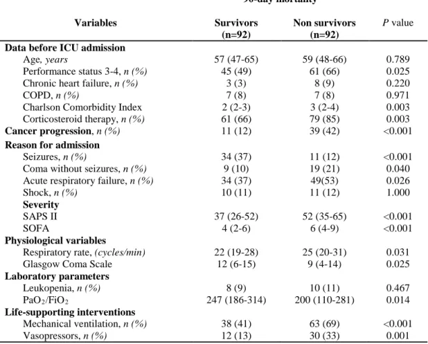
Table 3 displays the factors associated with 90-day mortality identified by univariate analysis. Multivariate forward or backward logistic regression analysis with missing data imputations showed that six of these factors independently predicted 90-day mortality. One factor, admission for seizures, was independently associated with lower 90-day mortality (OR 0.18, 95%CI 0.07-0.47, p<0.001). Five factors were independently associated with higher day-90 mortality: need for MV (OR 6.88, 95%CI 3.01-15.73, p<0.001), cancer progression (OR 7.87, 95%CI 3.26-18.99, p<0.001), Charlson Comorbidity Index (OR 1.44, 95%CI 1.10-1.88, p=0.007), systolic blood pressure on ICU admission (OR 1.02 95%CI 1.00-1.03, p=0.025) and respiratory rate (OR 1.05, 95%CI 1.00-1.10, p=0.041). The same seven factors were identified after excluding patients with missing data.
Subgroup of patients requiring invasive mechanical ventilation
Among the 108 patients who received MV, 18 (17%) received noninvasive ventilation (NIV) as first-line ventilatory support and 90 (83%) received invasive ventilation. Among the 18 patients who received NIV, 4 (22%) were intubated within 24 hours due to NIV failure. Among the 94 patients who finally received invasive mechanical ventilation (IMV), ICU mortality and 90-day mortality were 37% (n=35) and 60% (n=56), respectively.
Table 4 displays the factors associated with ICU mortality among patients who received IMV identified by univariate analysis. Multivariate forward or backward logistic regression analysis with missing data imputations showed that three of these factors independently predicted ICU mortality. Admission for seizures was the only factor independently associated with lower ICU mortality in patients who received IMV (OR 0.05, 95%CI 0.01-0.41, p=0.005), whereas two factors were associated with higher ICU mortality: cancer progression (OR 7.49, 95%CI 2.37-23.67, p<0.001) and respiratory rate (OR 1.08, 95%CI 1.01-1.15, p=0.022). The same three factors were identified after excluding patients with missing data.
DISCUSSION
The salient results of this study are: 1) mortality of PMBT patients admitted to the ICU was relatively low and has remained stable over the last 19 years, 2) cancer progression and the need for MV were associated with a poorer prognosis, 3) admission for seizures was a predictor of survival, even when invasive MV was required.
To the best of our knowledge, this is the largest cohort of neuro-oncological patients admitted to medical ICUs focusing on their short-term and medium-term prognosis[27].
The mortality of PMBT patients did not improve significantly during the study period, in contrast with the dramatic improvement of ICU survival rates observed in cancer patients over the last two decades[3]. This lack of improved survival cannot be attributed to increased severity of patients, as the SAPS II score of patients admitted to the ICU did not increase significantly over the study period. However, this mortality rate was similar to, if not lower than, that reported in patients with hematologic malignancies[17] and solid tumors[2]. The simple presence of a PMBT therefore should not be a sufficient argument for refusal of ICU admission. There are four possible explanations for this low mortality. First, the young age and limited comorbidities observed in PMBT patients may be protective factors. Second, since only patients with the best prognosis were considered for ICU admission, our population may be highly selected. Third, there was a high proportion of rapidly reversible causes of admission such as seizures. Fourth, progress in the management of malignancies over the past two decades[3], particularly in the subgroup of patient with primary CNS lymphoma, may also be beneficial for patients with PMBT.
In the present study, seizures appeared to be a predictive factor of short-term and medium-term survival, even in mechanically ventilated patients. Although the prognosis of tumor-associated status epilepticus appears to be poorer than that of status epilepticus due to other causes[28], it generally has a good prognosis and is associated with low mortality[29]. It should be stressed that the incidence of epilepsy is particularly high in gliomas, ranging from 60 to 100% for low-grade gliomas and 40 to 60% for high-grade gliomas[30]. A recent study reported similar results in PMBT patients admitted to the medical ICU: admission for a neurological cause (mainly seizures) was predictive of better ICU survival than non-neurological causes of ICU admission[27]. In contrast, in our study, cancer progression was a strong predictor of ICU and 90-day mortality regardless of the severity of the patients at the time of ICU admission. In most reports on cancer patients in the ICU, cancer progression appears to be an independent predictor of mortality[1, 17, 31, 34], highlighting the fact that cancer status is a crucial element that must be defined before any discussion regarding ICU admission.
Initiation of mechanical ventilation was an independent factor of poor prognosis, confirming numerous reports demonstrating the deleterious impact of mechanical ventilation on the prognosis of ICU patients with hematologic malignancies[17] or solid tumors[32, 33], particularly lung cancer[31, 32, 34]. In contrast with these series that reported a mean ICU mortality rate of 65% in mechanically ventilated patients[17, 31, 36], the ICU mortality rate in our PMBT patients was only 37%. This lower mortality in PMBT patients seemed to be directly related to the good prognosis of patients intubated for status epilepticus or seizure-related coma, as, when patients admitted for seizures were excluded, ICU mortality in mechanically ventilated patients increased to 49% (34/70). Interestingly, very few patients were treated by NIV. Although the potential benefit of NIV in cancer patients remains a matter of debate[37], NIV does not appear to be indicated in PMBT patients due to loss of upper airway control induced by disorders of consciousness and swallowing disorders[38]. Finally, the use of vasopressors was not an independent predictor of higher mortality, as previously reported in other types of cancer[31, 32, 34, 35], which could suggest that very brief use of vasopressors, for example to treat severe sepsis, should not be discouraged.
The present study has several limitations. First, this was a retrospective study, which involves a potential bias in patient selection or data collection. However, the rarity of the disease remains a major obstacle to prospective studies, even with a multicenter design. In addition, data were extracted from a prospectively managed database, which offers a higher reliability of data collection than simple chart examination. Second, the relevance of mortality as an outcome measure in this very specific population can be questioned in view of the potentially increased impairment of functional status and quality of life after an ICU stay. Unfortunately, such information was not available in our database. However, the vast majority of PMBT patients appeared to exhibit a stable or improved Karnofsky Performance Status after ICU discharge[27]. Third, the WHO classification of brain tumors has been updated in 2016, and without data regarding the molecular testing, further misclassification has been possible for the 2 patients with oligoastrocytoma of our cohort. Finally, we only considered patients admitted to the ICU. Patients who were not considered for ICU admission for any reason, such as poor prognosis or performance status, were therefore not included in this analysis.
In conclusion, patients with PMBT have a fairly good prognosis after ICU admission, as compared to patients with hematologic malignancies or solid tumors, especially when they are admitted for seizures. The presence of a PMBT therefore does not appear to be sufficient for ICU refusal. Prognostic factors such as cancer progression need for MV or seizures may help clinicians to make optimal triage decisions. In any case, ICU
transfer must always be based on multidisciplinary discussion between neuro-oncologists, intensivists, the patient and relatives, respecting the patient’s autonomy and willingness.
DECLARATIONS SECTION Ethical standards
Consent were not collected because of retrospective design
The study was approved by the Institutional Review Board of the French Intensive Care Society.
Conflict of interest:
Alexandre Demoule has signed research contracts with Covidien, Maquet, Philips and Ait Liquide Santé; he has also received personal fees from Covidien, Maquet and MSD.
Ahmed Idbaih reports research funding from La Fondation ARC pour la recherche sur le Cancer, Carthera, Beta-Innov, and Intselchimos; travel funding from Hoffmann-La Roche; and personal fees from Novartis, La Lettre du Cancérologue, BMS, and Cipla unrelated to the submitted work.
Nicolas Weiss has signed research contracts with Eumedica, BMS, MedDay pharmaceuticals; he has also received personal fees from Norgine and Alpha-Wasserman.
Thomas Similowski has received grant research from Coviden, Philips, Pierre Fabre Médicaments, Air Liquide Medical Systems; he has also received personal fees from Takeda, Teva Pharma, Lungpacer Inc, Almirall France, Pierre Fabre Médicaments, Novartis, Mundipharma, Invacare, Astra Zeneca, Boehringer Ingelheim and GlaxoSmithKline.
Julien Mayaux declares that he has no conflict of interest. Hélène Prodanovic declares that she has no conflict of interest.
Isabelle Rivals declares that she has no conflict of interest. Maxens Decavèle declares that he has no conflict of interest.
Research support/funding: none.
Author’s contributions to the manuscript:
Conception and design: Maxens Decavèle, Nicolas Weiss, Isabelle Rivals, Alexandre Demoule.
Collection and assembly of data: Maxens Decavèle, Julien Mayaux, Hélène Prodanovic, Alexandre Demoule Data analysis and interpretation: Maxens Decavèle, Isabelle Rivals, Alexandre Demoule, Thomas Similowski Manuscript writing: Maxens Decavèle, Alexandre Demoule
REFERENCES
1. Soares M, Caruso P, Silva E, Teles JM, Lobo SM, Friedman G et al (2010) Characteristics and outcomes of patients with cancer requiring admission to intensive care units: a prospective multicenter study. Crit Care Med 38:9-15.
2. Puxty K, McLoone P, Quasim T, Kinsella J, Morrison D (2014) Survival in solid cancer patients following intensive care unit admission. Intensive Care Med 40:1409-28.
3. Azoulay E, Soares M, Darmon M, Benoit D, Pastores S, Afessa B (2011) Intensive care of the cancer patient: recent achievements and remaining challenges. Ann Intensive Care 1:5.
4. Ferlay J, Steliarova-Foucher E, Lortet-Tieulent J, Rosso S, Coebergh JW, Comber H et al (2013) Cancer incidence and mortality patterns in Europe: estimates for 40 countries in 2012. Eur J Cancer 49:1374-403.
5. Suarez JI, Zaidat OO, Suri MF, Feen ES, Lynch G, Hickman J et al (2004) Length of stay and mortality in neurocritically ill patients: impact of a specialized neurocritical care team. Crit Care Med 32:2311-7.
6. Broessner G, Helbok R, Lackner P, Mitterberger M, Beer R, Engelhardt K et al (2007) Survival and long-term functional outcome in 1,155 consecutive neurocritical care patients. Crit Care Med 35:2025-30.
7. Kiphuth IC, Schellinger PD, Köhrmann M, Bardutzky J, Lücking H, Kloska S et al (2010) Predictors for good functional outcome after neurocritical care. Crit Care 14-R136.
8. Omuro A, DeAngelis LM (2013) Glioblastoma and other malignant gliomas: a clinical review. JAMA 310:1842-50.
9. Taphoorn MJB, Sizoo EM, Bottomley A (2010) Review on quality of life issues in patients with primary brain tumors. The Oncologist 15:618-26.
10. Giovagnoli AR, Silvani A, Colombo E, Boiardi A (2005) Facets and determinants of quality of life in
patients with recurrent high grade glioma. J Neurol Neurosurg Psychiatry 76:562-8.
11. Guidelines for intensive care unit admission, discharge, and triage. Task Force of the American College
12. Daniels TB, Brown PD, Felten SJ, Wu W, Buckner JC, Arusell RM et al (2011) Validation of EORTC prognostic factors for adults with low-grade glioma: a report using intergroup 86-72-51. Int J Radiat Oncol Biol Phys 81:218–24.
13. Stupp R, Mason WP, van den Bent MJ, Weller M, Fisher B, Taphoorn MJ et al (2005) Radiotherapy
plus concomitant and adjuvant temozolomide for glioblastoma. N Engl J Med 352:987-96.
14. Hegi ME, Diserens AC, Gorlia T, Hamou MF, de Tribolet N, Weller M et al (2005) MGMT gene
silencing and benefit from temozolomide in glioblastoma. N Engl J Med 352:997-1003.
15. Stupp R, Hegi ME, Mason WP, van den Bent MJ, Taphoorn MJ, Janzer RC et al (2009) Effects of
radiotherapy with concomitant and adjuvant temozolomide versus radiotherapy alone on survival in glioblastoma in a randomised phase III study: 5-year analysis of the EORTC-NCIC trial. Lancet Oncol 10:459-66.
16. Westphal M, Lamszus K (2015) Circulating biomarkers for gliomas. Nat Rev Neurol 11:556-66.
17. Azoulay E, Mokart D, Pène F, Lambert J, Kouatchet A, Mayaux J et al (2013) Outcomes of critically ill patients with hematologic malignancies: prospective multicenter data from France and Belgium-a groupe de recherche respiratoire en réanimation onco-hématologique study. J Clin Oncol 31:2810-8.
18. Louis DN, Ohgaki H, Wiestler OD, Cavenee WK, Burger PC, Jouvet A et al (2007) The 2007 WHO
classification of tumours of the central nervous system. Acta Neuropathol 114:97-109.
19. Oken MM, Creech RH, Tormey DC, Horton J, Davis TE, McFadden ET et al (1982) Toxicity and
response criteria of the Eastern Cooperative Oncology Group. Am J Clin Oncol 5:649-55.
20. Charlson M, Szatrowski TP, Peterson J, Gold J (1994) Validation of a combined comorbidity index. J
Clin Epidemiol 47:1245-51.
21. Vaquero J, Martínez R, Manrique M (2000) Stereotactic biopsy for brain tumors: is it always
necessary? Surg Neurol 53:432-437.
22. Ferreri AJM, Marturano E (2012) Primary CNS lymphoma. Best Pract Res Clin Haematol 25:119–30.
23. Le Gall JR, Lemeshow S, Saulnier FA (1993) new Simplified Acute Physiology Score (SAPS II) based
on a European/North American multicenter study. JAMA 270:2957-63.
24. Vincent JL, Moreno R, Takala J, Willatts S, De Mendonça A, Bruining H et al (1996) The SOFA
(Sepsis-related Organ Failure Assessment) score to describe organ dysfunction/failure. On behalf of the Working Group on Sepsis-Related Problems of the European Society of Intensive Care Medicine. Intensive Care Med 22:707-10.
25. Hosmer DW, Hosmer T, Le Cessie S, Lemeshow S (1997) A comparison of goodness-of-fit tests for the logistic regression model. Stat Med 16:965-80.
26. Hanley JA, McNeil BJ (1982) The meaning and use of the area under a receiver operating characteristic
(ROC) curve. Radiology 143:29-36.
27. Tabouret E, Boucard C, Devillier R, Barrie M, Boussen S, Autran D et al (2016) Neuro-oncological
patients admitted in intensive-care unit: predictive factors and functional outcome. J Neurooncol 127:111-7.
28. Arik Y, Leijten FS, Seute T, Robe PA, Snijders TJ (2014) Prognosis and therapy of tumor-related
versus non-tumor-related status epilepticus: a systematic review and meta-analysis. BMC Neurol 14:152.
29. Vooturi S, Jayalakshmi S, Sahu S, Mohandas S (2014) Prognosis and predictors of outcome of
refractory generalized convulsive status epilepticus in adults treated in neurointensive care unit. Clin Neurol Neurosurg 126:7-10.
30. Vecht CJ, Kerkhof M, Duran-Pena A (2014) Seizure prognosis in brain tumors: new insights and
evidence-based management. The Oncologist 19, 751–9.
31. Roques S, Parrot A, Lavole A, Ancel PY, Gounant V, Djibre M et al (2009) Six-month prognosis of
patients with lung cancer admitted to the intensive care unit. Intensive Care Med 35:2044-50.
32. Soares M, Toffart AC, Timsit JF, Burghi G, Irrazábal C, Pattison N et al (2014) Intensive Care in
Patients with Lung Cancer: A Multinational Study. Ann Oncol 25:1829-35.
33. Azoulay E, Thiéry G, Chevret S, Moreau D, Darmon M, Bergeron A et al (2004) The prognosis of
acute respiratory failure in critically ill cancer patients. Medicine (Baltimore) 83:360-70.
34. Soares M, Darmon M, Salluh JI, Ferreira CG, Thiéry G, Schlemmer B et al (2007) Prognosis of lung
cancer patients with life-threatening complications. Chest 131:840-6.
35. Azoulay E, Afessa B (2006) The intensive care support of patients with malignancy: do everything that
can be done. Intensive Care Med 32:3-5.
36. Slatore CG, Cecere LM, Letourneau JL, O'Neil ME, Duckart JP, Wiener RS et al (2012) Intensive care
unit outcomes among patients with lung cancer in the surveillance, epidemiology, and end results-medicare registry. J Clin Oncol 30:1686-91.
37. Lemiale V, Mokart D, Resche-Rigon M, Pène F, Mayaux J, Faucher E et al (2015) Effect of
Noninvasive Ventilation vs Oxygen Therapy on Mortality Among Immunocompromised Patients With Acute Respiratory Failure: A Randomized Clinical Trial. JAMA 314:1711-9.
38. Newton HB, Newton C, Pearl D, Davidson T (1994) Swallowing assessment in primary brain tumor patients with dysphagia. Neurology 44:1927-32.
Tables
Table 1. Characteristics of the 197 patients included in the study
Variables
Age, years 58 (47-66)
Gender (male) n (%) 132 (67)
Comorbidities
Chronic heart failure, n (%)
Chronic obstructive pulmonary disease, n (%)
11 (6) 14 (7)
Functional status
Performance Status 3-4, n (%) Charlson Comorbidity Index
114 (58) 2 (2-3)
Mode of admission
Intra-hospital transfer, n (%)
Transfer via emergency services, n (%) Transfer from another hospital, n (%)
119 (61) 62 (31)
15 (8)
Disease status on admission
Newly diagnosed, n (%) In progression n (%) Controlled n (%) Unknown, n (%) 17 (9) 53 (27) 101 (51) 25 (13)
Reason for admission
Acute respiratory failure, n (%) Seizures, n (%)
Coma without seizures, n (%) Shock, n (%) Other, n (%) 88 (45) 50 (26) 28 (14) 24 (12) 6 (3) Severity SAPS II SOFA 46 (31-59) 5 (3-7) Physiological variables
Systolic blood pressure, mmHg Heart rate, b.p.m.
Respiratory rate, cycles/min Glasgow Coma Scale Temperature, °C 120 (103-136) 100 (81-120) 24 (20-30) 11 (6-15) 37.7 (37.0-38.7) Laboratory parameters Leukocytes, /mm3 Leukopenia, n (%) Serum creatinine, μmol/l
7670 (4548-12143) 18 (9) 72 (53-97)
Arterial Blood Gases
pH PaCO2, mmHg PaO2/FiO2 Bicarbonate, mmol/l 7.43 (7.39-7.48) 37 (31-42) 231 (139-303) 24 (20-30) Life-supporting interventions Mechanical ventilation, n (%)
Non-Invasive Ventilation only, n (%) Invasive Ventilation only, n (%)
Total duration of mechanical ventilation, days Vasopressors, n (%)
Renal replacement therapy, n (%)
108 (55) 14 (7) 95 (48) 6 (2-11) 45 (23) 3 (2) End-of-life decision, n (%) 43 (22)
Mortality ICU mortality, n (%) Hospital mortality, n (%) 90-day mortality, n (%) 46 (23) 84 (43) 92 (50) SAPS II, Simplified Acute PhysiologyScore II; SOFA, Sequential Organ Failure Assessment.
Continuous variables are expressed as median (interquartile range) and categorical data are expressed as number (%).
Table 2. Univariate analysis: factors associated with ICU mortality
Continuous variables are expressed as median (interquartile range) and categorical data are expressed as number (%).
ICU, Intensive Care Unit; COPD, Chronic obstructive pulmonary disease; SAPS II, Simplified Acute
ICU mortality Variables Survivors (n=150) Non-survivors (n=46) P value
Data before ICU admission
Age, years
Performance status 3-4, n (%) Chronic heart failure
COPD, n (%)
Charlson Comorbidity Index Corticosteroid therapy, n (%) 58 (48-66) 82 (55) 3 (2) 9 (6) 2 (2-3) 111 (74) 58 (45-64) 32 (70) 8 (17) 5 (11) 2 (2-3) 39 (85) 0.964 0.073 <0.001 0.324 0.918 0.165 Cancer progression, n (%) 29 (19) 24 (52) <0.001
Reason for admission
Seizures, n (%)
Coma without seizures, n (%) Acute respiratory failure, n (%) Shock, n (%) 48 (32) 16 (11) 63 (42) 18 (12) 2 (4) 12 (26) 25 (54) 6 (13) <0.001 0.009 0.141 0.850 Severity SAPS II SOFA 41 (28-54) 4 (2-7) 63 (46-71) 8 (5-10) <0.001 <0.001 Physiological variables
Respiratory rate, (cycles/min) Glasgow Coma Scale
23 (20-30) 12 (7-15) 28 (20-33) 6 (3-13) 0.034 <0.001 Laboratory parameters Leukopenia, n (%) PaO2/FiO2 11 (7) 242 (162-324) 7 (15) 171 (106-252) 0.402 <0.001 Life-supporting interventions Mechanical ventilation, n (%) Vasopressors, n (%) 71 (47) 22 (15) 37 (80) 23 (50) <0.001 <0.001
Table 3. Univariate analysis: factors associated with 90-day mortality 90-day mortality Variables Survivors (n=92) Non survivors (n=92) P value
Data before ICU admission
Age, years
Performance status 3-4, n (%) Chronic heart failure, n (%) COPD, n (%)
Charlson Comorbidity Index Corticosteroid therapy, n (%) 57 (47-65) 45 (49) 3 (3) 7 (8) 2 (2-3) 61 (66) 59 (48-66) 61 (66) 8 (9) 7 (8) 3 (2-4) 79 (85) 0.789 0.025 0.220 0.971 0.003 0.003 Cancer progression, n (%) 11 (12) 39 (42) <0.001
Reason for admission
Seizures, n (%)
Coma without seizures, n (%) Acute respiratory failure, n (%) Shock, n (%) 34 (37) 9 (10) 34 (37) 10 (11) 11 (12) 19 (21) 49(53) 11 (12) <0.001 0.040 0.026 1.000 Severity SAPS II SOFA 37 (26-52) 4 (2-6) 52 (35-65) 6 (4-9) <0.001 <0.001 Physiological variables
Respiratory rate, (cycles/min) Glasgow Coma Scale
22 (19-28) 12 (6-15) 25 (20-31) 9 (4-14) 0.031 0.025 Laboratory parameters Leukopenia, n (%) PaO2/FiO2 8 (9) 247 (186-314) 10 (11) 200 (110-281) 0.467 0.014 Life-supporting interventions Mechanical ventilation, n (%) Vasopressors, n (%) 38 (41) 12 (13) 63 (69) 30 (33) <0.001 0.001
COPD, Chronic obstructive pulmonary disease; SAPS II, Simplified Acute Physiology Score II; SOFA, Sequential Organ Failure Assessment.
Continuous variables are expressed as median (interquartile range) and categorical data are expressed as number (%).
Table 4. Univariate analysis: factors associated with ICU-mortality among invasively ventilated patients.
ICU, Intensive Care Unit; COPD, Chronic obstructive pulmonary disease; SAPS II, Simplified Acute PhysiologyScore II; SOFA, Sequential Organ Failure Assessment.
Continuous variables are expressed as median (interquartile range) and categorical data are expressed as number (%). ICU mortality Survivors (n=59) Non survivors (n=35) P value
Data before ICU admission
Age, years
Performance status 3-4, n (%) Chronic heart failure, n (%) COPD, n (%)
Charlson Comorbidity Index Corticosteroid therapy, n (%) 59 (48-68) 30 (51) 3 (5) 2 (2) 2 (2-3) 41 (69) 58 (42-64) 23 (66) 3 (9) 4 (11) 2 (2-3) 29 (83) 0.743 0.223 0.587 0.191 0.587 0.151 Cancer progression, n (%) 12 (20) 19 (54) <0.001
Reason for admission
Seizure, n (%)
Coma without seizures, n (%) Acute respiratory failure, n (%) Shock, n (%) 24 (41) 7 (12) 23 (39) 5 (8) 1 (3) 11 (31) 16 (46) 6 (17) <0.001 0.020 0.512 0.319 Severity SAPS II SOFA 51 (38-59) 6 (5-8) 63 (52-72) 8 (6-11) 0.004 0.007 Physiological variables
Heart rate, beats/min Glasgow Coma Scale score
100 (79-115) 7 (3-12) 100 (86-130) 6 (3-10) 0.468 0.237 Biological variables Leukopenia, n (%) PaO2/FiO2 1 (2) 231 (146-322) 5 (14) 167 (103-273) 0.009 0.096
Life supporting interventions
Vasopressors, n (%)
Renal replacement therapy, n (%)
17 (29) 0 (0) 20 (57) 3 (9) 0.041 0.049
FIGURE LEGENDS
Figure 1. Study flow chart
ICU, intensive care unit.
Figure 2. Distribution of primary malignant brain tumor types
ICU, intensive care unit.
a other tumors included: 5 medulloblastomas, 3 malignant meningeal tumors, 2 germinomas and 5 gliomas with unknown malignancy grade because of missing data.
Figure 3. Changes in the number of admissions and death during the study period
Bars represent the number of admissions during each 2-year period. Solid line with filled circles represents the intensive care unit mortality rates during each 2-year period. Dashed line with open circles represents 90-day mortality rates during each 2-year period.