Angiotensin II downregulates the fatty acid oxidation
pathway in adult rat cardiomyocytes via release
of tumour necrosis factor-a
Corinne Pellieux*, Christophe Montessuit, Ire
`ne Papageorgiou, and Rene
´ Lerch
Cardiology Center, Department of Medicine, University Hospitals of Geneva, Foundation for Medical Research, 64, avenue de la Roseraie, CH-1211 Geneva 4, Switzerland
Received 26 August 2008; revised 18 December 2008; accepted 23 December 2008; online publish-ahead-of-print 8 January 2009
Time for primary review: 26 days
Aims Advanced heart failure is often associated with reduced myocardial fatty acid oxidation capacity. We have previously observed that failing hearts of mice with overexpression of angiotensinogen in the myocardium exhibit marked reduction of key regulatory proteins of fatty acid oxidation. In the present study, we determined whether exposure of adult rat cardiac (ARC) myocytes to angiotensin II (Ang II) influences expression of fatty acid translocase, muscle-type carnitine palmitoyl transferase-I, and medium-chain acyl-CoA dehydrogenase.
Methods and results Ang II reduced mRNA expression of the three regulatory proteins in ARC myocytes during the entire 14-days culture period. However, protein expression and palmitate oxidation rate remained unaltered for 7 days, but subsequently markedly decreased. The decrease of protein expression and of fatty acid oxidation coincided with the onset of increased protein expression of tumour necrosis factor-a (TNF-a). The effect of Ang II was completely abolished by either blocking TNF-a formation through inhibition of reactive oxygen species-mediated activation of nuclear factor-kB or by neutralizing TNF-a with a specific antibody. Activation of peroxisome proliferator-activated receptor-a (PPARa) and PPARb/d counteracted Ang II-mediated reduction of the fatty acid oxidation pathway.
Conclusion Prolonged exposure of cardiac myocytes to Ang II elicits downregulation of the fatty acid oxidation pathway mediated by enhanced synthesis of TNF-a.
KEYWORDS Myocytes; Cell culture; Energy metabolism; Angiotensin; Remodelling
1. Introduction
Cardiac myocytes exposed to chronic stress undergo a sequence of phenotypic changes, which eventually lead to heart failure. A number of observations indicate that adverse ventricular remodelling in response to disease conditions including myocardial infarction,1,2pressure3,4or
volume overload,5 and tachycardia6 is associated with changes in myocardial energy metabolism.4–8 There is
increasing evidence that fatty acid oxidation is reduced in advanced stages of ventricular remodelling, and more glucose is oxidized.7,9The consequences of reduction of the fatty acid oxidation pathway for disease progression remain controversial. On one hand, the shift to glucose oxidation may protect the myocardium by factors including enhance-ment of ATP yield per O2consumed,10support of ion pumps
by glycolysis-derived ATP,11 and reduction of proton
pro-duction by oxidation, instead of conversion to lactate, of glycolytically produced pyruvate.12 On the other hand, a
reduction of fatty acid oxidation may limit overall energy production and favour accumulation of potentially toxic lipid intermediates in the cytoplasm (‘lipotoxicity’).13
Reduction of fatty acid oxidation during ventricular remo-delling and heart failure seems to be mediated by reduction of protein levels of key regulatory proteins of fatty acid metabolism.1,6,7,14–16 This has been largely attributed to
inactivation and/or downregulation of the nuclear transcrip-tion factors peroxisome proliferator-activated receptors (PPARs) a17,18 and b/d19 or of the PPARg coactivator-1a (PGC-1a),20 which regulate the expression of multiple
genes controlling both fatty acid uptake and oxidation. However, a number of observations indicate that, in addition to reduced transcription, levels of regulatory proteins are also modulated by posttranscriptional mechanisms. Specifi-cally, in rodent models of pathological left ventricular remo-delling, mRNA expression of enzymes of fatty acid oxidation was reduced during compensated hypertrophy, but protein expression and fatty acid oxidation rate were maintained at control levels. However, both protein expression and fatty acid oxidation dropped dramatically when heart failure occurred.1,9,14 The signalling pathways driving
*Corresponding author. Tel: þ41 22 372 72 22; fax: þ41 22 372 72 29. E-mail address: [email protected]
Published on behalf of the European Society of Cardiology. All rights reserved.&The Author 2009. For permissions please email: [email protected].
downregulation of the fatty acid oxidation pathway during ventricular remodelling are largely unknown.
Activation of the renin–angiotensin system and increased formation of the main effector angiotensin II (Ang II) play a critical role in left ventricular remodelling and heart failure.21Ang II has been implicated in myocyte hypertrophy, interstitial fibrosis, and apoptosis.22 Furthermore, Ang II promotes the inflammatory response, which may be involved in the progression from compensated remodelling to heart failure.23 However, at present, little is known about whether Ang II contributes to altered metabolic regulation in the myocardium undergoing remodelling. We have recently observed that mice with targeted overexpression of angio-tensinogen in the myocardium, resulting in chronically elev-ated myocardial Ang II at unaltered blood pressure,24 progressively develop left ventricular hypertrophy associated with downregulation of mRNA expression of a number of regu-latory proteins of fatty acid oxidation.9 Protein expression and palmitate oxidation, measured during in vitro perfusion, did not differ from wild-type mice during compensated hypertrophy, but were significantly reduced after the onset of heart failure. Accordingly, chronic stimulation of the myo-cardium with Ang II elicits progressive myocardial remodel-ling, which results in reduction of regulatory proteins of the fatty acid oxidation pathway when heart failure occurs. However, it is not known whether Ang II stimulation of cardiac myocytes directly affects expression of the fatty acid oxidation pathway.
We therefore studied the effects of prolonged stimulation of adult rat cardiac (ARC) myocytes by Ang II on the expression of regulatory proteins of fatty acid oxidation. The results indicate that Ang II elicits downregulation of the fatty acid oxidation pathway. This effect is largely mediated by Ang II-induced generation of tumour necrosis factor-a (TNF-a).
2. Materials and methods
An expanded Materials and methods section is available in the online data supplement.
The investigation conforms to the Guide for the Care and Use of Laboratory Animals published by the US National Institutes of Health (NIH Publications No. 85-23, revised 1996) and was approved by the local animal protection authorities.
2.1 Cell isolation and culture
ARC myocytes were isolated from male OFA rats (Charles River, France) as previously described.25,26The culture medium was M199 supplemented with 20% (vol/vol) foetal calf serum (FCS; Invitrogen) throughout the culture period. The culture medium contained glucose (5.5 mM). Ang II (1–1000 nM), TNF-a (10 ng/mL), N-acetyl cystein (NAC; 10 mM), apocynin (1 mM), SN50 (18 mM), anti-rat TNF-a antibody (0.2 mg/mL), WY-14643 (100 nM), L165-041 (1 mM), or ciglitazone (10 nM) were added to the medium during the entire culture period. In selected experiments, palmitate plus oleate (0.05–0.25 mM) at equimolar concentrations complexed to bovine serum albumin (0.2 mM) were added.
2.2 Immunofluorescence and cell surface area
determination
Cells were fixed with paraformaldehyde and stained (a-actinin, F-actin, and DNA). Relative cell surface area was calculated from digitized images.
2.3 [
3H]-phenylalanine incorporation
To obtain an index for the rate of de novo protein synthesis, ARC were incubated with L-[2,3,4,5-3H]-phenylalanine. [3H]-phenylalanine incorporation was measured, and normalized to DNA content.
2.4 Measurement of intracellular reactive
oxygen species
Reactive oxygen species (ROS) were measured by the dichlorofluor-escein fluorescence method. Upon reaction with ROS, DCF becomes highly fluorescent and fluorescence was then recorded.
2.5 Protein and RNA expression
Western blot and quantitative reverse transcriptase–polymerase chain reaction were performed as in previous studies.26
2.6 Palmitate oxidation
Palmitate oxidation was estimated based on the release of14CO 2 from [1-14C]-palmitate.26
ARC were incubated in sealed flasks containing a suspended filter paper and medium containing palmitate (0.05 mM), oleate (0.05 mM), and 1 mCi/mL [1-14C]-palmitate complexed to bovine serum albumin (0.2 mM).14CO
2produced by [1-14C]-palmitate metab-olism was collected overnight on the filter paper and quantified.
3. Results
3.1 Ang II and tumour necrosis factor-a elicit
reduction of regulatory proteins of fatty acid
oxidation in adult rat cardiac myocytes
ARC cultured with 20% FCS underwent phenotypic changes during the 14 days culture period as described previously.26
Three to 4 days after isolation ARC resumed beating, gradu-ally increased in size, and reexpressed atrial natriuretic factor (ANF) mRNA. Supplementation of the culture medium with Ang II (100 nM) significantly enhanced the hypertrophic response as evidenced by a further increase of cell size, stimulation of protein synthesis, and increase of mRNA expression of both ANF and brain natriuretic peptide (BNP) (Figure 1). Since Ang II stimulates synthesis of TNF-a,27,28 we exposed, in parallel experiments, cells to TNF-a (10 ng/mL). TNF-a also increased all indices of the hypertrophic response, even more pronounced in extent compared with Ang II (Figure 1).
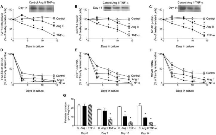
To determine the effects of both Ang II and TNF-a on key regulatory proteins of the fatty acid oxidation pathway, we monitored expression of fatty acid translocase (FAT/CD36), muscle-type carnitine palmitoyl transferase-I (mCPT-I) and medium-chain acyl-CoA dehydrogenase (MCAD). Protein expression of all three regulatory proteins remained stable in control ARC during the 14 days culture period. Both Ang II and TNF-a elicited a gradual reduction of the three regu-latory proteins (Figure 2A–C ). However, there was a clear difference in the time-course. Supplementation of the medium with Ang II during the entire culture period did not affect protein expression up to day 7. Thereafter, protein expression decreased to reach 60% of control on day 14. The reduction of protein expression by Ang II was concentration-dependent (Supplementary material online, Figure S2). In contrast to Ang II, TNF-a more rapidly reduced protein expression of FAT/CD36, mCPT-I, and MCAD, starting from the onset of the culture period, to
C. Pellieux et al. 342
levels as low as 10% of control after 14 days (Figure 2A–C ). Correspondingly, exposure of ARC to TNF-a for 7 days reduced palmitate oxidation (to 30%), whereas Ang II had no effect (Figure 2G). During exposure for more than 7 days both TNF-a and Ang II reduced palmitate oxidation (to 15% and 41%, respectively, after 14 days).
In contrast to protein expression, mRNA expression of FAT/CD36, mCPT-I, and MCAD rapidly decreased after iso-lation in untreated control ARC to 40 to 60% of values measured in freshly isolated cells (Figure 2D–F). Both Ang II and TNF-a elicited a further reduction of mRNA expression of all three regulatory proteins compared with untreated ARC. We have previously observed that the reduction of mRNA expression in control experiments is related to the low fatty acid concentration in the standard culture medium since it is prevented by the addition of fatty acid to the culture medium.26
To determine the role of the high concentration of FCS in the effects of Ang II, in additional experiments, ARC cul-tured in medium containing a low concentration of FCS (1%) were exposed to Ang II (100 nM) or TNF-a (10 ng/mL) for up to 7 days. Both Ang II and TNF-a reduced regulatory proteins of fatty acid oxidation and palmitate oxidation to a similar extent compared with results obtained in the pre-sence of 20% FCS. However, Ang II-induced reduction of both protein expression and palmitate oxidation was detectable earlier, already on day 4, and was reduced on day 7 to values comparable to those observed in the presence of 20% FCS on day 14. The decrease of both regulatory proteins and palmitate oxidation by TNF-a was also accelerated
compared to experiments in ARC cultured with 20% FCS (Supplementary material online, Figure S3).
3.2 Ang II-induced reduction of the fatty acid
oxidation pathway involves ROS-dependent
activation of NF-kB
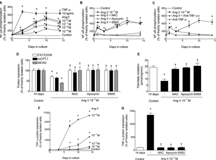
Because nuclear factor-kB (NF-kB) plays a central role in the Ang II-mediated hypertrophic response,29we first measured
phosphorylation of the p65 subunit of NF-kB at ser536, which is a marker of NF-kB activation.30Total NF-kB protein
level was not altered by Ang II (data not shown). However, phosphorylation of NF-kB progressively and concentration-dependently increased in the presence of Ang II until day 10 and then levelled off or slightly decreased (Figure 3A). Ang II at a concentration of 100 nM elicited a maximal response. The Ang II-induced increase of phospho-NF-kB was completely abolished if accumulation of ROS was prevented either by the NADPH oxidase inhibitor apocynin or the ROS scavenger NAC (Figure 3B). Prevention of ROS accumulation was validated in separate experiments by monitoring of dichlorofluorescin diacetate fluorescence (data not shown). Of note, TNF-a, in the absence of Ang II, rapidly and markedly enhanced phosphorylation of NF-kB (Figure 3A).
Inhibition of Ang II-induced ROS activity by apocynin or NAC completely prevented the reduction of protein expression of FAT/CD36, mCPT-I, and MCAD (Figure 3D), and the decrease of palmitate oxidation (Figure 3E). Reduction of both expression of regulatory proteins and pal-mitate oxidation rate was also prevented by SN50 (Figure 3D and E) which inhibits translocation of NF-kB to the nucleus,
Figure 1 Ang II and tumour necrosis factor-a (TNF-a) increase indices of myocyte hypertrophy in adult rat cardiac (ARC). (A) Relative difference of ARC size measured after 14 days of culture in control medium or medium supplemented with either Ang II (100 nM) or TNF-a (10 ng/mL). Cells were stained for a-actinin (green), F-actin (red) and DNA (blue). At least 100 cells in different cultures were measured for each condition. (B) De novo protein synthesis, estimated from [3H]-phenylalanine incorporation. (C and D) mRNA expression of ANF (C) or brain natriuretic peptide (D). n ¼ 4–8. Values are means + SEM. *P , 0.05 compared
without affecting phosphorylation (Figure 3B). Thus, ROS-mediated activation and translocation of NF-kB is necessary for Ang II-mediated downregulation of the fatty acid oxi-dation pathway.
3.3 Production of tumour necrosis factor-a
mediates Ang II-induced reduction of the fatty
acid oxidation pathway
To determine the role of TNF-a in Ang II-induced downregu-lation of regulatory proteins of fatty acid oxidation, we first measured the effect of Ang II on TNF-a expression in ARC. Ang II concentration-dependently stimulated protein expression of TNF-a (Figure 3F). However, TNF-a expression was absent or low during the first 7 days at Ang II concen-trations up to 100 nM. Induction of TNF-a protein expression was completely abolished by NAC, apocynin, and SN50 indi-cating the involvement of the ROS–NF-kB cascade in the stimulation of TNF-a expression (Figure 3G).
To further evaluate whether TNF-a secreted by ARC in response to Ang II stimulation was sufficient to reduce the fatty acid oxidation pathway, we added an antigen-affinity purified polyclonal rabbit anti-rat TNF-a antibody (0.2 mg/ mL) to the culture medium. Anti-TNF-a had no effect in unstimulated control cells (data not shown), but completely prevented the Ang II-induced reduction of regulatory pro-teins of fatty acid oxidation (Figure 4A) and of palmitate
oxidation rate (Figure 4B). Interestingly, anti-TNF-a mark-edly reduced Ang II-induced phosphorylation of NF-kB in ARC later than 7 days after isolation, suggesting that the generation of TNF-a largely mediated phosphorylation of NF-kB during the second week of culture (Figure 3C). Collec-tively, these data indicate that downregulation of the fatty acid oxidation pathway in ARC exposed to Ang II is mediated by TNF-a.
3.4 Ang II reduces protein expression of PPARa,
PPARb/d, and PPARg via tumour necrosis factor-a
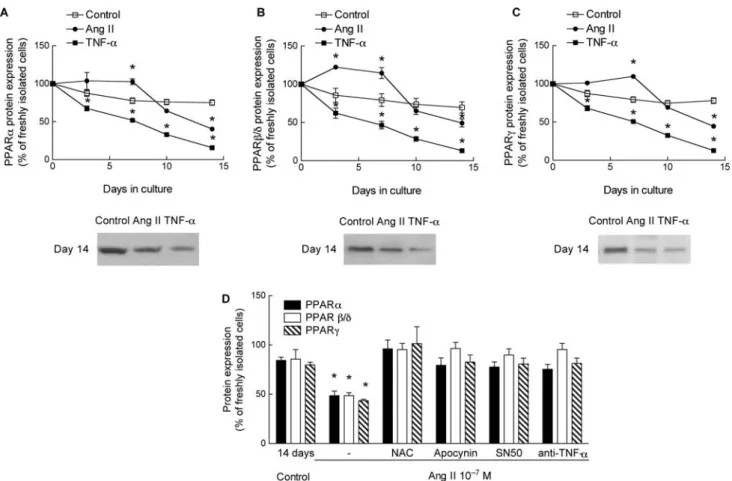
Because several previous studies have implicated reduction of transcriptional activity of PPARa17,18 and PPARb/d19 in the downregulation of the fatty acid
oxi-dation pathway during myocardial hypertrophy and heart failure, we next evaluated the effect of Ang II on the expression of three PPAR isoforms PPARa, PPARb/d, and PPARg. In control ARC, protein expression of PPARs did not appreciably change during the 14 days culture period (Figures 5A–C).
Ang II elicited a modest, dose-dependent, reduction of protein expression of PPARa, PPARb/d, and PPARg (Figures 5A–C; Supplementary material online, Figure S2) on day 14. Interestingly, during the first 5 to 7 days of culture expression of PPAR proteins was even slightly higher in ARC exposed to Ang II than in control ARC. In
Figure 2 Both Ang II and tumour necrosis factor-a (TNF-a) reduce expression of key regulatory proteins of fatty acid oxidation, but with different time-course. (A–C ) Time-course of protein expression of FAT/CD36 (A), mCPT-I (B), and MCAD (C) in control adult rat cardiac (ARC) (open squares), ARC exposed to Ang II (filled squares), or ARC exposed to TNF-a (filled circles) during the 14-day culture period. (D–F ) mRNA expression of FAT/CD36 (D), mCPT-I (E), and MCAD (F). Values in (A) to (F) are expressed as percentage of values measured in freshly isolated ARC. (G) Oxidation of [14C]-palmitate measured after different culture intervals in control ARC (open bars), ARC exposed to Ang II (black bars), or ARC exposed to TNF-a (hatched bars). n, at least three determinations for each culture interval and each condition. Values are means + SEM. *P , 0.05 compared with control.
C. Pellieux et al. 344
contrast to Ang II, TNF-a markedly reduced protein expression of the three PPAR isoforms during the entire culture period (Figures 5A–C).
In ARC exposed to Ang II, the addition of TNF-a anti-bodies, or blocking of TNF-a synthesis by apocynin, NAC, or SN50, prevented the late decrease of PPARa, PPARb/d, and
Figure 3 Ang II elicits phosphorylation of NF-kB, downregulation of the fatty acid oxidation pathway, and enhancement of expression of TNF-a in a ROS-dependent manner. (A) Time-course of Ang II- and TNF-a-mediated phosphorylation of the p65 subunit of NF-kB at ser536. (B) Inhibition of ROS accumulation by either NAC or apocynin-blocked phosphorylation of NF-kB. (C ) Binding of TNF-a by an antibody reduced NF-kB phosphorylation during the second week of culture. (D) Inhibition of ROS accumulation by either NAC or apocynin, or inhibition of translocation of NF-kB by SN50 prevented Ang II-mediated reduction of protein expression of FAT/CD36 (open bars), mCPT-I (filled bars), and MCAD (hatched bars). (E) The same interventions also prevented reduction of palmitate oxidation (shown are measurements on day 14). (F ) Ang II dose-and time-dependently stimulated protein expression of TNF-a in ARC. (G) Inhibition of ROS accumulation or blockade of NF-kB translocation by SN50 abolished Ang II-mediated synthesis of TNF-a. n, at least three determinations for each culture interval and each condition (F and G). All values are means + SEM. *P , 0.05 compared with control (14 days),†P , 0.05 compared with Ang II alone.
Figure 4 Neutralization of TNF-a by an antibody prevents downregulation of regulatory proteins of fatty acid oxidation. (A) Ang II-induced reduction of protein expression of FAT/CD36, mCPT-I, and MCAD was completely abolished by addition of anti-TNF-a antibodies to the culture medium during the entire culture period (shown is protein expression on day 14). (B) Reduction of palmitate oxidation was abolished by anti TNF-a (values of day 14). n, at least four determinations for each culture interval and each condition. Values are means + SEM. *P , 0.05 compared with control,†P , 0.05 compared with Ang II alone.
PPARg proteins (Figure 5D). Collectively, the results suggest that protein expression of PPARs is reduced during prolonged exposure to Ang II by release of TNF-a, analogous to obser-vations on FAT/CD36, mCPT-I, and MCAD. Ang II itself seems rather to enhance protein expression of PPARs and counter-act the effect of TNF-a.
3.5 Activation of PPARa and PPARb/d attenuates
the effects of Ang II on the fatty acid
oxidation pathway
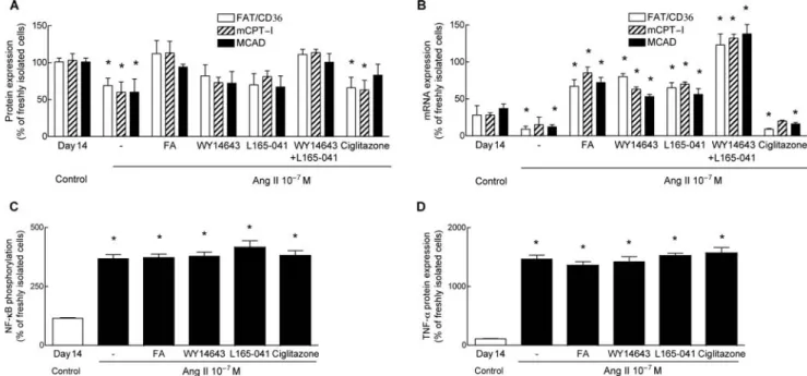
To determine whether ligand-mediated activation of PPARs influences Ang II-induced downregulation of the fatty acid oxidation pathway, we first examined the effect of sup-plementation of the culture medium with fatty acid.26In
ARC exposed to Ang II (100 nM) supplementation of the medium with fatty acid, concentration-dependently atte-nuated the reduction of PPAR protein expression (Sup-plementary material online, Figure S4). The addition of 0.5 mM fatty acid (0.25 mM palmitate plus 0.25 mM oleate) completely prevented the Ang II-induced reduction of protein expression of all three PPAR isoforms (Sup-plementary material online, Figure S4). Fatty acid also pre-vented the reduction of protein expression of FAT/CD36, MCAD, and mCPT-I concentration-dependently (Figure 6A; Supplementary material online, Figure S4), while mRNA expression of regulatory proteins was increased four- to five-fold compared with ARC exposed to Ang II alone (Figure 6B).
To identify the role of individual PPAR isoforms, we sup-plemented the culture medium with specific agonists of either PPARa (WY-14643), PPARb/d (L165-041) or PPARg (ciglitazone) in fatty acid-free standard medium. Specificity of these ligands has been shown previously.26
Activation of either PPARa by WY-14643 or of PPARb/d by L165-041 partially restored protein expression of FAT/CD36, mCPT-I, and MCAD, while combined stimulation of both iso-forms completely restored protein expression (Figure 6A). mRNA expression was concomitantly increased 4- to 10-fold to values exceeding those measured in untreated control ARC (Figure 6B). In contrast, activation of PPARg by ciglitazone had no effect on mRNA and protein expression of fatty acid oxidation genes. Neither fatty acid nor specific agonists prevented Ang II-mediated phosphorylation of NF-kB and enhancement of TNF-a protein expression (Figure 6C and D). The results indicate that combined acti-vation of PPARa and PPARb/d counteracts downregulation the fatty acid oxidation pathway by Ang II, independently of TNF-a synthesis, most likely by the stimulation of tran-scription of regulatory proteins of fatty acid oxidation.
3.6 Effect of inhibition Ang II-mediated generation
of tumour necrosis factor-a and activation of PPARs
on morphology and brain natriuretic peptide
expression of adult rat cardiac
In Ang II-treated ARC, scavenging of ROS by NAC, inhibition of NF-kB translocation to the nucleus by SN50, or binding
Figure 5 Effect of Ang II and TNF-a on protein expression of PPARa (A), PPARb/d (B), and PPARg (C ). n, at least three determinations for each culture interval and each condition. (D) Inhibition of ROS accumulation by NAC or apocynin, blockade of nuclear translocation of NF-kB by SN50, or addition of TNF-a anti-bodies prevented downregulation of all three PPAR isoforms (day 14 of culture). n, at least four determinations for each condition. Values are means + SEM. *P , 0.05 compared with control.
C. Pellieux et al. 346
of TNF-a by anti-TNF-a antibodies reduced cell surface and BNP mRNA to values comparable to untreated control ARC (Supplementary material online, Figure S5). Thus, interven-tions that prevent Ang II-mediated downregulation of the fatty acid oxidation pathway by inhibition of TNF-a expression also attenuate the hypertrophic response and BNP expression (Supplementary material online, Figure S5). On the other hand, selective activation of PPARa and PPARb/d, which prevent reduction of regulatory proteins of fatty acid oxidation, had no effect on cell size and BNP mRNA. However, selective activation of PPARg and non-selective activation of all PPAR isoforms by fatty acids pre-vented the increase in cell surface area and upregulation of mRNA expression of BNP and ANF (Supplementary material online, Figures S4 and S5) in ARC exposed to Ang II. Thus, in our study activity of PPARg, but not activity of PPARa and PPARb/d seem to modulate the hypertrophic phe-notype induced by Ang II.
4. Discussion
Ang II has been ascribed a central role in the phenotype modification of cardiac myocytes during pathological ventri-cular remodelling and heart failure. We have previously reported that failing hearts of mice with targeted overex-pression of angiotensinogen in the myocardium exhibited marked downregulation of fatty acid oxidation enzymes.9 Our present study indicates that prolonged exposure of ARC myocytes to Ang II is sufficient to reduce expression of key regulatory proteins of fatty acid oxidation including FAT/CD36, mCPT-I, and MCAD. This effect of Ang II on meta-bolic regulation is generated indirectly by stimulation of synthesis of TNF-a in the cardiac myocytes in response to ROS-induced activation of NF-kB.
The involvement of TNF-a in Ang II-mediated downregula-tion of the fatty acid oxidadownregula-tion pathway is supported by at least three observations of the present study: first, the onset of downregulation of regulatory proteins of fatty acid oxidation coincides with the onset of Ang II-induced generation of TNF-a. Second, the addition of TNF-a to the culture medium resulted in rapid reduction of protein expression of regulatory proteins of fatty acid oxidation. Third, addition of anti-TNF-a antibodies to Ang II-stimulated ARC completely blocked the effect of Ang II on the fatty acid oxidation pathway. To date, very little is known on the effects of TNF-a on metabolic regulation in the myocar-dium. Consistent with our results, Sekiguchi et al.31have observed in 8-week-old mice with cardiac-restricted overex-pression of TNF-a that mRNA exoverex-pression of FAT, mCPT-I, and very long-chain acyl-CoA dehydrogenase was reduced. Although protein expression has not been reported in their study, palmitate oxidation, measured in myocardial hom-ogenate, was reduced, compatible with concomitant reduction of regulatory proteins.
Previous studies have demonstrated that TNF-a biosyn-thesis is stimulated in cardiac myocytes in response to Ang II.27In contrast to observations in adult feline cardiac myo-cytes cultured in serum-free medium,27we did not detect
expression of TNF-a in ARC for 3 to 10 days after the onset of exposure to Ang II. The time-delay was dose-dependent and decreased with increasing concentration of Ang II. It needs to be emphasized that in the present study ARC were cultured with 20% FCS, which may alter the cellular response to Ang II. Using this culture condition, ARC undergo during the first 2 to 3 days a process of dedifferen-tiation with phenotypic changes, which include disassembly of the contractile elements,32followed by redifferentiation with resumption of contraction between day 4 and 7.33Ang II
Figure 6 Activation of PPARa and PPARb/d, but not of PPARg, increased protein and mRNA expression of regulatory proteins of fatty acid oxidation without reduction of TNF-a. (A) Supplementation of the culture medium with fatty acid (FA; 50% palmitate/50% oleate) or with specific ligands of PPARa (WY14643) or PPARb/d (L165-041) increased protein expression of FAT/CD36 (open bars), mCPT-I (hatched bars), and MCAD (filled bars). Activation of PPARg by ciglitazone had little effect. (B) Fatty acid, WY14643, and L165-041 markedly increased mRNA expression of regulatory proteins to levels exceeding control. (C and D) Acti-vation of PPARs did neither reduce Ang II-mediated phosphorylation of NF-kB (C), nor induction of TNF-a protein expression (D). Data of all panels were measured on day 14. n, at least four determinations for each condition. Values are means + SEM. *P , 0.05 compared with control.
signalling was not abrogated during the first week of culture, since Ang II-mediated phosphorylation of NF-kB and stimu-lation of protein synthesis were not abolished. Reduction of FCS to 1% shortened the time-delay of Ang II-induced reduction of regulatory proteins of fatty acid oxidation and palmitate oxidation rate, indicating that some component(s) of the serum may protect fatty acid oxidation from Ang II-induced downregulation. The mechanism remains unknown. Noteworthy, reduction of FCS to 1% did neither alter the ultimate extent of reduction of regulatory proteins of fatty acid oxidation and palmitate oxidation rate, nor the reversal by anti-TNF-a antibodies. Therefore, the concen-tration of FCS seems not critical for the ultimate effects of Ang II on the fatty acid oxidation pathway.
Consistent with observations in neonatal rat cardiac myo-cytes,19our results suggest a central role of activation of NF-kB in downregulation of the fatty acid oxidation pathway. Inhibition of Ang II-mediated activation or translo-cation of NF-kB by apocynin, NAC or SN50 completely pre-vented reduction of regulatory proteins of fatty acid oxidation. NF-kB has been proposed to lower fatty acid oxi-dation by direct interaction with PPARs which regulate tran-scription of target genes involved in fatty acid oxidation (transrepression).19
NF-kB is activated by receptor-initiated signalling cas-cades of both Ang II, via the AT-1 receptor,34 and TNF-a, via TNF-a receptor 1.35Furthermore, Ang II-induced acti-vation of NF-kB stimulates TNF-a synthesis in adult cardiac myocytes,27which may greatly amplify activation of NF-kB during exposure to Ang II. In fact, in our exper-iments, inactivation of TNF-a by anti-TNF-a antibodies did not diminish Ang II-induced phosphorylation of NF-kB on days 1 and 3, but markedly attenuated NF-kB phos-phorylation on day 7 and later, indicating a predominant role of TNF-a in NF-kB activation during the second week of culture. The finding that inactivation of TNF-a by anti-TNF-a antibodies prevents downregulation of the fatty acid oxidation pathway suggests that activation of NF-kB by Ang II alone is sufficient to trigger synthesis of TNF-a, but insufficient to reduce levels of regulatory pro-teins of fatty acid oxidation. The cellular mechanism underlying the necessity of TNF-a for downregulation of the fatty acid oxidation pathway is open to speculation. On one hand, activation of NF-kB by stimulation of the AT-1 receptor alone may be quantitatively insufficient. In fact, TNF-a-independent activation of NF-kB by Ang II, esti-mated during inhibition of TNF-a by antibodies, was mark-edly lower compared with activation without TNF-a inhibition. On the other hand, downregulation of regulatory proteins of fatty acid metabolism may be mediated by NF-kB-independent signals triggered by TNF-a.
Considerable evidence implicates reduction of transcrip-tional activity of PPARa and/or PPARb/d in the downregula-tion of the fatty acid oxidadownregula-tion pathway during progression of maladaptive myocardial remodelling.19Both PPARa and PPARb/d are involved in the regulation of transcriptional activity of genes encoding regulatory proteins of fatty acid metabolism, including FAT/CD36, mCPT-I, and MCAD. Pro-posed mechanisms for inactivation of transcriptional activity of PPARa, which is best characterized as yet, include reduced expression of PPARs,17 posttranslational inacti-vation by Erk1/2-mediated phosphorylation,17 reduced availability of cofactors including PGC-1,36 and increased
expression of the repressor protein chicken ovalbumin upstream promoter transcription factor.18Finally, transcrip-tional activity may be reduced by transrepression by nuclear transcription factors including NF-kB and activator protein-1 (AP-1), as has been shown in phenylephrine-stimulated neo-natal rat cardiac myocytes for PPARb/d.19
In our study, mRNA expression of FAT/CD36, mCPT-I, and MCAD was reduced by Ang II in ARC after 3 days of culture, indirectly suggesting that the transcription was decreased early during exposure to Ang II. Since protein expression of all three PPAR isoforms was decreased only during the second week of exposure to Ang II, the results are compati-ble with the interpretation that inactivation of PPARs pre-ceded reduction of PPAR protein expression.17,18,36Despite early reduction of mRNA expression of FAT/CD36, mCPT-I, and MCAD, protein expression was maintained for several days and dropped only during the second week of culture, when TNF-a was generated. Therefore, it is likely that post-transcriptional mechanisms are critically involved in the regulation of protein expression of FAT/CD36, mCPT-I, and MCAD in ARC and the reduction in response to Ang II. The mechanism of presumed posttranscriptional reduction of regulatory proteins of fatty acid oxidation by TNF-a is not known. However, there is evidence suggesting that TNF-a may enhance degradation of selected proteins by the ubiquitin-proteasome pathway.37
We have previously observed a similar dissociation between myocardial mRNA and protein expression of regulatory pro-teins of fatty acid metabolism in mice with targeted over-expression of angiotensinogen in the myocardium.9 mRNA expression of mCPT-I and MCAD progressively decreased during compensated hypertrophy, whereas protein expression did not change. However, protein expression and palmitate oxidation, measured during isolated heart perfusion in vitro, markedly dropped after the onset of heart failure. A similar observation has been reported by Sack et al.14in spon-taneously hypertensive SHHR/Mcc-facp rats. Although myo-cardial TNF-a content has not been measured in these in vivo studies, our results raise the possibility that the reduction of protein expression of regulatory proteins of fatty acid oxidation and consecutive reduction of metabolic flux may be related to enhanced exposure of the myocardium to TNF-a.38
Although our data indirectly suggest the involvement of posttranslational mechanisms, the results clearly indicate that PPAR activity critically influences both the hypertrophic response and expression of regulatory proteins of fatty acid oxidation in ARC exposed to Ang II. Supplementation of the culture medium with 0.5 mM fatty acid (50% oleate/50% pal-mitate) and selective activation of PPARa and PPARb/d, but not activation of PPARg, restored mRNA and protein expression of regulatory proteins of fatty acid metabolism, and prevented reduction of palmitate oxidation rate.
Activation of PPARa and PPARb/d may prevent reduction of regulatory proteins of fatty acid oxidation by at least two different mechanisms. The first mechanism is direct stimulation of transcription of target genes by binding to peroxisome proliferator responsive elements in the promo-ter region. Consistent with this possibility, mRNA expression of regulatory genes of fatty acid metabolism was increased 4- to 10-fold concomitantly with the restoration of protein expression. The second mechanism is direct interaction with transcription factors that are involved in the
C. Pellieux et al. 348
hypertrophic response, including NF-kB and AP-1, which potentially may, directly or indirectly, contribute to reduction of proteins of fatty acid oxidation.39Consistent with this concept, some studies have provided evidence that PPARa reduces TNF-a in the myocardium exposed to hypertrophic40 or inflammatory stimuli,41 apparently by inhibition of NF-kB.41 However, in our study, we did not observe a reduction of TNF-a during specific or non-specific stimulation of either PPAR isoform in ARC exposed to Ang II. Therefore, our observations are more consistent with the interpretation that stimulation of transcription during acti-vation of PPARa and PPARb/d compensated for both tran-scriptional and posttrantran-scriptional reduction of regulatory proteins of fatty acid oxidation. This mechanism of restor-ation of regulatory proteins of fatty acid metabolism differs from that effective during restoration of the fatty acid oxidation pathway by NAC, apocynin, or SN50, which all reduced synthesis of TNF-a by prevention of activation and nuclear translocation of NF-kB.
In summary, the findings of this study demonstrate for the first time that prolonged exposure of cardiac myocytes to Ang II may alter substrate metabolism in cardiac myocytes, characterized by reduction of the fatty acid oxidation capacity. The effect is largely mediated by the inflammatory cytokine TNF-a. The observation may be relevant for the understanding of the cellular mechanisms underlying the shift from fatty acid to glucose metabolism observed in the myocardium under conditions of ventricular remodelling and heart failure.7,9,10,14 A critical question remains whether limitation of fatty acid oxidation is an adaptive response or may contribute to further damage of the myocardium.
Supplementary material
Supplementary material is available at Cardiovascular Research online.
Conflict of interest: none declared.
Funding
This work was supported by the Swiss National Science Foun-dation (Grant 3200-067873 to R.L. and Grant 3100B0109212/ 1 to R.L.), the Swiss University Conference project ‘Heart remodellling in Health and Disease’, the Swiss Heart Foun-dation, the Foundation ‘Centre de Recherche Me´dicale Carlos et Elsie de Reuter’, and the Foundation ‘Valentine Gerbex-Bourget’.
References
1. Rosenblatt-Velin N, Montessuit C, Papageorgiou I, Terrand J, Lerch R. Postinfarction heart failure in rats is associated with upregulation of GLUT-1 and downregulation of genes of fatty acid metabolism. Cardio-vasc Res 2001;52:407–416.
2. Remondino A, Rosenblatt-Velin N, Montessuit C, Tardy I, Papageorgiou I, Dorsaz PA et al. Altered expression of proteins of metabolic regulation during remodeling of the left ventricle after myocardial infarction. J Mol Cell Cardiol 2000;32:2025–2034.
3. Young ME, Laws FA, Goodwin GW, Taegtmeyer H. Reactivation of peroxi-some proliferator-activated receptor alpha is associated with contractile dysfunction in hypertrophied rat heart. J Biol Chem 2001;276: 44390–44395.
4. Allard MF, Schonekess BO, Henning SL, English DR, Lopaschuk GD. Contri-bution of oxidative metabolism and glycolysis to ATP production in hyper-trophied hearts. Am J Physiol 1994;267:H742–H750.
5. el Alaoui-Talibi Z, Landormy S, Loireau A, Moravec J. Fatty acid oxidation and mechanical performance of volume-overloaded rat hearts. Am J Physiol 1992;262:H1068–H1074.
6. Osorio JC, Stanley WC, Linke A, Castellari M, Diep QN, Panchal AR et al. Impaired myocardial fatty acid oxidation and reduced protein expression of retinoid X receptor-alpha in pacing-induced heart failure. Circulation 2002;106:606–612.
7. Lei B, Lionetti V, Young ME, Chandler MP, d’Agostino C, Kang E et al. Paradoxical downregulation of the glucose oxidation pathway despite enhanced flux in severe heart failure. J Mol Cell Cardiol 2004;36: 567–576.
8. Chandler MP, Kerner J, Huang H, Vazquez E, Reszko A, Martini WZ et al. Moderate severity heart failure does not involve a downregulation of myocardial fatty acid oxidation. Am J Physiol Heart Circ Physiol 2004; 287:H1538–H1543.
9. Pellieux C, Aasum E, Larsen TS, Montessuit C, Papageorgiou I, Pedrazzini T et al. Overexpression of angiotensinogen in the myocardium induces downregulation of the fatty acid oxidation pathway. J Mol Cell Cardiol 2006;41:459–466.
10. Stanley WC, Recchia FA, Lopaschuk GD. Myocardial substrate metabolism in the normal and failing heart. Physiol Rev 2005;85:1093–1129. 11. Kusuoka H, Marban E. Mechanism of the diastolic dysfunction induced by
glycolytic inhibition. Does adenosine triphosphate derived from glycolysis play a favored role in cellular Ca2þ homeostasis in ferret myocardium? J Clin Invest 1994;93:1216–1223.
12. Lopaschuk GD. Cardiac energy metabolism alterations in angiotensin II induced hypertrophy. J Mol Cell Cardiol 2006;41:418–420.
13. Finck BN, Han X, Courtois M, Aimond F, Nerbonne JM, Kovacs A et al. A critical role for PPARalpha-mediated lipotoxicity in the pathogenesis of diabetic cardiomyopathy: modulation by dietary fat content. Proc Natl Acad Sci USA 2003;100:1226–1231.
14. Sack MN, Rader TA, Park S, Bastin J, McCune SA, Kelly DP. Fatty acid oxi-dation enzyme gene expression is downregulated in the failing heart. Cir-culation 1996;94:2837–2842.
15. Lionetti V, Linke A, Chandler MP, Young ME, Penn MS, Gupte S et al. Car-nitine palmitoyl transferase-I inhibition prevents ventricular remodeling and delays decompensation in pacing-induced heart failure. Cardiovasc Res 2005;66:454–461.
16. de Brouwer KFJ, Degens H, Aartsen WM, Lindhout M, Bitsch NJJE, Gilde AJ et al. Specific and sustained down-regulation of genes involved in fatty acid metabolism is not a hallmark of progression to cardiac failure in mice. J Mol Cell Cardiol 2006;40:838–845.
17. Barger PM, Brandt JM, Leone TC, Weinheimer CJ, Kelly DP. Deactivation of peroxisome proliferator-activated receptor-alpha during cardiac hypertrophic growth. J Clin Invest 2000;105:1723–1730.
18. Sack MN, Disch DL, Rockman HA, Kelly DP. A role for Sp and nuclear recep-tor transcription facrecep-tors in a cardiac hypertrophic growth program. Proc Natl Acad Sci USA 1997;94:6438–6443.
19. Planavila A, Laguna JC, Vazquez-Carrera M. Nuclear factor-kappaB acti-vation leads to down-regulation of fatty acid oxidation during cardiac hypertrophy. J Biol Chem 2005;280:17464–17471.
20. Finck BN, Kelly DP. Peroxisome proliferator-activated receptor gamma coactivator-1 (PGC-1) regulatory cascade in cardiac physiology and disease. Circulation 2007;115:2540–2548.
21. Serneri GG, Boddi M, Cecioni I, Vanni S, Coppo M, Papa ML et al. Cardiac angiotensin II formation in the clinical course of heart failure and its relationship with left ventricular function. Circ Res 2001;88: 961–968.
22. Bouzegrhane F, Thibault G. Is angiotensin II a proliferative factor of cardiac fibroblasts? Cardiovasc Res 2002;53:304–312.
23. Mann DL. Angiotensin II as an inflammatory mediator: evolving concepts in the role of the renin angiotensin system in the failing heart. Cardiovasc Drugs Ther 2002;16:7–9.
24. Mazzolai L, Pedrazzini T, Nicoud F, Gabbiani G, Brunner HR, Nussberger J. Increased cardiac angiotensin II levels induce right and left ventricular hypertrophy in normotensive mice. Hypertension 2000;35:985–991. 25. Montessuit C, Rosenblatt-Velin N, Papageorgiou I, Campos L, Pellieux C,
Palma T et al. Regulation of glucose transporter expression in cardiac myocytes: p38 MAPK is a strong inducer of GLUT4. Cardiovasc Res 2004;64:94–104.
26. Pellieux C, Montessuit C, Papageorgiou I, Lerch R. Inactivation of peroxi-some proliferator-activated receptor isoforms alpha, beta/delta, and
gamma mediate distinct facets of hypertrophic transformation of adult cardiac myocytes. Pflugers Arch 2007;455:443–454.
27. Kalra D, Sivasubramanian N, Mann DL. Angiotensin II induces tumor necrosis factor biosynthesis in the adult mammalian heart through a protein kinase C-dependent pathway. Circulation 2002;105:2198–2205. 28. Mann DL. Inflammatory mediators and the failing heart: past, present,
and the foreseeable future. Circ Res 2002;91:988–998.
29. Purcell NH, Tang G, Yu C, Mercurio F, DiDonato JA, Lin A. Activation of NF-kappa B is required for hypertrophic growth of primary rat neonatal ventricular cardiomyocytes. Proc Natl Acad Sci USA 2001;98:6668–6673. 30. Cui R, Tieu B, Recinos A, Tilton RG, Brasier AR. RhoA mediates angiotensin II-induced phospho-Ser536 nuclear factor kappaB/RelA subunit exchange on the interleukin-6 promoter in VSMCs. Circ Res 2006;99:723–730. 31. Sekiguchi K, Tian Q, Ishiyama M, Burchfield J, Gao F, Mann DL et al.
Inhi-bition of PPAR-alpha activity in mice with cardiac-restricted expression of tumor necrosis factor: potential role of TGF-beta/Smad3. Am J Physiol Heart Circ Physiol 2007;292:H1443–H1451.
32. Schaub MC, Hefti MA, Harder BA, Eppenberger HM. Various hypertrophic stimuli induce distinct phenotypes in cardiomyocytes. J Mol Med 1997; 75:901–920.
33. Rosenblatt-Velin N, Lerch R, Papageorgiou I, Montessuit C. Insulin resist-ance in adult cardiomyocytes undergoing dedifferentiation: role of GLUT4 expression and translocation. FASEB J 2004;18:872–874. 34. Zahradka P, Werner JP, Buhay S, Litchie B, Helwer G, Thomas S.
NF-kappaB activation is essential for angiotensin II-dependent
proliferation and migration of vascular smooth muscle cells. J Mol Cell Cardiol 2002;34:1609–1621.
35. Gloire G, Legrand-Poels S, Piette J. NF-kappaB activation by reactive oxygen species: fifteen years later. Biochem Pharmacol 2006;72:1493–1505. 36. Garnier A, Fortin D, Delomenie C, Momken I, Veksler V, Ventura-Clapier R.
Depressed mitochondrial transcription factors and oxidative capacity in rat failing cardiac and skeletal muscles. J Physiol 2003;551: 491–501.
37. Li YP, Lecker SH, Chen Y, Waddell ID, Goldberg AL, Reid MB. TNF-alpha increases ubiquitin-conjugating activity in skeletal muscle by up-regulating UbcH2/E220 k. FASEB J 2003;17:1048–1057.
38. Torre-Amione G, Kapadia S, Lee J, Durand JB, Bies RD, Young JB et al. Tumor necrosis factor-alpha and tumor necrosis factor receptors in the failing human heart. Circulation 1996;93:704–711.
39. Planavila A, Rodriguez-Calvo R, Jove M, Michalik L, Wahli W, Laguna JC et al. Peroxisome proliferator-activated receptor beta/delta activation inhibits hypertrophy in neonatal rat cardiomyocytes. Cardiovasc Res 2005;65:832–841.
40. Smeets PJ, Teunissen BE, Willemsen PH, van Nieuwenhoven FA, Brouns AE, Janssen BJ et al. Cardiac hypertrophy is enhanced in PPARfalphag2/2 mice in response to chronic pressure overload. Cardio-vasc Res 2008;78:79–89.
41. Takano H, Nagai T, Asakawa M, Toyozaki T, Oka T, Komuro I et al. Peroxisome proliferator-activated receptor activators inhibit lipopolysaccharide-induced tumor necrosis factor-a expression in neo-natal rat cardiac myocytes. Circ Res 2000;87:596–602.
C. Pellieux et al. 350