© 1985 by the Bishop Museum
CONCENTRATION OF HOST BLOOD PROTEIN DURING FEEDING BY
ANOPHELINE MOSQUITOES (DIPTERA: CULICIDAE)
1Hans Briegel and Licia Rezzonico2 Abstract. During blood feeding, Anopheles stephensi ingested
2 to 10 n\ of blood. It took such large meals by releasing large amounts of a red rectal fluid as it fed. Although the fluid con-tained intact erythrocytes, the female was able to concentrate the dietary protein in the midgut by a factor of about 2. Two larger species, An. albimanus and An. quadrimaculatus, consum-ing larger meals, released a clear rectal fluid and also concen-trated their blood meals without losing protein. Therefore, gravimetric determinations of blood consumption would be mis-leading in Anopheles. Concentration of blood proteins during the act of feeding, before diuresis, is unique for Anopheles and led to increased fecundity when compared to blood meals given by enema. Similar to results with Aedes, fecundity was signifi-cantly lower with human than with guinea pig blood. It was further demonstrated that female Anopheles actually refed on successive days when a host was offered. In contrast to Aedes, subsequent blood meals produced an increase in the number of maturing oocytes.
Despite their important role as primary vectors of plasmodia causing primate malaria, female
Anopheles have received comparatively little
atten-tion from insect physiologists, possibly because ae-dine and culicine mosquitoes are more easily reared and handled. Aedine and culicine mosquitoes gen-erally require 1 blood meal for each gonotrophic cycle, while anopheline mosquitoes in the labora-tory are routinely fed at least twice per gonotrophic cycle, sometimes even daily (Gerberg 1970, Hors-fall 1972). To our knowledge, however, the phys-iological implications of these feeding regimes have never been analyzed.
As in many hematophagous insects, e.g.,
Rhod-nius (Maddrell 1964) and Glossina (Gee 1975), a
conspicuous diuresis begins in mosquitoes shortly after ingestion of blood (Boorman 1960, Stobbart 1977, Jones & Brandt 1981, Williams et al. 1983). Nijhout & Carrow (1978) have investigated diuresis in Anopheles freeborni Aitken and ascribed its con-trol to a diuretic hormone. In all these studies the experimental analysis of diuresis began with ces-sation of blood feeding and continued for roughly 1 h thereafter. In Anopheles, however, excretion of
1. This project was supported by grants from the Swiss Na-tional Science Foundation.
2. Department of Zoology, University of Zurich, CH-8057 Zurich, Switzerland.
rectal fluid starts a few seconds after piercing the skin of the host and ingestion of blood. A notable occurrence during the feeding of colonies of An.
stephensi Liston is the excretion of a bright red
fluid. For convenience we will call these excretions rectal fluid, because their origin has not been in-vestigated. Excretion of rectal fluid during feeding is arbitrarily defined as prediuresis, and aqueous excretion after feeding has stopped as diuresis.
In this report we will analyze prediuretic excre-tion quantitatively and compare it among 3 anoph-eline species of different subgenera and body size. At the same time, the results will be related to fecundity of Anopheles, and possible behavioral ad-aptations will be discussed. It is particularly inter-esting to compare our results with quantitative data reported for Ae. aegypti, the yellow fever mosquito (Briegel 1985).
MATERIALS AND METHODS
The following species were used for our
exper-iments: Anopheles (Cellia) stephensi, An. (Nyssorhyn-chus) albimanus Wiedemann, and An. {Anopheles) quadrimaculatus Say. Larvae were reared on a
high-protein diet (Lea 1964), with 300 larvae per pan in 450 ml distilled water. The adults eclosed within a 2-day period and were of uniform size. They were kept continuously at 27 °C, 85% RH, under long-day conditions (16L:8D), with sunset and sunrise simulated by continuous dim light for 30 min.
Experimental blood meals were given either in measured volumes by enema (Briegel & Lea 1975) or by feeding the females individually on a re-strained guinea pig until repletion, i.e., until with-drawal of the proboscis (generally between 1 and
10 min).
For total nitrogen measurements, the excretory droplets were accumulated on a piece of cover slip held with forceps below the anus; evaporation was considered irrelevant for these determinations. For the hemoglobin determinations, however, a small glass vial containing 500 /zl of water was held near the anus to catch all rectal fluid released during feeding and to prevent denaturation of the
he-moglobin. After the experimental blood meals, fe-males were kept individually in reacting tubes (10 x 75 mm). For the first 24 h they were kept without water; then they were provided with cot-ton pads soaked in 10% sucrose solution for the rest of the experiment. This combination kept mortality below 5-10%. After 2 or 3 days the ova-ries were dissected in saline and the mature oocytes counted; in the experiments on fecundity, ovaries were subsequently used for nitrogen determina-tions.
In several experiments the volume of the blood meal taken by feeding was estimated by weighing individual females before and after on a micro-balance (Cahn DTL). For this purpose the females were anesthetized weakly with CO2; only those
re-covering instantly as indicated by avid biting activ-ity were used for the experiments. Some of the females were used for midgut dissection to measure total nitrogen. Dissection was carried out in saline (0.6%), and each midgut was analyzed individually after briefly washing it in a new drop of saline. Since the nitrogen content of the epithelium was below 2% of the blood bolus (unpubl. observ.), it was considered negligible.
Total nitrogen of the samples was determined through Nesslerization of a Kjeldahl digest (Minari & Zilversmit 1963). For the hemoglobin determi-nations we used a Drabkin reagent (Briegel et al. 1978) calibrated with a standard human hemoglo-bin (525-18, Sigma Chemical Co., St. Louis, MO). In most experiments the same guinea pig was used as a host. The variations in its blood nitrogen or hemoglobin titers were averaged through periodic sampling by heart puncture, performed under an-esthesia.
Linear regression lines for the data were com-puted using a commercially available program (HP-67).
RESULTS
Blood meal and simultaneous excretion of rectal fluid
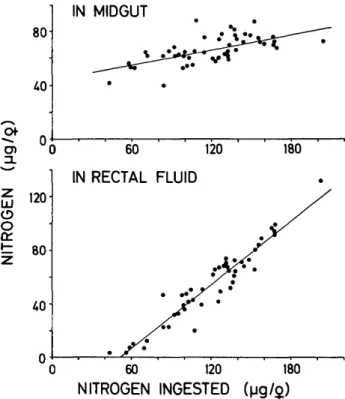
Forty-eight An. stephensi females were fed indi-vidually to repletion on a restrained guinea pig. The total nitrogen was compared in (1) the pooled droplets of rectal fluid collected during feeding, (2) the blood in the midgut, and (3) a sample of blood from the host. The data and the linear regressions are presented in Fig. 1, where the mid-gut nitrogen and excretory nitrogen are each plot-ted against the nitrogen content of the complete
80 40-IN MIDGUT ^ 0en 0
S
120 o o 1= 80 40-60 120 180 RECTAL FLUID 0 60 120 180 NITROGEN INGESTED (|jg/$)FIG. 1. Nitrogen retained in the midgut of An. stephensi or excreted with rectal fluid during feeding to repletion on a restrained guinea pig. Nitrogen ingested is the sum of the measurement from the midgut plus rectal fluid. Each point represents 19; linear regressions are given for midgut (r = 0.661) and for rectal fluid (r = 0.958) from 489.
blood meal (i.e., excretory plus intestinal nitrogen). We assumed that no substantial protein was de-graded or absorbed during the feeding and col-lecting period. The maximal amount of intestinal nitrogen was about 80 fxg/9 (Fig. 1). This figure further indicates that rectal fluid was not released before 40 to 60 ixg of nitrogen had been retained in the midgut. Since the nitrogen concentration of the host blood was 19.82 ± 1.44 Mg//il (n = 5), the maximal sum of nitrogen (about 200 /tig) indicated a blood consumption of up to 10 /il.
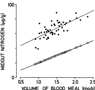
In Fig. 2 the midgut nitrogen (upper line) is com-pared with the volume of blood ingested, as de-termined by weight gain of these same females (lower line). Generally speaking, all midgut nitro-gen measurements were consistently about 30 ng higher than the extrapolation based on the weight increase and the host blood nitrogen (lower line). Thus blood meal nitrogen titers increased during feeding, and weighing the females underestimated the blood consumption substantially.
Total blood consumption per female was cal-culated by adding the average midgut and excre-tory nitrogen data and then dividing by the
nitro-1001 LU O O O Q a5 1.0 1.5 2.0 2.5 VOLUME OF BLOOD MEAL (mg/$) FIG. 2. Midgut nitrogen in An. stephensi after feeding to repletion on a guinea pig (•) and the amount of nitrogen ingested as calculated from the weight gains of 489 and the nitrogen content in the host blood (O). Each point represents 19 and linear regressions are given (r = 0.72 for • ; r = 0.96 for O).
gen concentration in the host blood (Table 1). This revealed an average blood consumption of 6 /nl/9
An. stephensi, with individual variations between 2
and 10
Mi-Dividing the average midgut nitrogen by the weight of the blood (after its conversion to nitro-gen) led to the concentration factor of 2.3. The loss of nitrogen through simultaneous excretion of rectal fluid varied tremendously between 2 and 130 Mg/2; the average of 54 ^g was 45% of the total blood consumption.
When we gave measured amounts of blood by enema to An. stephensi in 0.5-^1 increments from 0.5 to 3 ix\, we found that blood meals larger than 2 n\ could never be injected without rupturing the midgut. Obviously, this quantity was the maximal volume of the distended abdominal midgut for this species reared under our conditions. This volume also coincided with the amount of blood retained in the midgut before the prediuretic excretion of rectal fluid began (see above). Relating this to the range of blood consumption of 2 to 10 fi\ (Table 1), it is evident that a substantial concentration of the blood protein took place in most females.
Since 80% of the vertebrate blood nitrogen oc-curs in the hemoglobin (Bursell 1965), and because of the red color of the rectal fluid, this experiment was repeated, this time measuring the hemoglobin in the pooled fluid, the midgut, and host blood samples. Exactly the same result was obtained (Fig. 3). At the end of feeding, the midgut contained an average of 499.9 jtig hemoglobin (range 313-676
TABLE 1. Comparison of the blood meal concentration among 3 anopheline species. All values are expressed as microlitres or were converted to micrograms of nitrogen, where applicable. N = nitrogen.*
Ml/8 Wt. of blood/9** Mean (A) 1.4 Range 0.9-2.4 N/midgut Mean (B) Range
N excreted during feeding Mean (C)
Range
Amt. of blood ingested/9 Mean (B + C) 6.0 Range 2.2-10 Cone, factor Mean (B:A) Range An. stephensi (n = 489) MgN 28.5 65.7 38.9-87.1 53.8 2.3-130.5 119.5 .2 2.3 1.4-3.1 An. albimanus (n Ml/2 1.5 0.6-2.8 2.9 1.5-4.2 = 579) MgN 29.3 54.7 28.9-81.5 1.7*** 1.3-2.8 56.4 1.9 1.0-2.8 An. Ml/8 2.1 1.3-3.4 4.4 3.0-6.3 quadrimaculatus (n = 449) MgN 40.6 87.1 58.9-123.9 — 87.4 2.2 1.5-3.1 * All 99 were fed on the same guinea pig with a nitrogen content of 19.82 ± 1.44 ng/n\ blood (n = 5).
** Increase of 9 fresh weight assumed to equal volume of blood retained in the midgut. ***99.
8001 400" ZL IN MIDGUT 0 600 1200 IN RECTAL FLUID 1800 g 1200 ]
d
o LU 800 X 400 1800 0 600 1200 HEMOGLOBIN INGESTEDFIG. 3. Hemoglobin retained in the midgut of An. stephensi and excreted with rectal fluid during feeding to repletion on a restrained guinea pig. Hemoglobin ingested is the sum of the measurement from midgut plus rectal fluid. Each point represents 12; linear regressions are given for midgut (r = 0.633) and for rectal fluid (r = 0.949) from 839.
Mg, n = 30), while in the corresponding samples of rectal fluid we detected an average of 407.5 /tg (range 12-930 fig). Based on the hemoglobin titer in the host blood (142.6 ± 3.5 Mg/^l, n = 3), the average total hemoglobin ingested was roughly 6.5
fx\ of guinea pig blood (range 2.7-14.0 n\/2), while
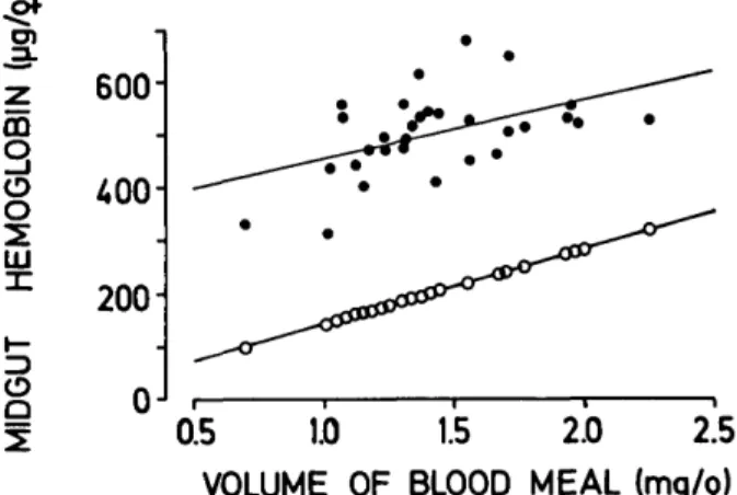
only a volume of 1.4 to 2 n\ was retained in the midgut. When the midgut hemoblogin was com-pared to the values obtained by weighing the fe-males (Fig. 4), it was ca. 300 ng higher than the extrapolated values. Thus, the error in estimating blood ingestion by gravimetry is comparable to the one noted before with nitrogen determinations.
To prove that the red rectal fluid excreted by
An. stephensi during feeding represented excess
blood, the following experiments were carried out. Giemsa-stained blood smears were prepared from the midgut contents immediately after feeding ended, as well as from the rectal fluid, and both were compared with smears from native host blood. Under the microscope, they all appeared identical, i.e., the rectal fluid contained intact erythrocytes, although at a much smaller density per optical field than the midgut preparation or the host blood (ca. 50%). Furthermore, at the end of feeding, the
z 180 " IEMOG I j _ DGU T 600 400 200 0 0.5 1.0 1.5 2.0 2.5 VOLUME OF BLOOD MEAL (mg/j) FIG. 4. Midgut hemoglobin in An. stephensi after feeding to repletion on a guinea pig (•) and the amount of hemoglobin ingested as calculated from the weight gains of 302 and the content in the host blood (O). Each point represents 12 and linear regressions are given (r = 0.47 for • ; r = 1.00 for O).
pooled rectal fluid, the entire midgut contents from the same female, and 2 fi\ of host blood were each subjected to an acid hydrolysis (6 N HC1, 130 °C, 13 h), and the molar distribution of the amino acids was compared by the percentage of their totals. All 3 samples had identical relative compositions, in-dicating the absence of selective absorption or re-tention of specific amino acids (or peptides or pro-teins) from the blood meal during feeding.
Comparison with other Anopheles species
Blood feeding and simultaneous prediuretic ex-cretion in An. stephensi was compared with An.
al-bimanus and An. quadrimaculatus. Since in both
species the rectal fluid is clear, apparently lacking any hemoglobin, we measured only total nitrogen in the samples.
Fifty-seven An. albimanus were fed individually on the same guinea pig, and the samples were col-lected as before. The total nitrogen per midgut at the end of feeding averaged 54.7 ng (Table 1), but only 1.7 fxg/9 (n = 9) was recovered in the large volume of rectal fluid, corresponding to less than 5% of the midgut content. The actual blood con-sumption in this species was between 1.5 and 4.2 /itl/2, with an average concentration factor of 1.9. Forty-four An. quadrimaculatus fed on the same guinea pig were treated and analyzed in the same way. The total nitrogen per midgut at the end of feeding was 87 ng, yielding an average blood con-sumption of 4.4 /ul/9; the concentration factor was 2.2 (Table 1). In this experiment the nitrogen ex-cretion was not quantified, but comparing the mid-gut nitrogen with blood consumption indicated that
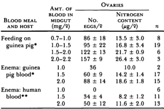
TABLE 2. Fecundity of An. stephensi after blood meal from guinea pig or man, administered by feeding on the host or
by enema (mean ± SE, n = no. determinations).
AMT. OF OVARIES BLOOD MEAL AND HOST Feeding on guinea pig* Enema: guinea pig blood* Enema: human blood** BLOOD IN MIDGUT (mg/9) 0.7-1.0 1.0-1.5 1.5-2.0 2.0-2.2 1.0 1.5 2.0 1.0 1.5 2.0 N o . EGGS/9 86 ± 18 95 ± 22 122 ± 13 157 ± 9 36 60 ± 9 88 ± 14 0 34 ± 4 50 ± 12 NITROGEN CONTENT 0*g/») 13.5 ± 3.0 16.8 ± 3.4 21.7 ± 0.9 26.4 ± 3.0 10.0 14.2 ± 1.4 18.6 ± 1.8 8.2 ± 1.2 11.6 ± 2.0 n 8 19 6 3 2 17 15 6 11 13 * Nitrogen content: 25.7 ± 0.9 Mg/f*l (n = 5). ** Nitrogen content: 33.4 ± 1.8 fig/fi\ (n = 5).
nitrogen excreted was less than 1% of the nitrogen ingested (Table 1).
Table 1 further demonstrates that the size range of blood meals ingested was related to the size of the mosquitoes. Anopheles quadrimaculatus, the largest species tested, ingested 1.3 to 3.4 mg of blood (50% of all females between 1.6 and 2.2 mg), whereas An. stephensi, the smallest species, retained only 0.9 to 2.4 mg blood (50% of all females be-tween 1.2 and 1.6 mg). Conversely, the smallest species showed the highest consumption (2 to 10
li\), accompanied by the highest loss of nitrogen
through prediuretic excretion, i.e., 2-131
Significance of blood meal concentration for fecundity
To test the effect of prediuretic blood meal con-centration on fecundity, 36 An. stephensi were fed to repletion on a guinea pig, while 34 other females were given enemas. In addition, 30 females were given human blood by enema. The number of ma-ture oocy tes was counted and compared to the mid-gut blood content and to the ovarian nitrogen (Ta-ble 2).
Fecundity was doubled when blood was taken by feeding as opposed to an enema of guinea pig blood. Enemas of human blood, on the other hand, led to a clearly reduced fecundity, despite its higher nitrogen content. Utilization of the dietary nitro-gen was 47 to 51 % with enemas of guinea pig blood, but only 16 to 17% with enemas of human blood. To our surprise, the average nitrogen content per egg varied considerably, depending on how the blood meal was acquired: lower after feeding
(guinea pig: 0.17 ± 0.02 Mg/egg, n = 36), and significantly higher when given by enema (guinea pig: 0.23 ± 0.03 Mg/egg, n = 34; human: 0.24 ± 0.03 Mg/egg, n = 24; P < 0.001 for both hosts).
To our knowledge, the usual practice of feeding
Anopheles in the laboratory daily never had been
subjected to a physiological analysis. Therefore, we tried to clarify whether females having fed once would actually refeed the following day, and whether multiple blood meals were required to complete 1 gonotrophic cycle. Of 25 An. stephensi fed to repletion on a guinea pig and thus containing a concentrated blood meal, 17 (68%) refed 24 h later. In another experiment, the 1st blood meals were given by enema (0.5, 1.0, 1.5, and 2.0 n\ of guinea pig blood). Twenty-four hours later these females were individually exposed to the guinea pig, and from 50% (initially given 2 /KI) to 100% (given 0.5 /ul) refed readily. Obviously, the lack of blood concentration owing to the enema technique did maintain biting activity.
In a similar experiment with An. albimanus, the 1st meals were enemas of 0.5 to 3.0 /xl guinea pig blood. None of the females receiving 2.5 or 3 /ul refed the next day; however, females initially given 0.5 to 1.5 /tl refed the following day, and even refed a 2nd time (i.e., 48 h after the enemas). Each of these blood meals led to an increase in the egg counts. For example, a female given only 0.5 /ul matured 17 eggs, while after a 2nd meal by feeding, her sister matured 82 eggs; a 3rd meal by feeding produced 128 eggs. In a case with a 1.5-/ul enema, the 3rd meal produced 185 eggs, in contrast to 52 with the enema alone. Although these results are too scant for statistical analysis, they demonstrate convincingly that fecundity is considerably en-hanced by multiple feedings.
DISCUSSION
In all 3 malaria vectors tested, the average ni-trogen or hemoglobin content of the midgut at the end of feeding was at least double that determined by weighing females. This is the result of predi-uretic excretion, i.e., excretion of serum or serum and erythrocytes as blood is ingested. Therefore, protein is accumulated substantially in the anoph-eline midgut, and the amount of protein available for oogenesis (Table 1, midgut) is comparable to that found in aedine mosquitoes (Briegel 1985). Since excretion starts during feeding as soon as the midgut is filled, it appears to be an overflow of blood from the distended midgut. The maximal volume that the abdominal midgut could hold
without rupturing was found to be 2 ^1 in An.
ste-phensi and 3 fil in An. albimanus, only xh or less of
the amount observed in Ae. aegypti (6-7 fi\; Briegel 1985). Regular diuresis, initiated only after ter-mination of feeding in Aedes (Boorman 1960, Stob-bart 1977, Jones & Brandt 1981, Williams et al.
1983) as well as in Anopheles (Nijhout & Carrow 1978), serves primarily to reduce the flight weight of the newly fed female. The prediuretic excretion reported here, however, seems to be an additional adaptation evolved by these mosquitoes primarily to compensate for the smaller volume of the mid-gut and/or its limited elasticity. Indeed, it is fol-lowed by normal diuresis as reported in the liter-ature.
There is, however, a considerable difference be-tween An. stephensi and the other 2 Anopheles tested:
An. albimanus and An. quadrimaculatus have an
ef-ficient "filtration system," producing a clear rectal fluid containing little or no nitrogen. This is in contrast to the rectal fluid of An. stephensi, which contains erythrocytes. Could this be interpreted as a more primitive evolutionary stage than in other
Anopheles, perhaps comparable to plant feeding in
aphids, which concentrate valuable nutrients by passing excessive fluids?
There are certain disadvantages to this concen-trating mechanism. Besides losing protein, the feeding time of An. stephensi is prolonged consid-erably (up to 10 min or more) when compared to the other Anopheles species or to Aedes (feeding times 1-2 min). A behavioral correlate to this extended feeding might be the biting activity during dusk or night, as is often encountered among anophelines (Horsfall 1972) because reduced defensive behav-ior of sleeping hosts is likely.
So far it remains an open question as to why An.
stephensi cannot prevent the loss of protein. One
possible explanation might be that the peritrophic membrane is formed much later in this than in other species (Freyvogel & Staubli 1965).
Accumulation of protein in the midgut through prediuretic excretion is clearly reflected by fecun-dity: the number of eggs produced per female
Anopheles after ingesting a "full meal" is similar to
or even higher than that in Ae. aegypti (Christo-phers 1960, Briegel 1985). The utilization of the blood meal nitrogen for oogenesis varied between 30 and 40% and was comparable to that in Ae.
aegypti. Fecundity was equally affected by the source
of the blood as it is in Aedes: with human blood, fecundity was reduced by xh as compared to rodent
blood, for the same reasons reported elsewhere
(Briegel 1985). Injecting blood meals by enema, on the other hand, also led to reduced fecundity as compared to feeding, in contrast to Aedes where no differences were observed between the 2 routes (Briegel 1985). This is explained by the absence of prediuretic concentration after enemas.
That in both An. stephensi and An. albimanus most females refed on a host 24 h after a previous blood meal points to 2 other basic differences compared to aedine mosquitoes. First, the humoral inhibition of another blood meal exerted by maturing oocytes (Klowden 1981) seems to be very weak or absent. No other time intervals have been tested yet, but in several cases even a 3rd meal was taken and utilized (see above). Second, as we have indicated before, these subsequent blood meals were utilized for vitellogenesis, which was initiated by the 1st meal. This observation points to different mech-anisms regulating fecundity than in Ae. aegypti, where oosorption was demonstrated to start about 8 to 12 h after the 1st (small) blood meal (Lea et al. 1978).
Acknowledgments. We thank Mr S. Zaba and Mrs R. Haigis
for technical assistance. The amino acid analyses were kindly performed by Dr P. Borner on an automatic amino acid ana-lyzer. We appreciate the critical reading of the manuscript by Dr A. O. Lea and the advice given by the reviewers.
LITERATURE CITED
Boorman, J.P.T. 1960. Observations on the feeding habits of
the mosquito Aedes (Stegomyia) aegypti (L.): the loss of fluid after a blood meal and the amount of blood taken during feeding. Ann. Trop. Med. Parasitol. 54: 8-14.
Briegel, H. 1985. Mosquito reproduction: incomplete
utili-zation of the blood meal protein for oogenesis. J. Insect
Physiol. 31: 15-21.
Briegel, H. & A.O. Lea. 1975. Relationship between protein
and proteolytic activity in the midgut of mosquitoes.y. Insect
Physiol. 21: 1597-1604.
Briegel, H., A.O. Lea&M.J. Klowden. 1978.
Hemoglobinom-etry as a method for measuring blood meal sizes of mos-quitoes (Diptera: Culicidae)./ Med. Entomol. 15: 235-38.
Bursell, E. 1965. Nitrogenous waste products of the tsetse fly, Glossina morsitans. J. Insect Physiol. 11: 993-1001. Christophers, S.R. 1960. Aedes aegypti (L.), the yellow fever
mosquito. Cambridge University Press, Cambridge. 739 p. Freyvogel, T.A. & W. Staubli. 1965. The formation of the
peritrophic membrane in Culicidae. Acta Trop. 22: 118-47.
Gee,J. D. 1975. The control of diuresis in the tsetse fly Glossina austeni: preliminary investigation of the diuretic hormone. J. Exp. Biol. 63: 391-401.
Gerberg, E. J. 1970. Manual for mosquito rearing and experimen-tal techniques. American Mosquito Control Assoc, Selma,
CA. 109 p.
Horsfall, W. R. 1972. Mosquitoes, their bionomics and relation to disease. Hafner Publishing Co., New York. 723 p.
Jones, J.C. & E. Brandt. 1981. Fluid excretion by adult Aedes aegypti mosquitoes./ Insect Physiol. 27: 545-49. Klowden, M. J. 1981. Initiation and termination of
host-seek-ing inhibition in Aedes aegypti durhost-seek-ing oocyte maturation./.
Insect Physiol. 27: 799-803.
Lea, A.O. 1964. Studies on the dietary and endocrine regu-lation of autogenous reproduction in Aedes taeniorhynchus (Wied.)./ Med. Entomol. 1: 40-44.
Lea, A.O., H. Briegel & H.M. Lea. 1978. Arrest, resorption,
or maturation of oocytes in Aedes aegypti: dependence on the quantity of blood and the interval between blood meals.
Physiol. Entomol. 3: 309-16.
Maddrell, S.H.P. 1964. Excretion in the blood-sucking bug, Rhodnius prolixus Stal. II. The normal course of diuresis
and the effect of temperature. J. Exp. Biol. 41: 163-76.
Minari, O. & D.B. Zilversmit. 1963. Use of KCN for
stabili-zation of color in direct Nessleristabili-zation of Kjeldahl digests.
Anal. Biochem. 6: 320-27.
Nijhout, H.F. & G.M. Carrow. 1978. Diuresis after a blood
meal in female Anopheles freeborni. J. Insect Physiol. 24:293-98.
Stobbart, R.H. 1977. The control of the diuresis following a
blood meal in females of the yellow fever mosquito Aedes
aegypti (L.)./. Exp. Biol. 69: 53-85.
Williams, J.C., H.H. Hagedorn & K.W. Beyenbach. 1983.
Dynamic changes in flow rate and composition of urine during post-bloodmeal diuresis in Aedes aegypti (L.).J. Comp.