J. Membrane Biol. 193, 67 78 (2003) DOI: 10.1007/s00232-002-2001-9
The Journal of
Membrane
Biology
9 Springer-Verlag New York Inc. 2003
Topical Review
Function o f K + Channels in the Intestinal Epithelium R. Warth l, J. Barhanin 2
Iphysiologisches lnstitut, Winterthurerstr. 190, 8057 ZUrich, Switzerland
2Institut de Pharmacologie du CNRS, 660 route de Lucioles, 06560 Valbonne Sophia-Antipolis, France Received: I I September 2002
Introduction
Potassium channels play an important role for reab- sorptive and secretory pathways in the gastro-intes- tinal tract. Over the last years molecular identification and functional characterization of K + channels led to a better understanding of physiology and disease of intestinal transport. In gastric parietal cells KCNJ10 (Kir4.1) and heteromeric KCNE2/KCNQI (MirP1/ KvLQT1), K + channels co-localize with the acid- producing H+/K + ATPase in the luminal membrane compartment. Specific K + channel blockage and ge- netical disruption of the KCNQ1 gene in mice inhibit acid secretion almost completely. In small intestine, K + channels provide the driving force for electrogenic reabsorption of nutrients in villus cells and secretion in intestinal crypt cells and they play an important role during cell volume regulation. In the colon, lu- minal K + channels repolarize the membrane and support electrogenic Na + reabsorption. In addition, they influence the final ionic composition of the feces. CaZ+-regulated KCNN4 K + channels and KCNE3/ KCNQ1 (MiRP2/KvLQT1) K + channels are local- ized in the basolateral membrane of crypt cells in small and large intestine. The KCNE3/KCNQ1 channel complex is activated by cAMP during secre- tory diarrhea, i.e., via cholera toxin. Tissue-specific modulation of intestinal K + channels could offer new therapeutic perspectives for the treatment of life- threatening forms of diarrhea or peptic ulcer disease.
Multifaceted Functions of K + Channels
Potassium channels are expressed in practically all mammalian cells. They are membrane proteins with
Correspondence to: R. Warth; email: [email protected]
Key words: Intestine - - Transport Potassium Reabsorp- tion - - Secretion
two, four or six transmembrane domains and have in common a pore-forming loop that is part of the K +-selective filter [22]. Additional regulatory subunits and hetero-multimerization are important for membrane targeting, pharmacology, and func- tional properties [1, 2, 5, 103, 114, 116, 140]. Very recently, fascinating structural data shed light on the molecular principles of K + channel structure and function [58, 59]. Helpful overviews on the molecular diversity and nomenclature of the about 85 different K + channel genes are available online at http://www, gene.ucl.ac.uk/nomenclature/gene- family/KCN.shtml and at http://www.ipmc.cnrs.fr/ ~duprat/.
This review focuses on the physiological and pathophysiological role of K + channels in epithelial cells of the gastro-intestinal tract. In intestinal epi- thelia K + channels are involved in different cellular function.
HYPERPOLARIZATION AND GENERATION OF DRIVING FORCE FOR ELECTROGENIC TRANSPORT
Opening of basolateral K + channels leads to a shift of the membrane voltage towards the Nernst potential for K +, which at the basolateral side is in the range of - 9 0 inV. K + is then recycled by the Na+/K + ATPase. The hyperpolarization increases the driving force for electrogenic transport: Since the paracellular path- way of the intestinal epithelium is relatively leaky and does not completely isolate the luminal from the ba- solateral side, a paracellular short-circuit current (Isc) is induced by the transepithelial voltage difference (Vto) [36]. Thus, the basolateral K + channel-induced hyperpolarization is conducted to the luminal mem- brane and hyperpolarizes this membrane. By this mechanism, opening of basolateral K + channels supports basolateral and luminal electrogenic trans- port (Fig. 1) [41, 75]. Besides Vte, the paracellular ion flux depends on the specific ionic permeability of the paracellular barrier, which differs along the intestinal
68 R. Warth and J. Barhanin: K ~ Channels of the Intestinal Epithelium
Fig. 1. Role of K + channels during electrogenic transport. (A) A simplified circuit model for an epithelial cell layer (modified from [40, 41]). For simplification, Na+/K § ATPase pump current and para- cellular diffusion potential have been omitted. Abbreviations: Vt~, transepithelial voltage; Va, apical membrane voltage, Vbl, basotateral membrane voltage; l~c, short circuit current over the paracellular resistance; Ra apical membrane resistance; Rbl , basolateral mem- brane resistance; Rs, paracellular shunt resistance; E~, electrochem- ical driving force o f the apical membrane; Ebl, electrochemical driving force of the basolateral membrane. (B) In a theoretic epi- thelium having only luminal CI , basolateral K + channels, and no paracellular conductance, Isc equals 0. Va and Vbl are determined by the Nernst equilibrium for CI- and K § , respectively. Neither C1 nor K* leaves the cell because the driving force is missing. Despite a high
Vte no transepithelial ion flux can occur. (C) Addition of a paracel- lular Na § permeability to model B leads to Na + flux to the lumen driven by Vte. This 1s~ attenuates Vte, hyperpolarizes the luminal and depolarizes the basolateral membrane, resulting in C l - and K ~ exit to the luminal and basolateral side, respectively: The epithelium se- cretes NaC1. (D) Additional activation o f a luminal K + conductance ensues in electroneutral transcellular KCI secretion and further re- duces V~e, thereby diminishing paracellular Na + flux. For a reab- sorptive epithelium the luminal CI conductance in models C and D has to be replaced by electrogenic transport systems such as Na ~- dependent glucose or amino-acid transport. In such an epithelium, a C1 -selective paracellular conductance would cause paracellular CI reabsorption, resulting in transepithelial reabsorption of substrates, Na ~ , and CI-. Water follows along the osmotic gradient.
R. Warth and J. Barhanin: K + Channels of the Intestinal Epithelium 69
tract, depending on the composition of barrier- forming proteins [23, 105].
K + SECRETION AND FINE-TUNING OF SALT METABOLISM
Luminal K + channels directly hyperpolarize the apical membrane and lead to K + secretion into the lumen. In contrast to basolateral K + channels, ac- tivation of luminal K + channels decreases Vte, thereby reducing the driving force for paracellular transport of ions. Luminal K + channel activity in distal colon and distal renal nephron segments plays an important role for K + excretion and Na + reab- sorption [3, 76]. These segments determine the final ion composition of feces and urine and are mainly regulated by the mineralocorticoid aldosterone and K + intake.
RECYCLING OF K + ACROSS THE LUMINAL MEMBRANE
In gastric parietal cells and to less extent in colonic surface cells, luminal K + channels are required to recycle K + that has been taken up by H+/K + ATP- ases [37, 137]. In gastric glands, inhibition of the K +- recycling channel blocks H+/K + ATPase activity and hence gastric acid secretion almost completely [38]. CELL VOLUME REGULATION
Transport of osmolytes and osmotic gradients across the luminal membrane represent a continuous chal- lenge for volume regulation in intestinal cells. Upon hypotonic swelling, activation of K + channels allows reduction of the concentration of intracellular osm- olytes, thereby performing regulatory volume de- crease [121]. In contrast, cell shrinkage leads to inhibition of K + channels. Thus, volume regulation- induced changes of K + channel activity can directly affect transepithelial ion transport [109, 137]. Several cloned K + channels that are expressed in intestinal epithelium are affected by changes in cell volume (i.e., KCNN4 [44, 137], KCNK5 [96], and KCNQ1 [73]). An excellent review on cell volume regulating mech- anisms is given in [68].
PROLIFERATION AND DIFFERENTIATION
K + channel activity is dependent on cell cycle, pro- liferation and differentiation. There is some evidence that K + channels might be directly involved in the regulation of these processes [100, 141] and that pharmacological modulation of K § channels can change cellular fate [60]. Recently, activation of K + channels was observed in intestinal epithelial cells after wounding, which probably plays an important role for epithelial cell migration [107, 131]. During
cancer genesis in colonic epithelium, changes in ion channel expression have been observed [12], It is of great interest to gain more insight into the mecha- nisms regulating the rapid cell renewal in the gastro- intestinal tract. However, at present it is not clear whether the changes in ionic conductances are mainly secondary effects or whether the ionic properties in- terfere with the cellular fate in a more direct way.
Although all of the above mentioned different functions of K + channels are of importance, we will focus in this review on the major task of the gastro- intestinal epithelium: transport of substrates, salt and water.
Luminal K + Channels Are Required for Acid Secretion in Gastric Parietal Cells
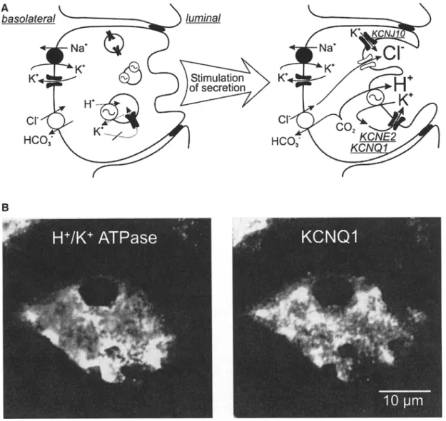
In stomach mucosa, acid secretion occurs in a spe- cialized cell type, the parietal cell. These mitochon- dria-rich cells have a unique morphology with invaginations of the luminal membrane into the cy- toplasm and complex vesicular structures, so-called tubulocisternae, which amalgamate with the luminal membrane upon secretory stimuli [31]. The acid- producing enzyme H+/K+-ATPase is localized in these tubulocisternae. After amalgamation with the luminal membrane, the H+/K+-ATPase pumps H + out of the cell, paralleled by an uptake of K +. C1- is secreted into the lumen via apical C1C-2 C1- chan- nels [82, 123]. To avoid K + depletion of the luminal fluid due to H+/K + ATPase activity, a recycling pathway for K + is required (Fig. 2A). There is good experimental evidence that K + channels are the respective pathway [46, 138, 139]. In immunofluo- rescence experiments the KCNQ1 K + channel (for- merly named KvLQT1) showed an H+/K+-ATPase - like distribution pattern in human parietal cells (Fig. 2B) [24, 38]. Its pharmacological inhibition by the chromanol 293B abolished acid secretion almost completely in vitro and in vivo. Most likely, KCNQ1 is associated with its regulatory subunit KCNE2 (MiRP1) to form the native channel complex [24, 38]. The functional role of KCNQ1 for acid secretion was underlined by observations in KCNQ1 knockout mice, which have an impaired acid secretion leading to hypergastrinemia and glandular gastric hypertro- phy [69]. At present, it is not known whether homo- zygous KCNQ1 mutations in patients suffering from so-called Jervell-Lange-Nielsen syndrome [95] also lead to defects in gastric acid secretion. Very recently, another K + channel was described in parietal cells: Immunofluorescence experiments and electron mi- croscopy revealed a localization of KCNJ10 (Kir4.1) in the H+/K+-ATPase-containing membrane com- partment, suggesting that besides KCNQI also KCNJ10 might play a role during acid secretion [37]. To date no data are available concerning the gastric
70 R. Warth and J. Barhanin: K + Channels o f the Intestinal Epithelium
Fig. 2. K + recycling is required for acid secretion in parietal cells. (A) Functional and morphological changes of a parietal cell during stimulation o f acid secretion. In the absence o f secretory stimuli H+/K + ATPase is located in intracellular vesicular structures (so- called tubulocisternae [31]). At present, it is not known whether K + channels, required for K + recyling, are located in H+/K + ATPase- containing vesicles or in separate vesicular structures. A connection o f the tubulocisternae to the lumen does not yet exist. U p o n stimulation via c A M P and IP3/Ca 2+ pathways, tubulocisternae get fused with the luminal membrane, thereby enlarging the luminal
surface and targeting H+/K+-ATPases and K + channels to the apical side. Now K + channels get activated by increasing concen- trations o f c A M P and cytosolic Ca 2+, and allow K + recycling, which is necessary for ongoing H+/K+-ATPase activity. (B) lm- munofluorescence of a resting human parietal cell with labeling of H+/K +-ATPases (left) and K C N Q 1 K + channels (right). Both show a granular distribution pattern within the cell, suggesting a locali- zation of the proteins in vesicles and tubulocisternae, which belong to the luminal membrane compartment.
phenotype of KCNJ10 knockout mice [85]. Further studies are required to elucidate nature, function and membrane targeting of luminal K + channels during gastric acid secretion.
In the basolateral membrane the C1-/HCO 3 ex- changer serves C1- uptake and HCO 3 extrusion. Basolateral Na+/H + exchangers stabilize intra- cellular pH and cell volume [110, 118]. With the patch-clamp technique, K + conductances have been observed in the basolateral membrane and in whole- cell experiments [25, 90]; in whole-cell experiments [63], however, it is at present not clear whether they
provide the driving force for voltage-dependent ba- solateral transport or for luminal C1- exit. For the latter function paracellular permeability would be required
(see
Fig. 1), which has been shown to be rather low [36].Function of K + Channels for Reabsorption and Secretion in Small Intestine
In small intestine most of the nutrients are digested and consecutively absorbed. The largest part of the
R. Warth and J. Barhanin: K + Channels of the Intestinal Epithelium 71
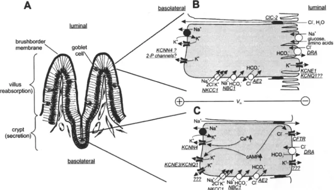
Fig. 3. Electrogenic transport in small intestine. (A) The mucosa of small intestine consists of crypt and villus cells. Reabsorption takes mainly place at the brushborder membrane of villus enterocytes. Goblet cells produce mucus, crypt cells secrete HCO 3-rich fluid to neutralize gastric acid. Stem cells for the rapid regeneration of enterocytes are localized at the crypt basis. (B) In the luminal membrane of villus cells, glucose and amino acids are reabsorbed in a secondary active way together with Na +. This depolarizes the luminal membrane, leading to the generation of a lumen-negative transepithelial voltage (Vte), which can drive reabsorption of anions via the paracellular pathway. Luminal K + channels could play a role for repolarization [134]. There is also some evidence for an H+/K + exchange mechanism, probably via H+/K + ATPase [9]. The molecular identity of luminal and basolateral K + channels is not known. The intermediate-conductance K + channel is probably less expressed in the basolateral membrane of villus cells when corn-
pared with crypt cells [47]. C1C-2 C1 channels seem to mediate paraceltular C1- fluxes [45] driven by the electro-chemical gradient. (C) Secretion in crypt cells requires activation of luminal C1- channels (CFTR), basolateral (and luminal) K + channels, NKCC1 and Na+/K + ATPase [42, 125]. Heteromultimeric KCNE3/KCNQ1 channels represent a cAMP-activated K + conductance. Their in- hibition largely attenuates electrogenic C1 /HCO 3 secretion. The intermediate-conductance K + channel (probably KCNN4) is acti- vated via increases in cytosolic Ca 2+. HCO 3 leaves the cell via CFTR and via luminal anion exchangers (probably DRA). On the basolateral side, CI can be taken up by NKCC1 and by Na +- dependent and -independent anion exchangers (AE2 and NBC) [130]. Luminal C1 and basolateral K + conductance create a lu- men-negative Vte, that possibly induces paracellular Na + fluxes. The mechanisms for HCO 3 secretion are probably present in villus cells, too (B).
fluid is r e a b s o r b e d b y the small i n t e s t i n a l m u c o s a a n d o n l y s o m e 20% e n t e r the c o l o n . T h e m a s s t r a n s p o r t in the s m a l l i n t e s t i n e involves b o t h t r a n s c e l l u l a r a n d p a r a c e l l u l a r t r a n s p o r t o f solutes, i o n s a n d water. I n o r d e r to fulfill different p h y s i o l o g i c a l d e m a n d s , the r e a b s o r p t i v e c a p a c i t y o f small i n t e s t i n a l m u c o s a is r e g u l a t e d b y t r a n s c r i p t i o n a l a n d n o n - t r a n s c r i p t i o n a l m e c h a n i s m s . T h u s , e x p r e s s i o n o f t r a n s p o r t e r s a n d i o n c h a n n e l s c a n v a r y o v e r a b r o a d r a n g e , d e p e n d i n g on the needs [53, 64, 97, 99]. A m o n g the t r a n s c e l l u l a r m e c h a n i s m s , s e c o n d a r y active t r a n s p o r t a l l o w s a b s o r p t i o n o f s u b s t r a t e s even a g a i n s t a c o n c e n t r a t i o n g r a d i e n t , i.e., b y N a + - c o u - p l e d t r a n s p o r t e r s for glucose a n d a m i n o acids. M a n y o f the t r a n s p o r t e r s use the c h e m i c a l g r a d i e n t for N a § o r H § a n d the n e g a t i v e m e m b r a n e v o l t a g e as d r i v i n g forces [86, 98]. T h e e n t r y o f a p o s i t i v e n e t c h a r g e via these systems d e p o l a r i z e s the l u m i n a l m e m b r a n e o f e n t e r o c y t e s , l e a d i n g to a t r a n s e p i t h e l i a l v o l t a g e dif-
ference (Vte, s e e Fig. 1 a n d Fig. 3) a n d c o n s e c u t i v e l y to p a r a c e l l u l a r e l e c t r o g e n i c t r a n s p o r t , i.e., t r a n s p o r t o f C1- [8, 45]. H o w e v e r , the d e p o l a r i z a t i o n o f the l u m i n a l m e m b r a n e reduces f u r t h e r v o l t a g e - d e p e n d e n t t r a n s c e l l u l a r t r a n s p o r t . T o r e s t o r e the t r a n s p o r t ca- p a c i t y o f the e n t e r o c y t e s , K § c h a n n e l s are r e q u i r e d to r e p o l a r i z e the m e m b r a n e voltage. T h e y c a n be local- ized in e i t h e r the l u m i n a l o r the b a s o l a t e r a l m e m - b r a n e o f villus cells (Fig. 3). It has been s h o w n t h a t a c t i v a t i o n o f b a s o l a t e r a l K § c h a n n e l s b y p i n a c i d i l o r B R L 38227 increases NaC1 r e a b s o r p t i o n , p a r a l l e l e d b y a n e n h a n c e d s h o r t - c i r c u i t c u r r e n t [51]. S t r o n g ac- t i v a t i o n o f l u m i n a l K § c h a n n e l s , e.g., b y cell swelling [80], c o u l d even p r o d u c e a l u m e n - p o s i t i v e t r a n s e p i - thelial v o l t a g e , w h i c h in t u r n w o u l d i n v e r t the direc- t i o n o f p a r a c e l l u l a r i o n fluxes [45]. I n a n o t h e r s t u d y , a nitric o x i d e - a c t i v a t e d b a s o l a t e r a l K § c o n d u c t a n c e was d e s c r i b e d , w h o s e a c t i v a t i o n b y the K § c h a n n e l o p e n e r c r o m a k a l i m e n h a n c e d net fluid r e a b s o r p t i o n
72 R. Warth and J. Barhanin: K ~ Channels of the Intestinal Epithelium
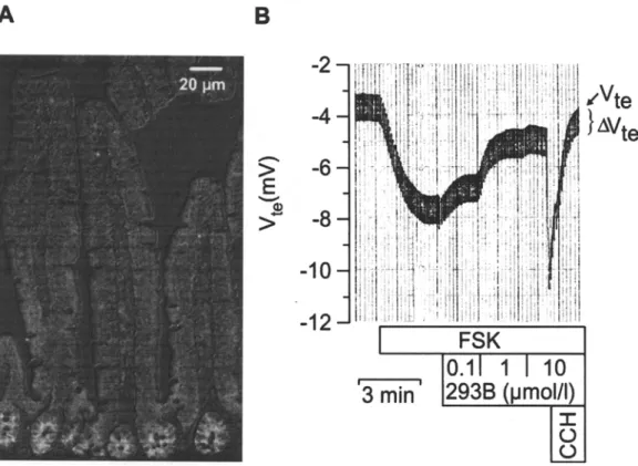
Fig. 4. KCNE3/KCNQI K + channels in mouse jejunum. (A) Im- munolocalization of KCNQI in mouse jejunum (overlay with dif- ferential interference contrast image). The KCNQl-specific antibody [134] preferentially labeled the basolateral membrane of jejunal crypts cells. (B) Effect of the chromanol 293B on cAMP- activated secretion in mouse jejunum. Forskolin at 5 gM (FSK)
increased transepithelial voltage (Vte) mainly by activation of lu- minal C1 and basolateral KCNE3/KCNQ1 K ~ channels. 293B reduced Vtc concentration-dependently by some 70%. Carbochol 100 gM (CCH) led to a rise in Vte by transiently activating Ca 2+ activated K § channels (probably KCNN4). AVte is a measure for transepithelial resistance.
[115], indicating that luminal and basolateral K + conductances support reabsorption in small intestinal villus cells.
In the crypts of the small intestine, net secretion of bicarbonate, mucus, salt and water occurs, which allows buffering of acidic gastric content and facili- tates propulsion of the intestinal content. Under pathological conditions, overwhelming activation of secretion, i.e., by cholera toxin, can lead to severe diarrhea. Transcellular transport in crypt cells is mainly a serosal-mucosal flux of CI-, HCO 3 and K + [130], which is activated via cAMP and C a 2+ path- ways and to some extent via nitric oxide/cGMP [6]. Bicarbonate secretion plays an important role for mucosal protection and neutralization of gastric acid. However, the luminal pathway for bicarbonate exit is controversially discussed: It probably involves elec- trogenic systems such as C F T R channels (cystic fi- brosis transmembrane conductance regulator) [21] and electroneutral C1 /HCO 3 exchangers such as D R A (down-regulated in adenoma) [50, 55, 93, 104]. The amount of HCO 3 secreted via electrogenic transport systems is dependent on the membrane voltage and, thus, on K + channel activity. For the maintenance of electro-neutrality, Na + enters the lumen via the paracellular pathway driven by the
transepithelial voltage, and K + can be secreted via luminal K + channels (Fig. 3C).
Transcellular mass transport of nutrients, salt, and water is a continuous challenge for cellular vol- ume regulation of enterocytes in small intestine. There is good evidence that K + channels play a role during regulatory volume decrease after swelling of villus enterocytes [79]. Moreover, it has been shown that Na+-dependent absorption of amino acids in enterocytes leads to activation of Na+/K + ATPase and of basolateral K + channels. The increase in ba- solateral K + conductance prevents rises in intracel- lular K + concentration (due to enhanced Na+/K + ATPase activity) and counteracts the depolarization caused by luminal electrogenic transport activity [39]. Compared with colonic K + channels, relatively little is known about the functional properties and the molecular identity of K + channels in the small in- testine (Fig. 3) [124]. With electrophysiological methods several basolateral K + channels could be
distinguished: an abundant large conductance
"maxi" K + channel (90 to 250 pS) [15, 92, 122], an intermediate-conductance, CaZ+-activated K + chan- nel, small-conductance channels [15], KATp-like channels [30] and a chromanol 293B-sensitive K + conductance [134]. The regulation of the large-con-
R. Warth and J. Barhanin: K + Channels of the Intestinal Epithelium 73
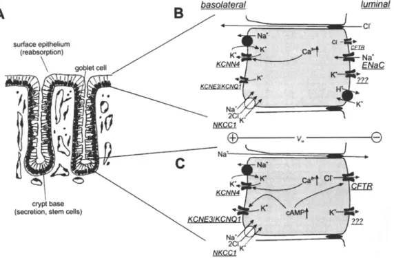
Fig. 5. Role of K + channels for secretion and absorption in distal colonic mucosa. (A) Colonic crypts consist of enterocytes, entero- endocrine cells and goblet cells. Absorption of short-chain fatty acids, salt, and water takes place in surface cells and the upper part of the crypt. At the crypt base, secretion and mitosis take place. (B) In surface cells Na § enters the cell via ENaC and is exported via basolateral Na+/K + ATPase. CFTR CI channels are less abun- dant than in crypt base cells. Without luminal K + channel activity, ENaC-induced luminal depolarization leads to CI reabsorption via CFTR. Activation of luminal K + channels results in K + se- cretion paralleled by luminal hyperpolarization. If the luminal membrane voltage is more negative than the CI equilibrium po- tential, the direction of the CFTR C1 flux would invert: the cell
secretes KC1 and reabsorbs Na +. Luminal H+/K + ATPase serves mainly K + reabsorption. Basolateral K + channels provide a part of the driving force for luminal ion movement. (B) In crypt base cells, secretion requires cAMP-dependent activation of luminal CFTR C1- channels and of basolateral KCNE3/KCNQ1 K + channels. Ca 2+ leads to activation of basolateral KCNN4 K + channels but requires a certain level of luminal CFTR activity to induce C1 secretion, because CFTR seems not to be directly Ca2+-activated. Ca 2 +-activated CI- channels play a minor role in normal tissue but can be observed during tumor genesis in colonic mucosa [12]. Driven by the transepithelial voltage, Na + follows via the para- cellular pathway.
ductance K + channel by cAMP and Ca 2+ is con- troversially discussed [15, 87, 92, 122]. The interme- diate-conductance K + channel is strongly activated by rises in cytosolic Ca 2+ and by 1-ethyl-2-benzimi- dazolinone (1-EBIO) and shows functional charac- teristics similar to cloned IK1 channels (KCNN4) [47], suggesting that KCNN4 is the intermediate- conductance K + channel as found in rat colonic crypts [135]. There is some evidence for a regulation of the intermediate-conductance K + channel by cAMP [87], however, in another study no such reg- ulation was observed [15]. Activation of this channel by 1-EBIO leads to a strong increase in C1 secretion but does not affect phloridzin-sensitive glucose ab- sorption indicating that the 1-EBIO-activated K + channel is mainly expressed in intestinal crypts and not or only little in villus cells [47].
The 293B-sensitive conductance is mainly located in crypt cells and formed by KCNQ1/KCNE3 channel proteins (Figs. 3C and 4A) [24, 134]. The KCNE3/KCNQ1 channel complex is activated dur- ing cAMP-stimulated secretion. Its inhibition blocks
electrogenic jejunal secretion by some 50-70% (Fig. 4B). Thus, KCNE3/KCNQ1 channels are a possible target for the treatment of life-threatening forms of secretory diarrhea, such as cholera. The 293B deriv- ative IKs224 was shown to inhibit jejunal secretion
with an IC5o in the nanomolar range [134]. However,
clinical use of KCNE3/KCNQ1 blockers would re- quire subunit specificity and/or locally restricted de- livery of the drug, since one has to avoid inhibition of KCNE1/KCNQ1 channels in heart and inner ear.
K + Channels in Colonic M u c o s a
In contrast to mass transport in small intestine, the colonic mucosa serves the fine tuning of salt and water metabolism. Under physiological conditions absorption largely exceeds secretion: Some 90% of 1.51 of intestinal content entering the colon are absorbed and only 150-200 g are excreted under physiological conditions. However, a certain level of secretion of mucins, electrolytes and water is
74 R. Warth and J. Barhanin: K + Channels of the Intestinal Epithelium ..= 9 et0 O z ~ ~ . t'N r z ~ ~ Z ~ g z ~" z g W ,~ ,-.a ~' W 8 ~ ~ z ~ ~ C" + + + +~ -~ + + + +~ + + ~. + x .-. +~ + +" + + I + + + + + + ~ • . + + ~ + + ~ + + ~+ . ~ + ~ ~ + + + + + + + ~ ~ ~ + + .~+ ~, + + ~ ~ +~+ + + + ~ 1 ~ I + ~ + + ~ + ~ + + + + + + + , z a , . ~ + + + + + + + + + + + + + + E + + + a= .~+ ~ § . r ~ + ~ + ~ ~ § ~ +~ ~ + + ~++~ + + ~ + x + + + ~ +~ + + + + ;=+=+ + + + 5 + ~=+=+=++~+~t + ? ~ Z Z Z Z Z Z Z Z Z Z Z Z Z Z Z Z oa ..= E O t,> ..= O *6 < Z 9 9 Z i e ~ e ~ O ' ( y ..Q "-~, O O . ~ r = . = . r i .
R. Warth and J. Barhanin: K + Channels of the Intestinal Epithelium 75
required for normal transport and composition of feces. Thus, defects in secretion, i.e., in cystic fibrosis, can lead to severe constipation. On the other hand, impairment of colonic reabsorption leads to diarrhea, affecting salt/water and acid/base metabolism [67].
In the proximal part, Na + is mainly reabsorbed via Na +/H + exchangers and in distal colon, via Na + channels (ENaC). Depending on the needs of me- tabolism and hormone concentrations, K + can be net-secreted via channels or reabsorbed by a luminal H+/K+-ATPase (Fig. 5) [10, 34, 89, 120]. A detailed list of hormones and mediators regulating colonic transport is given in [133]. Moreover, bacterial deg- radation of undigested nutrients leads to generation of short-chain fatty acids and ammonia, which are taken up by cells located at the surface of colonic crypts [17, 18, 48].
K + channels play a role for secretory and re- absorptive pathways. In particular the activity of luminal K + channels affects the K + balance of the' body and therefore is regulated via K + intake and different hormones. In the presence of high potas- sium diet [16, 113] or high aldosterone concentra- tions [35, 43, 76, 81], K + channels hyperpolarize the luminal membrane of crypt cells, leading to electro- neutral transcellular secretion of KC1 instead of transcellular C1 exit and paracellular Na + flux (Fig. ID). In surface epithelial cells, the K + channel-in- duced hyperpolarization increases the driving force for Na + uptake via ENaC in distal colon. These K + conductances are modulated by cholinergic and cAMP-mediated pathways [29, 49, 84, 119]. Direct functional characterization of luminal K + channels with the patch-clamp technique is hampered by mucus and microvilli, which make measurements very difficult. There is evidence for large-conduc- tance K + channels with single-channel conductances from 120-230 pS [16, 113, 133] and for a pH-sensi- tive ROMK-type channel [52, 62, 133]. Data from KCNMA1 knockout mice indicate that maxi K + channels play a role in K + secretion after purinergic receptor stimulation [70]. The inhibitory effect of the chromanol 293B on cAMP-activated serosal-to-mu- cosal Rb + flux suggests that KCNQ1 might be lo- calized in luminal and basolateral membranes [29]. In the luminal membrane, KCNQ1 could co-as- semble with the small regulatory proteins KCNE1 or KCNE2 [1321.
In the basolateral membrane of colonic mucosa at least three different types of K + channels are present: (1) a large-conductance K + channel that is very abundant in rabbit colon [77, 78, 127]; (2) KCNN4 underlying the intermediate-conductance CaZ+-activated K + channel [14, 27, 135], and (3) KCNQ1, a very small-conductance K + channel that is stimulated via cAMP and blocked by 293B [11, 24, 26, 65, 75, 117, 136].
Upon cholinergic stimulation of rat colonic mu- cosa, a strong but transient opening of KCNN4 channels is observed, leading to hyperpolarization of the basolateral membrane. The basolateral hyperpo- larization increases the transepithelial potential dif- ference if the luminal membrane is depolarized by C1- channels [13, 49]. Interestingly, the Ca z+ path- way alone seems not to activate sufficiently the lu- minal C1- conductance in normal colonic mucosa: in the absence of cAMP-mediated activation of luminal C1- channels, cholinergic stimulation by carbachol inverts the polarity of the Vte and leads to a lumen- positive transepithelial voltage via activation of luminal K + channels [83]. However, during carcino- genesis, Ca2+-activated C1- channels are expressed, counteracting the hyperpolarizing effect of KCNN4 channel opening [12] and thereby mimicking the ef- fect of carbachol in some colonic cell lines. Colonic KCNN4 channels are potently blocked by the anti- fungal drug clotrimazole and activated by 1-EBIO [28, 102, 129, 135]. Pharmacological activation and inhibition of colonic KCNN4 channels have been discussed as strategies for the treatment of cystic fi- brosis and diarrhea, respectively [28, 57, 111, 126]. Basolateral K + channel inhibition was claimed to mediate the effect of the antidiarrheal drug lopera- mide [33].
In large intestine cAMP-mediated secretion plays an important role for normal transport of feces, which is impaired in cystic fibrosis. On the other hand, some bacterial toxins, such as cholera toxin, induce an overwhelming secretion in small and large intestine [133]. It has been shown that cAMP de- creases intracellular Ca 2 + activity of colonocytes via depolarization-induced reduction of Ca z+ influx. Thus, KCNN4-type CaZ+-dependent K + channels are closed during cAMP-mediated secretion [13]. Under these conditions, the driving force for luminal C1- exit mainly depends on cAMP-activated KCNQ1 K + channels. The KCNQ1 inhibition by 293B was shown to block the electrogenic part of C1 secretion (Fig. 1C) [75] and the minor electroneutral secretion of KC1 (Fig. 1D) [29]. Like in small intestine, KCNQ1 is associated with its regulatory subunit KCNE3 in the basolateral membrane of colonic enterocytes. In contrast to the cardiac KCNE1/KCNQ1 channel complex, which is voltage-dependent and slowly ac- tivating, KCNE3/KCNQ1 channels are voltage-in- dependent and constitutively open [117].
More Intestinal K + Channels in the Future?
During the last years a number of new K + channel transcripts, i.e., for several maxi K + channels and 2-p-domain K + channels, have been found in the gastrointestinal tract and the list of intestinal K ~ channel genes (Table 1) is probably not complete.
76 R. Warth and J. Barhanin: K + Channels of the Intestinal Epithelium On the other hand, for expression analysis, often
commercially available samples of total tissue cDNA or RNA (including muscle layers and neu- ronal cells) were used. In these cases it is not proven that the respective K + channels are loca- lized in the epithelial cells. Only for a very limited number of channels epithelial expression, cellular localization and a functional role in the native tis- sue could be shown. However, these data led to a better understanding of transport in the intestinal tract and these channels could be targets of drug development for the treatment of transport-related diseases.
Supported by a "Forschungskredit der Universitfit Ztirich" to R.W.
References
1. Abbott, G.W., Sesti, F., Splawski, I., Buck, ME., Lehmann, M.H., Timothy, K.W., Keating, M.T., Goldstein, S.A. 1999.
Cell 97:175-187
2. Aguilar-Bryan, L., Nichols, C.G., Wechsler, S.W., Clement, J.P., Boyd, A.E., Gonzfilez, G., Herrera-Sosa, H., Nguy, K., Bryan, J., Nelson, D.A. 1995. Science 268:423~,26
3. Arrighi, I., Bloch-Faure, M., Grahammer, F., Bleich, M., Warth, R., Mengual, R., Drici, M.D., Barhanin, J., Meneton, P. 2001. Proc. Natl. Acad. Sci. USA 98:8792-8797
4. Arrighi, l., Lesage, F., Scimeca, J.C., Carle, G.F., Barhanin, J. 1998. FEBS Lett. 425:310-316
5. Barhanin, J., Lesage, F., Guillemare, E., Fink, M., Lazdun- ski, M., Romey, G. 1996. Nature 384:78-80
6. Barry, M.K., Aloisi, J.D., Yeo, C.J. 1995. J. Surg. Res. 59:681-686
7. Behrens, R., Nolting, A., Reimann, F., Schwarz, M., Waldschutz, R., Pongs, O. 2000. F E B S Lett. 474:99-106 8. Bijlsma, P.B., Bakker, R., Groot, J.A. 1997. J. Membrane
Biol. 157:127-137
9. Binder, H.J., Murer, H. 1986. J. Membrane Biol. 91:71 84 10. Binder, H.J., Sangan, P., Rajendran, V.M. 1999. Semin.
Nephrol. 19:405414
11. Bleich, M., Briel, M., Busch, A.E., Lang, H.J., Gerlach, U., G6gelein, H., Greger, R., Kunzelmann, K. 1997. Pfluegers
Arch. 34:499-501
12. Bleich, M., Ecke, D., Schwartz, B., Fraser, G., Greger, R. 1997. Pfluegers Arch. 433:254-259
13. Bleich, M., Riedemann, N., Warth, R., Kerstan, D., Leipzi- ger, J., H6r, M., Van Driessche, W., Greger, R. 1996.
Pfluegers" Arch. 432:1011-1022
14. Burckhardt, B.-C., G6gelein, H. 1992. Pfluegers Arch. 420:54-60
15. Butt, A.G., Hamilton, K.L. 1998. Pfluegers Arch. 435:528- 538
16. Bunerfield, I., Warhurst, G., Jones, M.N., Sandle, G.I. 1997.
J. Physiol. 501:537-547
17. Castell, D.O., Moore, E.W. 1971. Gastroenterology 60:33M.2 18. Charney, A.N., Micic, L., Egnor, R.W. 1998. Am. J. Physiol.
274:G518-24
19. Chavez, R.A., Gray, A.T., Zhao, B.B., Kindler, C.H., Ma- zurek, M.J., Mehta, Y., Forsayeth, J.R., Yost, C.S. 1999.
J. Biol. Chem. 274:7887-7892
20. Chouabe, C., Neyroud, N., Guicheney, P., Lazdunski, M., Romey, G., Barhanin, J. 1997. E M B O J. 16:5472-5479 21. Clarke, L.L., Stien, X., Walker, N.M. 2001. J. Pancreas
2:263 267
22. Coetzee, W.A., Amarillo, Y., Chiu, J., Chow, A., Lau, D., McCormack, T., Moreno, H., Nadal, M.S., Ozaita, A., Po- untney, D., Saganich, M., Vega-Saenz, d.M., Rudy, B. 1999.
Ann. N. Y. Acad. Sei. 868:233-285
23. Colegio, OR., Van Itallie, C.M., McCrea, H.J., Rahner, C., Anderson, J.M. 2002. Am. J. Physiol. 283:C142-C147 24. Dedek, K., Waldegger, S. 2001. Pfluegers Arch. 442:896-902 25. Demarest, J.R., Loo, D.D. 1990. Annu. Rev. Physiol. 52:307-
319
26. Demolombe, S., Franco, D., de Boer, P., Kuperschmidt, S., Roden, D., Pereon, Y., Jarry, A., Moorman, A.F., Escande, D. 2001. Am. J. Physiol. 280:C359-C372
27. Devor, D.C., Frizzell, R.A. 1998. Am. J. Physiol. 274:C138 C148
28. Devor, D.C., Singh, A.K., Frizzell, R.A., Bridges, R.J. 1996.
Am. J. Physiol. 271:L775 784
29. Diener, M., Hug, F,, Strabel, D., Scharrer~ E. 1996, Br. J.
Pharmacol. 118:1477-1487
30. Dubinsky, W.P., Mayorga-Wark, O., Schultz, S.G. 1998.
Am. J. Physiol. 27g:c1653 C1659
31. Duman, J.G., Pathak, N.J., Ladinsky, M.S., McDonald, K.L., Forte, J.G. 2002. J. Cell Sci. 115:1251-1258
32. Duprat, F., Lesage, F., Fink, M., Reyes, R., Heurteaux, C., Lazdunski, M. 1997. E M B O 16:5464-5471
33. Eppte, H.J., Fromm, M., Riecken, E.O., Schulzke, J.D. 2001.
Seand. J. Gastroenterol. 36:731-737
34. Foster, E.S., Budinger, M.E., Hayslett, J.P., Binder, H.J. 1986. J. Clin. Invest. 77:228-235
35. Fromm, M., Schulzke, J.D., Hegel, U. 1990. Pfluegers Arch. 416:573-579
36. Fr6mter, E., Diamond, J. 1972. Nature 235:9-13
37. Fujita, A., Horio, Y., Higashi, K., Mouri, T., Hata, F., Takeguchi, N., Kurachi, Y. 2002. J. Physiol. 540:85-92 38. Grahammer, F., Herling, A.W., Lang, H.J., Schmitt-Gr/iff,
A., Wittekindt, O.H., Nitschke, R., Bleich, M., Barhanin, J., Warm, R. 2001. Gastroenterology 120:1363-1371
39. Grasset, E., Gunter-Smith, P., Schultz, S.G., 1983. J. Mem-
brane Biol. 71:89-94
40. Greger, R. 1996. Epithelial transport. In: Comprehensive Human Physiology. 1217 1232. R. Greger, U. Windhorst, ed., Springer, Berlin, Heidelberg, New York
41. Greger, R., Schlatter, E. 1983. Pfluegers Arch. 396:325-334 42. Greger, R., Schlatter, E., G6gelein, H. 1986. News Physiol.
Sci. 1:134-136
43. Grotjoharm, I., Gitter, A.H., Kockerling, A., Bertog, M., Schulzke, J.D., Fromm, M. 1998. J. Physiol. 507:561-570 44. Grunnet, M., MacAulay, N., Jorgensen, N.K., Jensen, S.,
Olesen, S.P., Klaerke, D.A. 2002. Pfluegers Arch. 444:167-177 45. Gyomorey, K., Yeger, H., Ackerley, C., Garami, E., Bear,
C.E. 2000. Am. J. Physiol. 279:C1787-C1794
46. Hagen, SJ., Wu, H., Morrison, S.W. 2000. Am. J. Physiol. 279:G400 G410
47. Hamilton, K.L., Meads, L., Butt, A.G. 1999. pfluegers Arch. 439:158 166
48. Hasselblatt, P., Warth, R., Schulz-Baldes, A., Greger, R., Bleich, M. 2000. Pfluegers Arch. 441:118-124
49. Heinke, B., H6rger, S., Diener, M. 1998. Eur~ J. Pharmacol. 362:199 206
50. Hoglund, P., Haila, S., Socha, J., Tomaszewski, L., Sa- arialho-Kere, U., Karjalainen-Lindsberg, M.L., Airola, K., Holmberg, C., de la Chapelle, A., Kere, J. 1996. Nat. Genet. 14:316-319
R. Warth and J. Barhanin: K + Channels of the Intestinal Epithelium 77 51. Homaidan, F.R., Broutman, G. 1994. Ear. J. Pharmacol.
271:561 565
52. Huang, Y., Wong, P.Y.D. 1996. J. Cell. Physiol. 168:678 683 53. Ihara, T., Tsujikawa, T., Fujiyama, Y., Bamba, T. 2000.
Digestion 61:59 67
54. Ishii, T.M., Silvia, C., Hirschberg, B., Bond, C.T., Adelman, J.P., Maylie, J. 1997. Proc. Natl. Acad. Sci. USA 94:11651-
11656
55. Jacob, P., Rossmann, H., Lamprecht, G., Kretz, A., Neff, C., Lin-Wu, E., Gregor, M., Groneberg, D.A., Kere, J., Seidler, U. 2002. Gastroenterology 122:709-724
56. Jensen, B.S., Strobaek, D., Christophersen, P., Jorgensen, T.D., Hansen, C., Silahtaroglu, A., Olesen, S.P., Ahring, P.K. 1998. Am. J. Physiol. 27fi:C848 C856
57. Jensen, B.S., Strobaek, D., Olesen, S.P., Christophersen, P. 2001. (Turr. Drug Targets 2:401~422
58. Jiang, Y., Lee, A., Chen, J., Cadene, M., Chait, B.T., MacKinnon, R. 2002. Nature 417:515-522
59. Jiang, Y., Lee, A., Chen, J., Cadene, M., Chait, B.T., MacKinnon, R. 2002. Nature 417:523-526
60. Khanna, R., Chang, M.C., Joiner, W.J., Kaczmarek, L.K., Schlichter, L.C. 1999. J. Biol. Chem. 274.14838-14849 61. Kim, D., Fujita, A., Horio, Y., Kurachi, Y. 1998. Circ. Res.
82:513-518
62. Kondo, C., Isomoto, S., Matsumoto, S., Yamada, M., Horio, Y., Yamashita, S., Takemura-Kameda, K., Matsuzawa, Y., Kurachi, Y. 1996. F E B S Lett. 399:122-126
63. Kotera, T., Hashimoto, A., Ueda, S., Okada, Y. 1991.
J. Membrane Biol. 124:43 52
64. Koyama, K., Sasaki, I., Naito, H., Funayama, Y., Fuku- shima, K., Unno, M., Matsuno, S., Hayashi, H., Suzuki, Y. 1999. Am. J. Physiol. 276:G975 G984
65. Kunzelmann, K., Bleich, M., Warm, R., Levy-Holzman, R., Garty, H., Schreiber, R. 2001. Clin. Exp. Pharmacol. Physiol.
28:79 83
66. Kunzehnann, K., Htibner, M., Schreiber, R., Levy-Holzman, R., Garty, H., Bleich, M., Warth, R., Slavik, M., von Hahn, T., Greger, R. 2001. J. Memb. Biol. 119-155-164
67. Kunzelmann, K., Mall, M. 2002. Physiol. Rev. 82"245-289 68. Lang, F., Busch, G.L., Ritter, M., Volkl, H., Waldegger, S.,
Gulbins, E., Haussinger, D. 1998. Physiol. Rev. 18:247-306 69. Lee, M.P., Ravenel, J.D., Hu, R.J., Lustig, L.R., Tomaselli,
G., Berger, R.D., Brandenburg, S.A., Litzi, T.J., Bunton, T.E., Limb, C., Francis, H., Gorelikow, M., Gu, H., Wash- ington, K., Argani, P., Goldenring, J.R., Coffey, R.J., Fein- berg, A.P. 2000. J. Clin. Invest. 106:1447-1455
70. Leipziger, J., Matos, J., Sausbier, M., Ruth, P. 2003. Pflue- gers Arch. in press
71. Lesage, F., Lazdunski, M. 2000. Am. J. Physiol. 279:F793- F801
72. Lesage, F., Terrenoire, C., Romey, G., Lazdunski, M. 2000.
J. Biol. Chem. 275:28398-28405
73. Lock, H., Valverde, M.A. 2000. J. Biol. Chem. 275:34849- 34852
74. Logsdon, N.J., Kang, J., Togo, J.A., Christian, E.P., Aiyar, J. 1997. J. Biol. Chem. 272:32723-32726
75. Lohrmann, E., Burhoff, I., Nitschke, R.B., Lang, H.J., Mania, D., Englert, H.C., Hropot, M., Warth, R., Rohm, W., Bleich, M., Greger, R. 1995. Pfluegers Arch. 429.'517- 530
76. Lomax, R.B., McNicholas, C.M., Lombes, M., Sandle, G T 1994. Am. J. Physiol. 266:G71 G82
77. Lomax, R.B., Warhurst, G., Sandle, G.I. 1996. Gut 38:243 247
78. Loo, D.D.F., Kaunitz, J.D. 1989. J. Membrane Biol. 110:19 28
79. MacLeod, RJ., Hamilton, J.R. 1999. J. Membrane Biol.
172:59-66
80. MacLeod, R.J., Hamilton, J.R. 1999. J. Membrane Biol.
172:47-58
81. Maguire, D., MacNamara, B., Cuffe, J.E., Winter, D., Doolan, C.M., Urbach, V., O'Sullivan, G.C., Harvey, B.J. 1999. Steroid~ 64:51 63
82. Malinowska, D.H., Kupert, E.Y., Bahinski, A., Sherry, A.M., Cuppoletti, J. 1995. Am. J. Physiol. 268"C191-C200 83. Mall, M., Bleich, M., Schfirlein, M., K,ahr, J., Seydewitz,
H.H., Brandis, M., Greger, R., Kunzelmann, K. 1998. Am. J.
Physiol. 275:G 1274~G 1281
84. Mall, M., Wissner, A., Seydewitz, H.H., Kuehr, J., Brandis, M., Greger, R., Kunzelmann, K. 2000. Am. J. Physiol.
278:G617-G624
85. Marcus, D.C., Wu, T., Wangemann, P., Kofuji, P. 2002. Am.
J. Physiol. 282:C403 C407
86. Matthews, J.C., Anderson, K.J. 2002. Curr. Opin. Clin. Nutr.
Metab. Care. 5:77-84
87. McNicholas, C.M., Fraser, G., Sandle, G.I. 1994. or. Physiol.
477:381-392
88. Medhurst, A.D., Rennie, G., Chapman, C.G., Meadows, H., Duckworth, M.D., Kelsell, R.E., Gloger, l.I., Pangalos, M.N. 2001. Brain Res. Mol. Brain Res. 86"101-114 89. Meneton, P., Schultheis, P.J., Greeb, J., Nieman, M.L., Liu,
L.H., Clarke, L.L., Daffy, J.J., Doetschman, T., Lorenz, J.N., Shull, G.E. 1998. J. Clin. Invest. 101:53(~542 90. Mieno, H., Kajiyama, G. 1991. Am. J. Physiol. 261:G206-
G212
91. Morita, T., Hanaoka, K., Morales, M.M., Montrose-Rafi- zadeh, C., Guggino, W.B. 1997. Am. J. Physiol. 273:F615- F624
92. Morris, A.P., Gallacher, D.V., Lee, J.A. 1986. F E B S Lett.
206:87-92
93. Moseley, R.H., H6glund, P., Wu, G.D., Silberg, D.G., Haila, S., de la Chapelle, A., Holmberg, C., Kere, J. 1999. Am. J.
Physiol. 276:G185 G192
94. Nakamura, N., Suzuki, Y., Sakuta, H., Ookata, K., Kawa- hara, K., Hirose, S. 1999. Biochem. J. 342:329 336 95. Neyroud, N., Tesson, F., Denjoy, I., Leibovici, M., Don-
ger, C., Barhanin, J., Faure, S., Gary, F., Coumel, P., Petit, C., Schwartz, K., Guicheney, P. 1997. Nature Genet.
1g:186 189
96. Niemeyer, M.I., Cid, L.P., Barros, L.F., Sepulveda, F.V. 2001. J. Biol. Chem.
97. Pacha, J. 2000. Physiol. Rev. 80:1633-1667
98. Palacin, M., Estevez, R., Bertran, J., Zorzano, A. 1998.
Physiol. Rev. 78:969-1054
99. Pan, X., Terada, T., Irie, M., Saito, H., Inui, K. 2002. Am. J.
Physiol. 283:G57-G64
100. Pardo, L.A., del Camino, D., Sanchez, A., Alves, F., Bru- ggemann, A., Beckh, S., Stuhmer, W. 1999. E M B O J.
18:5540 5547
101. Partiseti, M., Collura, V., Agnel, M., Culouscou, J.M., Graham, D. 1998. F E B S Lett. 434:171-176
102. Pedersen, K.A., Schroder, R.L., Skaaning-Jensen, B., Stro- baek, D., Olesen, S.-P., Christophersen, P. 1999. Biochim.
Biophys. Acta 1420:231-240
103. Pongs, O., Leicher, T., Berger, M., Roeper, J., Bahring, R., Wray, D., Giese, K.P., Silva, A.J., Storm, J.F. 1999. Ann. N.
Y. Acad. Sci. 868:344~355
104. Praetorius, J., Friis, U.G., Ainsworth, M.A., Schaffalitzky de Muckadell, O.B., Johansen, T. 2002. Acta Physiol. Scand.
174:327-336
105, Rahner, C., Mitic, L.L., Anderson, J.M. 2001. Gastroenter-
78 R. Warth and J. Barhanin: K + Channels of the Intestinal Epithelium 106. Rajan, S., Wischmeyer, E., Karschin, C., Preisig-Muller, R.,
Grzeschik, K.H., Daut, J., Karschin, A., Derst, C. 2000.
J. Biol. Chem. 276:7302-73ll
107. Rao, J.N., Platoshyn, O., Li, L., Guo, X., Golovina, V.A., Yuan, J.X., Wang, J.Y. 2002. Am. J. Physiol. 282:C885 C898 108. Reyes, R., Duprat, F., Lesage, F., Fink, M., Salinas, M., Farman, N., Lazdunski, M. 1998. J. Biol. Chem. 273:30863 30869
109. Ribeiro, R., Heinke, B., Diener, M. 2001. Acta Physiol.
Scand. 171:445-458
110. Rossmann, H., Sonnentag, T., Heinzmann, A., Seidler, B., Bachmann, O., Vieillard-Baron, D., Gregor, M., Seidler, U. 2001. Am. J. Physiol. 281:G447-G458
111. Rufo, P.A., Merlin, D., Riegler, M., Ferguson-Maltzman, M.H., Dickinson, B.L., Brugnara, C., Alper, S.L., Lencer, W.I. 1997. J. Clin. Invest. 100:3111 3120
112. Salinas, M., Reyes, R., Lesage, F., Fosset, M., Heurteaux, C., Romey, G., Lazdunski, M. 1999. J. Biol. Chem. 274:11751 11760
113. Sandle, G.I., Butterfield, I. 1999. Gut 44:4(>46
114. Sanguinetti, M.C., Curran, M.E., Zou, A., Shen, J., Spector, P.S., Atkinson, D.L., Keating, M.T. 1996. Nature 384:80-83 115. Schirgi-Degen, A., Beubler, E. 1996. Eur. J. Pharmacol.
316:257-262
116. Schroeder, B.C., Kubisch, C,, Stein, V., Jentsch, TJ. 1998.
Nature 396:687-690
117. Schroeder, B.C., Waldegger, S., Fehr, S., Bleich, M., Warth, R., Greger, R., Jentsch, T.J. 2000. Nature 403:196 199 118. Schultheis, P.J., Clarke, L.L., Meneton, P., Harline, M.,
Boivin, G.P., Stemmermann, G., Duffy, J.J., Doetschman, T., Miller, M.L., Shull, G.E. 1998. J. Clin. Invest. 101:1243 1253 119. Schultheil3, G., Diener, M. 1997. Br. J. Pharmacol. 122:87-94 120. Schultz, S.G. 1984. Annu. Rev. Physiol. 46:435~451 121. Schultz, S.G., Dubinsky, W.P. 2001. J. Membrane Biol.
184:255-261
122. Sepulveda, F.V., Mason, W.T. 1985. F E B S Lett. 191:87-91 123. Sherry, A.M., Malinowska, D.H., Morris, R.E., Ciraolo,
G.M., Cuppoletti, J. 2001. Am. J. Physiol. 280:C1599-C1606 124. Shieh, C.C., Coghlan, M., Sullivan, J.P., Gopalakrishnan, M.
2000. Pharmacol. Rev. 52:557-594
125. Silva, P., Stoff, J., Field, M., Fine, L., Forrest, J.N., Epstein, F.H. 1977. Am. J. Physiol. 233:F298 F306
126. Singh, S., Syme, C.A., Singh, A.K., Devor, D.C., Bridges, R.J. 2001. J. Pharmacol. Exp. Ther. 296:600-611
127. Turnheim, K., Plass, H., Wyskovsky, W. 2002. Biochim.
Biophys. Acta 1560:51 66
128. Vandorpe, D.H., Shmukler, B.E., Jiang, L., Lim, B., Maylie, J., Adelman, J.P., de Franceschi, L., Cappellini, M.D., Brugnara, C., Alper, S.L. 1998. J. Biol. Chem. 273:21542-21553 129. yon Hahn, T., Thiele, I., Zingaro, L., Hamm, K., Garcia
Alzamora, M., K6ttgen, M., Bleich, M., Warth, R. 2001. Cell
Physiol. Biochem. 11:219-230
130. Walker, N.M., Flagella, M., Gawenis, L.R., Shull, G.E., Clarke, L.L. 2002. Gastroenterology 123:531-541
131. Wang, J.Y., Wang, J., Golovina, V.A., Li, L., Platoshyn, O., Yuan, J.X. 2000. Am. J. Physiol. 278:C303-C314
132. Warth, R., Barhanin, J. 2002. Am. J. Physiol. 282:R639- R648
133. Warth, R., Bleich, M. 2000. Rev. Physiol. Biochem. Phar-
macol. 140:1 62
134. Warth, R., Garcia, A.M., Kim, K., Zdebik, A., Nitschke, R., Bleich, M., Gerlach, U., Barhanin, J., Kim, J. 2002. Pfluegers
Arch. 443:822-828
135. Warth, R., Hamm, K., Bleich, M., Kunzelmann, K., von Hahn, T., Schreiber, R., Ullrich, E., Mengel, M., Trautmann, N., Kindle, P., Schwab, A., Greger, R. 1999. Pfluegers Arch.
438:437~444
136. Warth, R., Riedemann, N., Bleich, M., Van Driessche, W., Busch, A.E., Greger, R. 1996. Pfluegers Arch. 432:81 88 137. Weyand, B., Warth, R., Bleich, M., Greger, R. 1998. Pflue-
gers Arch. 436:227 232
138. Wolosin, J.M., Forte, J.G. 1984. Am. J. Physiol. 246:C537- C545
139. Wolosin, J.M., Forte, J.G. 1985. J. Membrane Biol. 83:261 272
140. Xia, X.-M., Fakler, B., Rivard, A., Wayman, G., Johnson- Pais, T., Keen, J.E., Ishii, T., Hirschberg, B., Bond, C.T., Lutsenko, S., Maylie, J., Adelman, J.P. 1998. Nature
395:503-507
![Fig. 1. Role of K + channels during electrogenic transport. (A) A simplified circuit model for an epithelial cell layer (modified from [40, 41])](https://thumb-eu.123doks.com/thumbv2/123doknet/14836624.622673/2.886.87.650.111.860/role-channels-electrogenic-transport-simplified-circuit-epithelial-modified.webp)