HAL Id: hal-01951194
https://hal-amu.archives-ouvertes.fr/hal-01951194
Submitted on 11 Dec 2018HAL is a multi-disciplinary open access archive for the deposit and dissemination of sci-entific research documents, whether they are pub-lished or not. The documents may come from teaching and research institutions in France or abroad, or from public or private research centers.
L’archive ouverte pluridisciplinaire HAL, est destinée au dépôt et à la diffusion de documents scientifiques de niveau recherche, publiés ou non, émanant des établissements d’enseignement et de recherche français ou étrangers, des laboratoires publics ou privés.
Distributed under a Creative Commons Attribution| 4.0 International License
Fast and unconditionally safe in vivo MR head protocol
for home-made coil prototype assessment at 7T
Alexandre Vignaud, Franck Mauconduit, Vincent Gras, Nicolas Boulant,
Olivier Girard, Alexander Raaijmakers, Frank Kober, Denis Le Bihan, Redha
Abdeddaim
To cite this version:
Alexandre Vignaud, Franck Mauconduit, Vincent Gras, Nicolas Boulant, Olivier Girard, et al.. Fast and unconditionally safe in vivo MR head protocol for home-made coil prototype assessment at 7T. Journal of Physics: Conference Series, IOP Publishing, 2018, 1092, pp.012159. �10.1088/1742-6596/1092/1/012159�. �hal-01951194�
HAL Id: hal-01951194
https://hal-amu.archives-ouvertes.fr/hal-01951194
Submitted on 11 Dec 2018HAL is a multi-disciplinary open access archive for the deposit and dissemination of sci-entific research documents, whether they are pub-lished or not. The documents may come from teaching and research institutions in France or abroad, or from public or private research centers.
L’archive ouverte pluridisciplinaire HAL, est destinée au dépôt et à la diffusion de documents scientifiques de niveau recherche, publiés ou non, émanant des établissements d’enseignement et de recherche français ou étrangers, des laboratoires publics ou privés.
Distributed under a Creative Commons Attribution| 4.0 International License
Fast and unconditionally safe in vivo MR head protocol
for home-made coil prototype assessment at 7T
Alexandre Vignaud, Franck Mauconduit, Vincent Gras, Nicolas Boulant,
Olivier Girard, Alexander Raaijmakers, Frank Kober, Denis Le Bihan, Redha
Abdeddaim
To cite this version:
Alexandre Vignaud, Franck Mauconduit, Vincent Gras, Nicolas Boulant, Olivier Girard, et al.. Fast and unconditionally safe in vivo MR head protocol for home-made coil prototype assessment at 7T. Journal of Physics: Conference Series, IOP Publishing, 2018, 1092, pp.012159. <10.1088/1742-6596/1092/1/012159>. <hal-01951194>
1
Content from this work may be used under the terms of theCreative Commons Attribution 3.0 licence. Any further distribution of this work must maintain attribution to the author(s) and the title of the work, journal citation and DOI.
Published under licence by IOP Publishing Ltd
1234567890 ‘’“”
METANANO 2018 IOP Publishing
IOP Conf. Series: Journal of Physics: Conf. Series 1092 (2018) 012159 doi :10.1088/1742-6596/1092/1/012159
Fast and unconditionally safe in vivo MR head protocol for
home-made coil prototype assessment at 7T
Alexandre Vignaud1, Franck Mauconduit2, Vincent Gras1, Nicolas Boulant1,
Olivier Girard3, Alexander Raaijmakers4, Frank Kober3, Denis Le Bihan1, Redha
Abdeddaim5
1CEA, NeuroSpin, UNIRS & Université Paris-Saclay, Gif-Sur-Yvette, France ; 2Siemens Healthineers, Saint-Denis, France; 3CEMEREM, Université Aix-Marseille, Marseille, France; 4University Medical Center, Department of Radiology, Utrecht, The Netherlands/Eindhoven University of Technology, Department of Biomedical
Engineering, Eindhoven, The Netherlands; 5Aix Marseille Univ, CNRS, Centrale Marseille, Institut Fresnel, Marseille, France
Abstract. Depending on the local IRB (Internal Regulation Board) regulations for safety reasons, homemade RF coil prototype assessment through an in vivo experiment can be painful and lengthy administrative process. It includes to document simulations and experimental validations on phantom before being able to proceed. The situation can be even worse, if for some reasons, once it has passed all the acceptance stages, the coil does not deliver as expected in vivo. The process to maybe then rebooted.
In this work, we introduce the concept of unconditional safe MR protocol allowing to safely use homemade coil in vivo, at any step of the RF coil development, securing valuable information for the developer, and making sure that neither local nor global SAR limits will be ever reached anywhere in the organ to image. The protocol includes in particular a fast B1+ mapping, which is essential to assess coil behaviour. The strategy can be easily extended to more contrasts, other organs and other magnetic field strengths.
1. Introduction
Ultra-High-Field (UHF) Magnetic Resonance Imaging (MRI) scanners hold great promises for clinical and neuroscientific researches. RF coil technologies, at the state of the art, have had hard time to deliver all the expectations put in UHF. The first reason, why they failed, is certainly the heterogeneous excitation of the nuclear spins obtained with a classic coil design, which typically leads to shadows across the human brain images, making the impacted regions non-exploitable. Parallel Transmission (pTx) is the dominant strategy to solve the issue, but such system holds also some weaknesses including long calibration time and a relative inefficiency because of strong coupling between coils [1,2]. Attempts to mitigate the B1+ field has also been done including in the RF coil high dielectric constant materials [3] or metamaterials [4,5]. Whatever the chosen strategy, RF coil delivers energy inside human body to excite the nuclear spins that will be used to form an image. This energy is also partially absorbed by the
2
1234567890 ‘’“”
METANANO 2018 IOP Publishing
IOP Conf. Series: Journal of Physics: Conf. Series 1092 (2018) 012159 doi :10.1088/1742-6596/1092/1/012159
biological tissues and translates into a rise of body temperature. Therefore, it is necessary to be sure that globally and locally temperature stays within safe limitations. Because it is not possible to measure the temperature in real-time during an MR examination, regulation relies on Specific Absorption Ratio (SAR) limitations. The norm is based on IEC 60601-2-33 recommendations [6]. This question is specially scrutinized at UHF because local SAR can be reached faster than global SAR promoting insidious hotspots in the imaged organ [7]. Depending on the local IRB (Internal Regulation Board) regulations, homemade RF coil prototype assessment though an in vivo experiment can be painful and lengthy administrative process. It includes to document simulations and experimental validations on phantom. It relies on very well engineered processes which warrant that the SAR limitations are never exceeded [8,9] with any RF excitation scenarios for a given RF coil.
In this work, we propose unconditional MR protocol allowing to safely use home-made coil in vivo, at any step of the RF coil development, securing valuable information for the developer, and making sure that neither local nor global SAR limits will be ever reached. The idea is to warrant safety regardless of the coil to be used by adapting sequence parameters to deliver ultra-low averaged power. Within this mode if the whole RF power would focus in a single 10g of biological tissue, it would not exceed the regulatory global and local SAR limits even for this piece of tissue. Hence, the goal is to identify MR sequence parameters that could be played with such low input power and still deliver valuable MR images within a decent acquisition time (TA). In such conditions, it becomes possible to acquire MR image in vivo without any prior coil validations.
2. Materials and Methods
Demonstration has been carried on a Magnetom 7T MRI scanner (Siemens Healthineers, Erlangen, Germany) equipped with a SC72 whole body gradient (max gradient amplitude 100mT/m, and slewrate 200 T/m/s) with a standard transceiver birdcage (Invivo Corp, Gainesville, FL, USA). In vivo acquisitions have been done under local IRB rules on one healthy volunteer.
The calculation of the relevant sequence parameters has been done based on the fact that the calibration of the reference pulse (REF), a 500us boxcar 90° pulse, would require the full power available at the port of the coil (e.g. 8x1kW for configuration we used) which is again very restrictive because the coil would be excessively inefficient in this situation. Four sequences have been identified that can be played within 7min as described in the Table for single channel system. For pTx system, 1min per additional channel is required to monitor B1+. The so-called restricted SAR mode protocol relied on gradient recalled echo (GRE) for B0 shimming, XFL [10] for B1+ mapping and localizer and Echo-Planar (EPI) for Eddy current investigations.
3. Results
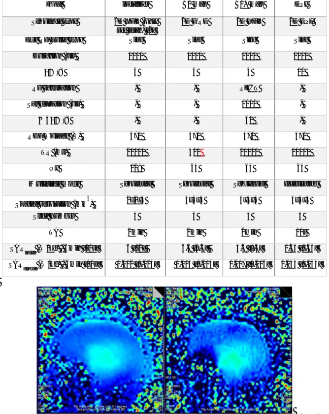
In Table, the sequences parameters are displayed with an estimation of the worst global and local SAR possible while using the worst possible REF. It is well below the limit even in the case where all the power would focus inside 10g of tissue. On Figure, the optimal setup of the XFL and the restricted mode version shows a very good agreement.
3
1234567890 ‘’“”
METANANO 2018 IOP Publishing
IOP Conf. Series: Journal of Physics: Conf. Series 1092 (2018) 012159 doi :10.1088/1742-6596/1092/1/012159
Table: Parameters of the identified “restricted SAR mode” sequences. They have been validated on
phantom and in vivo using a single channel volume coil to ensure to be sensitive enough to get reliable information on coil prototype behaviors.
Goal localizer B0 map B1+ map EPI
Sequence type 2D XFL (only
ref scan) [2]
2D GRE 2D XFL 2D EPI
Exc RF pulse type Sinc Sinc Sinc Sinc
Duration (us) 1000 1000 1000 1000
FA (°) 3 5 3 10
RF saturation - - RECT -
Sat duration (us) - - 1000 -
Sat FA (°) - - 60 -
REF Voltage (V) 470 470 470 470
TR (ms) 20000 400 20000 10000
Ny 128 64 64 64
Multislice mode Sequential Sequential Sequential Interleaved
Spatial resolution (mm
3
) 2x2x4 4x4x4 4x4x4 4x4x4
Slice number 3 5 3 3
TA 2min 2min 2min 10s
SAR
local (W/kg) - 6min [10s] 6 [12] 6.6 [6.6] 3.6 [8.3] 1.63 [1.63]
SAR
global (W/kg) - 6min [10s] 0.012 [0.024] 0.013 [0.013] 0.007 [0.016] 0.033 [0.033]
Figure : In vivo sagittal views of a B1+ head maps acquired respectively from left to right with optimal
XFL protocol and with “Restricted SAR mode” setting with InVivo Corp transceiver birdcage coil at the same resolution. The restricted SAR mode version brings SAR estimation toward zero. If all power focused into 10g of tissue, local SAR would only achieve 3.6W/kg.
4
1234567890 ‘’“”
METANANO 2018 IOP Publishing
IOP Conf. Series: Journal of Physics: Conf. Series 1092 (2018) 012159 doi :10.1088/1742-6596/1092/1/012159
4. Discussion and conclusions
We successfully demonstrated the possibility to acquire reliable information using sequence powering very limited RF deposition with a short TA making it compatible with in vivo acquisition. Using this protocol, no prior simulation of any kind is needed but no change in the sequence parameters is allowed during the examination. The sequences should therefore be compiled into a “Restricted SAR mode” version to lock the sequence parameters.
More contrasts could be considered in this mode especially T1 using Turbo FLASH (TFL) like sequence and T2* weighted with GRE sequence. T2 might be more challenging in a decent time. The demonstration was done on purpose with a transceiver head birdcage, which is known to be weak in Signal to Noise Ratio. Thus, any tests on state of the art receiving coil array should return even better results.
References
[1] Katscher U., Börnert, P., Parallel RF transmission in MRI. NMR Biomed 2006; 19: 393–400 [2] Grissom W., Yip, C., Zhang, Z., Stenger, V.A., Fessler, J.A., Noll, D.C., Spatial domain method for the design of RF pulses in multicoil parallel excitation. Magn Reson Med 2006; 56: 620–629
[3] Webb A.G. Dielectric materials in magnetic resonance. Concept Magn Reson A 2011; 38: 148-184 [4] Slobozhanyuk A.P., Poddubny A.N., Raaijmakers A.J., van den Berg C.A., Kozachenko A.V., Dubrovina I.A., Melchakova I.V., Kivshar Y.S., Belov P.A. Enhancement of Magnetic Resonance Imaging with Metasurfaces. Adv Mater. 2016 Mar 2;28(9):1832-8.
[5] Marc Dubois, Lisa Leroi, Zo Raolison, Redha Abdeddaim, Tryfon Antonakakis, Julien de Rosny, Alexandre Vignaud, Pierre Sabouroux, Elodie Georget, Benoit Larrat, Gerard Tayeb, Nicolas Bonod, Alexis Amadon, Franck Mauconduit, Cyril Poupon, Denis Le Bihan, Stefan Enoch. Kerker Effect in Ultra High Field Magnetic Resonance Imaging. Nature Communication 2018 under review
[6] International Electrotechnical Commission Medical electrical equipment-part 2–33: particular requirements for the basic safety and essential performance of magnetic resonance equipment for medical diagnosis - 3rd ed. Geneva: International Electrotechnical Commission: 2010- 2–33 :601 [7] Seifert, F., Wubbeler, G., Junge, S., Ittermann, B., Rinneberg, H., Patient safety concept for multichannel transmit coils. J. Magn. Reson. Imaging 2007; 26: 1315–1321
[8] N. Boulant, V. Gras, A. Amadon, M. Luong, G. Ferrand, A. Vignaud. Workflow proposal for defining SAR safety margins in parallel transmission. Proc Int Soc Magn Reson Med 2018;
[9] E.F. Meliado, A.J.E. Raaijmakers, P. Luijten, C.A.T. van den Berg, Magn Reson Med 2018 under review
[10] Klose U. Mapping of the radio frequency magnetic field with a MR snapshot FLASH technique. Medical Physics 1992; 19: 1099–1104
5
1234567890 ‘’“”
METANANO 2018 IOP Publishing
IOP Conf. Series: Journal of Physics: Conf. Series 1092 (2018) 012159 doi :10.1088/1742-6596/1092/1/012159
Acknowledgements:
The project leading to this publication has received funding through the M-CUBE project by the European Unions Horizon 2020 Research and Innovation programme under Grant Agreement No 736937 and by France Life Imaging grant ANR-11-INBS-0006.
This work has also received funding support from the ERPT equipment program of the Leducq Foundation and DIM “Cerveau et pensée” from Région Ile-de-France.