HAL Id: hal-00084437
https://hal.archives-ouvertes.fr/hal-00084437
Submitted on 7 Jul 2006
HAL is a multi-disciplinary open access archive for the deposit and dissemination of sci-entific research documents, whether they are pub-lished or not. The documents may come from teaching and research institutions in France or abroad, or from public or private research centers.
L’archive ouverte pluridisciplinaire HAL, est destinée au dépôt et à la diffusion de documents scientifiques de niveau recherche, publiés ou non, émanant des établissements d’enseignement et de recherche français ou étrangers, des laboratoires publics ou privés.
Alternating dCdG Sequences
E. Emanuele, K. Zakrzewska, D. Markovitsi, R. Lavery, P. Millie
To cite this version:
E. Emanuele, K. Zakrzewska, D. Markovitsi, R. Lavery, P. Millie. Exciton States of Dynamic DNA Double Helices: Alternating dCdG Sequences. Journal of Physical Chemistry B, American Chemical Society, 2005, 109, pp.16109 -16118. �10.1021/jp051833k�. �hal-00084437�
Exciton states of dynamic DNA double helices: alternating dCdG
sequences
Emanuela Emanuele,a Krystyna Zakrzewska,b Dimitra Markovitsi,a1 Richard
Laveryb and Philippe Milliéa
Laboratoire Francis Perrin CEA/DSM/DRECAM/SPAM - CNRS URA 2453, CEA Saclay, 91191 Gif-sur-Yvette, France
Laboratoire de Biochimie Théorique, CNRS UPR 9080, Institut de Biologie Physico-Chimique, 13, rue Pierre et Marie Curie, 75005 Paris, France
1
Corresponding author: Dimitra Markovitsi (dimitra.markovitsi@cea.fr) a
Laboratoire Francis Perrin b
Abstract
The present communication deals with the excited states of the alternating DNA
oligomer (dCdG)5.(dCdG)5 which correspond to the UV absorption band around 260 nm.
Their properties are studied in the frame of the exciton theory, combining molecular dynamics
simulations and quantum chemistry data. It is shown that the dipolar coupling undergoes
important variations with the site and the helix geometry. In contrast, the energy of the
monomer transitions within the double helix is not sensitive to the local environment. It is thus
considered to be distributed over Gaussian curves whose maximum and width are derived
from the experimental absorption spectra of nucleosides in aqueous solution. The influence of
the spectral width on the excited states delocalization and the absorption spectra is much
stronger than that of the oligomer plasticity. About half of the excited states are delocalized
over at least two bases. Many of them result from mixing of different monomer states and
extend on both strands. The trends found in the simulated spectra, when going from
non-interacting monomers to the duplex, are in agreement with experimental observations.
Conformational changes enhance the diversity of the states which can be populated upon
excitation at a given energy. The states with larger spatial extent are located close to the
1. Introduction
UV irradiation absorbed by DNA bases induces photochemical reactions leading to
carcinogenic mutations1-2. The major products of those photoreactions, such as bipyrimidine
dimers, cytosine hydrate or oxo-dihydroxoguanine, are now well characterized.3,4 In contrast,
very little is known about the excited states responsible for their formation. Thus, the energy
of the lowest excited states of the bases within the helix is sometimes assumed to be different
than that determined for the unstacked monomeric chromophore in solution. Attempts have
been made to determine experimentally the absorption spectrum of adenine and thymine
within the synthetic double helix (dA)n.(dT)n,5 or the energy of the thymine triplet within
native DNA.6,7 Two assumptions underlie the analysis of these experimental results: excited
states are localized on single bases and their energy is not site dependent.
The commonly accepted idea regarding the localization of the DNA singlet excited
states goes back to the sixties. Theoretical calculations, performed in the frame of the exciton
theory, concluded that the excited states in DNA should be delocalized.7-9 At the same time,
they predicted that the formation of exciton states induces large spectral shifts and a visible
splitting of the absorption band around 260 nm.8 Since those features are not observed in the
experimental absorption spectra of DNA, which were found to closely resemble the sum of the
spectra of the constituent bases, the hypothesis of localized excited states prevailed10 and
guided subsequent studies dealing with the photophysics and the photochemistry of DNA. The
possibility of excitation delocalization having been ruled out, any difference in the spectra or
the excited state reactivity between double-stranded and monomeric nucleic acids was
implicitly attributed to the influence of the local environment. Such a “solvation” effect is
In parallel with those considerations, it has been discovered that the base sequence
does play a role in various processes occurring after photon absorption. This is the case, for
example, with energy transfer11 or pyrimidine dimer formation in synthetic oligonucleotides.12
It is also observed in the lesion distribution around mutational hot spots.13 Moreover,
delocalization of singlet excited states is suggested by the steady-state fluorescence
polarization found for oligonucleotides14 and the fluorescence excitation spectra of
aminopurines incorporated in a double-stranded oligonucleotide.15 Such findings incite to a
theoretical reexamination of DNA excited states, taking advantage of progress made during
the past decades.
Our knowledge of the excited states of the components of DNA (bases, nucleosides,
nucleotides) has improved, in particular thanks to femtosecond spectroscopy.16-18 Molecular
dynamics simulations, which take into account counter-ions and water molecules, provide now
the possibility to obtain a detailed description of the local environment for a multitude of
conformations.19 Quantum chemistry data allow a very accurate calculation of dipolar
interactions which constitute the major component of the electronic coupling in the case of
allowed transitions. Moreover, the difference in the interaction energy between the permanent
atomic charges of a chromophore with the surrounding molecules in its ground and excited
states represents an important term in the energy changes due to the local environment, in
particular when excitation induces large changes in the dipole moment. Finally, the
development of computational techniques allows data extracted from different areas to be
combined in order to identify factors that affect the excited state properties of complex
systems and determine their footprint in the absorption spectra.
excited states related to the UV absorption by model double-stranded oligonucleotides.20,21 We
are interested in the way that the local environment may affect the excitation energy localized
on each base and we address the question of excitation delocalization. To this end, we perform
calculations combining molecular dynamics simulations, quantum chemistry data and the
exciton theory. 22-24 According to the exciton theory the states of a multichromophoric system
are linear combinations of the excited states of each chromophore.22-24 The properties of the
exciton states are obtained by diagonalization of the Hamiltonian matrix, in which the diagonal
and off-diagonal terms represent the excitation energy of the monomer transitions within the
system and the electronic coupling, respectively.
Our previous studies20,21 involved helices composed of only adenine-thymine base
pairs, (dA)n.(dT)n and (dAdT)n/2.(dAdT)n/2. We showed that the local environment (base
sequence, conformational changes, water molecules and counter-ions) affects only slightly the
excitation energy of adenine and thymine within these double helices. In contrast, drastic
changes in the excited state properties are induced by the dipolar coupling which leads to
delocalization of the electronic excitation with double helices having an idealized B-DNA
geometry.20 Structural fluctuations reduce the spatial extent of the excited states, but
excitations still remain delocalized over several bases.21
These studies neglected the fact that the UV absorption bands of nucleic acids are quite
large. In other terms, it was assumed that the energy of the electronic transitions of the
monomeric chromophores is not affected by changes in their structure. A general criterion for
formation of delocalized excited states is the relative magnitude of the strength of the
electronic coupling compared to the spectral width.25 The dipolar coupling between
whereas the spectral width is about one order of magnitude larger.20 Accordingly, one would
expect complete localization of the excited states. However, this effect may be compensated
by the existence of more than one monomer electronic transition, with different polarizations,
which can be coupled.26 This indeed occurs in the case of the (dA)10.(dT)10.27
The present communication focuses on the alternating oligomer (dCdG)5.(dCdG)5. We
have three objectives. First, we study the influence of the local environment on the dipolar
coupling and the excitation energy of the cytosine and guanine transitions within the double
helix. To this end, we build the exciton Hamiltonian matrix combining ground state, excited
state and transition atomic charges, extracted from quantum chemical calculations, with
ground state conformations extracted from molecular dynamics simulations. In this way, we
correlate the coupling fluctuations with the site and conformational variations of structural
parameters. Second, we examine how the spectral width, combined with conformational
changes, affects the exciton states related to photon absorption (Franck-Condon states). We
illustrate their spatial extent and we quantify the various types of delocalization (spatial,
electronic, intrastrand, interstrand). Third, we focus on the absorption spectrum. We compare
the spectrum of the double helix to that of non-interacting monomers and we associate the
observed trend to experimental data. Finally, we establish a correspondence between the
absorption spectrum and the properties of the singlet excited states providing some guidelines
for experimental photophysical and photochemical studies.
2. Methodology
2.1. Ground state molecular dynamics. The ground state conformations of
(dCdG)5.(dCdG)5 used for the calculation of its excited states were extracted from molecular
the oligomer used in the simulations was 12 base pairs long.
Model building and simulations were performed using the AMBER 6 program28 and
the Parm98 parameter set.29 The oligomer (dCdG)6.(dCdG)6 was constructed using a standard
B-DNA conformation and was neutralized with 22 Na+ ions (placed using electrostatic
potentials) and solvated with more than 6000 TIP3P water molecules in a truncated octahedral
box. Molecular dynamics simulations were performed at constant temperature (300 K) and
pressure (1 bar) using the Berendsen algorithm.30 An integration time step of 2 fs was used and
all bond lengths involving hydrogens were constrained using SHAKE.31 Long-range
electrostatic interactions were treated using the particle mesh Ewald (PME) approach32 with a
9 Å direct space cut-off. The non-bonded pair-list was updated heuristically and the center of
mass motion was removed every 10 ps during the simulation. Initially, the water molecules
and ions were relaxed by energy minimization and allowed to equilibrate at 300 K around the
fixed DNA for 100 ps at constant volume; the entire system was then heated from 100 to 300
K during 10 ps and equilibrated during 40 ps with harmonic restraints of 5.0 kcal/mol/Å2 on
the solute atoms at constant volume. Subsequently, the simulation was continued at constant
pressure; the restraints were gradually removed over a period of 250 ps and an unrestrained
simulation followed for over 4 ns. The coordinates were saved every 1 ps. The last nanosecond
was used for the further study. 100 snapshots spaced by 10 ps were selected. In order to
minimize bond length and valence angle distortions the snapshots were minimized in AMBER
for 1000 cycles before being used for Poisson-Boltzmann calculations of the electrostatic
energy. Non-linear solutions of the Poisson-Boltzmann equation were obtained with the
DELPHI program (version 2.1).33
the lowest transition of cytosine S0 → S1 and two close lying transitions of guanine S0 → S1
and S0 → S2.
The excitation energy Eim corresponding to the transition S0 → Si of a chromophore m
within the double helix is given by:
(
)
4
4
4
3
4
4
4
2
1
4
4
4
3
4
4
4
2
1
4
3
42
1
III m n m p n p 0 , 0 p , n m n 0 , 0 n , m II m n m p n p 0 , 0 p , n m n 0 , i n , m I 0 m i m i mE
E
E
E
E
⎟⎟
⎟
⎠
⎞
⎜⎜
⎜
⎝
⎛
+
−
⎟⎟
⎟
⎠
⎞
⎜⎜
⎜
⎝
⎛
+
+
ε
−
ε
=
∑
∑ ∑
∑
∑ ∑
≠ ≠ < ≠ ≠ ≠ < ≠ (1)Term (I) represents the excitation energy of free monomer m from its ground to its ith
electronic state. Term (II) corresponds to the interaction energy of the system in which
monomer m is in its ith state and all others in their respective ground states. It is computed as
the electrostatic energy of the system in water by solving the non-linear Poisson-Boltzmann
equation with AMBER atomic charges. In this calculation, atomic charge distributions
associated with the excited states of the monomers were used. Finally, term (III) is the energy
of the ground-state system, and is calculated by the Poisson-Boltzmann method as well.
Atomic charges for the excited states were constructed from ab initio calculations. We
described the change in the monomers’ electronic wavefunction upon excitation as a set of
atomic charge differences (Figure 1), computed by subtraction of the CASSCF/RESP charges
on the atoms of the ground state molecule from those corresponding to the excited state. The
active space choose for CASSCF/RESP calculation is 10 electrons in 10 molecular orbitals for
cytosine and 14 electrons in 12 molecular orbitals for guanine (we include all the valence
pi-orbitals and the lone pairs pi-orbitals of the heteroatoms). The basis set used is cc-pVZ.
The charges for the excited states of cytosine and guanine were obtained by adding the
account for the reorganization of the electronic system of the monomers upon excitation, while
retaining the generality of the AMBER force field, which is very well suited to the study of
nucleic acids.19 The calculation procedure is described in detail in the appendix of reference 21.
Figure 1
The values attributed to term ε in Equation 1 are chosen in two different ways. First, im i
m
ε are given constant values: 37 500 cm-1
for the S0 → S1 transition of cytosine; 36 000 cm-1
and 37 500 cm-1 for the S0 → S1 and S0 → S2 transitions of guanine, respectively. Second, i
m
ε obey Gaussian distributions whose width (fwhm) is 3 750, 3 600 and 3 750 cm-1
for the S0
→ S1 cytosine transition and the S0 → S1 and S0 → S2 guaninetransitions, respectively (Table
1). Both the maximum excitation energies and the spectral width of the Gaussian functions are
derived from the experimental absorption spectra of nucleosides in aqueous solutions.20
Table 1
2.3. Off-diagonal terms. The dipolar coupling was calculated using the atomic
transition charge distribution model according to which the off-diagonal terms are subjected to
a dipolar development.34 The resulting molecular transition dipoles m0 k
m r Ψ
Ψ =
μr r are then decomposed onto the atomic orbitals of the molecule, in the framework of the INDO
approximation.35 Atomic charges of the three transitions were derived from quantum
rescaled so that the computed transition moments to match the experimental transition
moments (Table 1). The coupling corresponding to all the pairs of different bases forming the
double helix was calculated.
2.4. Eigenstate properties. The detailed formalism associated with the calculation of
the eigenstates is described in reference 20. Diagonalization of the exciton matrix
corresponding to a given helical conformation and a given distribution of monomer excitation
energies yields the k eigenstates of the system which are linear combinations of the
wavefunctions <Ψn> corresponding to the monomer transitions
∑
= Ψ = N n n n k C k 1 , . Since the
double-helix studied contains ten cytosines, with one transition each, and ten guanines, with
two transitions each, it has thirty eigenstates <k>, whose energy increases from <1> to <30>.
The degree of delocalization of the exciton states is usually quantified by the
participation ratio PR=1/Lk which represents the number of coherently coupled
chromophores.36,37 When there is more than one electronic transition per chromophore, Lk is
written as follows:
∑
⎢⎣
⎡
∑
⎥⎦
⎤
=
m monomer i i m k,(C
2 states 2 k)
L
.The sum within the square brackets represents the contribution to the eigenstate <k> of
different electronic states belonging to the same monomer (base), e.g. the S1 and S2 states of
3. Results and Discussion
3.1 Local environment
The heterogeneity in the local environment of a specific base arises from its position
within the double helix and the structural fluctuations of the helix. In the present section, we
examine the effect of these factors on the dipolar coupling and the excitation energy.
During the nanosecond of simulation, from which we extracted 100 snapshots, the
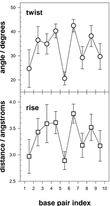
oligomer conformation shows the global characteristics of B-DNA, with an average twist of
33° and average rise of 3.34 Å. To illustrate the structural fluctuations of the oligomer, Figure
2 shows these parameters along the double helix, together with their standard deviations
calculated over the 100 conformations. It can be seen that every step has a different average
value. The curves show a dinucleotide character with both rise and twist being higher for the
GC steps than for the CG steps.
Figure 2
To further illustrate the variability of the relative position of the chromophores, we
have plotted in Figure 3 the two intra base pair parameters, propeller and buckle. The propeller
describes contra rotation angle around the long axis of the base pair and varies from -12.5° to
7.0°. The buckle describes contra rotation angle around the short axis of the base pair, and
ranges from -14.6° to 2.7°. The propeller and buckle fluctuations exhibited by a given base
pair due to conformational changes are also important.
Conformational changes of the DNA double helix have an important impact on the
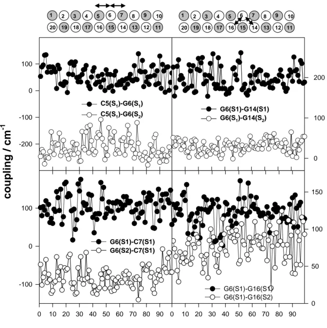
dipolar coupling. The coupling variations observed for the central part of the oligomer are
shown in Figure 4. By comparing the left side plots concerning C5-G6 and G6-C7, we remark
that the coupling between two given transitions (S0 → S1 of cytosine and S0 → S2 guanine) of
neighboring chromophores, located on the same strand, may differ by a factor of two. This is
also true for neighboring chromophores located on different strands (compare the right side
plots concerning G6-G14 and G6-G16). Moreover, for certain conformations and sites
(G6-G16), the coupling of the S0 → S1 with the S0 → S2 transition may be as high as that between
two S0 → S1 transitions whereas for other sites (G6-C14) the two coupling values differ by at
least 180 cm-1. Finally, the amplitude of fluctuations observed for the coupling involving two
specific bases depends on the type of the transitions. For example, for C5 and G6, the
fluctuations in the coupling between S0 → S1 transitions amount to 62%, but they are limited
to 18% when the S0 → S2 guanine transition is involved.
The coupling between two given transitions within a base pair varies with the position
of this pair along the double helix. For example, the average coupling between the S0 → S1
transitions of C5 and G16, obtained for 100 conformations, is 122 cm-1, whereas that
associated with the bases of the C3-G18 pair is only 95 cm-1. Similar site variations concern
the coupling between transitions of neighboring chromophores located on either the same
strand or on different strands.
To further analyze the dependence of the dipolar coupling on DNA flexibility, we
calculated the correlation coefficient between the conformation series of dipolar coupling
shown in Figure 4 and all the structural parameters associated with the relevant bases and base
pairs. To our surprise, the strongest correlation was found with the slide parameter, which
describes the relative displacement of neighboring bases or base pairs along their long axis.
The highest correlation coefficient (0.88) was obtained for the dipolar coupling between S0 →
S1 transition of G6 and S0 → S2 transition of G14 and the displacement of the base pairs
G6-C15 and C7-G14. The corresponding plots are presented in Figure 5.
Figure 5
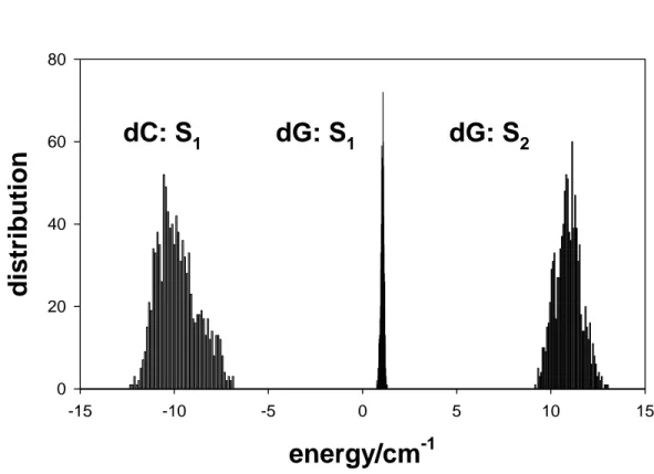
The effect of the local environment on the diagonal energy is represented in Figure 6
where the values obtained for the ten different sites corresponding to each monomer transition
within each of the 100 conformations of the double helix, that is, a total of 1000 values, are
plotted. The values found for cytosine are of opposite sign and more widely spread than those
for guanine. This trend is in line with the photo-induced changes observed in the permanent
dipole moments (Table 2) for the three transitions: -1.78 D for the S0 → S1 transition of
cytosine, +1.67 D and +0.25 D for S0 → S1 and S0 → S2 transitions of guanine, respectively. In
fact, the larger the photoinduced change in the atomic charges, the more sensitive the
excitation energy will be to the local environment as far as electrostatic interactions are
concerned. In this respect, the present data, together with those found previously for adenine
and thymine,21 show that the lowest pyrimidine transitions are more sensitive than the purine
Figure 6
In spite the fact that the fluctuations of the monomer excitation energies clearly depend
on the type of transition, they all remain very weak (< 15 cm-1) as compared to the spectral
width of room temperature solution spectra. The same result was found for (dA)10.(dT)10 and
(dAdT)5.(dAdT)5.21 This general behavior shows that modification of the excitation energy
due to electrostatic interactions with the local environment cannot account for any difference
between the absorption spectra of double helices and those of non interacting monomers
observed at room temperature.
Table 2
3.2. Eigenstate properties
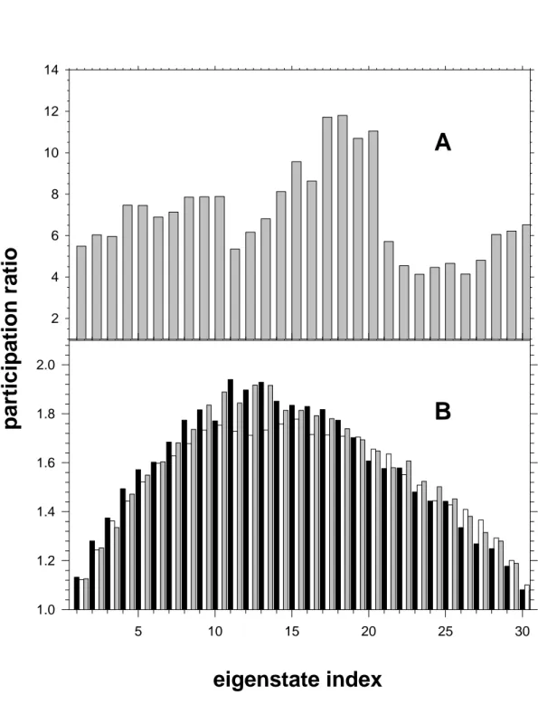
The spatial extent of the (dCdG)5.(dCdG)5 excited states obtained by diagonalization of
the exciton matrix is determined using the participation ratio PR. First, we consider only the
effect of conformational changes assuming than the electronic transitions are devoid of any
spectral width. To this end, the term ε in Equation 1 is given constant values (Table 1). The im
participation ratio calculated for each eigenstate, averaged over 100 conformations, is shown
in Figure 7A. It ranges from 4 to about 12. The highest values are encountered for <k> =
17-21. Interestingly, the same eigenstates were found to be the most delocalized in the case of the
alternating oligomer (dAdT)5.(dAdT)5 (Figure 7 in reference 21). Despite this similarity, the PR
the double helix. For example, the transition moment of the S0 → S1 transition of thymine
(3.68) is similar to that of guanine (3.31) (Table 1 in reference 20) but the coupling in a pair of
guanines is higher than the coupling in a pair of thymines located at an analogous position
within (dAdT)5.(dAdT)5 duplex. Thus, the average coupling values found for the G6-G14 and
G6-G16 guanine pairs are 122 and 198, respectively, whereas those obtained for the
corresponding thymine pairs are only 64 and 94, respectively.
Figure 7
Next, we consider the excited states of a single conformation of (dCdG)5.(dCdG)5,
associated with a corresponding set of coupling terms. The diagonal terms are obtained from
equation 1 where ε obey Gaussian distributions. Since the effect of the local environment on im
the monomer excitation energy was found to be negligible (Figure 6), the Gaussian
distributions account for homogeneous broadening. The average PR found values for three
different conformations, each one averaged over 500 sets of diagonal energy are plotted in
Figure 7B. We observe that the average PR distribution over the thirty eigenstates is similar
for the three conformations examined. The three plots in Figure 7B are quite different from
that in Figure 7A. The position of the larger PR values has moved to lower eigenstates. The
eigenstates located at the edges of the exciton band have the smallest PR. Most importantly, all
PR values have drastically decreased, ranging between 1.1 and 1.9.
We thus conclude that diagonal disorder related to the homogeneous broadening plays
a more decisive role in the spatial extent of the eigenstates of the double helix than
that the eigenstates are only weakly delocalized. However, this picture results from PR values
averaged over 500 sets of diagonal energy. Therefore, we examine below the delocalization
behavior of the duplex excited states in more detail.
Delocalization of the excitation, even weak, may manifest itself in different ways. This
is illustrated in Figure 8, where the topography of three typical eigenstates, associated with a
single conformation and a single set of monomer excitation energies, is shown. The
contribution of the excited state i, associated with the chromophore m, to eigenstate <k> is
given by the coefficient (Cik,m) which is drawn upwards or downwards, depending on the
strand to which m belongs. We observe that each eigenstate exhibits a specific topography. In
the case of <24>, 97% of the excitation is located on the same base pair; 75 % is born by the
S2 state of guanine and 22% by the S1 state of cytosine. The eigenstate <18> is mainly built on
the S1 and S2 states of a guanine, but the S1 state of the neighboring cytosine also bears 17% of
the excitation. Despite their different topography, the states <24> and <18> have similar
participation ratios, 1.6 and 1.7, respectively. Finally, the eigenstate <9> is built on only one
type of monomer state, the S2 state of guanine, and it is delocalized over several bases located
on both strands. The corresponding participation ratio is 3.7.
Figure 8
The topographies shown in Figure 8 indicate that delocalization may concern different
monomer electronic states (S1 and S2 states of the same guanine), different bases (guanines
belonging to various pairs), different types of bases (guanine or cytosine) and, finally, different
these types of delocalization, we consider (Cik,m)2max,that is the coefficient ( i m , k C )2 having the
highest value among the thirty coefficients describing a given eigenstate <k>. In Figure 9 we
show the distribution of the excited state population obtained for 500 sets of monomer
excitation energy, that is a total of 15 000 eigenstates, as a function of (Cik,m)2max. We remark
that, for 42% of the eigenstates, (Cik,m)2max is located within the interval 0.90-1.0. These
eigenstates are almost completely localized both electronically and spatially, since more than
90% of the excitation is concentrated on one excited state of a single base. For the remaining
58% of the excited state population, an increasing part of the excitation energy is shared with
at least a second electronic transition belonging either to the same or to different bases.
Figure 9
Now, we focus on the way that minor part of the excitation, corresponding to (1
-( i m , k C )2max ), is distributed. If (Cik,m) 2
max is related to a guanine (or cytosine), the probability to
find the remaining excitation on a cytosine (or guanine) is given by
)
(
max 2 i m , k e sin cyto 2 i m , kC
1
)
C
(
−
∑
and isequal to 0.55. This means that 55% of the excitation shared is encountered on a different type
of base. Following the same reasoning, 73% of the excitation shared is located on the
complementary strand.
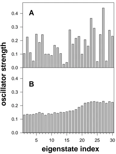
Although the double helix eigenstates are only weakly delocalized, their oscillator
strength f differs a lot with respect to that of non-interacting chromophores. This can be seen
corresponding to one conformation is plotted. We observe that the f values are quite dispersed.
They may be one order of magnitude smaller or twice as large in comparison to those of the
monomers (0.19-0.22; Table 1). If we consider many sets of diagonal energy and different
conformations, we observe that the differences between the oscillator strength of the various
eigenstates are strongly reduced. Nevertheless, on average, the f values associated with the
eigenstates located at the bottom of the exciton band are lower (ca. 0.14) than those of the
eigenstates at the top of the band for which f is around 0.22 (Figure 10B).
Figure 10
3.3. Spectral properties
The question now arises whether the weak delocalization of the excitation energy
found the double helix excited states is detectable in the absorption spectra. In order to answer
it, we compare the spectra of the double helix with those of non-interacting chromophores.
The results are shown in Figure 11 where both calculated and experimental absorption spectra
are presented on a nanometer scale, for comparison with usual experimental conditions.
The experimental spectrum of the double helix is that recorded for the synthetic
polymer poly(dCdG).poly(dCdG) in a phosphate buffer at room temperature.2 It is compared
with that of an equimolar mixture of nucleosides, 2’-deoxycytosine and 2’-deoxyguanosine. The molar extinction coefficient ε is given per base. The ε at 260 nm of
2
The nucleotides were purchased from Sigma Aldrich and dissolved in ultrapure water. The double stranded polymer was obtained from Amersham and dissolved in a phosphate buffer (pH=6.8; 0.1 M NaH2PO4, 0.1 M Na2HPO4, 0.25 M NaCl). Absorption spectra were recorded using a Perkin Lamba 900 spectrophotometer.
poly(dCdG).poly(dCdG) is taken from reference 38.
The spectrum calculated for a single conformation of (dCdG)5.(dCdG)5, was
constructed by plotting the oscillator strength of the thirty eigenstates obtained for each one of
the 500 sets of monomer transition energy values. The spectrum profiles corresponding to
different conformations are quite similar, in line with the participation ratio. The calculated
spectrum of non-interacting monomers was obtained by adding the three Gaussian curves
which represent the energy distribution of three monomer transitions, the area under each
Gaussian being proportional to the associated oscillator strength.
Figure 11
The calculated spectra of the double helix are not expected to strictly reproduce the
experimental spectra. Notably, symmetric Gaussian curves were used to simulate the monomer
transitions, whereas experimental bands are asymmetric. Moreover, at short wavelengths,
higher order transitions overlap with those taken into account in the simulations. For those
reasons, the calculated spectrum of non-interacting chromophores is different from the
experimental one. Finally, charge transfer interactions which may operate in real systems are
neglected in the present calculation of the exciton states. Despite the above limitations, it is
interesting to compare the spectral changes occurring when going from non interacting
chromophores to the double helix.
In both plots in Figure 11, we remark following trends. The monomer and double helix
spectra are located in the same spectral region; no important shifts are observed. The profiles
both cases, the relative intensity of the spectrum below 260 nm is more important for the
double helix than for non-interacting monomers and its maximum intensity is lower than that
of non-interacting monomers.
The calculated spectra of (dCdG)5.(dCdG)5 constitute an envelop corresponding to the
absorption of 30 different eigenstates. Due to both diagonal disorder, induced by the spectral
width, and off-diagonal disorder, generated by the plasticity of the double helix, the energy of
each eigenstate and, consequently, its footprint in the absorption spectrum are quite spread.
This is illustrated in Figure 12A, where the position of the thirty eigenstates <k>, obtained for
a single conformation and 500 sets of diagonal energy distributions, is represented in the form
of linear segments, together with the absorption spectrum of the double helix. We observe that
the positions of the various eigenstates overlap. Thus, excitation at a given wavelength will
populate eigenstates with different indexes, corresponding to various distributions of monomer
energy transitions. Their relative proportion depends on the associated oscillator strength. The
diversity of the excited state population formed upon photon absorption of a given energy is
further amplified if various conformations are considered. Thus, although conformational
changes do not have an important influence on the average properties of the eigenstates, they
enhance the variety of the excited state populated at a given energy.
If internal conversion among the various eigenstates (intraband scattering) is faster than
any other relaxation process, fluorescence is expected to result only from the states located at
the bottom of the exciton band. Consequently, the fluorescence spectra of the double helix
should not depend on the excitation wavelength. This is precisely what is experimentally
observed for poly(dCdG).poly(dCdG).39
Figure 12B. We note that although the average PR values do not exceed 1.9 (Figure 7), the PR
associated with some states may reach values around five. The more extended eigenstates are
located close to the absorption maximum. In contrast, the eigenstates located near the spectral
edges are mainly localized on single bases.
4. Conclusions and Comments
The main conclusions of the present theoretical study on the alternating DNA oligomer
(dCdG)5.(dCdG)5 are summarized as follows.
Our molecular dynamics simulations have shown that the oligomer exhibits typical
B-DNA geometry with important variations of intra- and inter-base pair parameters. The dipolar
coupling, calculated using atomic transition charges, is highly perturbed by conformational
changes. In contrast, the energy of the monomer transitions (S0 → S1 for cytosine, S0 → S1 and
S0 → S2 for guanine) within the double helix, calculated by combining ground state and
excited state transition charges with molecular dynamics simulations, including explicit water
molecules and counter-ions, is not sensitive to the local environment.
The properties of the (dCdG)5.(dCdG)5 excited states, calculated in the frame of exciton
theory, are less affected by off-diagonal disorder associated with the plasticity of the double
helix than by diagonal disorder associated with homogeneous broadening. The latter is
approximated by considering that the energy of the monomer transitions is distributed over
Gaussian curves, whose maxima and widths are derived from the experimental absorption
spectra of nucleosides in aqueous solution. About half of the excited states are delocalized
over at least two bases. Many of them result from mixing of different monomer states and
The trends found in the simulated spectra, when going from non-interacting monomers
to the double helix, are in agreement with experimental findings: although no important
spectral shift is observed in the oligomer spectra, their profile changes. The states with a larger
spatial extent are located close to the maximum of the absorption spectrum. The plasticity of
the double helix contributes to increased heterogeneity of the absorption spectra.
The conclusions drawn in the present study for (dCdG)5.(dCdG)5 regarding the
influence of the local environment on the monomer excitation energy within the double helix
and the dipolar coupling, are quite similar to those drawn previously in the case of oligomers
composed of only adenine-thymine base pairs.21 The dominant role of homogenous
broadening in the spatial extent of excited states within the double helix found here confirms
our findings on (dA)10.(dT)10.27 In both cases, an important part of the excited states are
delocalized over at least two bases. Consequently, all these features seem to be sequence
independent properties of DNA double helices.
It is important to stress that the degree of delocalization found in the present study for
the excited states of (dCdG)5.(dCdG)5 corresponds in fact to a lower limit of spatial extent.
This is due to two reasons. First, we have overestimated diagonal disorder by assuming that
the homogeneous spectral width is equal to the width of the experimental spectra obtained for
aqueous solutions of nucleotides. In fact, the experimental width contains both homogeneous
and heterogeneous contributions which cannot be separated at the present stage of our
knowledge. Second, we have underestimated the electronic coupling by considering only
dipolar interactions and neglecting interchromophore orbital overlap and charge transfer
interactions.
energy transfer in double helices, which so far has been supposed to proceed via excitation
hopping.5,39-41 Intraband scattering should rapidly lead to eigenstates located to the bottom of
the band. However, the properties of the latter states, whose lifetime may reach hundreds of
picoseconds or nanoseconds,42 should be altered by coupling fluctuations occurring at the
same time-scale. If the present study has shed some light onto the excited states directly
created upon photon absorption, further work, both experimental and theoretical, is needed in
order to understand the time-dependence of the DNA excited states and their relationship to
photodamage.
Acknowledgment. Financial support from the ACI “Physicochimie de la Matière
Complexe” is gratefully acknowledged. We thank Dr B. Bouvier for helpful discussions and
TABLE 1. Properties of the Gaussian curves representing the monomer transitions used
in the calculation of the (dCdG)5.(dCdG)5 eigenstates.20
Monomer transition Transition
moment (D) Area (f) Maximum (cm-1) Width (fwhm/cm-1) Cytosine S0→S1 3.45 0.21 36800 3750 S0→S1 3.31 0.19 36700 3600 Guanine S0→S2 3.31 0.21 40300 3750
TABLE 2: Norms of the permanent dipole moments (in Debye) of the electronic states of
cytosine and guanine calculated from atomic charges
Cytosine S0 Cytosine S1 Guanine S0 Guanine S1 Guanine S2
Figure Captions
1. Difference between the excited state and the ground state atomic charges
corresponding to the S0→S1 transition of cytosine and the S0→S1 and S0→S2 transitions of
guanine; negative and positive charges appear as black and grey disks, respectively. The
area of the disks is proportional to the absolute value of the change in charge.
2. Intrer-base pair parameters of (dCdG)5.(dCdG)5. Twist is the rotation around
the helical axis. Rise is the distance between the successive base pairs along the helical
axis. The values are averaged over 100 conformations. The error bars correspond to
standard deviation.
3. Intra-base pair parameters of (dCdG)5.(dCdG)5. Propeller and buckle are
contra-rotations around the long and short axes of the base pair, respectively. Average
values over 100 conformations. The error bars correspond to standard deviation.
4. Dipolar coupling determined for 100 conformations of (dCdG)5.(dCdG)5
extracted from the molecular dynamics simulations. Left side plots: coupling between
transitions of neighboring cytosine and guanine pairs located in the same strand. Right side
plots: coupling between transitions of two neighboring guanines located on different
strands. The double strand is schematically represented by grey (cytosine) and white
5. Dependence of dipolar coupling on the (dCdG)5.(dCdG)5 conformation.
Coupling between the S0→S1 transitions of G6 and G14 (grey line; left axis). Slide
parameter between the base pairs G6-C15 et C7-G14 (black line; right axis). Slide
describes the relative displacement of the successive base pairs along their long axis.
6. Modification of the monomer excitation energy due to electrostatic interactions
with the local environment. Energy distribution determined for the ten cytosines and ten
guanines of (dCdG)5.(dCdG)5 in 100 conformations extracted from the molecular dynamics
simulations.
7. Participation ratio corresponding to the 30 eigenstates of the (dCdG)5.(dCdG)5.
(A) Only the local environment effect on the diagonal energy is considered (average values
over 100 conformations). (B) Average values over 500 distributions of monomer transition
energies; black, grey and white bars correspond to different oligomer conformations.
8. Topography of the eigenstates <24>, <18>, and <9> obtained for a same
conformation of (dCdG)5.(dCdG)5 and the same diagonal energy distribution. The
coefficients ( i m , k
C ) represent the contribution of chromophore m in its ith excited state to
eigenstate <k>. The upper and lower parts of each histogram refer to chromophores located
9. Distribution of the eigenstate population of (dCdG)5.(dCdG)5 as a function of
the maximum value encountered among the 30 (Cik,m)2 coefficients. A single conformation
and 500 diagonal energy sets are considered.
10. Oscillator strength corresponding to the 30 eigenstates of (dCdG)5.(dCdG)5. (A)
A single conformation and a single distribution of the monomer transition energies, both
chosen randomly, are considered. (B) Average values over 500 distributions of monomer
transition energies obtained for a single conformation of the double helix.
11. Comparison between the absorption spectra of the double helix (solid lines)and
those of non-interacting monomers (dashed lines). The experimental spectra were obtained
for poly(dCdG).poly(dCdG) and an equimolar mixture of nucleosides dC and dG; the molar extinction coefficients (ε) are given per base. The calculated spectra of (dCdG)5.(dCdG)5 were obtained for two different conformations (500 diagonal energy
distributions per conformation); the total oscillator strength was normalized per base pair.
The monomer calculated spectrum is the sum of the Gaussians corresponding to the three
monomer transitions considered.
12. Position of the eigenstates (linear segments) and their participation ratio
(circles) with respect to the absorption spectrum obtained for a single conformation of
REFERENCES
(1) Cadet, J.; Vigny, P. The Photochemistry of Nucleic Acids. In Bioorganic Photochemistry;
Morrison, H., Ed.; John Wiley & Sons: New York, 1990; pp 1.
(2) Dumaz, N.; Van Kranen, H. J.; De Vries, A.; Berg, R. J. W.; Wester, P. W.; Van Kreijl, C.
F.; Sarasin, A.; Daya-Grosjean, L.; De Gruijl, F. R. Carcinogenesis 1997, 18, 897.
(3) Cadet, J.; Berger, M.; Douki, T.; Morin, B.; S., R.; Ravanat, J. L.; Spinelli, S. Biol. Chem.
1997, 378, 1275.
(4) Cadet, J.; Douki, T.; Pouget, J. P.; Ravanat, J. L.; Sauvaigo, S. Curr Prob. Dermat. 2001,
29, 62.
(5) Georghiou, S.; Phillips, G. R.; Ge, G. Biopolymers 1992, 32, 1417.
(6) Gut, I. G.; Wood, P. D.; Redmond, R., W. J. Am. Chem. Soc. 1996, 118, 2366.
(7) Tinoco Jr., I. J. Am. Chem. Soc. 1960, 82, 4785.
(8) Rhodes, W. J. Am. Chem. Soc. 1961, 83, 3609.
(9) Miyata, T.; Yomosa, S. J. Phys. Soc. Jpn. 1969, 27, 727.
(10) Eisinger, J.; Shulman, R. G. Science 1968, 161, 1311.
(11) Xu, D.-G.; Nordlund, T. M. Biophys. J. 2000, 78, 1042.
(12) Douki, T.; Zalizniak, T.; Cadet, J. Photochem. Photobiol. 1997, 66, 171.
(13) Sage, E. Photochem. Photobiol 1993, 57, 163.
(14) Wilson, R. W.; Callis, P. R. J. Phys. Chem. 1976, 80, 2280.
(15) Rist, M.; Wagenknecht, H.-A.; Fiebig, T. ChemPhysChem 2002, 8, 704.
(16) Pecourt, J.-M. L.; Peon, J.; Kohler, B. J. Am. Chem. Soc. 2001, 123, 10370.
(18) Onidas, D.; Markovitsi, D.; Marguet, S.; Sharonov, A.; Gustavsson, T. J. Phys. Chem. B
2002, 106, 11367.
(19) Giudice, E.; Lavery, R. Acc. Chem. Res. 2002, 35, 350.
(20) Bouvier, B.; Gustavsson, T.; Markovitsi, D.; Millié, P. Chem. Phys. 2002, 275, 75.
(21) Bouvier, B.; Dognon, J. P.; Lavery, R.; Markovitsi, D.; Millié, P.; Onidas, D.;
Zakrzewska, K. J. Phys. Chem. B 2003, 107, 13512.
(22) Frenkel, J. Phys. Rev. 1931, 37, 1276.
(23) Davydov, A. S. Theory of Molecular Excitons. In Theory of Molecular Excitons; Plenum
Press: New York, 1971.
(24) Rashbah, E. I.; Sturge, M. D. Excitons; North-Holland: Amsterdam, 1982.
(25) Förster, T. Delocalization excitation and excitation transfer. In Modern Quantum
Chemistry; Sinanoglu, O., Ed.; Academic Press: New York, 1965; Vol. 3; pp 93.
(26) Markovitsi, D.; Gallos, L. K.; Lemaistre, J. P.; Argyrakis, P. Chem. Phys. 2001, 269, 147.
(27) Emanuele, E.; Markovitsi, D.; Millié, P.; Zakrzewska, K. ChemPhysChem 2005, in press.
(28) Case, D. A.; Pearlman, D. A.; Caldwell, J. W.; Cheatham III, T. E.; Ross, W. S.;
Simmerling, C. L.; Darden, T. A.; Metz, K. M.; Stanton, R. V.; Cheng, A. L.; Vincent, J. J.;
Crowley, M.; Tsui, V.; Radmer, R. J.; Duan, Y.; Pitera, J.; Massova, I.; Seibel, G. L.; Singh,
U. C.; Weimer, P. K.; Kollman, P. A. AMBER 6; University of California: San Francisco.
1999.
(29) Cheatham, T. E.; Cieplak, P.; Kollman, P. A. J. Biomol. Struct. Dyn. 1999, 16, 845.
(30) Berendsen, H. J. C.; Postma, J. P. M.; van Gunsteren, W. F.; DiNola, A.; Haak, J. R. J.
Chem. Phys. 1984, 81, 3684.
(32) Darden, T.; York, D.; Pedersen, L. J. Chem. Phys. 1993, 98, 10089.
(33) Sharp, K. A.; Honig, B. Annu. Rev. Biophys. Biophys. Chem. 1990, 19, 301.
(34) Claverie, P. Persp. Quant. Chem. Biochem. 1978, 2(Intermolecular Interactions - From
Diatomics to Biopolymers), 69.
(35) Pople, J. A.; Beveridge, D. L.; Dobosh, P. A. J. Chem. Phys. 1967, 47, 2026.
(36) Dean, P. Rev. Mod. Phys. 1972, 44, 127.
(37) Schreiber, M.; Toyosawa, Y. J. Phys. Soc. Jpn. 1982, 51, 1537.
(38) Allen, F. S.; Gray, D. M.; Roberts, G. P.; Tinoco Jr., I. Biopolymers 1972, 11, 853.
(39) Huang, C.-R.; Georghiou, S. Photochem. Photobiol. 1992, 56, 95.
(40) Georghiou, S.; Zhu, S.; Weidner, R.; Huang, C.-R.; Ge, G. J. Biomol. Struct. 1990, 8, 657.
(41) Ge, G.; Georghiou, S. Photochem. Photobiol. 1991, 54, 477.
Figure 1
N N O N H H H H H N H N N N O N H H H H N H N N N O N H H H H 0.1eGuanine S
0→S
1Guanine S
0→S
2Cytosine
S
0→S
1Figure 2
1 2 3 4 5 6 7 8 9 10angle /
degrees
20 30 40 50 1 2 3 4 5 6 7 8 9 10dist
ance / angst
röms
2.5 3.0 3.5 4.0twist
rise
Figure 3
base pair index
-20 -10 0 10 20
base pair index
1 2 3 4 5 6 7 8 9 10
angle /
degrees
-20 -10 0 10 20propeller
buckle
Figure 4
0 10 20 30 40 50 60 70 80 90 0 50 100 150 G6(S1)-G16(S1) G6(S1)-G16(S2) -200 -100 0 100 C5(S1)-G6(S1) C5(S1)-G6(S2) 0 10 20 30 40 50 60 70 80 90co
up
lin
g
/ cm
-1 -100 0 100 G6(S1)-C7(S1) G6(S2)-C7(S1) 0 100 200 G6(S1)-G14(S1) G6(S1)-G14(S2) 1 2 3 4 5 6 7 8 9 10 11 12 13 14 15 16 17 18 19 20 1 2 3 4 5 6 7 8 9 10 11 12 13 14 15 16 17 18 19 20conformation number
Figure 5
0 10 20 30 40 50 60 70 80 90 100coupl
ing / cm
-1 -100 0 100 200 300 -1 0 1 2 3conformation number
slid
e
/ Å
1 2 3 4 5 6 7 8 9 10 11 12 13 14 15 16 17 18 19 20Figure 6
energy/cm
-1 -15 -10 -5 0 5 10 15distr
ibution
0 20 40 60 80dC: S
1dG: S
1dG: S
2Figure 7
5 10 15 20 25 30 2 4 6 8 10 12 14eigenstate index
participati
on ratio
5 10 15 20 25 30 1.0 1.2 1.4 1.6 1.8 2.0A
B
Figure 8
base pair index
1 2 3 4 5 6 7 8 9 10 -0.9 -0.6 -0.3 0.0 0.3 0.6 0.9 -0.9 -0.6 -0.3 0.0 0.3 0.6 0.9 -0.9 -0.6 -0.3 0.0 0.3 0.6 0.9 Guanine S1 Guanine S2 Cytosine S1 <k> = 18 PR = 1.7 <k> = 24 PR = 1.6 <k> = 9 PR = 3.7


