HAL Id: tel-01285303
https://tel.archives-ouvertes.fr/tel-01285303
Submitted on 9 Mar 2016HAL is a multi-disciplinary open access
archive for the deposit and dissemination of sci-entific research documents, whether they are pub-lished or not. The documents may come from teaching and research institutions in France or abroad, or from public or private research centers.
L’archive ouverte pluridisciplinaire HAL, est destinée au dépôt et à la diffusion de documents scientifiques de niveau recherche, publiés ou non, émanant des établissements d’enseignement et de recherche français ou étrangers, des laboratoires publics ou privés.
Mechanisms of delayed wound healing in various models
of human diseases
van Tuan Nguyen
To cite this version:
van Tuan Nguyen. Mechanisms of delayed wound healing in various models of human diseases. Hu-man health and pathology. Université Pierre et Marie Curie - Paris VI, 2015. English. �NNT : 2015PA066280�. �tel-01285303�
1
PIERRE AND MARIE CURIE UNIVERSITY
Graduate school of Physiology, Physiopathology and Therapeutics
Mechanisms of delayed wound healing in various models
of human diseases
Presented by
Van Tuan NGUYEN
PhD Thesis
Specialty: Physiology, Physiopathology and Therapeutics
Directed by
Prof. Sélim ARACTINGIPresented on 7th September 2015
In front of the jury consist of:
Pr Stephane Hatem President Dr Kiarash Khosrotehrani Reviewer Dr Michel Simon Reviewer Dr Charles Durand Examinator Dr Nicolette Farman Examinator
2
Acknowledgment:
I am happy to say a thank to the jury members for taking the time to evaluate my thesis. I especially thank to the reviewers have reviewed my manuscript and helping me to improve it through their remarks and proposals.
I acknowledge Vietnamese government for supporting me the scholarship during 3 years.
I also acknowledge FRM for giving me the funding for the important fourth year of thesis.
I would like say a huge thank to my supervisor, Sélim for supporting and taking care my life during four years in Paris. I always see you as my father.
I would like to say a big thank to my advisor, Nicolette who always beside me during four years. I particularly thank you for your huge help and putting up me during the months of writing the manuscript and thesis. Thank you for the world formed research, for giving an excellent job and for sending me your knowledge.
Karine Raymond, many thanks for teaching me techniques and giving me useful suggestions and comment. But also for the long hours of discussions during manipulations as well as motivation and support you have given me during these years. Especially, I would acknowledge you for your important help in applying the funding for the fourth year of my thesis.
Happy to thank Frederic have mounted the MR and wound healing project and help us in our reflections. Thanks to members from your lab: Basile, Soumaya, Christian. I would also like to send a thank to my friend, Dany who has always offered extremely useful suggestions and comment for my work.
Thanks to Michele, Marie-Jo and Aude forever taking the time to help me.
Thanks to my colleagues from the laboratory both past and present: Sarah, Thomas, Geoffroy, Zhe, Mathieu, Romain, for taking the time to give me a help for discussion and manipulations in the laboratory.
3
I thank the friends, colleagues in Saint Antoine Research Center for the help you have given me at all levels.
Thanks to my friends in UEVF for the funny time.
Thanks to all my friends and family for your support during these four years.
Finally, I especially thank my wife, Lĩnh for take caring all of house work and never complain each time I leave home from early and come back very late at night. Thanks to my daughter, Thảo Anh who always happy with us in our most difficult time.
4
Mechanisms of delayed wound healing
in various models of human diseases
5
Abstract :
Impaired wound healing is a major unsolved clinical problem in aging, diabetes, sickle cell disease (SCD), glucocorticoid (GC) therapy or Cushing syndrome.
First, we investigated the mechanisms underlying SCD ulcers, using SAD transgenic mouse model with mutated human beta globin. Old SAD mice displayed delayed healing correlated with the severity of anemia and hemolysis, as in human SCD, related to impaired cutaneous angiogenesis and poor endothelial progenitor cell (EPC) mobilization from the bone marrow. CXCL12 secretion by keratinocytes and inflammatory cells in SAD wounds was low. Noticeably, local wound therapy with EPCs or with recombinant CXCL12 restored wound angiogenesis and healing in old SAD mice.
Second, we questioned the role of mineralocorticoid receptor (MR) in wound healing and proposed that MR blockade could improve reepithelialization in pathological situations (GC-treated skin, diabetic mouse models); I also participated to the demonstration that MR blockade limits GC-induced epidermal atrophy in human skin. We used skin explants from mouse and humans, full thickness skin wounds on the back of mice, and analyzed wound closure after skin biopsy in healthy volunteers pre-treated with GC, as a post-hoc study of the SPIREPI clinical trial. We found that MR blockers can rescue the pathological delayed wound healing while they did not modify wound closure in normal condition. We show that the improvement of GC-induced wound healing delay by MR blockade involves restoration of keratinocyte proliferation and down-regulation of the epithelial sodium channel. These results suggest an important and entirely novel role for MR in to improve delayed wound closure.
6
Résumé:
Le retard de cicatrisation (RETCIC) est un problème clinique majeur, touchant les gens âgés, les diabétiques, drépanocytaires (DR), ou traités par les glucocorticoides (GC). Nous avons abordé les mécanismes des ulcères DR dans un modèle transgénique ayant un gène beta globine humain muté (SAD). Les souris SAD âgées ont un RETCIC corrélé au degré d’anémie et d’hemolyse, comme dans la DR humaine ; le RETCIC est lié à l’altération de l’angiogenèse cutanée et un faible recrutement de cellules endothéliales progénitrices (EPC) depuis la moelle. La sécrétion de CXCL12 dans la plaie par les kératinocytes et les cellules inflammatoires est diminuée. Fait notable, le traitement local de la plaie par des EPC ou du CXCL12 recombinant restaurent l’angiogenèse et une cicatrisation des souris SAD âgées.
Nous avons ensuite abordé le rôle du récepteur minéralocorticoide dans la cicatrisation cutanée, et proposé ses antagonistes (MRA) pour améliorer le RETCIC en pathologie (traitement local GC, diabète murin); j’ai aussi participé à la démonstration de l’effet bénéfique des MRA sur l’atrophie épidermique GC-induite chez l’homme. A l’aide d’explants de peau murine et humaine, de plaies cutanées crées sur le dos de souris, et par l’analyse de la cicatrisation de biopsies chez le volontaire sain après traitement GC (étude post-hoc de l’essai SPIREPI), nous montrons que les MRA améliorent le RETCIC pathologique, et n’affectent pas la peau saine normale. Nous avons montré que l’effet bénéfique des MRA topiques implique une augmentation de la prolifération kératinocytaire et le canal sodium épithelial. Ces résultats montrent un rôle inédit et important du MR cutané dans la cicatrisation.
7
Key words
:
SkinWound healing
Sickle cell disease
Leg ulcers
SAD
Angiogenesis
Endothelial progenitor cells
Inflammation Proliferation Re-epithelialization Mineralocorticoid receptor Glucocorticoid Aldosterone Antagonist CXCL12
8
Laboratory attachment:
All works of my thesis have been done at the "Team of progenitors and endothelial cells during and after pregnancy", led by Professor Sélim Aractingi.
Address:
Laboratory of progenitors and endothelial cells during and after pregnancy Saint Antoine research center, UPMC/INSERM UMR S 938
27 rue de Chaligny 75012 Paris
9
CONTENTS
CHAPTER 1 - INTRODUCTION ... 10
Mechanisms implicated in normal skin wound healing ... 11
I A Endothelial progenitor cell- a key actor of angiogenesis and wound repair ... 28
I B Re-epithelialization: role of nuclear receptors and ligands ... 33
I C Abnormal wound healing in human and/or mice context ... 35
I D -II-AIMS OF WORK ... 39
CHAPTER 2 - DELAYED WOUND HEALING IN MURINE MODEL OF SICKLE CELL DISEASE 40 I-INTRODUCTION ... 40
Sickle cell disease ... 40
I A Sickle cell Leg ulcers ... 45
I B Murine model of sickle cell disease ... 46
I C -II-ARTICLE 1:SUBMITTED ... 48
CHAPTER 3 - ROLE OF MINERALOCORTICOID RECEPTOR IN SKIN WOUND HEALING .... 83
I-INTRODUCTION:BIOLOGY OF MINERALOCORTICOID RECEPTOR ... 83
Structure of mineralocorticoid receptor ... 84
I A The ligands of mineralocorticoid receptor ... 85
I B Mineralocorticoid receptor specificity ... 86
I C Mineralocorticoid receptor and pathology of skin ... 88
I D -II-ARTICLE 2: ... 91
III-ARTICLE 3: ... 121
GENERAL DISCUSSION, CONCLUSIONS AND PERSPECTIVES ... 132
10
CHAPTER 1 - INTRODUCTION
The primary function of the skin is to serve as a protective barrier against the environment. Loss of the integrity of large portions of this barrier -as a result of injury or disease- may lead to major complications or even death. The ability of the skin to repair any wound is therefore a main skin “duty” in order to keep a proper homeostasis. Wound healing represents therefore a major organ functional ability of the skin in humans as well as in mammals.
Skin repair is a multistep complex biological process that requires a close interaction of multiple cell types in a highly coordinated program. It involves hemostasis, inflammation, angiogenesis, migration and proliferation of progenitor cells, as well as production and remodeling of the extracellular matrix (ECM). As a peculiar example recent studies have allowed a better understanding of the intervention of distant stem cells derived from distant tissues such as bone marrow in order to allow cutaneous healing.
Abnormalities of wound healing are very common. These lead mainly to impaired repair that itself provokes chronic ulcers. The causes of such healing delays are multiple. During the work of this PhD, we chose to focus on the mechanisms implicated in a severe circumstance of wound healing delay, namely sickle cell disease. We also extended my work toward the implication of a new pathway implicated in cutaneous repair, namely mineralocorticoid receptor (MR) activation. Such pathway may play a beneficial role in important disease with healing delay such as diabetes and skin adverse reactions to glucocorticoid treatments.
11
Mechanisms implicated in normal skin wound
I - A -
healing
The normal mammalian response to cutaneous injury occurs in overlapping stages: detersion, inflammation, new tissue formation, and remodeling (Singer and Clark 1999; Gurtner et al. 2008). Full healing is achieved only after a long lag time that may reach one year in humans. Many cellular and molecular players are involved in well-orchestrated dynamic and very complex processes. These phenomena include phagocytosis, production of enzymes and reactive oxygen species, cell proliferation, cell apoptosis, and migration that occur at different time points. Recent discoveries have led to many attempts to cell and stem cell therapies for pathological wound healing (Lau et al. 2009). The main achievements concerning the four phases of cutaneous wound healing will be summarized here.
12
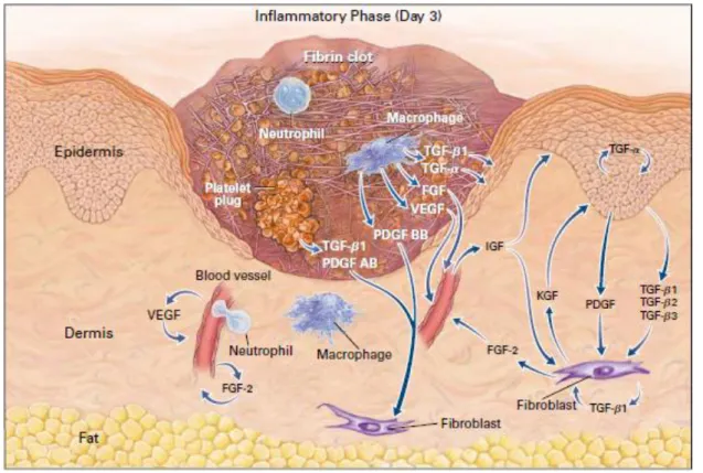
Figure 1. Stages of wound healing process.
Schematic representation of the three different phases of wound repair (Werner and Grose 2003).
A- Inflammatory phase: 12–24 h after injury the wounded area is filled with a blood clot. Neutrophils have invaded into the clot.
B- New tissue formation: at days 3–7 after injury, the majority of neutrophils have undergone apoptosis. Instead, macrophages are abundant in the wound tissue at this stage of repair. Endothelial cells migrate into the clot; they proliferate and form new blood vessels. Fibroblasts migrate into the wound tissue, where they proliferate and deposit extracellular matrix. The new tissue is called granulation tissue.
13
Keratinocytes proliferate at the wound edge and migrate down the injured dermis and above the provisional matrix.
C- Tissue remodeling: 1–2 weeks after injury the wound is completely filled with granulation tissue. Fibroblasts have transformed into myofibroblasts, leading to wound contraction and collagen deposition. The wound is completely covered with a neo-epidermis.
I - A - 1 - Inflammatory phase
This stage corresponds to the classical early stage of wound repair although all phases are redundant (Figure 2). Components of the coagulation cascade, inflammatory pathways and immune system are needed to prevent ongoing blood and fluid losses, to remove dead and dying tissues and to prevent infection. This stage involves hemostasis first, and mechanisms of inflammatory response as the second step.
Figure 2. The first stage of wound healing – inflammation.
Schematic representation of main cellular actors involved in wound inflammation (Singer and Clark 1999).
14
1- Hemostasis
Skin injury causes vascular disruption and extravasation of blood constituents. The first step of wound healing is thus hemostasis to stop the bleeding. During this initial phase, three steps occur in a rapid sequence. Vascular spasm is the first response as the blood vessels constrict in the earlier minutes to prevent ongoing blood and fluid losses. In the second step, the activated platelets and endothelial cells from existing but altered dermal capillaries will play a major role. The formation of the platelet plug results from the platelet aggregation at the sites of vessel breaks. Platelets stick together to form a temporary seal that covers the break in the vessel wall. This plug is fragile. Is strengthening requires the formation of a blood clot which, leading to the formation of a fibrin clot. Such aggregated platelets on altered endothelia provides an important matrix scaffold for neutrophils and monocytes (Tonnesen et al. 2000). Indeed such scaffold reinforced by the fibrin clot also serves as a gradient of cytokines, chemokines and growth factors that are able to direct inflammatory cell recruitment. To do so, platelets secrete several chemokines and growth factors implicated in early wound repair stage such as IL-1, TNF, TGF-α, TGF-β, PDGF and VEGF, platelet factor IV (PF4) and complement proteins (Broughton et al. 2006; Barrientos et al. 2008). In addition, activated endothelial cells activate COX-2 and -synthesize products of arachidonic acid degradation, namely various prostaglandins that cause vasodilation and an augmentation of vascular permeability, and leukotrienes, particularly LTB4, which activate and recruit neutrophils and macrophages (Broughton et al. 2006).
2- Inflammatory response
Shortly after plug formation, polymorphonuclear leukocytes (neutrophils, PMN) begin to transmigrate into the wound site, followed later by monocytes/macrophages. They are recruited to the wound in response to IL-1, TNF, TGF-β, PF4 mainly coming from platelets, and later from activated endothelial cells.
As said, neutrophils invade the wound in large numbers, within a few minutes of injury, as a result of IL-1, TNF, TGF-β, PF4 secretion locally (Singer and Clark 1999). Neutrophils transmigrate through the vascular wall by diapedesis, this being
15
facilitated by the modifications of permeability induced by secreted prostaglandins. The primary function of recruited neutrophils is the debridement of devitalized tissue and phagocytosis of foreign particles, as well as bacteria. These neutrophils also secrete proteolytic enzymes such as serine proteases (cathepsin G, proteinase-3, elastase, urokinase-type plasminogen activator) and metalloproteases (Weiss 1989). As a consequence of the functionality of myeloperoxidase (MPO), PMN generate cationic peptides and oxygen free radicals, which display anti-microbial activity (Broughton et al. 2006; Eming et al. 2007). Furthermore, some studies showed that neutrophils are an important source for pro-angiogenic factors, including VEGF-A and IL-8 (Li et al. 2003; Ancelin et al. 2004; Ohki et al. 2005; Schruefer et al. 2006). During normal wound healing in mice, the number of PMN is maximal at day 2 of the wound and then gradually decreases and disappears, even before complete re-epithelialization (Kim et al. 2008). Their presence in large numbers is observed in chronic wounds (Simpson and Ross 1972; Diegelmann 2003). Neutrophils are eliminated primarily by apoptosis and then by phagocytosis by macrophages (Witko-Sarsat et al. 2011).
The influx of neutrophils is quickly followed by the migration of monocytes. These are recruited from the circulating blood and marrow to the wound site in response to chemo-attractants and differentiate locally into macrophages (Werner and Grose 2003). In addition to these major newly recruited monocytes/macrophages, another population of resident skin macrophage is also presented at the wound side, although with a low density and minor role (MacDonald et al. 2010). The infiltration of macrophages increase during the inflammatory stage, peak at day 2 of the wound and their number gradually decreases during maturation stage, but these cells are massively remained at later time (Martin and Leibovich 2005). There are several evidences for characteristic of macrophages. Some reports have demonstrated macrophages in wound as the IL4 activated cells since these macrophages express various markers of tissue remodeling after activated by IL4 (Raes et al. 2002; Wynes and Riches 2003; Mosser and Edwards 2008; Martinez et al. 2009). In contrast, a work on aseptic wounds described wound macrophages as cells bearing the activation markers such as TNFα, mannose receptor (Daley et al. 2010).
The importance of inflammatory cells in wound healing is a hot topic. Macrophages are the cells that are responsible for ending the local inflammation steps as well as
16
the initiation of the following steps of proliferation, angiogenesis, granulation tissue formation, fibroblasts recruitment, and remodeling of the wound (DiPietro 1995). However all these tasks relate on two types of macrophages, called the M1 and M2 macrophages. Of note, the M1 macrophages are activated in response to microbial agents like lipopolysaccharide (LPS) and inflammatory cytokines like IFN. In turn, these cells exhibit antimicrobial properties by release of many important pro-inflammatory cytokines such as TNFα, IL-1β, IL-6, or IL-12, and overexpress MHC class II molecules. On the other hand, M2 macrophages, which seen as a result of activation macrophages by IL4 and IL13, are activated macrophages. They are a much more heterogeneous population, composed of all macrophages that do not correspond to M1 characteristics (Gordon 2003; Mantovani et al. 2004). It has been reported that M2 macrophages play an important role in angiogenesis, wound healing and the protection against parasitic infection (Gordon 2003). However, these cells are also involved in several diseases, such as fibrosis, allergy (Duffield 2003). Thus, both M1 and M2 macrophages are pivotal for healing process. Therefore, the balance between the two phenotypes is important in the different stage of wound healing. More M1 macrophages are needed to remove debris and to kill invading pathogens, while M2 macrophages may have more important role in later phases. In this context, study of Sindrilaru et al indicate that an unrestrained proinflammatory M1 macrophage population lead to alteration of wound healing in humans and mice (Sindrilaru et al. 2011).
The infiltration of macrophages into wound bed is changed during the healing process suggesting different roles of these inflammatory cells in the diverse phases of wound repair. Indeed, it has recently been demonstrated that early depletion of macrophages during the inflammation phase of a surgical wound leads to a reduction of angiogenesis and the formation of granulation tissue together with an alteration of the re-epithelialization (Mirza et al. 2009; Lucas et al. 2010). In contrast, an invalidation of macrophages during the intermediate stage of proliferation causes a hemorrhage in wound and prevents the transition to the maturation and remodeling phase. Finally, depletion of macrophages during the late phase of remodeling does not affect the final result (Goren et al. 2009; Lucas et al. 2010). However, it has been suggested that inflammatory cells are not needed for wound closure since the study showed that wound healing on PU.1 null mice (which lacked macrophages, mast
17
cells and functional neutrophils) treated with antibiotic is similar to wild types. The actions of macrophages are mediated by the secretion of a large number of cytokines and growth factors including EGF, TGF-α, TGF-β, PDGF and VEGF, IL-1, IL-6 and TNF-α (Barrientos et al. 2008). Macrophages also participate to the debridement of wounds by releasing NO in the extracellular medium by the action of iNOS induced by IL-1 and TNF-α (Goldman et al. 2004).
Of note, in addition to neutrophils and macrophages, other innate immune cells, such as mast cells, dendritic cells, eosinophils and basophils can be recruited in the wound bed (Noble and Jiang 2006). Moreover, a role for T cells and B cells in the wound healing process has also been demonstrated. Indeed, in the absence of dendritic cell-derived T cells, healing is delayed (Jameson et al. 2002), and epidermally-derived T cells locally secrete growth factors in the skin during the healing process (Toulon et al. 2009). Mice that are deficient in B-1 cells exhibit increased inflammatory cell infiltration, particularly neutrophils, and have a delayed healing response (Oliveira et al. 2010).
I - A - 2 - New tissue formation
This proliferative phase includes, angiogenesis, and proliferation and migration of fibroblasts, synthesis of components of the extracellular matrix of the dermis and finally keratinocyte proliferation. It is characterized by the formation of granulation tissue and results in healing of the wound.
1- Angiogenesis and vasculogenesis
Angiogenesis is a vital component of the normal healing process. In addition to angiogenesis which consists a sprouting of capillaries from preexisting blood vessels, several studies suggest that the vasculogenesis process also participates in the vascular regeneration during wound healing. It consists of generation of new vessels, from endothelial progenitor cells (EPCs) mostly derived from the marrow or even resident skin (Amoh et al. 2004; Eming et al. 2007) (figure 4).
18
Figure 4. Angiogenesis and vasculogenesis: This is part of the proliferative
phase. Angiogenesis is predominantly regulated by macrophages, endothelial cells and keratinocytes. The most important mediator during this phase is VEGF (Mahdavian Delavary et al. 2011).
In setting of injury, resident skin endothelial cells (ECs) migrate into the wound at the leading tip of capillaries, forming tube-like structures (ref) that continue to extend, branch, and creating networks. These events are first promoted by the resident skin progenitors, and of course require a dynamic temporally and spatially regulated interaction between ECs various angiogenic factors, and surrounding ECM proteins (Tonnesen et al. 2000). The angiogenic factors that can stimulate wound angiogenesis are gradually being elucidated. Indeed, FGF-1 and FGF-2 are the first members of FGF family to be discovered that have potent angiogenic activity (ref). Fibrin and fibrinogen bound FGF-2 could enhance the proliferation of ECs through induction of intergrin v3 and FGF-R1 (Sahni et al. 1999; Sahni and Francis 2004). In addition, induction of v3 by FGF-2 can also facilitate localization of MMP-2 to ECs that can subsequent promote EC migration (Brooks et al. 1996). Furthermore, FGF-2 together with VEGF, TNF-a and hypoxia could promote increase of plasmin levels, which activates invasion of ECs into fibrin matrix, modulating EC migration and angiogenesis (Koolwijk et al. 1996).
19
In addition to FGF, VEGF is vital angiogenic factor in wound angiogenesis. This protein is secreted by numerous cell types including keratinocytes, macrophages, ECs, fibroblasts. Regarding to functional activity, it has been demonstrated that VEGF can bind to fibrin or fibrinogen, that in turn facilitates localization and mitogenic activity of VEGF at wound sites in modulating EC proliferation (Sahni and Francis 2000). Of note, functional activity of VEGF is also enhanced through the interaction with it receptor VEGFR. Indeed, VEGFR2 that express on surface of ECs, interacts with VEGF and induce the activity of ECs, stimulate the formation of new capillary. Moreover, the activity of VEGF is modulated by up-regulation of several intergrin receptors (v3, 11, and 21) and enhanced expression of the anti-apoptotic protein Bcl-2 (Senger et al. 1996; Senger et al. 1997). The expression of VEGF during wound healing is variety and is regulated by many factors including hypoxia, eNOS derived NO, and inflammatory cytokines (Murohara et al. 1998; Murohara et al. 1998). Of note, VEGF is highly expressed during granulation tissue formation from day 4 through 7 after wounding, promoting the EC proliferation, migration and modulate angiogenesis, while FGF-2 expression peak after initial wound and declines towards day 7, suggesting the different role of these factors in wound angiogenesis (Nissen et al. 1998).
Furthermore, there are many other molecules that are known to be involved in angiogenesis during wound healing, such as Angiopoietins (Angs), Tie-1, Tie-2,
TGF-. Angs comprise a family of four ligands that are EC-specific growth factors, exhibiting separate but synergistic effects with VEGF on angiogenesis. They act by means of the Tie2 receptor, which is mainly expressed on surface of ECs (Papapetropoulos et al. 2000; Tonnesen et al. 2000). Ang-1 induces the association of new vessels with pericytes and vascular smooth muscle cells, resulting in their stabilization. In contrast, Ang-2 acts as an antagonistic ligand of Tie-2 receptor and induces destabilization and remodeling vessels (Saharinen et al. 2011). Both Ang-1 and Ang-2 as well as Tie-2 are expressed in wound tissue, especially the upregulation of Ang-2 and Tie-2 was observed upon injury. Interestingly, overexpression of Ang-1 in wound of diabetic mice stimulated various events of wound healing including angiogenesis (Bitto et al. 2008). TGF-b could promotes angiogenesis through stimulate the recruitment of macrophages that then would produce other active angiogenesis factors (Tonnesen et al. 2000).
20
In addition to angiogenesis and vasculogenesis derived from local prognenitors, several studies have shown the recruitment of bone marrow-originated cells into cutaneous wounds (Asahara et al. 1999; Fathke et al. 2004; Galiano et al. 2004; Bluff et al. 2007; Okuno et al. 2011). Fathke et al. using marrow chimeric mice demonstrated the presence, of endothelial cells deriving from the marrow in healing skin specimens. These authors reported that less than 0.1% of total cells from the marrow are present in wounds and co-express CD31 by day 28 after wounding (Fathke et al. 2004). Asahara et al. studied the bone marrow-derived cells (BMDCs) in the skin wounds of wild-type (WT) mice transplanted with bone marrow of transgenic lacZ protein, which was under the control of an endothelial-specific promoter flk-1 or tie-2. The number of endothelial progenitor cells (EPCs) CD34+ CD31+ from the marrow was maximal at day 4 after surgical wound and decreased to a very low level after 4 weeks once the healing has ended (Asahara et al. 1999). In the same way, using the Tie2/lacZ bone marrow transplant (BMT) model, Bluff et al. also found BM-derived EPCs in skin wounds. The kinetics was not the same, the number of marrow derived EPC increased gradually from day 5 and peaked at 14 days post wounding (Bluff et al. 2007). This may relate to wounding differences. In contrast, using a GFP-bone marrow chimeric mice, Okuno et al. showed that BMDCs found in acute and chronic wounds did not express CD31, but rather the leukocyte antigens CD45 (70%) and CD11b (45%) by flow cytometry, and F4/80 by immunohistochemistry. These wound recruited marrow- cells appear to be M2 macrophages, recruited under the chemotactic action of CSF. They express metalloproteinases. Indeed, pharmacological inhibition of CSF or invalidation of CSF-1 gene led to a delayed angiogenesis and wound healing (Okuno et al. 20CSF-1CSF-1). In the same study, when the recipient mice were grafted with full VEGFR2-GFP bone marrow cells, GFP cells co-expressing CD31 were detected in the skin wounds. In contrast, when mice were grafted with the LysM-GFP bone marrow, GFP cells detected in the wounds expressed F4/80 (Okuno et al. 2011). Therefore there seem to be in the marrow a population of progenitors able to form vessels in the wounded skin as well as another population of myeloid cells that secrete VEGFa and stimulate paracrine angiogenesis. In addition, bone marrow-derived mesenchymal stem cells (MSCs) are capable of differentiating into endothelial cells (Wu et al. 2007). These reports are therefore at least partly contradictory and open the question of the degree
21
of involvement of bone marrow-derived EPCs in angiogenesis and vasculogenesis during skin wound healing.
Several recent studies have demonstrated the impact of EPCs during wound healing process (Montesinos et al. 2004; Bauer et al. 2006; Bluff et al. 2007). By using Tie2/GFP BMT model, Montesinos et al. have found very few (approximately 1%) BM-derived EPCs in the newly formed vasculature of excisional wounds at day 3, although this proportion increased to 3% by 6 days post wounding, with 3.34 ± 0.63 BM-derived EPCs per mm² at day 6 (Montesinos et al. 2004). In agreement with this, studies of Bluff et al. showed the low frequency and temporal expression of EPCs in newly formed vessels in granulation tissue, suggesting that they do not make a significant contribution to the neovascularization of cutaneous incisional wounds. Indeed, EPCs contributed only 4.4% ± 1.5% of total ECs in the granulation tissue (Bluff et al. 2007). In contrast, other investigators have shown a larger EPC recruitment to this wound type (Bauer et al. 2006)
2- Migration and proliferation of fibroblasts
Fibroblasts proliferate, migrate to the center of wound and invade the granulation tissue. They are activated essentially by growth factors PDGF, EGF and FGF and inflammatory cytokines IL-1 and TNF-α secreted by platelets, macrophages and keratinocytes (Broughton et al. 2006) (Figure 5).
22
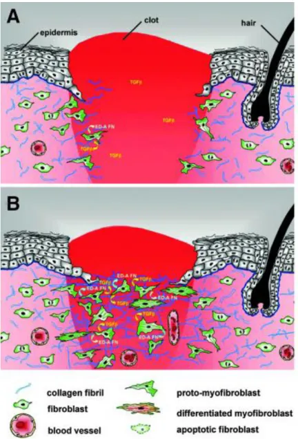
Figure 5. Migration and proliferation of fibroblast in wound
Source: (Van De Water et al. 2013)
(A) In normal tissues, fibroblasts are shielded from “routine” external mechanical
perturbations in the skin by ECM. Following a skin injury, the wound is filled with a provisional matrix of fibrin, fibronectin and a complement of growth factors and cytokines. Fibroblasts, along with blood vessels, are stimulated to migrate into center of wound, forming granulation tissue. (B) Tractional forces accompanying fibroblast
migration are responsible for local areas of increased stiffness in newly made collagen. Focal adhesion assembly is increased with increasing stiffness and this is accompanied by clustering of integrins within focal adhesions and increased stress fiber assembly resulting in fibroblast acquisition of the proto-myofibroblast phenotype.
23
In response to platelet, endothelial and macrophages cytokines, fibroblasts synthesize initially a provisional extracellular matrix composed of collagen type III glycosamicoglycanes, elastin, and fibronectin (Broughton et al. 2006). During this process, cellularity at the wound site increases and this proliferative phase lasts for several days until the wound area is filled to restore tissue integrity. The dermal-epidermal interaction is crucial during this phase and during all the remaining healing process. In incisional wounds, TGF-β expression is maximal at day 7 and modulates remodeling of the extracellular matrix. TGF-β stimulates the synthesis of type I collagen by fibroblasts, inhibits the production of matrix metalloproteases (MMPs) and increases the production of tissue inhibitors of metalloproteinases (TIMPs) (Barrientos et al. 2008). In excisional wounds, TGF-β stimulates the differentiation of fibroblasts into myofibroblasts. Such cells express smooth muscle actin (α-SMA), have contractile activity and cause contraction of the wound (Darby et al. 1990; Sethi et al. 2002).
There are conflicting data on the origin of fibroblasts repopulating skin wounds. In their model of chimeric transplanted GFP marrow mouse, Fathke et al. showed the presence of BMDCs in the skin wounds at a late time (day 42), having a fusiform fibroblast-like appearance, capable of contracting collagen matrix in vitro and expressing collagen I and III. They also showed that collagen I was also expressed by dermal non-bone marrow-derived cells (GFP- cells) (Fathke et al. 2004). These results suggest that at least part of the dermal cells secreting collagen I derived from bone marrow. In contrast to these results, it was recently shown using another model, that wound fibroblasts expressing collagen type I were never of marrow origin (Higashiyama et al. 2011). All these data leaves the subject of the origin of fibroblasts involved in wound healing pending for further works.
3- Re-epithelialization
Re-epithelialization of injured skin is a major and later step in the formation of new tissue. It generally involves the migration of adjacent epidermal keratinocytes into center of wound, the proliferation of keratinocytes from wound edges and or hair follicles remnants. The wound re-epithelialization is ensured by local epidermal
24
progenitoes from the wound edges as well as by epithelial stem cells from hair follicles or sweat glands (Martin 1997; Miller et al. 1998; Roh and Lyle 2006; Lau et al. 2009). This process is activated by signaling pathways of epithelial and non-epithelial cells at the wound edges, which release a myriad of different cytokines and growth factors, e.g. hepatocyte growth factor (HGF), fibroblast growth factor (FGF), epidermal growth factors (EGF), insulin-like growth factor-1 (IGF-1) (Werner and Grose 2003). Wound re-epithelialization is also promoted by various receptors such as G-protein coupled receptors, tyrosine kinase receptors (Sano et al. 1999; Amendt et al. 2002; Ashcroft et al. 2003; Chmielowiec et al. 2007; Raja et al. 2007), or nuclear receptors as peroxysome-proliferator-activated receptors, androgen, estrogen and glucocorticoid receptors (Icre et al. 2006; Michalik and Wahli 2006; Pullar et al. 2006)
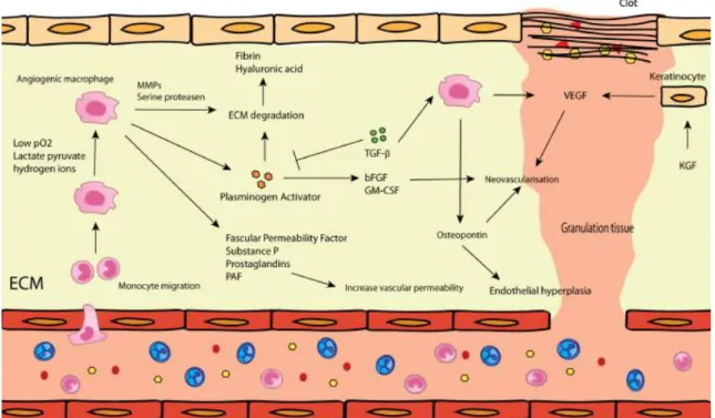
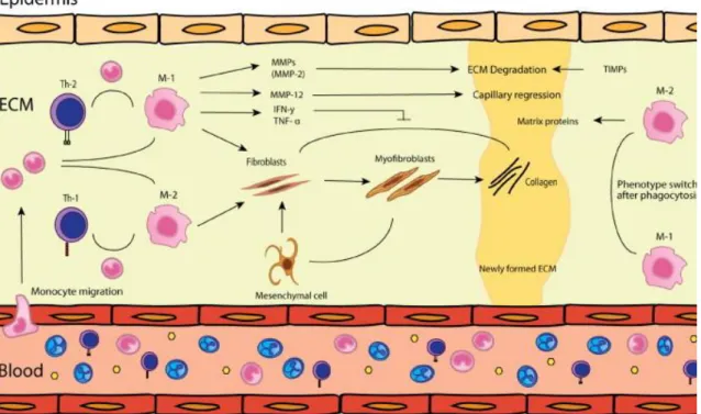
Figure 3. The new tissue formation phase (including reepithelialization): during
this phase active fibroplasia, neovascularization and reepithelization occurs. Dependent on the macrophage phenotype more or less ECM is produced via fibroblasts and myofibroblasts (Mahdavian Delavary et al. 2011)
25
Keratinocyte migration is an early event in wound re-epithelialization (Hell E et al 1979). The keratinocytes initially respond to an injury by migrating from the edges of the wound. Desmosomes are cell-cell junctions that provide the tensile strength of the tissue. Basal epidermal keratinocytes at the wound edge change morphologically: they become flattened and desmosomes, are altered (Matsuzaki et al. 2004). As a result of the morphological changes, epidermal cells at the edge of a wound display lateral mobility and an ability to migrate. Hemidesmosomes, are junctions that link the epidermal cells to the basement membrane. They becomes also altered in order to allow keratinocytes to migrate (Matsuzaki et al. 2004). In addition to wound edges, the keratinocyte migration occurs also from remaining skin appendages.
Multiple elements are involved in keratinocyte migration and re-epithelialization, including the ECM, MMPs, integrin receptors, and growth factors. The effect of the keratinocyte growth factor (KGF) on re-epithelialization and wound healing is still discussed. KGF mRNA is highly expressed during wound healing (Werner et al. 1992) and KGF stimulates keratinocyte proliferation and migration (Tsuboi et al. 1993). Sanz Garcia et al. identified that EGF, KGF and basic FGF (bFGF) all promote wound re-epithelialisation (Sanz Garcia et al. 2000). However, Guo et al. reported that mice null for the KGF gene showed no delay in healing of dorsal incisional wounds and tail wounds (Guo et al. 1996). In mice where FGF function had been blocked in the epidermis using a truncated FGFR2-IIIb, Werner et al. reported
healing of incisional dorsal wounds was delayed (Werner et al. 1994). TGF-1 is also one of the most important players for epithelial cell migration during
re-epithelialization (O'Kane and Ferguson 1997), as it stimulates the expression of integrin subunits that promote keratinocyte migration on the provisional ECM.
Keratinocytes use their surface integrin receptors to interact with a fibronectin-rich provisional matrix. The direction of migration is also regulated by the binding of keratinocytes integrin receptors on the newly formed collagen molecules in the wound bed. Dissociation of this binding allows the keratinocytes to migrate from the edges toward the wound center. MMPs also play an important role in keratinocyte migration by their involvement in this dissociation. Migrating keratinocytes produce MMPs, such as MMP-9, which specifically degrades type IV collagen and laminins in the basement membrane. This allows cells to leave the basement membrane and migrate into the wound (Parks 1999).
26
It has been shown that the decrease in caspase-8 expression in keratinocytes of wound edges raises keratinocyte proliferation (Lee et al. 2009). Migrating keratinocytes express keratins 6 and 16, loose the expression of gap junction proteins as connexin 43 (Brandner et al. 2004), and overexpress β1 integrin, allowing them to interact with components of the extracellular matrix (ECM) of granulation tissue and to migrate to cover the wound (Margadant et al. 2010). Once keratinocytes cover all the granulation tissue, they loose the expression of keratin 6 and 16 and start to express keratins 14 and 5 of the basal layer. These keratinocytes synthetize proteases such as MMP-1 and basement membrane proteins such as laminin-5, which allow them to re-epithelialize the wound (Eming et al. 2007). Basal keratinocytes differentiate thereafter following the physiological epidermal program, giving suprabasal layers expressing keratins 1 and 10, and subsequently the terminal differentiation markers including fillagrin, loricrin and involucrin (Singer and Clark 1999). It has been nicely shown that in addition to interfollicular epidermal progenitors, hair follicle stem cells were also involved in wound re-epidermization (Ito et al. 2005).
A restoration of an intact basement membrane that connects the epidermis and dermis occurs through proteins secretion by keratinocytes and fibroblasts (Li et al. 2007; Gurtner et al. 2008) (Figure 3).
I - A - 3 - Tissue remodelling phase
After the recruitment of inflammatory cells, the angiogenesis, cell proliferation, and re-epithelialization steps, have finished; we are facing healed skin but that still is far from a normal skin. At that time point, begins the remodeling phase. This stage aims to restore a better tissue structural integrity and functional competence. This last phase consists in the synthesis of the dermal extra cellular matrix and its subsequent changes over time. During this period, there is a balance between the synthesis of new components of the extracellular matrix and their degradation by proteases. The processes of regression of granulation tissue are not well known. Key elements of this stage are the regression of vascular structures, decrease of leukocyte infiltrate which returns progressively to a normal level and substitution of transient components of the extracellular matrix as vitronectin, fibronectin and type III collagen
27
by the permanent collagenous fibrous components such as type I collagen (Li et al. 2007). The apoptosis of leukocytes and endothelial cells is a major mechanism, responsible for the decrease of their number in the granulation tissue (Desmouliere et al. 1995). Other mechanisms involve the reduction of local expression of growth factors and chemokines, decreasing the recruitment and proliferation, and the augmentation of expression of anti-inflammatory and anti-angiogenic cytokines (DiPietro 1995).
The formation of the extracellular matrix follows a specific process during wound healing. Initially, the matrix is composed of fibrin and fibronectin as a result from hemostasis. Glycosaminoglycans and proteoglycans are then synthesized by fibroblasts (Broughton et al. 2006). This primary matrix will then be replaced by a more resistant and organized matrix composed of collagen. In healthy skin, collagen in the dermis consists of 80-90% of type I and 10-20% type III. However, during early steps of wound healing, type III collagen becomes the predominant collagen synthesized by fibroblast in granulation tissue. Col III first appears after 48 to 72 hours (30%) and is maximally secreted between 5 and 7 days. The peak of type III collagen is precocious and matches with a peak of fibronectin (Ehrlich and Krummel 1996; Li et al. 2007). MMPs and their inhibitors (TIMPs) are responsible for the remodeling of extracellular matrix components. Their activity and synthesis is influenced by changes in local concentrations of TGF, PDGF, IL-1 and EGF (Henry and Garner 2003). Collagen synthesis is continued for 4 to 5 weeks after wound. The initially deposited collagen is thinner than that in a non-injured normal skin and oriented parallel to the skin surface. It will be subsequently digested, and new, thicker and organized collagen is deposited in bunches along the skin tension lines. Nevertheless, the dermis will never recover its original structure and the wound will not recover its strength at 100%: at one week, the healing skin displays 3% of its original strength. It increases progressively to 30% in 3 weeks, and 80% after 3 months (Diegelmann 2003; Broughton et al. 2006).
28
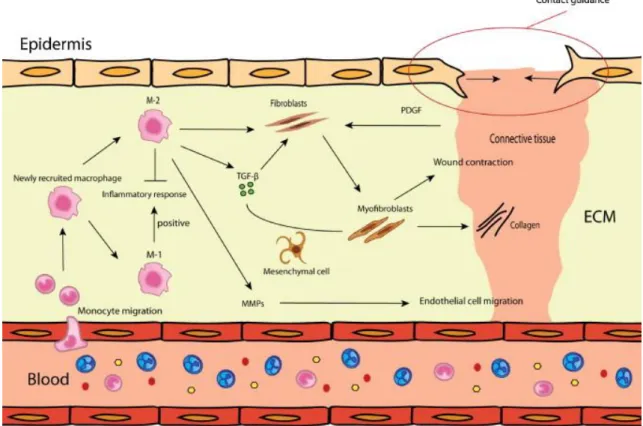
Figure 6. Tissue remodeling phase: During this phase the balance between
ECM-breakdown and -formation is important and determines the eventual scar result. The balance is determined by among others the micro-environment, macrophage phenotype and T-cell response (Mahdavian Delavary et al. 2011).
Endothelial progenitor cell- a key actor of
I - B -
angiogenesis and wound repair
EPCs are bone marrow-derived progenitor cells that were first discovered as significant contributors to neovascularization by Asahara et al. (Asahara et al. 1997). In response to injury, these cells are recruited into circulation and homed to ischemia sites; these cells undergo in situ differentiation, and ultimately participate to the formation of new vessels (Asahara et al. 1999; Carmeliet 2004) (Figure 7).
29
Figure 7 : Implication of EPCs in wound angiogenesis and vasculogenesis
Light microscopic detection of EPCs in wound-healing site created in skin of Flk-1/LZ/ BMT and Tie-2/LZ/BMT mice. Flk-1–expressing EPCs in granulation tissue (A) of
Flk-1/LZ/BMT and Tie-2–expressing EPCs in blood vessels (D) of Tie-2/LZ/BMT were
observed 4 days after wounding (Asahara et al. 1999).
Although the molecular identification of EPCs remains a topic of debate, studies suggest that two functionally distinct subpopulations exist based on in vitro isolation techniques: early outgrowth EPCs and late outgrowth EPCs (Sieveking et al. 2008; Asahara et al. 2011). Specifically, early outgrowth EPCs appear to function in a paracrine role in promoting neovascularization whereas late growth EPCs directly differentiate into endothelial tubes (Sieveking et al. 2008). EPCs have been characterized and classified based on multiple markers. However, none of the markers used are specific for EPCs (Timmermans et al. 2009). However, a common feature of EPCs is their functional ability to mobilize and home to injured areas and promote vessel formation (Lamping 2007). Although the mobilization of EPCs is a complex event and that the mechanisms that specifically recruit these cells to wound sites remain under investigation, numerous molecules and signaling pathways have already been addressed and showed to be implicated in this process. EPCs are thought to mobilize from BM and home to areas of endothelial damage via adhesion molecules. Alternatively, resident EPC may be present at low levels in tissues (ref).
The secreted proteases cathepsin L and MMP2 regulate the transmigration of EPCs. These cells may subsequently mature and differentiate towards the endothelial lineage (Urbich et al. 2005; Cheng et al. 2007).
30
Of note, nitric oxide (NO) signaling and reactive oxygen species (ROS) have also been implicated in EPC mobilization and activity (Gallagher et al. 2007; Hamed et al. 2009). Moreover, previous studies indicate that EPCs are mobilized in response to tissue ischemia. This probably results through cytokine and/or chemokine secretion from ischemic tissue, thereby augmenting neovascularization of ischemic tissues (Takahashi, T 1999; Ishida, Y 2012).
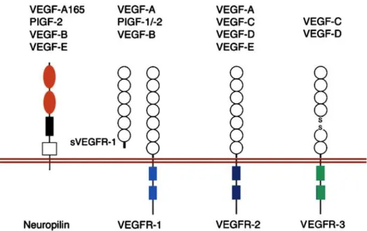
Among cytokines implicated in EPC mobilization and angiogenesis, the family of vascular endothelial growth factors (VEGF) and their receptors have been identified as the most potent and predominant ones (Roskoski 2008; Carmeliet and Jain 2011). VEGFs are endothelial cell specific mitogens and potent inducers of vascular permeability. They play a crucial role in endothelial cell motility; stimulate vascular endothelial cell growth, survival, and proliferation and eventually increase the number of capillaries. This wide range of effects is mediated in part by the multiple VEGF members, VEGF-A through VEGF-D and in associated with three VEGF receptor-tyrosine kinases, known as VEGFR-1, -2 and -3, as well as with co-receptors including neuropilins (Nrp) (Figure 8) (Ferrara 2004; Olsson et al. 2006; Eming et al. 2007).
Figure 8. Schematic representation of interactions between VEGF family members and their receptors. This is a complex network of interactions of VEGF
family members with transmembrane and soluble VEGFR-1 (sVEGFR-1), VEGFR-2, VEGFR-3, and Nrp (Eming et al. 2007) .
31
Each subtype of VEGF is secreted by one or several cell types and appears to be involved in angiogenesis and vasculogenesis via different signaling pathways. VEGF-A, the first and the most important member of VEGF family, has been shown to promote vasculogenesis, by signaling through VEGF receptor-2 (Asahara et al. 1999). Studying a diabetic murine model, Frank et al. have provided for the first time a causative link between decreased VEGF-A activity, impaired wound angiogenesis and delayed wound healing (Frank et al. 1995). Since then, several studies have shown that VEGF levels are indeed increased following tissue injury, and that this level correlated with the migration of EPCs to ischemic area (Asahara et al. 1999; Tepper et al. 2005). In addition, it has been shown that topical VEGF-A application improves the delayed wound healing in diabetic mice through local upregulation of other angiogenic cytokines and recruitment of BMDCs (Galiano et al. 2004). VEGF-A has also been reported to regulate the expression of endothelial cell surface proteins, known as integrins, that link cells with various members of the ECM. Integrins, which comprise a family of transmembrane heterodimeric proteins, play a major role in controlling EPC mobilization and homing to injury and ischemic areas (Caiado and Dias 2012). According to its powerful angiogenic ability, VEGF-A even has been applied in clinical therapy for some human ischemic diseases (you need reference here).
There are various sources for VEGF-A secretion during wound healing. These include keratinocytes, macrophages, platelets, neutrophils and mast cells. The expression of these proteins can be regulated by numerous factors including growth factors, pro-inflammatory cytokines, hormones and cellular stress (Fukumura et al. 1998; Kishimoto et al. 2000; Pages and Pouyssegur 2005). One of the best characterized mechanisms of VEGF-A synthesis in wound healing as well as other settings is hypoxia-inducible factor-1 (HIF-1). HIF-1 is a transcription factor that exists as a dimeric complex, consisting of a cytoplasmic 𝛼 subunit and a nuclear subunit (Ceradini and Gurtner 2005). During hypoxia, HIF-1 translocates into the nucleus to form a complex with HIF-1, initiating the transcription of genes inducing angiogenesis including VEGF and chemokine stromal cell–derived factor-1 (SDF-1. Thus, strategies to stabilize HIF-1 may enhance EPC mobilization and function
32
(Ceradini et al. 2004; Hoenig et al. 2008) and therefore improve delayed wound healing (Botusan et al. 2008; Thangarajah et al. 2009).
Besides VEGF, other peptides have been shown to modulate EPC recruitement during wound healing. Chemokines also crucial players in EPC and hematopoietic stem cell (HSC) trafficking and angiogenesis during wound healing process.CCL5 is one of these. An elegant study by Ishida et al. was able to demonstrate that CCL5/CCR5 interaction is an important molecular pathway for modulation of EPCs and eventual neovascularization during wound healing process. Indeed, CCL5 is secreted at day 1 post wounding by F480 infiltrating macrophages and allows mobilization of CCR5+ EPC from marrow to granulation tissue. (Ishida et al. 2012). This study also demonstrated that EPCs are important not only as the progenitors of endothelial cells, but also as the source of growth factors during tissue repair important for other healing steps (Ishida et al. 2012).
The chemokine CXCL12 (also known as SDF-1) and its specific receptor CXCR4 is another important factor mediating recruitment of various stem/progenitor cells like EPCs or Hematopoietic stem cells. Indeed, CXCL12 that express in different tissues can stimulate the adhesion, migration and homing of progenitor cells from circulation to ischemic tissues. In contrast, blockade of this chemokine in ischemic tissue or CXCR4 on circulating cells reduce the mobilization of progenitors to sites of injury (Ceradini et al. 2004). Importantly, the level of SDF-1 expression in injured tissue correlates with stem cell recruitment and healing. The EPC recruitment into tissue depends on the upregulation of SDF-1 (Askari et al. 2003; Yamaguchi et al. 2003; Ceradini et al. 2004). Consistent with this view, it has been reported that the impaired EPC homing and poor angiogenesis in diabetic wounds are due to decreased level of SDF-1. And this defect could be reversed by therapy of this chemokine into wounds (Gallagher et al. 2007). In contrast, inhibition of SDF-1 leads to further impairment of wound healing, decrease angiogenesis in the diabetic mice (Bermudez et al. 2011). Of note, the expression of SDF-1 is regulated by many factors, such as hypoxia or the activity of eNOS (Ceradini et al. 2004; Gallagher et al. 2007).
33
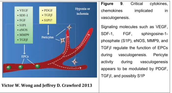
Victor W. Wong and Jeffrey D. Crawford 2013
Figure 9. Critical cytokines,
chemokines implicated in vasculogenesis.
Signaling molecules such as VEGF, SDF-1, FGF, sphingosine-1-phosphate (S1P), eNOS, MMP9, and TGF𝛽 regulate the function of EPCs during vasculogenesis. Pericyte activity during vasculogenesis appears to be modulated by PDGF, TGF𝛽, and possibly S1P
Re-epithelialization: role of nuclear receptors
I - C -
and ligands
Re-epithelialization is an important step in the formation of new tissue after wounding, involving both proliferation and migration of keratinocytes from the edges of wound to reconstitute a complete barrier. This process is promoted by many signaling pathways with the critical involvement of various families of receptors including nuclear receptors (NR), such as peroxysome-proliferator-activated receptors (PPAR), androgen (AR), estrogen (ER) and glucocorticoid (GR) receptors and their ligands (Icre et al. 2006; Michalik and Wahli 2006; Pullar et al. 2006). It is generally considered that estrogens enhance wound repair, while androgens impair wound healing. Role of estrogens and its receptor are different between male and female. Activation of estrogen receptor by its ligand estrogen markedly delays wound re-epithelialization in male mice (Gilliver et al. 2010), while systemic or topical estrogen treatment can reverse age-related impaired healing in animal models and humans females, in association with accelerated re-epithelialization, reduced local inflammation and enhanced matrix deposition (Ashcroft et al. 1997; Ashcroft et al. 1999; Hardman et al. 2008). The sex difference in the effects of systemic estrogen treatment on wound re-epithelialization may partly be explained by a fundamental difference in the response of keratinocytes to estrogens: cells cultured from female
34
mice migrate more rapidly following treatment with an estrogen 17--estradiol (Emmerson et al. 2009); those from male donors were unresponsive to estrogen in the same assay. Another critical actor in wound re-epithelialization is AR. Lai and colleagues have demonstrated that monocytes/macrophages-specific deletion of AR lead to improvement of wound healing, whereas keratinocyte- and fibroblast-specific ARKO mice do not. Even though AR in keratinocytes and fibroblasts is dispensable for overall wound healing, AR in these 2 cell types plays key but opposing roles to regulate re-epithelialization. Keratinocyte AR promoted re-epithelialization, while fibroblast AR suppressed it (Lai et al. 2009). Among three subtypes of PPAR, PPAR
and are important modulators for the re-epithelialization during wound healing and each of them plays a specific role in this process. PPAR is mainly involved in the early inflammation phase of the healing, whereas PPAR is implicated in the control of keratinocyte proliferation (Michalik et al. 2001). In contrast, PPARhas been reported to contribute to the wound healing, not only by accelerating the migration of keratinocytes but also by promoting extracellular matrix-mediated cellular interactions at the wound edge through TGF-1/Smad3 signaling-dependent or - independent pathway (Ham et al. 2010). Moreover, according to Nakamura et al. this receptor is also involved in the corneal epithelial wound healing (Nakamura et al. 2012). Vitamin D hormone metabolites and analogues inhibit the proliferation of cultured keratinocytes that may bring benefits in human hyperproliferative skin diseases such as psoriasis (Lehmann 2009). In contrast, the thyroid hormones stimulate keratinocyte proliferation, and therefore accelerate wound healing (Safer et al. 2005). Finally, GR and its ligands are classical modulators of skin proliferation and differentiation. Glucocorticoids derivatives are frequently used in acute and chronic inflammatory diseases; however, unwanted side-effects, such as epidermal atrophy or delayed wound healing are common, limiting their long-term use (Schacke et al. 2002; Barnes and Adcock 2009; De Bosscher and Haegeman 2009). Study of Sanchis et al. showed that overexpression of GR in keratinocytes lead to a reduced re-epithelialization and delayed wound healing, in correlation with repressed KGF expression and decreased ERK activity (Sanchis et al. 2012). The role of GR and glucocorticoids in skin and cutaneous wound healing will be detailed later (MR chapter).
35
Abnormal wound healing in human and/or mice
I - D -
context
As detailed above, normal wound healing is crucial in restoring the cutaneous barrier in order to allow the skin to perform its dedicated functions, such as preventing infection by foreign agents and fluid loss. The healing process is not only complex but also fragile. The kinetics and quality of skin healing are major parameters in the clinical practice. They are under the influence of several factors. Various abnormalities in any of these may alter normal healing leading to chronic wounds. Again, such situation can be responsible for a significant morbidity and mortality that place chronic wounds as a major public health problems that we will discuss below.
I - D - 1 - Age-related delayed wound healing
The ageing process in humans is well known to be detrimental to wound healing. Although the exact mechanisms of this age related process are remained unclear, it has been demonstrated that an increase in fragility of aged skin together with impaired immune response and cellular ageing contribute to impaired wound healing in the elderly, associated to disruption of the normal phases of repair, leading to an increase in healing time following injury (Thomas 2001).
Archival studies highlight several events which contribute to age-related delayed healing. First, platelet function was found to be impaired in the elderly patients, indicated by a significantly longer “closure time”, and assessed by measuring the time taken for platelets to occlude a Platelet Function Analyser-PFA 100™ (Boldt et al. 2001). In addition, Fukaya et al. determined that platelet aggregation is increased with age, indicating a delayed wound hemostasis in aged individuals (Fukaya et al. 2000). Second, the impaired healing of the elderly has been strongly linked to excessive inflammation; especially neutrophil recruitment and protease production in response to the increased pro-inflammatory cytokine production (Ashcroft et al. 1998; Ashcroft and Ashworth 2003). Moreover, study of Reed et al. showed that together with aging, dermal fibroblast proliferation and migration are impaired (Reed et al. 2001). The proliferation and migration of epithelial cells during wound healing is also delayed with intrinsic ageing in both healthy human subjects and animal models (Holt et al. 1992; Ashcroft et al. 1997). More interestingly, the majority of studies have
36
associated ageing with a decreased angiogenesis in relation with a defect in the production of VEGF (Swift et al. 1999) and TGF-β (Rivard et al. 1999) or with an alteration in matrix synthesis and reduced proliferation of progenitor endothelial cells (Reed and Edelberg 2004).
I - D - 2 - Chronic wounds
Chronic wounds are wounds that have failed to progress through the normal stages of healing leading to a prolonged loss of substance. In the human setting, chronic wounds are usually defined by a persistence of an unhealed wound for at least 3 months. However, such classification does not fit with all situation since normal healing depends also of the age, surface of initial wound as well as the medical care that has been given.. .
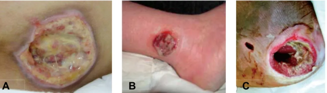
Figure 10. Types of chronic wounds: A) pressure ulcer (Inui et al. 2010), B)
venous leg ulcer (Grey et al. 2006), C) diabetic foot ulcer (Olea et al. 2014)
In Western countries, the most common causes of chronic ulcers are vascular diseases that are responsible for up to 70% affected patients (Nwomeh et al. 1998) (Figure 10). The vascular disorders causing chronic wounds can either derive from large diameter vessels diseases or in contrast small diameter vessels diseases. Disorders of lower limbs large diameter arteries or veins can indeed lead to ischemia. Once ischemia having led to tissue necrosis, a complete wound repair cannot occur because of the same ischemic mechanisms. Causes of lower limbs atheroma are mainly (a) tobacco intake, (b) overweight, (c) elevated cholesterol levels, (d) diabetes, (e) hypertension. Causes of venous insufficiency are (a) genetic background, (b) repeated pregnancies in females (c) overweight, (d) prolonged
37
standing at work. More rarely, ischemia can result from microangiopathies which are small vessels diseases. These are found mainly in diabetes and more rarely in neuropathies, nutritional deficiencies, some genetic diseases, systemic auto-immune diseases (Fonder et al. 2008). In developing countries, the most common causes of chronic wounds are specific infections, arterial disease and traumatisms (Sibanda et al. 2009).
As mentioned above, diabetes is responsible for frequent delayed healing. Diabetes can induce tissue hypoxemia through the induction of large vessels atheroma, neuropathy induced pressure ulcers, increased susceptibility to infections or microangiopathy. The mechanisms of microangiopathy have been addressed by animal model studies or in humans. Microangiopathy has been shown in db/db mice to result from an alteration of EPC mobilization from marrow to wounds (Gallagher et al. 2007; Marrotte et al. 2010). Therefore the importance of these EPC mobilization is illustrated by its implication in diabetic wounds.
In addition to the cases of the chronic wounds mentioned above, many issues of delayed wound healing have been reported are steroids related. Indeed, glucocorticoids, that are wide used to treat chronic inflammatory or auto-immune diseases, usually accompanied by side-effects including skin atrophy and delayed wound healing (Hengge et al. 2006). In the same context of wound healing, while estrogen is considered to enhance wound repair and therefore the deprivation of this steroid as the major factor controlling delayed healing in elderly subjects; androgen, incontrast, is important factor of the delayed wound healing. Indeed, many genes, that are known to be associated with delayed healing, such as growth factor alpha (TGFα), arginase 1 (ARG1) have been found to be altered in elderly male subjects, are also known to be estrogen regulated (Hardman and Ashcroft 2008), while the deletion of androgen receptor in monocytes/macrophages lead to improvement of wound healing (Lai et al. 2009).
Chronic ulcers are sources of various and sometimes serious complications in patients. These may include skin infections, contact dermatitis, subcutaneous fibrosis with secondary functional limitation of the adjacent joints (ankylosis), and moreover chronic pain with risk of depression or mood disorders. These complications, in addition to functional impairment, have psychological impact and lead to a worsening in the quality of life of affected patients.
38
When chronic wounds are severe and worsen despite adapted treatment, amputation may be necessary. In the United States, the incidence of secondary amputation to a diabetic leg ulcer is estimated to reach 11.3/1000 patient-years (Adler et al. 1999). Importantly, prolonged ulcers may be complicated by the development of squamous cell carcinoma arising on one part of the wound (Menke et al. 2007). The incidence of burn scars undergoing malignant transformation has been reported to be 0.77 to 2% of affected cases. All parts of the body can be concerned, but the extremities and the scalp are most frequently affected (Copcu 2009).
In USA currently 1 to 2% of individuals will be affected by leg ulceration during their lifetime, and this figure will likely increase with the aging population (Fonder et al. 2008; Piel et al. 2013). Every year in the United States, about 6.5 million people present one or several chronic skin ulcers secondary to venous insufficiency, diabetes or pressure ulcers (Singer and Clark 1999). In Europe, 5-15% of individuals between 30 and 70 years suffer from venous insufficiency, including at least 1% with varicose ulcer (Welt et al. 2009). Similarly, the prevalence of type II diabetes in the United States is 2% and the incidence of leg ulcers in these diabetic patients is 15% (Reiber 1996). In addition, an increase of these numbers is expected due to the aging of the population and the high prevalence of the causes of poor healing in the elderly (Nelzen et al. 1996). Indeed, healing mechanisms are modified in aging skin. Even, in the absence of any specific disorder, skin in old patients will heal in a longer kinetics (Taylor et al. 2005). This relates with various mechanisms including a reduction in stem cell compartment.
Medical care for chronic ulcers has a very high cost from society. In the US, the total cost of care and morbidity of skin wounds is estimated 13 to 15 billion USD per year and the treatment of venous ulcers alone amounted to 3 billion (Bergan et al. 2006; Fonder et al. 2008). It has become mandatory to develop new treatments that are effective and easily accessible for wound healing problems. This makes it crucial to understand the cellular and molecular mechanisms involved in this phenomenon.
39
II - Aims of work
As detailed above, impaired wound healing is a major clinical problem in various situations including in subjects treated by corticosteroids/dcorticoids, in the elderly and in pathological conditions, such as diabetes, sickle cell disease or Cushing syndrome. Efforts towards improving wound healing represent a major challenge, to improve quality of life. It is therefore important to further improve the understanding about the mechanisms underlying the normal and abnormal wound healing in those situations. In this work, we aimed to address two questions :
1. Develop a murine model for SCD ulcers to improve the understanding of the cellular and molecular pathways responsible for a long-lasting and severe sickle cell leg ulcers.
2. Investigate specific targets in delayed cutaneous wound healing in various pathological situations such as glucocorticoid treated patients, diabetes) and more specifically the implication of the mineralocorticoid receptor inhibition.
40
CHAPTER 2 - DELAYED WOUND HEALING IN MURINE
MODEL OF SICKLE CELL DISEASE
I - Introduction
Sickle cell disease
I - A -
I - A - 1 - Genetics
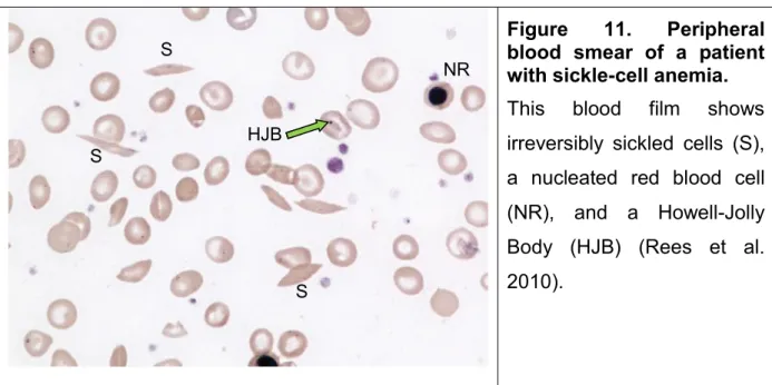
Sickle cell disease (SCD) is a recessive autosomic disorder of the b globin chain of hemoglobin. It is one the most common severe genetic disease reported in many countries worldwide (Weatherall et al. 2005; Piel et al. 2010). It results from a single mutation at the -globin gene in which the 17th nucleotide thymine is replaced by adenine and as a consequence, the sixth amino acid in the β-globin chain changed to valine instead of glutamic acid (Pauling et al. 1949; Bunn 1997). Hemoglobin S (HbS) produced as a result of this defect, is poorly soluble and polymerizes under deoxygenated condition, giving sickle-shaped red blood cells (RBCs) (Bunn 1997). These cells are heterogeneity in genotypes and phenotypes as Herrick first described in 1910 (Figure 11) (Herrick 2001; Rees et al. 2010).
Figure 11. Peripheral blood smear of a patient with sickle-cell anemia.
This blood film shows irreversibly sickled cells (S), a nucleated red blood cell (NR), and a Howell-Jolly Body (HJB) (Rees et al. 2010). S S NR S HJB