HAL Id: inserm-02441720
https://www.hal.inserm.fr/inserm-02441720
Submitted on 16 Jan 2020
HAL is a multi-disciplinary open access
archive for the deposit and dissemination of
sci-entific research documents, whether they are
pub-lished or not. The documents may come from
teaching and research institutions in France or
abroad, or from public or private research centers.
L’archive ouverte pluridisciplinaire HAL, est
destinée au dépôt et à la diffusion de documents
scientifiques de niveau recherche, publiés ou non,
émanant des établissements d’enseignement et de
recherche français ou étrangers, des laboratoires
publics ou privés.
Lucille Stuani, Marie Sabatier, Jean-Emmanuel Sarry
To cite this version:
Lucille Stuani, Marie Sabatier, Jean-Emmanuel Sarry. Exploiting metabolic vulnerabilities for
per-sonalized therapy in acute myeloid leukemia. BMC Biology, BioMed Central, 2019, 17 (1), pp.57.
�10.1186/s12915-019-0670-4�. �inserm-02441720�
R E V I E W
Open Access
Exploiting metabolic vulnerabilities for
personalized therapy in acute myeloid
leukemia
Lucille Stuani
*, Marie Sabatier and Jean-Emmanuel Sarry
*Abstract
Changes in cell metabolism and metabolic adaptation
are hallmark features of many cancers, including
leukemia, that support biological processes involved
into tumor initiation, growth, and response to
therapeutics. The discovery of mutations in key
metabolic enzymes has highlighted the importance of
metabolism in cancer biology and how these changes
might constitute an Achilles heel for cancer treatment.
In this Review, we discuss the role of metabolic and
mitochondrial pathways dysregulated in acute
myeloid leukemia, and the potential of therapeutic
intervention targeting these metabolic dependencies
on the proliferation, differentiation, stem cell function
and cell survival to improve patient stratification and
outcomes.
Acute myeloid leukemia (AML) is a heterogeneous
group of hematological malignancies and represents the
most frequent cause of leukemia-related deaths [
1
]. It
arises from genetic abnormalities in hematopoietic stem
or progenitor cells, inducing uncontrolled growth and
an accumulation of abnormal myeloblasts, leading to
bone marrow failure and often death. For the past three
decades, standard intensive induction therapy involved a
combination of cytarabine plus anthracycline cytotoxic
chemotherapy. Despite a high rate (70–80%) of complete
remission after standard front-line chemotherapy, the
prognosis remains poor, especially for older patients.
This mainly results from the high frequency of distant
relapses caused by tumor regrowth initiated by
chemore-sistant leukemic clones after chemotherapy [
2
,
3
].
© The Author(s). 2019 Open Access This article is distributed under the terms of the Creative Commons Attribution 4.0 International License (http://creativecommons.org/licenses/by/4.0/), which permits unrestricted use, distribution, and reproduction in any medium, provided you give appropriate credit to the original author(s) and the source, provide a link to the Creative Commons license, and indicate if changes were made. The Creative Commons Public Domain Dedication waiver (http://creativecommons.org/publicdomain/zero/1.0/) applies to the data made available in this article, unless otherwise stated.
* Correspondence:[email protected];[email protected]
Centre de Recherches en Cancérologie de Toulouse, UMR1037, Inserm, Université de Toulouse 3 Paul Sabatier, Equipe Labellisée LIGUE 2018, F-31037 Toulouse, France
Therefore, more specific and safe therapeutics are
ur-gently needed. One area of high interest and potential is
targeting metabolic and mitochondrial pathways that are
important in AML biology and that may constitute an
Achilles heel of AML cells. This review focuses on
meta-bolic pathways dysregulated in AML, and especially in
several cytogenetically defined patient subgroups, and
how targeting these metabolic dependencies impacts
proliferation and cell survival in this disease.
Major metabolic dysregulations in acute myeloid
leukemia
Metabolism is altered in most, if not all, cancer cells,
re-gardless of the tumor type [
4
]. A key alteration in cancer
metabolism is the increase in glucose uptake required to
satisfy energetic and anabolic demands. It is now well
established that the metabolic reprogramming
under-gone by transformed cells extends far beyond glycolysis
and the Warburg effect, and changes in cell metabolism
have fundamental implications for tumor biology and
therapy [
5
,
6
].
Glucose metabolism
Higher aerobic glycolysis in cancer cells, reported almost
one century ago by Otto Warburg and known as the
Warburg effect [
7
,
8
], has sparked debate over the role
of glycolysis and oxidative phosphorylation in normal
and cancer cells. Since Warburg’s discovery and
espe-cially during the past 20 years, considerable efforts have
been made to better understand glucose utilization in
cancer cells, in particular to determine if inhibiting
glycolysis or other glucose-dependent pathways could
represent promising therapeutic approaches. It has been
suggested that AML patients exhibit a high glycolytic
metabolism at diagnosis that is potentially associated
with favorable outcomes [
9
], even if the number of
patients in this study remains small. Another study
re-ported
that
a
six-metabolite
signature
(including
pyruvate and lactate) related to the crosstalk between
glycolysis and mitochondria was specifically enriched in
the serum of patients at diagnosis compared to healthy
controls and demonstrated prognostic value in
cytoge-netically normal AML (CN-AML) patients as it could
predict poor survival for these patients [
10
].
Interest-ingly, deletions of the two glycolytic enzymes PKM2 and
LDHA, which catalyze the production of cytosolic
pyru-vate and lactate, respectively, inhibit leukemia initiation
in vivo in AML mice models while preserving normal
hematopoietic stem cell function [
11
] (Fig.
1
).
Glucose metabolism is also involved in other crucial
metabolic pathways such as the pentose phosphate
path-way (PPP) coupled to NADPH production, glutathione/
redox recycling, and nucleotide biosynthesis (Fig.
1
).
Overexpression of glucose-6-phosphate dehydrogenase
(G6PD) has been reported to correlate with an adverse
prognosis in an AML cohort [
12
]. Moreover, in vitro
and in vivo inhibition of 6-phosphogluconate
dehydro-genase (6PGD) and G6PD demonstrated anti-leukemic
activities and synergized with cytarabine [
12
–
15
].
Inhib-ition of 6PGD leads to impaired lipogenesis through
re-activation of LKB1-AMPK signaling [
14
]. Sensitivity to
G6PD inhibition is driven by mTORC1 activity as
mTORC1 activation leads to glucose addiction in AML.
Inhibition of mTORC1 induces a switch toward
oxida-tive metabolism and survival of AML cells [
12
].
Further-more, the anti-leukemic effects of mTOR inhibitors are
enhanced when combined with anti-glycolytic agents,
underscoring the strong interconnection between mTOR
activity
and
leukemic
metabolism
[
16
].
Better
characterization of mTOR-associated metabolic
alter-ations would help in the design of new combinatory
therapeutic approaches and/or help distinguish patients
who could better benefit from these treatments. This
will be even more important since no clear evidence of
clinical efficacy has been found by several clinical trials
of agents targeting mTOR kinase in myeloid leukemia
[
17
–
22
] (Table
1
). This modest efficacy is due to
multi-factorial aspects of mTOR biology and AML
heterogen-eity. The anti-leukemic effect of mTOR inhibition
depends on the level of constitutive PI3K/Akt/mTOR
pathway activation, leukemia-microenvironment
cross-talk, and the release of mediators by both AML and
stromal cells [
71
].
Amino acid metabolism
Of note, Willems et al. have shown that glutamine
avail-ability is a limiting step for mTORC1 activation and that
the anti-tumor effect of L-asparaginase is mainly due to
its glutaminase activity in AML [
72
], highlighting a major
role for amino acids in leukemia biology. Indeed,
intracel-lular glutamine concentration controls the uptake of
leu-cine as leuleu-cine is imported into the cell in exchange for
glutamine by the SLC7A5/3A2 transporter and leucine is
required for Rheb-mediated mTOR activation at the
lyso-somal surface [
73
,
74
]. Glutamine is a non-essential amino
acid and one of the major carbon sources used by cancer
cells for proliferation in vitro [
75
,
76
]. It is also an
import-ant nitrogen donor for amino acids and nucleotides and a
major substrate for TCA cycle intermediates as well as
glutamate and aspartate [
77
–
79
] (Fig.
1
). Dependence of
leukemic cells on glutamine for tumor growth has been
reported, and knockdown of the glutamine transporter
SLC1A5 abrogates tumor development in mice [
72
].
An approach to extend therapeutic opportunities
beyond glycolysis and glutaminolysis may be found
in the identification of auxotrophic amino acids
re-quired by AML cells. It has been reported that most
AML
patients
are
deficient
in
arginosuccinate
synthetase-1 (ASS1), an enzyme that allows the
con-version of citrulline and aspartate into the arginine
precursor argininosuccinate [
29
] (Fig.
1
). The loss of
ASS1 has been reported in other tumor types where
it is required to support cell proliferation and
nu-cleotide synthesis by sustaining the intracellular
as-partate level [
80
]. A decrease in ASS1 can also lead
to a dependence on arginine, which has been
ex-plored as a potential vulnerability in different cancer
types, including AML [
29
].
Lipid and sterol metabolism
De novo lipid biosynthesis is another metabolic pathway
highly reprogrammed in cancer and leukemic cells, in
particular to increase biomass. Numerous studies
sup-port targeting lipid synthesis for therapeutic benefit [
81
,
82
]. Inhibition of key lipogenic enzymes, fatty acid
syn-thase (FASN) [
83
] and stearoyl CoA desaturase 1
(SCD1) [
68
], have been shown to disrupt lipid synthesis
and induce apoptosis in AML (Fig.
1
). SCD1 inhibition
was obtained through treatment with BaP, a combination
of lipid-regulating bezafibrate and the sex hormone
medroxyprogesterone acetate [
68
] (Table
1
). BaP
dis-rupts prostaglandin metabolism, leading to AML growth
arrest and differentiation [
68
–
70
]. Interestingly, it was
re-ported that BaP treatment caused redirection of pyruvate
utilization leading to conversion of
α-ketoglutarate (α-KG)
to succinate and of oxaloacetate into malonate to cope with
oxidative stress [
68
,
84
–
86
]. This pyruvate reprogramming
by BaP includes preferential activation of pyruvate
carb-oxylase (PC) over pyruvate dehydrogenase (PDH) to
pro-duce malonate, a competitive inhibitor of the succinate
dehydrogenase [
87
–
89
] (Fig.
1
). PC has been shown to play
a key role in different solid tumors, in particular through in
vivo reprogramming of glucose utilization to support
ana-pleurosis [
90
–
95
]. Further investigations of PC activity in
leukemia, especially in vivo, would be highly valuable and
provide a better understanding of pyruvate metabolism and
channeling between glycolysis, TCA cycle, and amino acid
pathways.
Various studies have focused on the mevalonate
path-way and the inhibition of the rate-limiting enzyme
3-hydroxy-3-methylglutaryl-coenzyme
A
(HMG-CoA)
with statins in AML [
63
,
96
] (Fig.
1
). The end-products
of the mevalonate pathway include cholesterol, a major
constituent of cell membranes, but also ubiquinone,
which is involved in electron transfer between the
Elec-tron transfer chain (ETC) complexes I to III (see below;
Fig.
2
), geranylgeranyl and farnesyl pyrophosphate,
which are necessary for post-translational modification
of oncogenic proteins, and tyrosine kinase (TK)
recep-tors [
97
].
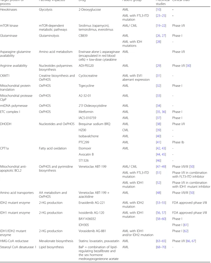
Fig. 1 Metabolic pathways relative to deregulated reactions in myeloid leukemia. Enzymes discussed in this review are in blue. Compound abbreviations: F1P fructose-1-phosphate, G1P glucose-1-phosphate, G6P glucose-6-phosphate, F6P fructose-6-phosphate, F1,6BP fructose-1,6-biphosphate, GA3P glyceraldehyde 3-phosphate, DHAP dihydroxyacetone phosphate, 3PG 3-phosphoglycerate, P-Serine phosphoserine, 2PG 2-phosphoglycerate, PEP phosphoenolpyruvate, 6PGL 6-phosphogluconolactone, 6PG 6-phosphogluconic acid, Rib5P ribulose-5-phosphate, X5P xylulose-5-phosphate, R5P ribose-5-xylulose-5-phosphate, Sed7P sedoheptulose-7-xylulose-5-phosphate, E4P erythrose-4-xylulose-5-phosphate, PRPP phosphoribosyl pyroxylulose-5-phosphate, Carbamoyl-P carbamoyl phosphate, DHO dihydroorotate, THF tetrahydrofolate, OAA oxaloacetate, α-KG α-ketoglutarate, 2-HG 2-hydroxyglutarate, BCAA branched-chain amino acid
Table 1 Drugs targeting metabolic activities in myeloid leukemia
Target protein orprocess
Pathway impacted Drug Patient group Preclinical studies
Clinical trials
Hexokinases Glycolysis 2-Deoxyglucose AML [10]
-AML with FTL3-ITD mutation
[23–25] -mTOR kinase mTOR-dependent
metabolic pathways
Sirolimus (rapamycin), temsirolimus, everolimus
AML/ CML [19–22] Phase I/II
Glutaminase Glutaminolysis CB839 AML [26,27] Phase I
AML with IDH mutations
[28]
-Asparagine glutamine availability
Amino acid metabolism Erwinase alone L-asparaginase (encapsulated in red blood cells) + low-dose cytarabine
AML Phase I/II
Arginine availability Nucleotides polyamines biosynthesis
ADI-PEG20 AML [29] Phase I/II [30]
CKMT1 Creatine biosynthesis and OxPHOS
Cyclocreatine AML with EVI1 aberrant expression
[31]
-Mitochondrial protein translation
OxPHOS Tigecycline AML [32] Phase I
Mitochondrial protease ClpP
OxPHOS A2-32-01 AML [33]
-mtDNA polymerase OxPHOS 2'3'-Dideoxycytidine AML [34]
-ETC complex I OxPHOS Metformin AML [35,36] Phase I
IACS-010759 AML [37] Phase I
DHODH Nucleotides and OxPHOS Brequinar sodium BRQ AML [38] Phase I/II
HZ00 CML [39]
-Isobavalchone AML [40]
-PTC299 AML [41] Phase Ib
CPT1a Fatty acid oxidation Etomoxir AML [42,43]
-Avocatin B [44,45]
-ST1326 [46]
-Mitochondrial anti-apoptotic BCL2
OxPHOS and pyrimidine biosynthesis
Venetoclax ABT-199 AML/ CML [47–49] Phase I/II/III [50] AML with FTL3-ITD
mutation
[51] Phase I/II in combination with FLT3-ITD inhibitor AML with IDH1
mutation
[52] Phase I/II in combination with IDH1 mutant inhibitor Amino acid transporters AA metabolism and
OxPHOS
Venetoclax ABT-199 + azacitidine
AML [48] Phase I/II/III [50]
IDH2 mutant enzyme 2-HG production Enasidenib AG-221 AML with IDH2 mutation
[53–55] FDA approved phase I/III IDH1 mutant enzyme 2-HG production Ivosidenib AG-120 AML with IDH1
mutation
[56,57] FDA approved phase I/III
BAY1436032 [58–60] Phase I
IDH305 Phase I [61]
IDH1/IDH2 mutant enzyme
2-HG production Vorasidenib AG-881 AML with IDH1 and/or IDH2 mutation
Phase I [62]
HMG-CoA reductase Mevalonate biosynthesis Statins: lovastatin, pravastatin AML [63–65] Phase I/II [66,67] Stearoyl CoA desaturase 1 Lipid biosynthesis BaP = combination of
lipid-regulating bezafibrate and the sex hormone
medroxyprogesterone acetate
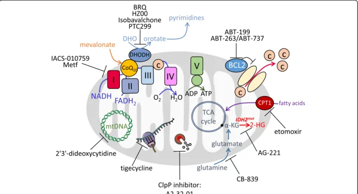
-Oxidative phosphorylation and mitochondrial metabolism
Originally, observations by Otto Warburg that cancer cells
exhibited higher glycolytic activity than normal cells even
in the presence of oxygen led to the assumption that
can-cer cell mitochondrial respiration may be impaired. Since
then, major studies have clearly demonstrated that cancer
cells are able to use oxygen via oxidative phosphorylation
(OxPHOS) [
98
–
101
] and mitochondria are essential for
cancer cell survival. In myeloid leukemia,
krtić et al.
ob-served that AML cells had higher mitochondrial mass and
an increased oxygen consumption rate compared to
nor-mal hematopoietic progenitors [
32
]. Of note, bulk cell
populations had higher mitochondrial mass than an
im-mature CD34
+CD38
−cell population, suggesting unique
mitochondrial characteristics of leukemic stem cells
(LSCs). However, the increased mitochondrial mass in
AML did not translate into an increase in ETC complex I,
III, IV, and V activities, resulting in a lower capability of
AML compared to normal cells to enhance their maximal
respiration with higher electron flux, known as the spare
reserve capacity, suggesting a decreased ability to cope
with oxidative stress [
102
]. In addition, different studies
have reported an amplification of mitochondrial DNA
(mtDNA) levels in AML [
34
,
103
] that correlates with
en-hanced cytoplasmic nucleoside kinase expression [
34
,
104
]. Almost 20 years ago, Beuneu et al. reported that
dihydro-orotate dehydrogenase (DHODH), a
mitochon-drial enzyme of de novo pyrimidine biosynthesis that
cata-lyzes the ubiquinone-mediated conversion of
dihydro-orotate (DHO) to dihydro-orotate, could provide electrons to the
ETC via ubiquinone in AML cells [
105
]. Therefore,
inhib-ition of DHODH could represent another promising
ap-proach to tackle mitochondria in cancer.
Fatty acids can be a major source for TCA cycle
pre-cursors and mitochondrial respiration, especially during
and following metabolic challenges or limitations of
other oxidizable substrates [
82
,
106
] (Fig.
2
). Increased
fatty acid oxidation (FAO) and high carnitine
palmitoyl-transferase 1 (CPT1a) expression have been associated
with a poor prognosis in normal karyotype AML
pa-tients [
107
,
108
]. German et al. [
109
] observed a key role
of prolyl-hydroxylase 3 (PHD3) in FAO regulation in
AML. They reported that, in the setting of high nutrient
abundance, PHD3 activates acetyl-CoA carboxylase 2
(ACC2) via hydroxylation, causing inhibition of CPT1a
and FAO. Accordingly, when nutrients are scarce and
energetic stress is induced, AMPK phosphorylates and
inhibits ACC2 to activate FAO [
110
,
111
]. Reduced
ex-pression of PHD3 could therefore represent a marker of
good responders to FAO inhibitors in AML.
Targeting metabolic vulnerabilities in acute
myeloid leukemia
As metabolic alterations are part of oncogenesis and
tumor progression, cancer cell metabolism offers
prom-ising targets for therapeutic intervention. Hereafter, we
discuss several key metabolic pathways that might be
therapeutically targetable for AML treatment.
Tackling aerobic glycolysis
Treatment with 2-deoxyglucose (2-DG) to inhibit
aer-obic glycolysis and related glycosylation of oncogenic
proteins exerts an anti-proliferative effect in different
AML cell lines and patients and synergizes with
conven-tional cytarabine chemotherapy [
10
,
23
]. However,
tar-geting aerobic glycolysis has not shown great success in
clinical settings as 2-DG treatment necessitates high
dosing that might induce hypoglycemia and cardiac and
red blood cell toxicities due to PPP alteration. Moreover,
LDH inhibitors have never progressed into clinical trials
(Table
1
). Another way to approach high glycolytic
me-tabolism in myeloid leukemia could be through direct
targeting of the glucose storage pathway or inhibition of
other glycolytic sources such as glycogen and fructose
(Fig.
1
). It is notable that mRNA levels of glycogen
bio-synthetic enzymes GYS1/2 and GBE1 were associated
with poor survival in AML and that invalidation of
GYS1 delayed tumor growth in vivo [
112
]. AML cells
may additionally rely on fructose under low glucose
conditions through upregulation of the GLUT5
trans-porter to maintain glycolytic flux and overcome glucose
restriction. Expression of SLC25A5, which encodes
GLUT5, is associated with poor AML patient outcome
and pharmacological inhibition of GLUT5 eliminates
leukemic phenotypes and potentiates the effect of
cytar-abine in vivo [
113
].
Glutaminolysis inhibition and amino acid depletion
Targeting glutaminolysis has been investigated as a
prom-ising therapeutic target in myeloid leukemia [
26
,
114
,
115
]. Of particular interest, inhibition of glutaminase with
CB-839 reduces mitochondrial activities and TCA cycle
intermediate levels, suggesting that glutamine exerts
con-trol on mitochondrial oxidative metabolism in AML [
26
,
116
] (Fig.
2
). Clinical trials are currently in progress to
as-sess the benefit of the allosteric glutaminase inhibitor
CB-839 (Table
1
) with mixed evidence of clinical efficacy.
Willems et al. have shown that the ability of
L-asparaginase (kidrolase and erwinase) to transform
extra-cellular glutamine into glutamate leads to inhibition of
mTORC1 and protein translation in AML cells and that
L-asparaginase exhibits anti-leukemic activities [
72
].
asparaginase, which mainly catalyzes the hydrolysis of
L-asparagine to L-aspartic acid, is one of the standard drugs
for treatment of acute lymphoblastic leukemia (ALL).
These patients lack L-asparagine synthetase (ASNS), the
enzyme that catalyzes the biosynthesis of L-asparagine,
leading to a higher dependency on this amino acid [
117
].
However, AML patients harbor variable expression of
ASNS that could explain their reduced sensitivity to
L-asparaginase. Another recent study proposed another
ex-planation linked to the bone marrow microenvironment
[
118
]. The study by Michelozzi et al. suggests that while
AML cells are sensitive to L-asparaginase, mesenchymal
stromal cells (MSCs) and monocytes/macrophages
pro-duce lysosomal cysteine protease cathepsin B able to
in-activate L-asparaginase. This contribution of the bone
marrow microenvironment to asparaginase resistance was
also described in ALL through release of asparagine and
glutamine by adipocytes [
119
].
Depletion of arginine using a mycoplasma-derived
en-zyme of arginine deiminase formulated with
polyethyl-ene glycol (ADI-PEG20) that degrades arginine to
citrulline reduces tumor burden in AML and synergizes
with cytarabine in vitro and in vivo. Normal human
hematopoietic stem-progenitor cells express higher
ASS1 than AML cells, supporting the idea of selective
targeting of leukemia cells and highlighting a potential
therapeutic window for ADI-PEG20 [
29
,
30
], currently
under phase 2 clinical evaluation (Table
1
).
Inhibition of the mevalonate pathway
The anti-leukemic effects of statins, HMG-CoA inhibitors,
have been studied [
96
,
120
] and found to be additive with
conventional chemotherapies such as cytarabine and
daunorubicin in primary AML samples compared to
healthy donors [
63
–
65
]. Phase I and then phase II clinical
trials combining pravastatin with idarubicin and
cytara-bine for relapse cases of AML have shown an encouraging
response rate of 75% [
66
,
67
] (Table
1
). However, a
subse-quent investigation of this regimen has not confirmed
these encouraging results in patients with newly diagnosed
AML or MDS [
121
]. These differences between response
in newly diagnosed AML or patients at relapse could be
due to rewiring of intracellular cholesterol metabolism
and sterol membrane transport following chemotherapy
and suggest that statins could play a role in overcoming
chemoresistance rather than synergizing with frontline
therapies. The focus of this review does not include
deci-phering all the adaptive mechanisms induced by
chemo-therapeutic agents or new drugs in AML, although this is
important for understanding the clinical relevance of these
metabolic inhibitors.
Hitting at OxPHOS, BCL2, and mitochondrial
dependencies
Mitochondria are dynamic organelles that play a crucial
role in several fundamental signaling and metabolic
pro-cesses such as reactive oxygen species (ROS) regulation,
energy production, calcium signaling, TCA cycle, and
pyrimidine or heme biosynthesis. Mitochondrial
metab-olism represents a targetable vulnerability due to the
en-hanced dependency on mitochondrial energetics of
AML cells. Various strategies to disable mitochondrial
function have been investigated in myeloid leukemia,
in-cluding inhibition of mitochondrial translation with
tige-cycline [
32
], inhibition of the mitochondrial protease
ClpP, thereby decreasing ETC complex II activity [
33
],
and inhibition of mtDNA polymerase using
2’,3’-dideoxy-cytidine, a drug already used in the treatment of AIDS
[
34
] (Fig.
2
; Table
1
). Each of these treatments had
anti-leukemic properties in vitro and in vivo. Moreover, we
and other investigators have shown that metformin, a
common biguanide used to treat type 2 diabetes,
ex-hibits anti-leukemic activities in AML [
35
,
36
] (Table
1
).
However, metformin pharmacokinetics and its
max-imum efficient dose do not allow its use as an anti-AML
agent alone in a clinical setting. Nevertheless, metformin
(or other biguanides) might be promising in
combin-ation with chemotherapies or other targeted therapies,
as recently shown in diffuse large B cell lymphoma
re-fractory to all anti-CD20-based therapies using
L-asparaginase, mTOR inhibitor, and metformin (called
KTM therapy) [
122
]. Whereas metformin inhibits ETC
complex I activity and thus mitochondrial oxygen
con-sumption, high basal glucose consumption and Akt
levels can also affect metformin sensitivity, suggesting
combinatory therapies with AKT inhibitors may be
ef-fective [
35
]. More recently, the new ETC complex I
in-hibitor IACS-010759, which inhibits OxPHOS and
nucleotide biosynthesis by decreasing aspartate levels
[
37
], is in a phase I clinical trial for AML and solid
tu-mors [
37
] (Fig.
2
; Table
1
).
FAO is a key catabolic pathway involved in the
gener-ation of NADH and FADH
2, which are the electron
do-nors of complex I and complex II of the ETC,
respectively, and leading to the production of
acetyl-CoA. This latter plays a crucial role in energy
gener-ation, biosynthesis, and epigenetic control through
post-translational protein modifications. Inhibiting FAO has
been investigated in myeloid leukemia [
42
,
44
,
46
,
114
,
123
]. Inhibition of CPT1a, which catalyzes the transfer
of the acyl group from fatty acyl CoA to carnitine and
constitutes the rate limiting step of FAO, with the
ami-nocarnitine derivative ST1326 [
45
,
46
], lipid Avocatin B
[
44
], or etomoxir [
42
,
43
] has shown anti-leukemic
prop-erties (Table
1
).
As an inner mitochondrial membrane protein associated
with the ETC, DHODH links de novo pyrimidine
biosyn-thesis to mitochondrial bioenergetics. In this context,
Sykes et al. [
38
] found that its inhibition with brequinar
sodium (BRQ) abrogates the myeloid differentiation
blockade and leads to anti-leukemic activities in a diverse
range of AML subtypes. This can be rescued by addition
of extracellular uridine. Very recently, two other newly
de-veloped DHODH inhibitors for AML and one for chronic
myeloid leukemia (CML) have been described [
39
–
41
]
(Fig.
2
; Table
1
). Although BRQ has not shown benefits in
early phase clinical trials with solid cancers, it has not yet
been studied in hematological malignancies [
124
–
128
].
Because BRQ has been shown to lead to a potent
induction
of
myeloid
differentiation
and
decrease
leukemic burden, the role of DHODH in AML
metabol-ism merits further study.
Another very exciting approach to trigger
mitochon-drial priming of cell death is through treatment with
anti-apoptotic BCL2 inhibitors [
52
,
129
] (Fig.
2
).
Lagadi-nou et al. demonstrated that LSCs are characterized by
low levels of ROS. These ROS-low LSCs are dependent
on OxPHOS via amino acid uptake for respiration rather
than glycolysis and overexpress BCL2 anti-apoptotic
proteins [
47
,
48
]. Thus, pharmacological inhibition of
BCL2 with the drug ABT-199 (venetoclax) impairs
mito-chondrial respiration and selectively targets ROS-low
LSCs unable to switch to glycolysis/glucose or FAO to
maintain energy production [
47
]. Clinical trials with
venetoclax monotherapy in relapsed/refractory AML
have shown a very low response rate due to a lack of
apoptosis induction while mitochondrial priming is
acti-vated by this treatment to induce cell death. However,
results from a phase 1b study in elderly patients with
previously untreated AML on venetoclax treatment in
combination with hypomethylating agents (azacitidine
and decitabine) reported a 61% overall response [
50
]
(Table
1
). Treatment with venetoclax plus azacitidine
inhibited amino acid uptake and induced disruption of
the TCA cycle, inhibition of ETC complex II, and
im-pairment of OxPHOS in ROS-low LSCs [
48
,
49
].
Pharmacological inhibition of amino acid metabolism
also decreased OxPHOS and induced cell death in AML
[
48
] (Table
1
). Previous work has suggested that FAO
could be involved in BCL2 regulation and BAX- and
BAK-dependent mitochondrial permeability transition
pore formation through interactions between CPT1 and
the pro-apoptotic BH3-only protein Bid [
130
] or BCL2
[
131
], highlighting a dual interest in FAO inhibition and
synergy with BH3 mimetics in AML. Because many of
the reported manipulations of metabolic pathways have
been shown to modulate BCL2 expression or
depend-ence, combinations of metabolic inhibitors and BCL2
in-hibitors are of special interest. For example, statins also
enhanced ABT-199 efficacy in AML through the
inhib-ition of protein geranyl-geranylation, which leads to
BCL2 modulation and upregulation of pro-apoptotic
BH3 only proteins PUMA [
132
] and etomoxir,
increas-ing the therapeutic efficacy of ABT-737 in vivo [
43
].
Very recently, a study has elegantly mapped metabolic
pathways that are specifically implicated in
ABT-199-induced apoptotic cell death, and demonstrated that the
heme biosynthetic pathway is the major regulator of
mitochondrial priming of apoptosis through ETC and
OxPHOS in AML [
133
]. Altogether, these studies
strengthen the scientific rationale for clinical
develop-ment of new combinations of venetoclax and OxPHOS
(or FAO) inhibitors (Table
1
).
Metabolic stratification to decipher specific
vulnerabilities and develop more efficient
therapies in patient genetic subgroups
For diagnosis and management of AML, a prognostic
stratification has been proposed based on criteria for
progressive disease and for the genomic landscape of the
disease [
134
]. However, metabolic features have not been
taken into consideration yet. As more and more studies
are highlighting metabolic specificities driven by
muta-tions in AML and as specific inhibitors of some of these
mutations are displaying very promising results in
clin-ical trials, investigating the link between genetic
stratifi-cation, metabolic dependencies, and response to these
specific inhibitors is particularly important. This may be
crucial in order to propose better combinations of these
new drugs, understand mechanisms of resistance to
them, and potentially identify early markers of response.
Isocitrate dehydrogenase mutations
In 2009, recurrent mutations in genes of two crucial
metabolic enzymes, cytosolic isocitrate dehydrogenase
(IDH)1 and mitochondrial IDH2, were observed in about
20% of AML patients [
135
–
138
], reinforcing the
import-ance of furthering metabolic investigations in AML.
While wild-type IDH (IDH WT) catalyzes the
conver-sion of isocitrate to
α-KG and generates NADPH,
mu-tant IDH catalyzes a neomorphic enzyme activity that
oxidizes NADPH and produces the oncometabolite
2-hydroxyglutarate (2-HG) from
α-KG [
139
,
140
]. The
im-pact of monoallelic IDH mutation and the related
accu-mulation of 2-HG have been well documented, in
particular its effect on
α-KG-dependent dioxygenase
ac-tivity and subsequent effects on numerous cellular
func-tions in these cancers, such as alteration of DNA and
histone methylation and biased myeloid/erythroid
differ-entiation [
141
–
152
].
Beyond epigenetic modifications and chromatin
re-modeling, 2-HG has multi-faceted roles in AML biology
and leukemic transformation by competitively inhibiting
multiple classes of
αKG-dependent dioxygenases
in-volved in metabolic reprogramming, BCL2-dependent
cell survival, and cellular defense against oxidative stress.
As IDH mutations are early events in oncogenesis and
are systematically conserved at relapse [
153
,
154
], IDH1/
2 mutated enzymes represent attractive therapeutic
tar-gets [
53
,
144
,
155
–
157
] and small molecules selectively
inhibiting the mutated forms of these enzymes have
been developed and very recently approved for clinical
studies [
54
,
56
,
58
–
62
] (Table
1
). Both the IDH2m- and
IDH1m-specific inhibitors promote differentiation and
reduce methylation levels as well as significantly
de-creasing 2-HG levels [
53
,
54
,
57
,
157
,
158
]. However,
while clinical trials are highly encouraging (up to 40%
overall response rate in monotherapy in phase I/II for
relapsed or refractory AML patients), resistance is
rou-tinely observed [
54
–
57
,
159
].
Moreover, suppression of serum 2-HG levels alone did
not predict response in AML patients, as
non-responders also displayed a significant decrease in the
amount of 2-HG [
54
,
55
,
57
,
160
,
161
]. Thus, targeting
IDH mutant activity alone is not sufficient to achieve a
durable clinical response in relapsed AML and new
combinatory approaches need to be designed. Given the
crucial roles of wild type IDH1/2 in cell metabolism (e.g.
Krebs cycle, OxPHOS, cytosolic and mitochondrial
redox, anabolism including lipid biosynthesis), a better
understanding of the contribution of oncogenic IDH
mutations to AML cell intermediary metabolism and
α-KG homeostasis is expected to lead to new therapeutic
strategies.
Because
α-KG is the direct precursor of 2-HG,
vari-ous studies have investigated the glutaminolysis
path-way in IDH mutant cells and reported that glutamine
was indeed the main source of 2-HG production [
139
,
162
]. Therefore, inhibition of glutaminolysis with
differ-ent glutaminase inhibitors (BPTES, CB-839) has shown
higher in vitro anti-leukemic activities in IDH mutant
cells than in IDH wild-type cells [
28
,
114
], in line with
the results obtained in gliomas [
163
]. However,
al-though CB-839 clinical efficiency is currently being
assessed in a phase 1 study in patients with AML
(NCT02071927), in vivo preclinical studies have not
been highly encouraging [
27
].
Interestingly, in IDH1 mutant glioma, 2-HG has been
shown to inhibit branched-chain amino acid
transami-nases BCAT1 and BCAT2, which catalyze the degradation
of BCAA into glutamate, increasing the dependency on
glutamine to sustain glutamate and glutathione
produc-tion and leading to synergy between glutaminase
inhib-ition with CB-839 and radiation therapy [
164
,
165
]. It
would be particularly relevant to investigate BCAA in
IDH mutant cells as Raffel et al. have already shown that
BCAT1 mediates
α-KG homeostasis in IDH WT AML
and could represent a good therapeutic opportunity [
166
].
As demonstrated in gliomas, investigating the
conse-quences of decreasing the BCAA pathway in IDH mutant
AML and/or following treatments with IDH mutant
in-hibitors could pave the way toward a more efficient
com-binatory approach in myeloid leukemia. Furthermore,
IDH mutation leads to higher mitochondrial activities in
various solid cancers [
162
,
167
–
169
], and the decreased
NADPH levels associated with reduced wild-type activity
in brain tumors and colorectal carcinomas [
170
–
172
] was
partly restored by enhanced PPP activity in mutant
astro-cytes [
173
]. However, no detailed investigations of redox
homeostasis in IDH mutant cells in AML have been
re-ported to date, though Ward et al. suggested an increase
in the activity of IDH wild-type enzyme may make a
significant contribution to maintaining cellular and
sub-cellular NADPH levels [
140
].
Key metabolic differences such as sensitivity to
OxPHOS inhibitors seem to emerge in regard to cell
lineage or cell types. Indeed, it has recently been
re-ported that IDH1 mutant glioma cells were more
resist-ant to rotenone (ETC complex I inhibitor) due to
enhanced activity of pyrroline 5-carboxylate reductase 1
(PYCR1), which can oxidize NADH and produce proline
as a
‘metabolic bypass’ of ETC complex I [
174
] (Fig.
1
),
while breast and colon cancer IDH1 mutant cells have
been reported to be more sensitive to ETC complex I
in-hibition by metformin [
167
]. Of particular interest,
over-all response to a combination of venetoclax with
azacitidine increased to 33% in IDH mutant subgroups
of AML patients [
15
]. Chan et al. observed that (R)-2-HG
inhibited cytochrome c oxidase activity (ETC complex
IV), increasing the dependence on BCL2, and this led to
higher sensitivity to ABT-199 in AML primary cells with
an IDH mutation [
175
]. Notably, they observed a partial
rescue of ABT-199 sensitivity with addition of specific
IDH mutant inhibitors, which lower 2-HG levels [
175
].
FMS-like tyrosine kinase 3 mutations
FMS-like tyrosine kinase 3 (FLT3) mutations,
predomin-antly including internal tandem duplication defect
(FLT3-ITD), are found in 30% of AML patients and
con-fer a poor prognosis with enhanced relapse rate [
176
–
179
]. Clinical success of tyrosine kinase inhibitors (TKIs)
against the oncogenic kinase BCR-ABL for CML
treat-ment raised great expectations for FLT3 inhibitors in
AML. However, although the initial response to
mono-therapy was promising (44% response in FLT3-ITD
pa-tients with relapsed/refractory AML treated with AC220,
quizartinib [
180
,
181
]), this did not result in prolonged
disease-free survival [
182
]. The necessity to find new
combinations has thus become apparent, underscoring
the importance of better understanding FLT3-ITD
speci-ficities and linking this with inhibitor resistance (Table
1
).
Ju et al. first compared murine BaF3 cells with BaF3 cells
overexpressing FLT3-ITD and observed enhanced
glyco-lytic activity in FLT3-ITD cells, which was associated with
higher phosphorylation of HK2 localized preferentially to
mitochondria, favoring ATP transfer from OxPHOS to
promote glycolysis. This also provides mitochondrial
pro-tection against mitochondrial death pathways by
prevent-ing openprevent-ing of the mitochondrial permeability transition
pore. Thus, a combination of glycolytic inhibitors with
FLT3-ITD inhibitors produced encouraging results in vivo
[
24
,
25
], corroborating previous observations about
2-DG antileukemic activity in AML with FLT3-ITD or
KIT mutations through glycosylation of oncogenic
pro-teins [
23
].
Gregory et al. performed a synthetic lethality screen in
AML cell line MOLM13 harboring a FLT3-ITD mutation
and found that a number of the genes able to sensitize AML
FLT3-ITD cells to FLT3 inhibitors were involved in
meta-bolic processes [
183
], in particular the ataxia telangiectasia
mutated (ATM) gene shown to activate G6PD to maintain
redox homeostasis [
184
]. Furthermore, while AC220
treat-ment largely reverses the glycolytic phenotype, it also
in-duces decreased glutathione metabolism, accumulation of
mitochondrial ROS, and higher mitochondrial membrane
potential, leading to an increased dependency on glutamine
uptake to compensate. Thus, while not conveying benefit
alone, AC220 efficacy in FLT3-ITD AML in vivo was
in-creased by the addition of OxPHOS inhibitors or
glutamin-ase inhibitors [
51
,
183
,
185
–
187
].
While described in many cancer types as a key deregulated
metabolic pathway and promising therapeutic target [
188
–
192
], one-carbon metabolism in myeloid leukemia remains
mostly unexplored. One carbon metabolism plays a crucial
role in nucleotide synthesis, methylation processes, and
redox homeostasis. Serine availability resulting from both
in-creased uptake and de novo synthesis also appears to be a
key player in tumorigenesis for various cancers [
188
,
193
–
195
] but, to date, has not been reported in myeloid leukemia.
However, Pikman et al. demonstrated that inhibition of
methylenetetrahydrofolate dehydrogenase-cyclohydrolase 2
(MTHFD2) decreased AML growth, in particular in the
FLT3-ITD subgroup [
196
]. MTHFD2 catalyzes the
mito-chondrial conversion of methylene-THF to formyl-THF
using either NAD
+or NADP
+and is thus involved in purine
biosynthesis, OxPHOS, redox homeostasis, and lipogenesis
(Fig.
1
).
Interestingly, an increasing number of studies focus on
using current preclinical and clinical trials of these new
drugs to better define their mechanisms of action and
propose combinations with already FDA-approved
treat-ments. In this context, the relevance of combining IDH
mutant inhibitors with inhibition of oncogenic kinase
sig-naling using TKIs has been demonstrated in two studies
in AML [
197
,
198
]. In one hand, Shih et al. have shown
that combination of AC220 with the IDH2 mutant
inhibi-tor
AG-221
promotes
better
recovery
of
normal
hematopoiesis and a reduction in mutant allele burden,
targeting the mutant clone in vivo in Idh2
R140QFlt3
ITDAML mice [
197
]. On the other hand, Chen et al. recently
pinpointed that both FLT3 WT and FLT3-ITD mutation
increased the activity of IDH1 mutant AMLs through the
activation of JAK2 by phosphorylation, providing a clinical
rationale to combine FLT3 inhibitor and IDH1 mutant
in-hibitor regardless of FLT3 mutational status [
198
].
Other AML patient mutational and cytogenetic subgroups
Surprisingly, metabolic dysregulation and/or specific
biochemical
characteristics
are
almost
completely
unknown in other karyotype and mutational patient
sub-groups with adverse risks, such as patients with p53,
RAS, or CEBPα mutations, or monosomic complex
kar-yotypes in AML. Notably, Fenouille et al. have shown
that mitochondrial function was specifically driven by
the creatine kinase pathway in the EVI1 subgroup of
pa-tients associated with poor prognosis [
31
]. EVI1
re-presses the myeloid differentiation regulator RUNX1,
thus promoting expression of creatine kinase
mitochon-drial 1 (CKMT1). CKMT1 contributes to the conversion
of arginine into creatinine. Pharmacological inactivation
or genetic invalidation of CKMT1 abrogates ATP
pro-duction and mitochondrial respiration, decreases
viabil-ity of EVI1 AML, and prolongs the survival of the mice
engrafted with high EVI1-expressing AML cells
com-pared to xenograft with low EVI1-expressing AML cells.
These observations highlight the therapeutic potential of
targeting metabolic dependency specific to this EVI1
pa-tient subgroup and show the necessity of identifying
spe-cific liabilities to achieve the best clinical outcome
(Table
1
).
Current limitations in cancer metabolism studies
and metabolism-based therapeutic strategies
Over the last 10 years, a number of increasing concerns
emerged in cancer (metabolism) research about 1)
re-producibility of published data [
199
–
201
], 2) differences
of efficacy between in vitro and in vivo studies [
93
,
94
,
202
,
203
] and 3) high attrition rates for cancer drugs
[
200
,
204
]. The models to use, the culture conditions,
and the experimental design are undoubtedly at the
heart of these discussions.
The example of striking discrepancies in anticancer
ef-ficacy of glutaminase inhibitor CB839 observed in vitro
and in vivo highlights the crucial importance of tumor
cell environment. Indeed, human non-small cell lung
cancer (NSCLC) cells exhibit high sensitivity to CB839
treatment and displayed enhanced glutamine catabolism
in vitro, while resistance to this inhibitor was observed
in vivo [
94
]. Isotopic profiling experiments using
13C-glucose and/or
13C-glutamine performed in vivo in
mouse KRAS-driven NSCLC and directly in patients
using
intraoperative
13C-glucose infusions
revealed
NSCLC tumors rely much more on glucose than on
glu-tamine for TCA cycle anaplerosis in vivo [
93
,
94
,
202
].
Interestingly, Muir et al. cultured NSCLC cells in adult
bovine serum, a medium in which component
concen-trations are much closer to in vivo models. In this
cul-ture medium, they observed that glutamine contribution
to TCA was significantly lower compared to the classic
in vitro conditions using fetal bovine serum, and thus
comparable to in vivo data on glutamine metabolism
and response to CB839 [
203
]. They went further,
dem-onstrating that these differences relied on the level of a
single nutrient, cystine (the oxidized dimer of the amino
acid cysteine), present in classic in vitro conditions in
concentrations 100-fold higher than in in vivo
condi-tions. As the cystine level regulates glutamate export
through the cystine/glutamate antiporter xCT, high
levels of cystine in vitro lead to an increased export of
intracellular glutamate and therefore a higher
depend-ence on glutaminase activity to maintain glutamate level,
and thus ultimately to enhanced sensitivity to CB839.
Accordingly, these in vitro observations were not
trans-latable to mouse and patient models [
203
]. These crucial
studies highlighted the importance of taking into
ac-count how nutrient conditions can impact cell
metabol-ism and response to therapies.
In this same vein, various efforts have been made to
develop media with nutrient levels closer to those found
in human serum such as human plasma-like medium
(HPLM) [
205
] and Plasmax [
206
]. Cultures with these
two media revealed that nutrient compositions of
rou-tinely used culture media can induce metabolic
depend-encies and rewiring that are not observed in vivo. One
example of this is that growth of cancer and AML cells
in HPLM containing human plasma levels of uric acid
led to the inhibition of de novo pyrimidine synthesis.
In-deed, uric acid is tenfold higher in human blood than in
culture media and mice serum and can inhibit uridine
monophosphate synthase (UMPS), and consequently
re-duces the sensitivity of cancer cells to the
chemothera-peutic agent 5-fluorouracil [
205
]. Finally, large-scale
RNAi and CRISPR screens are powerful tools to identify
metabolic genes essential for cancer/AML cell
prolifera-tion and response to therapies. However, metabolic gene
essentiality depends on cell culture medium, which is
the major confounding factor affecting the
reproducibil-ity of such approaches [
207
]. This should especially be
accounted for when investigating metabolic
abnormal-ities in the context of tumor metabolic heterogeneity
and to develop more effective metabolism-focused
treat-ment strategies.
The above-mentioned studies indicate the importance
of addressing metabolic reprogramming in the context of
the microenvironment and developing combinatory
thera-peutic strategies. Directly linked to nutrient amounts and
substrate availability in the niche, the notion of crosstalk
between cancer cells and their neighbors should be taken
into account. As we briefly mentioned in the previous
sec-tion, MSCs and adipocytes have been shown to participate
in and modulate the response to several therapies in
AML, in particular through nutrient and metabolite
re-leases or transfers. Co-cultures of AML cells with MSCs
or with bone marrow adipocytes significantly reduced the
sensitivity to CPT1a inhibitors [
43
,
208
], reinforcing the
major role of the microenvironment in sustaining
ener-getic and anabolic demands. Notably, Tabe et al. reported
that inhibition of CPT1a in AML increases free fatty acids
and glucose uptake only in bone marrow adipocyte
co-cultures, allowing blasts to preserve their viability [
208
].
A consideration of tumor metabolic systems biology is
also allowing a better understanding of metabolic
regula-tion, substrate utilizaregula-tion, and energy balance in whole
organisms and will ultimately lead to better therapeutic
strategies. Interestingly, AML cells were recently shown
to hijack systemic glucose metabolism, inducing an
insu-lin resistance with aberrant homeostasis in adipose
tis-sues, pancreas, gut, and microbiota to desensitize
normal tissues to glucose and support their own growth
[
209
]. This study strongly suggests that 1) AML cells
have a parasitic behavior in systemic host metabolism
and that 2) organismal metabolic status is a key
compo-nent of cancer/AML progression. Accordingly, recent
studies have shown that nutrient availability from the
environment/host, dietary regimens, and hormonal
sta-tus can affect host insulin homeostasis and cancer cell
metabolism to enhance drug efficacy [
210
,
211
].
More-over,
non-cell
autonomous
autophagy,
also
called
secretory autophagy (i.e., autophagy of cells in the tumor
microenvironment), has recently been implicated in
can-cer metabolism by providing nutrients required to
sup-port anabolic cell growth and to satisfy cell demands in
vivo for proliferation [
212
–
214
].
Finally, chemoresistance is the main cause of poor
prognosis in AML patients and assessing the metabolic
reprogramming of resistant LSCs after conventional
chemotherapy or new treatments is an area of intensive
research. A crucial point is that cells at relapse have
been shown to be dramatically different in terms of
phenotype or metabolism [
48
,
108
,
215
,
216
]. Strikingly,
Jones et al. have reported fundamental differences
be-tween therapy-naïve LSCs and LSCs at relapse [
48
]. As
mentioned previously, they demonstrated that naïve
LSCs are more dependent on amino acid uptake for
OxPHOS maintenance and cannot up-regulate FA
me-tabolism to preserve TCA cycle fueling in the absence of
amino acids. However, they indicated that LSCs from
re-lapse patients after conventional chemotherapy exhibit a
new ability to compensate amino acid loss by enhancing
FAO [
48
]. This could explain the decreased overall
re-sponse to a combination of venetoclax with azacitidine
in clinical trials for relapsed patients [
217
] compared
to previously untreated patients [
50
]. Therefore, if de
novo AML LSCs seem to be metabolically inflexible,
at least regarding OxPHOS dependency, the ones
re-sistant to chemotherapy and contributing to relapse
are AML cells able to acquire metabolic flexibility
and adapt [
48
,
218
].
Altogether, these studies highlight the importance of
better defining, better characterizing, and better
de-signing our in vitro and preclinical studies as cell
culture medium composition can significantly affect
the response to metabolic pathway inhibition.
Interest-ingly, comparisons between classic in vitro and more
physiological medium also led to understanding some
tumor metabolic specificities and dependencies and to
propose new combinations of standard
chemothera-peutic treatment or newly FDA-approved targeted
therapies with metabolism-based drugs. Such studies
should be conducted in AML and could, at least in
part, explain the unsuccessful clinical translation of
glutaminase or metabolic inhibitors, even though they
displayed promising results in vitro and even in some
studies in mice. These also show two major points in
studying metabolic reprogramming to identify efficient
clinical targets: 1) understanding metabolic
cooper-ation, competition and symbiosis in the tumor
microenvironment/niche is fundamental to tackling
flexibility; and 2) primary tumor cell culture
condi-tions impose critical experimental limitacondi-tions to the
study of cancer.
Conclusion and perspectives
In the past decade, tremendous research efforts have
un-covered key metabolic specificities and Achilles heels of
cancer cells, including AML cells. These studies strongly
suggest that myeloid leukemias are metabolic disorders
and should be regarded in this light for metabolic-based
personalized medicine treatments as well as for
monitor-ing clinical responses to treatment. Several studies have
further shown that AML cells, like other normal and
cancer cells, are able to undergo compensatory
meta-bolic and energetic adaptations in response to the
inhib-ition of metabolic pathways, indicating that AML cells
display complex metabolic capacities and flexibility that
limit sustained drug efficacy, especially when challenged
by chemotherapeutic drugs. However, targeting
meta-bolic flexibility per se is not a feasible approach. By
contrast, non-exclusive therapeutic strategies, which
im-pede this metabolic flexibility by targeting its
conse-quence(s), such as mitochondrial dependency, blocking
the utilization of nutrients from the microenvironment,
and/or targeting metabolic checkpoints, are emerging.
Most of the metabolic pathways described in this review
also occur in normal cells, although they are frequently
less active, making the determination of the right
thera-peutic window difficult. Thus, if we are able to
distin-guish particular requirements of cancer cells to take up
and utilize or eliminate certain metabolites, specifically
targeting these exchanges may provide more effective
treatment strategies. Finally, as already described in
sev-eral solid tumors, an in vitro examination of metabolic
flux networks does not reflect what occurs in situ, in
vivo, and in patients due mainly to the enormous
plasti-city and heterogeneity of their metabolism [
219
,
220
,
202
]. AML, in common with many tumors, is highly
genetically heterogeneous and its metabolism should be
directly studied in patients in situ.
Abbreviations
2-DG:2-Deoxyglucose; 2-HG: 2-Hydroxyglutarate; 6PGD: 6-Phosphogluconate dehydrogenase;α-KG: α-Ketoglutarate; ACC2: Acetyl-CoA carboxylase 2; Akt: Protein kinase B; ALL: Acute lymphoblastic leukemia; AML: Acute myeloid leukemia; AMPK: 5' Adenosine monophosphate-activated protein kinase; ASNS: L-asparagine synthetase; ASS1: Arginosuccinate synthetase-1; ATM: Ataxia telangiectasia mutated; ATP: Adenosine triphosphate; BAK: Bcl-2 homologous antagonist/killer; BAX: Bcl-2-associated X protein;
BCAA: Branched-chain amino acid; BCAT1/2: Branched-chain amino acid transaminases; BCL2: B-cell lymphoma 2; BH3: Bcl-2 homology domain 3; BRQ: Brequinar sodium; CKMT1: Creatine kinase mitochondrial 1; CML: Chronic myeloid leukemia; CN-AML: Cytogenetically normal AML; CPT1: Carnitine palmitoyltransferase 1; DHODH: Dihydro-orotate dehydrogenase; DHO: Dihydro-orotate; ETC: Electron transfer chain; EVI1: Ecotropic virus integration site 1 protein homolog; FADH2: Flavin
adenine dinucleotide (hydroquinone form); FAO: Fatty acid oxidation; FASN: Fatty acid synthase; FDA: Food and Drug Administration; FLT3: FMS-like tyrosine kinase 3; G6PD: Glucose-6-phosphate dehydrogenase; GBE1: Glycogen branching enzyme; GYS1/2: Glycogen synthase 1/2; HMG-CoA: 3-Hydroxy-3-methylglutaryl-coenzyme A; HPLM: Human plasma-like medium; IDH: Isocitrate dehydrogenase; JAK2: Janus kinase 2 non-receptor tyrosine kinase; LDHA: Lactate dehydrogenase A; LKB1: Liver kinase B1 protein; LSCs: Leukemic stem cell; MDS: Myelodysplastic syndrome; MSC: Mesenchymal stromal cell; mtDNA: Mitochondrial DNA; mTORC1: Mammalian target of rapamycin complex 1 protein; MTHF2: Methylenetetrahydrofolate dehydrogenase-cyclohydrolase 2; NADH: Nicotinamide adenine dinucleotide; NADPH: Nicotinamide adenine dinucleotide phosphate; NSCLC: Non-small cell lung cancer;
OxPHOS: Oxidative phosphorylation; PC: Pyruvate carboxylase; PDH: Pyruvate dehydrogenase; PHD3: Prolyl-hydroxylase 3; PI3K: Phosphoinositide 3-kinase; PKM2: Pyruvate kinase PKM; PPP: Pentose phosphate pathway; PUMA: p53 upregulated modulator of apoptosis; PYCR1: Pyrroline 5-carboxylate reduc-tase 1; ROS: Reactive oxygen species; SCD1: Stearoyl CoA desaturase 1; TCA: Tricarboxylic acid cycle or Krebs cycle; TK: Tyrosine kinase; TKI: Tyrosine kinase inhibitor; UMPS: Uridine monophosphate synthase; WT: Wild type
Acknowledgements
The authors thank Dr. M.A. Selak for critical reading of the manuscript.
Authors’ contributions
LS, MS, and J-ES wrote the manuscript and read and approved the final version.
Funding
The authors thank the Région Midi-Pyrénées, Plan Cancer 2014-BioSys (FLEX-AML), and the Institut national de la santé et de la recherche médicale (Inserm) for their support.
Availability of data and materials Not applicable.
Competing interests
The authors declare that they have no competing interests.
References
1. Montalban-Bravo G, Garcia-Manero G. Novel drugs for older patients with acute myeloid leukemia. Leukemia. 2015;29:760–9.https://doi.org/10.1038/ leu.2014.244.
2. Dombret H, Gardin C. An update of current treatments for adult acute myeloid leukemia. Blood. 2016;127:53–61. https://doi.org/10.1182/blood-2015-08-604520.
3. Döhner H, Weisdorf DJ, Bloomfield CD. Acute myeloid leukemia. N Engl J Med. 2015;373:1136–52.https://doi.org/10.1056/NEJMra1406184. 4. Hanahan D, Weinberg RA. Hallmarks of cancer: The next generation. Cell.
2011;144:646–74.https://doi.org/10.1016/j.cell.2011.02.013.
5. Sullivan LB, Gui DY, Vander HMG. Altered metabolite levels in cancer: implications for tumour biology and cancer therapy. Nat Rev Cancer. 2016; 16:680–93.https://doi.org/10.1038/nrc.2016.85.
6. Pavlova NN, Thompson CB. The emerging hallmarks of cancer metabolism. Cell Metab. 2016;23:27–47.https://doi.org/10.1016/j.cmet.2015.12.006. 7. Warburg O, Wind F, Negelein E. The metabolism of tumors in the body. J
Gen Physiol. 1927;8:519–30.https://doi.org/10.1085/JGP.8.6.519.
8. Warburg O. On the origin of cancer cells. Science. 1956;123:309–14https:// doi.org/10.1126/science.123.3191.309.
9. Herst PM, Howman RA, Neeson PJ, Berridge MV, Ritchie DS. The level of glycolytic metabolism in acute myeloid leukemia blasts at diagnosis is prognostic for clinical outcome. J Leukoc Biol. 2011;89:51–5.https://doi.org/ 10.1189/jlb.0710417.
10. Chen W-L, Wang J-H, Zhao A-H, Xu X, Wang Y-H, Chen T-L, et al. A distinct glucose metabolism signature of acute myeloid leukemia with prognostic value. Blood. 2014;124:1645–54. https://doi.org/10.1182/blood-2014-02-554204.
11. Wang Y-H, Israelsen WJ, Lee D, Yu VWC, Jeanson NT, Clish CB, et al. Cell-state-specific metabolic dependency in hematopoiesis and leukemogenesis. Cell. 2014;158:1309–23.https://doi.org/10.1016/j.cell.2014.07.048.
12. Poulain L, Sujobert P, Zylbersztejn F, Barreau S, Stuani L, Lambert M, et al. High mTORC1 activity drives glycolysis addiction and sensitivity to G6PD inhibition in acute myeloid leukemia cells. Leukemia. 2017;31:2326–35. https://doi.org/10.1038/leu.2017.81.
13. Bhanot H, Weisberg EL, Reddy MM, Nonami A, Neuberg D, Stone RM, et al. Acute myeloid leukemia cells require 6-phosphogluconate dehydrogenase for cell growth and NADPH-dependent metabolic reprogramming. Oncotarget. 2017;8:67639–50.https://doi.org/10.18632/oncotarget.18797. 14. Lin R, Elf S, Shan C, Kang H-B, Ji Q, Zhou L, et al. 6-Phosphogluconate
dehydrogenase links oxidative PPP, lipogenesis and tumour growth by inhibiting LKB1–AMPK signalling. Nat Cell Biol. 2015;17:1484–96.https://doi. org/10.1038/ncb3255.
15. Elf S, Lin R, Xia S, Pan Y, Shan C, Wu S, et al. Targeting 6-phosphogluconate dehydrogenase in the oxidative PPP sensitizes leukemia cells to antimalarial agent dihydroartemisinin. Oncogene. 2017;36:254–62.https://doi.org/10. 1038/onc.2016.196.
16. Akers LJ, Fang W, Levy AG, Franklin AR, Huang P, Zweidler-McKay PA. Targeting glycolysis in leukemia: a novel inhibitor 3-BrOP in combination with rapamycin. Leuk Res. 2011;35:814–20.https://doi.org/10.1016/j.leukres. 2010.12.028.
17. Tabe Y, Tafuri A, Sekihara K, Yang H, Konopleva M. Inhibition of mTOR kinase as a therapeutic target for acute myeloid leukemia. Expert Opin Ther Targets. 2017;21:705–14.https://doi.org/10.1080/14728222.2017.1333600. 18. Carneiro BA, Kaplan JB, Altman JK, Giles FJ, Platanias LC. Targeting mTOR
signaling pathways and related negative feedback loops for the treatment of acute myeloid leukemia. Cancer Biol Ther. 2015;16:648–56.https://doi. org/10.1080/15384047.2015.1026510.
19. Recher C, Beyne-Rauzy O, Demur C, Chicanne G, Dos Santos C, VM-D M, et al. Antileukemic activity of rapamycin in acute myeloid leukemia. Blood. 2005;105:2527–34.https://doi.org/10.1182/blood-2004-06-2494. 20. Willems L, Chapuis N, Puissant A, Maciel TT, Green AS, Jacque N, et al. The
dual mTORC1 and mTORC2 inhibitor AZD8055 has anti-tumor activity in acute myeloid leukemia. Leukemia. 2012;26:1195–202.https://doi.org/10. 1038/leu.2011.339.
21. Tamburini J, Green AS, Bardet V, Chapuis N, Park S, Willems L, et al. Protein synthesis is resistant to rapamycin and constitutes a promising therapeutic target in acute myeloid leukemia. Blood. 2009;114:1618–27.https://doi.org/ 10.1182/blood-2008-10-184515.
22. Altman BJ, Jacobs SR, Mason EF, Michalek RD, MacIntyre AN, Coloff JL, et al. Autophagy is essential to suppress cell stress and to allow BCR-Abl-mediated leukemogenesis. Oncogene. 2011;30:1855–67.https://doi.org/10. 1038/onc.2010.561.
23. Larrue C, Saland E, Vergez F, Serhan N, Delabesse E, Mansat-De Mas V, et al. Antileukemic activity of 2-deoxy-D-glucose through inhibition of N-linked glycosylation in acute myeloid leukemia with FLT3-ITD or c-KIT mutations. Mol Cancer Ther. 2015;14:2364–73. https://doi.org/10.1158/1535-7163.MCT-15-0163.
24. Ju H-Q, Zhan G, Huang A, Sun Y, Wen S, Yang J, et al. ITD mutation in FLT3 tyrosine kinase promotes Warburg effect and renders therapeutic sensitivity to glycolytic inhibition. Leukemia. 2017;31:2143–50.https://doi.org/10.1038/ leu.2017.45.
![Fig. 2), geranylgeranyl and farnesyl pyrophosphate, which are necessary for post-translational modification of oncogenic proteins, and tyrosine kinase (TK) recep-tors [97].](https://thumb-eu.123doks.com/thumbv2/123doknet/14364978.503356/4.892.86.807.132.803/geranylgeranyl-farnesyl-pyrophosphate-necessary-translational-modification-oncogenic-proteins.webp)