Université de Montréal
Implicating the mechanisms of ADP-ribosylation factor activation in
the resistance of invasive breast cancer cells to EGFR tyrosine
kinase inhibitors
Par Eric Haines
Département de pharmacologie Faculté de Médecine
Thèse présentée à la Faculté des études supérieures en vue de l’obtention du grade de PhD
en Pharmacologie
March 2015
Université de Montréal Faculté des études supérieures
Cette thèse intitulée :
p66Shc-mediated ARF1 activation in invasive breast cancer cells:
implications in EGFR tyrosine kinase inhibitor resistance
présentée par : Eric Haines
a été évaluée par un jury composé des personnes suivantes : Noël Raynal, président-rapporteur
Audrey Claing, directeur de recherche Christian Beauséjour, membre du jury Louise Larose, examinateur externe Sylvie Mader, représentant du doyen de la FES
Résumé
ADP-ribosylation factor-1 (ARF1) est une petite GTPase principalement connue pour son rôle dans la formation de vésicules au niveau de l’appareil de Golgi. Récemment, dans des cellules de cancer du sein, nous avons démontré qu’ARF1 est aussi un médiateur important de la signalisation du récepteur du facteur de croissance épidermique (EGFR) contrôlant la prolifération, la migration et l'invasion cellulaire. Cependant, le mécanisme par lequel l’EGFR active la GTPase ainsi que le rôle de cette dernière dans la régulation de la fonction du récepteur demeure inconnue. Dans cette thèse, nous avions comme objectifs de définir le mécanisme d'activation de ARF1 dans les cellules de cancer du sein hautement invasif et démontrer que l’activation de cette isoforme de ARF joue un rôle essentiel dans la résistance de ces cellules aux inhibiteurs de l'EGFR. Nos études démontrent que les protéines d’adaptatrices Grb2 et p66Shc jouent un rôle important dans l'activation de ARF1. Alors que Grb2 favorise le recrutement d’ARF1 à l'EGFR ainsi que l'activation de cette petite GTPase, p66Shc inhibe le recrutement du complexe Grb2-ARF1 au récepteur et donc contribue à limiter l’activation d’ARF1.
De plus, nous démontrons que ARF1 favorise la résistance aux inhibiteurs des tyrosines kinases dans les cellules de cancer du sein hautement invasif. En effet, une diminution de l’expression de ARF1 a augmenté la sensibilité des cellules aux inhibiteurs de l'EGFR. Nous montrons également que de hauts niveaux de ARF1 contribuent à la résistance des cellules à ces médicaments en améliorant la survie et les signaux prolifératifs à travers ERK1/2, Src et AKT, tout en bloquant les voies apoptotiques (p38MAPK et JNK). Enfin, nous mettons en évidence le rôle de la protéine ARF1 dans l’apoptose en réponse aux traitements des inhibiteurs de l’EGFR. Nos résultats indiquent que la dépletion d’ARF1 promeut la mort cellulaire induite par gefitinib, en augmentant l'expression de facteurs pro-apoptotiques (p66shc, Bax), en altérant le potentiel de la membrane mitochondriale et la libération du cytochrome C.
Ensemble, nos résultats délimitent un nouveau mécanisme d'activation de ARF1 dans les cellules du cancer du sein hautement invasif et impliquent l’activité d’ARF1 comme un médiateur important de la résistance aux inhibiteurs EGFR.
Abstract
The small GTPase ADP-ribosylation factor-1 (ARF1) has been well described for its role in regulating transport within the Golgi. Recently, in breast cancer cells, we have characterized ARF1 as important mediator of epidermal growth factor receptor (EGFR) signals leading to cell proliferation, migration and invasion. However, the mechanisms regulating ARF1 activity downstream of the EGFR had yet to be defined. Here, we aim to characterize these mechanisms of ARF1 activation in invasive breast cancer cells and demonstrate that activated ARF1 plays an essential role in mediating the resistance of breast cancer cells to EGFR tyrosine kinase inhibitors. We show that the adaptor proteins Grb2 and p66Shc regulate EGF-dependent ARF1 activation. While Grb2 was shown to be essential in the recruitment of ARF1 to the EGFR as well as the activation of this small GTPase, p66Shc blocked the recruitment of this Grb2-ARF1 complex to the receptor and thus suppressed EGF-induced ARF1 activation.
Additionally, we demonstrate that ARF1 promotes EGFR tyrosine kinase inhibitor resistance in invasive breast cancer cells. Indeed, the depletion of ARF1 was associated with an increased sensitivity to EGFR inhibition. We show that ARF1 promotes resistance by enhancing survival and proliferative signals through Erk1/2, Src and AKT, while blocking the apoptotic p38MAPK and JNK pathways. Furthermore, ARF1 was shown to stabilize EGFR dynamics (Expression, activation, dimerization and down-regulation) in response to treatment with EGFR inhibitors Finally, we highlight the role of ARF1 in mediating mitochondrial-dependent apoptosis in response to EGFR tyrosine kinase inhibitor treatment. The depletion of ARF1 was shown to promote gefitinib-induced cell death as measured by increase expression of pro-apoptotic factors (p66Shc, Bax), altered mitochondrial membrane potential and cytochrome C release.
Together, our results delineate a novel mechanism of ARF1 activation in breast cancer cells and implicate ARF1 activity as an important mediator of EGFR inhibitor resistance further supporting the importance of targeting this GTPase in breast cancer patients.
Table of contents
CHAPTER I. Introduction
I.1 Cancer ...1
I.2 Breast cancer ...3
I.2.1 Luminal A/B breast cancer ...4
I.2.2 HER2-positive breast cancer ...4
I.2.3 Triple negative breast cancer ...5
I.3 EGFR inhibitors ...6
I.3.1 Monoclonal antibodies ...7
I.3.2 Tyrosine kinase inhibitors ...8
I.4 EGFR inhibitor resistance ...11
I.4.1 Intrinsic resistance ...11
I.4.2 Acquired resistance ...11
I.5 Membrane receptors ...13
I.5.1 Receptor tyrosine kinases ...13
I.5.2 EGFR family...14
I.5.3 EGFR ...15
I.5.3.1 EGFR structure ...16
I.5.3.2 EGFR activation mechanism ...18
I.5.3.3 EGFR in resistance ...18
I.5.4 HER2 ...19
I.5.5 HER3 ...20
I.5.5 HER4 ...21
I.6 Signaling adaptors ...22
I.6.1 Grb2 ...22
I.6.1.1 Grb2 structure ...23
I.6.2 Shc family of adaptors ...24
I.6.2.1 ShcA isoform structures ...25
I.6.2.2 p52Shc ...27
I.6.2.3 p46Shc ...28
I.6.2.4 p66Shc ...28
I.6.2.5 Mitochondrial p66Shc ...30
I.6.2.6 p66Shc in resistance ...31
I.7 Ras GTPase superfamily ...32
I.7.1 ADP ribosylation factors (ARFs) ...33
I.7.2 ARF structure ...34
I.7.3 ARF activation ...35
I.7.3.1 ARFGEFs ...36
I.7.3.2 ARFGAPs ...37
I.7.3.3 Brefeldin A – ARFGEF inhibitor ...38
I.7.4 ARF1 ...39
I.7.5 ARF6 ...40
I.7.6 ARF GTPases and breast cancer ...41
I.7.7 Other ARF isoforms ...42
I.8 Signal transduction ...43
I.8.1 Ras/ERK1/2 pathway ...45
I.8.1.1 MEK inhibitors ...46
I.8.2 PI3K/AKT pathway ...48
I.8.2.1 PI3K inhibitors ...49
I.8.2.2 PI3K pathway and EGFRTKi resistance ...50
I.8.3 p38MAPK pathway ...51
I.8.3.1 p38MAPK inhibitors ...51
I.8.3.2 p38MAPK pathway and EGFRTKi resistance ...52
I.8.4 JNK pathway ...53
I.8.4.1 JNK inhibitors...54
I.8.4.2 JNK pathway and EGFRTKi resistance ...54
I.8.5 Src kinase ...55
I.8.5.1 Src kinase inhibitors ...56
I.8.5.2 Src kinase and EGFRTKi resistance ...56
I.9 Cell death ...57
I.9.1 Apoptosis ...58
I.9.2 The mitochondria ...60
I.9.3 The mitochondria and cancer ...60
I.9.4 Bcl family ...61
I.9.5 Mitochondrial membrane potential ...62
I.9.6 Cytochrome C ...64
I.9.7 Caspases...64
I.9.8 Apoptosis and resistance ...65
CHAPTER II. The adaptor proteins p66Shc and Grb2 regulate the activation of
the GTPases ARF1 and ARF6 in invasive breast cancer cells
II.1 Abstract ...70
II.2 Introduction ...71
II.3 Materials and methods ...73
II.4 Results ...77
II.5 Discussion ...85
II.6 Figure legends ...90
II.7 References ...109
CHAPTER III. The small GTPase ARF1 mediates sensitivity to EGFR tyrosine
kinase inhibitors in triple negative breast cancer cells
III.1 Abstract ...117III.2 Introduction ...118
III.3 Materials and methods ...120
III.4 Results ...124
III.5 Discussion ...133
III.6 Figure legends ...136
III.7 Tables ...148
III.8 Supplementary figure legends ...150
III.9 References ...155
CHAPTER IV: Further characterization of the role of ARF GTPases in mediating
gefitinib sensitivity in breast cancer cells
IV.1 Abstract ...163IV.2 Introduction...164
IV.3 Materials and methods ...166
IV.5 Discussion ...178
IV.6 Figure legends ...181
IV.7 References ...195
CHAPTER V. Discussion
V.1 Adaptor protein modulate EGFR-dependent ARF activation in breast cancer cells ...200V.2 p66Shc modulates breast cancer cell migration and apoptosis ...202
V.3 ARF1 mediates sensitivity of breast cancer cells to EGFR inhibition ...204
V.4 ARF1 and oncogene addiction ...206
V.5 A role for mitochondrial ARF1 in regulating gefitinib sensitivity ...208
V.6 ARF1 promotes mitochondrial-dependent apoptosis ...209
V.7 Conclusion ...210
CHAPTER VI. Future perspectives
VI.1 Characterize the association between p66Shc and ARF1 ...211VI.2 Identify the GEFs and GAPs involved in p66Shc-mediated ARF activation ...211
VI.3 Determine the role of ARF1 in gefitinib-induced EGFR down-regulation ...212
VI.4 Characterize the role of ARF1 within the mitochondria ...213
VI.5 Identify and characterize novel ARF1 inhibitors ...213
List of tables
CHAPTER III:List of figures
CHAPTER I:Figure 1 Cancer development and progression ...2
Figure 2 Targeting the EGFR ...7
Figure 3 Crystalized structure of gefitinib binding the ATP-binding domain of the EGFR ...9
Figure 4 Chemical structures of EGFR tyrosine kinase inhibitors ...10
Figure 5 Mechanisms of EGFRTKi resistance ...12
Figure 6 EGFR family of receptor tyrosine kinases ...15
Figure 7 EGFR structure ...17
Figure 8 Grb2 structural domains ...24
Figure 9 Shc isoforms ...27
Figure 10 The role of p66Shc in the mitochondria ...31
Figure 11 The structure of ARF GTPases ...35
Figure 12 Regulation of ARF activity ...36
Figure 13 Signaling cascades activated downstream of the EGFR in cancer ...44
Figure 14 Intrinsic versus extrinsic apoptotic pathways ...59
Figure 15 Mediation of apoptosis by mitochondrial membrane potential ...63
CHAPTER II: Figure 1 p66Shc negatively regulates ARF1 activation ...99
Figure 2 p66Shc regulates the activation of the Ras/MAPK and AKT pathways ...100
Figure 3 p66Shc mediates MDA-MB-231 cell growth and migration ...101
Figure 4 Signals downstream of the HER3 receptor are mediated by p66Shc ...102
Figure 5 The recruitment of ARF1 to the EGFR is attenuated by p66Shc ...103
Figure 6 p66Shc blocks the recruitment of Grb2 to the activated EGFR ...104
Figure 8 p66Shc enhances ARF6 activation and the recruitment of this GTPase to the EGFR ..106
Figure 9 Grb2 is also essential for ARF6 activation and its recruitment to the EGFR ...107
Figure 10 Model of ARF1 and ARF6 activation downstream of the EGFR in MDA-MB-231 ..108
CHAPTER III: Figure 1 ARF1 mediates gefitinib sensitivity in invasive breast cancer cells ...141
Figure 2 Gefitinib-induced survival signaling is altered in ARF1 depleted cells ...142
Figure 3 Enhanced gefitinib-mediated apoptotic signals in ARF1 depleted cells ...143
Figure 4 Gefitinib-induced EGFR family member expression is mediated by ARF1 ...144
Figure 5 Gefitinib-dependent EGFR internalization and degradation is enhanced by ARF1 depletion ...145
Figure 6 Gefitinib promotes ARF1 activation through the recruitment of this GTPase to AXL 146 Figure 7 The role of ARF1 in mediating gefitinib sensitivity in breast cancer cells ...147
Supplementary Figure 1 ARF1 mediates the sensitivity of breast cancer cells to EGFR inhibitors ...152
Supplementary Figure 2 Gefitinib blocks EGF-dependent activation of the EGFR, ERK1/2 and AKT ...153
Supplementary Figure 3 The ARF inhibitor, Brefeldin A, sensitizes MDA-MB-231 cells to EGFR inhibition ...154
CHAPTER IV: Figure 1 p66Shc mediates gefitinib sensitivity of MDA-MB-231 cells ...188
Figure 2 Gefitinib induces the translocation of ARF1 into the mitochondria ...189
Figure 3 ARF1 is required for p66Shc translocation into the mitochondria ...190
Figure 4 ARF1 depletion promotes gefitinib-induced mitochondrial apoptosis ...191
Figure 5 ARF6 depletion enhances gefitinib sensitivity ...192
Figure 6 ARF1 depletion mediates gefitinib-dependent EGFR activation and dimerization ...193
Figure 8 ARF1 is required for heregulin-induced proliferation and migration ...195 Figure 9 Gefitinib resistance is abolished upon ARF1 depletion ...196 Figure 10 The receptor and signaling profile of gefitinib-resistant cells is regulated by ARF1 .197
List of Abbreviations
AKT: Protein kinase B
APAF-1: Apoptotic protease activating factor-1 ARF: ADP-ribosylation-factor
ARFGAP: ARF GTPase-activating protein ARFGEF: ARF guanine exchange factor ATP: Adenosine triphosphate
AXL: AXL receptor tyrosine kinase Bax: Bcl2-associated X protein Bcl2: B-cell lymphoma 2 BFA: Brefeldin A
BH3: Bcl2-homology domain 2 Bim: Bcl2-like protein 11 (Bcl2L11) CB: Cytochrome C binding domain Cbl: Casitas B-lineage Lymphoma CH: Collagen homology domain
cMET: Hepatocyte growth factor receptor (HGFR) CytC: Cytochrome C
EGF: Epidermal growth factor
EGFR: Epidermal growth factor receptor EGFRTKi: EGFR tyrosine kinase inhibitor EMT: Epithelial-mesenchymal transformation ER: Estrogen receptor
ERK1/2: Extracellular signal-regulated kinase 1/2
ERMES: Endoplasmic reticulum-mitochondria encounter structure complex FGFR: Fibroblast growth factor receptor
Gab1: Grb2-associated binding protein 1 GAP: GTPase activating protein
GDP: guanosine diphosphate GEF: Guanine exchange factor
GGA3: Golgi-localized, gamma adaptin ear-containing, ARF-binding protein 3 Grb2: Growth factor receptor-binding protein 2
GTP: Guanosine triphosphate
HER2: Epidermal growth factor receptor-2 (ErbB2) HER3: Epidermal growth factor receptor-3 (ErbB3) HER4: Epidermal growth factor receptor-4 (ErbB4) HRG: Heregulin
HSP70: Heat shock protein 70
IC50: Half maximal inhibitory concentration
IGF1R: Insulin growth factor-1 receptor JNK: c-Jun N-terminal kinase
Mab: Monoclonal antibody
MAPK: Mitogen-activated protein kinase MEK: Mitogen-activated protein kinase kinase mTor: mammalian target of rapamycin
MTT: 3-(4, 5-Dimethylthiazol-2-Yl)-2, 5-Diphenyltetrazolium Bromide OMM: Outer mitochondrial membrane
PIP2: Phosphatidylinositol 4, 5-bisphosphate PIP3: Phosphatidylinositol (3, 4, 5)-trisphosphate PI3K: Phosphatidylinositol-4, 5-bisphosphate 3-kinase PKC: Protein kinase C
PLD: Phospholipase D PR: Progesterone receptor
PTEN: Phosphatase and tensin homolog PTB: Protein tyrosine binding domain PTPC: Permeability transition pore complex
PUMA: p53 upregulated modulator of apoptosis p38MAPK: P38 mitogen-activated protein kinase pY: phosphotyrosine residue
Raf: Rapidly accelerated fibrosarcoma kinase Ras: Rat sarcoma protein
ROS: Reactive oxygen species RTK: Receptor tyrosine kinase Shc: SH2-containing protein SH2: Src homology-2 domain SH3: Src homology-3 domain siRNA: Small interference RNA SOS: Son of sevenless
Src: Src tyrosine kinase
STAT: Signal transducer and activator of transcription VEGFR: Vascular endothelial growth factor receptor
I would like to dedicate this thesis to my wife Sevine and our children Spencer and Logan
Acknowledgements
It has been a long journey filled with many ups and downs. But looking back, I regret none of it. I have had the opportunity to meet a lot of great people over the years. Some have come and gone but others I will consider good friends for the rest of my life. I would like to thank you all for your support, help, laughs and countless pints of beer. Cheers!
Firstly, I would like to thank Audrey for always being there for me to bounce ideas off of and for giving me the creative liberties to go off on tangents and seek answer that might have been a little outside of the realm of the laboratory. I thank you for your supervision and guidance and I’ll do you proud in Boston. I would also like to thank my fellow lab members for being there to hear and discuss my sometime crazy scientific ideas, putting up with my not always enjoyable music selections and for making each day a little more entertaining.
Next, I would like to briefly thank some of the important people that have helped me get through this long journey: Dr. Giovanni Hernandez for taking me under his wing and introducing me to the pleasures of scientific research, the Ali Lab, especially Naila, Jenny and Noor for keeping me semi-sane, H5 and H7 for the many good times had, GLG communications – you may of worked me crazy but you significantly improved my writing skills and the Piekny lab – Paul, Madhav, Husni and Neetha you have become my best of friends. I raise my pint to you all. I would also like to thank Ye Olde Orchards and Mr Man for the great service and cold beers.
I would like to now thank my parents. No matter my decision you have always supported me. Even though you have no clue what I do, I know that you are proud me and that is all that matters. I only hope to be as good as a parent to my boys as you are to me
Finally, I would like to thank my wife, Sevine. When we first met, I was lost. You gave me the direction needed to start this journey. More, importantly, you were there to support me through every step of the way. You put up with me bouncing around from lab to lab and if it wasn’t for you I wouldn’t have made it through it. I thank you and I love you.
CHAPTER I. Introduction
I.1 CancerAlterations in a cell’s physiology are often associated with the development of disease, including cancer. Cancer is a complex pathology that effects multiple organ systems and tissues within the human body. While it is a dynamic disease, all cancers have similar properties and are characterized by an abnormal cell growth and increased capacity to invade both local and distant tissues. Many factors have been shown to contribute to the development of this disease. These include: tobacco/alcohol use, obesity, diet, physical inactivity as well as environmental and occupational risk factors. On a molecular level, cancer is caused by DNA damage. While DNA damage occurs in normal cells, cells have mechanisms to repair this damage. Furthermore, when these repair mechanisms fail, the cell undergoes programmed cell death, apoptosis, to ensure that cells with damaged DNA do not propagate. However, in cancer cells, DNA damage repair and cell death mechanisms are inhibited or impaired. This gives rise to a cancerous population of cells with mutated DNA.
Throughout our body, genes play an important role in mediating the functions of normal cells such as cell growth, maturity and death. However, alterations in these genes as simple as a single nucleotide point mutation or as broad as a gain or loss of entire chromosomes are associated with the development of disease. Genetic alterations have been linked to the development of cancer. Three genetic alterations leading to cancer have been proposed: 1- Genomic amplification where a cell gains many copies of a small chromosomal locus. Examples include: amplification of the
myc oncogene in a variety of tumors and HER2 in breast and ovarian cancer. 2- DNA mutations
characterized by single nucleotide mutations and nucleotide deletions or insertions that alter the functionality or expression of their coded proteins. For instance, BRCA1 and BRCA2 mutations in breast and ovarian cancers or p53 mutations and 3- Translocation where two separate chromosomal regions become abnormally fused. The best known example is the BCR-ABL fusion protein in chronic myelogenous leukemia.
There are two broad categories of genes that are affected by these alterations: 1- Oncogenes or genes known to cause cancer. While many oncogenes are expressed in normal/non-cancerous cells, cancer patients have been shown to have an abnormal increase in the expression or activity of these oncogenes. Furthermore, mutations in normal genes can alter the functionality of these genes and
promote oncogenesis. 2- Tumor suppressor genes or genes that generally inhibit the cell division and survival of cells that have damaged DNA. In cancer patients, these genes are disabled either through gene deletion or mutation. This allows for the growth and survival of cancer cells (Weinberg 1995).
The majority of rapidly dividing cancer cells will form solid tumors in their target tissues. However, blood cancers, such as leukemia, generally do not form solid tumors. Tumors are considered to be malignant because they can invade into nearby tissues. Additionally, as the tumor grows, some cancerous cells can travel through either the circulatory or lymphatic systems and form tumors in distant organs and/or tissues. This is known as a metastasis. The development and progression of cancer is depicted in the Figure 1 below.
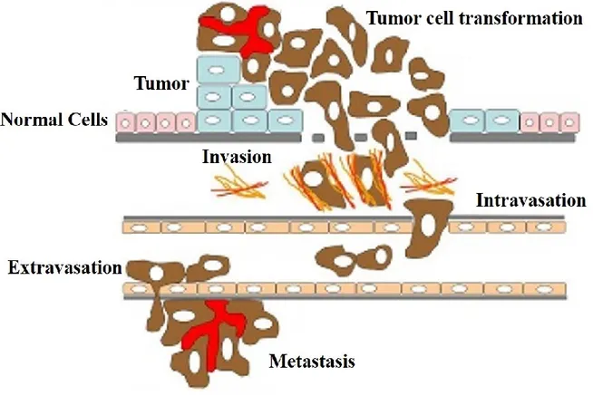
Figure 1. Cancer development and progression
Genetic alterations in normal cells results in the rapid division of cells and the formation of a primary tumor. As the tumor grows some cells transform into an invasive phenotype through a process known as epithelial-mesenchymal transformation. These cells can invade the basal membrane and enter the blood vessels, a process known as intravasation. Once in the circulatory system the tumor cell to transported to a distant organ or tissue. The cell exits the circulatory and invades this secondary site though a process known as extravasation. The tumor cell can now proliferation to form a secondary tumor or metastasis. Adapted from: (Freire-de-Lima 2014)
Within the last few decades, there has been an increased interest in cancer research and the development of therapeutics to counter this disease. Over these years, there has been significant progress which has led to the thorough characterization of the disease and the development of effective therapies. However, a large proportion of cancer patients are unresponsive to current available therapies and the majority of cancers remain incurable. Thus, it is important to further characterize the mechanisms that regulate the development and progression of cancer and to develop improved therapeutic agents.
I.2 Breast cancer
The genetic and cellular alterations described in the section above can result in the development of cancer in multiple organ systems. The most commonly affected tissues include: the lung, colon, breast and prostate (Canadian Cancer Society) In fact, breast cancer is the most commonly diagnosed cancer in Canadian women. It is estimated that in 2014, close to 24000 new cases of breast cancer were diagnosed. In fact, 1 in 9 women will be diagnosed with breast cancer within their lifetime and 1 in 30 women will succumb to the disease. However, improved diagnostics, screening techniques and therapeutics have decreased the overall mortality rate by almost 50% since 1986. Furthermore, approximately 90% of breast cancer patients survive for at least 5 years (Canadian Breast Cancer Foundation).
Breast cancer is a heterogeneous disease that is generally classified into four subtypes; Luminal A, Luminal B, HER2-positive and Triple negative (Basal-like) breast cancers. However, a 5th
subtype, Normal-like breast cancer, has emerged. This subtype is characterized by a genetic profile similar to that of normal breast tissue, small tumor size and good prognosis (Carey, Perou et al. 2006). However, it is still unclear whether normal-like breast cancer is a distinct molecular subtype or just a collection of tumors that are difficult to classify in another subtype (Prat, Carey et al. 2014). The classification of cancers into these subtypes is based on the expression of therapeutically important genetic markers such as the estrogen receptor (ER), progesterone receptor (PR) and the epidermal growth factor-2 receptor (HER2)(Yersal and Barutca 2014).
I.2.1 Luminal A/B breast cancer
Luminal A breast cancer is the most common form of breast cancer, representing 50-60% of all diagnosed cases. Whereas. Luminal B breast cancer represents 15-20% of diagnosed cancers. These subtypes are characterized by a high ER and/or PR expression(Carey, Perou et al. 2006). In general, patients with luminal A/B breast cancer have a good prognosis, lower relapse rate and low incidence of metastatic disease (Kennecke, Yerushalmi et al. 2010). The primary difference between Luminal A and Luminal B breast cancer stems from the increased expression of proliferative genes in the Luminal B subtype (Reis-Filho, Weigelt et al. 2010). This increased proliferative index is associated with a worse prognosis, increased grade and aggressivity, higher recurrence and lower survival rate compared to Luminal A breast cancer (Ellis, Tao et al. 2008; Creighton 2012). Hormonal therapy is commonly used in the treatment of these subtypes (Ignatiadis and Sotiriou 2013).
I.2.2 HER2-positive breast cancer
The third subtype is known as HER2-positive breast cancer and is characterized by a high expression of the HER2 gene. It represents 15-20% of diagnosed breast cancers and is associated with a poor prognosis, high proliferative index and aggressivity and low survival rate when left untreated (Tsutsui, Ohno et al. 2003; Staaf, Ringner et al. 2010). However, these patients are generally responsive to HER2-directed therapies (Ross, Fletcher et al. 2003). In fact, 75-80% of women diagnosed with HER2-poistive metastatic breast cancer have been shown to be responsive HER2-inhibition when treated in combination with chemotherapy(Slamon, Leyland-Jones et al. 2001). However, the overall survival of patients treated with HER2-targeted therapies after adjuvant chemotherapy was not different to that of patients left untreated (Piccart-Gebhart, Procter et al. 2005). Furthermore, there are many limitations to targeting the HER2 receptor. These limitations include: 1- Effective only in tumors expressing high levels of HER2 (low response rate in moderate and low HER2 expressing tumors) (Albanell, Codony et al. 2003), 2- Large proportion of patients develop resistance to HER2-targeted therapies (Romond, Perez et al. 2005) and 3- Generally an expensive therapy, but still considered cost-effective(Dedes, Szucs et al. 2007). Therefore, substantial work is required in this subtype to improve therapeutic outcomes.
I.2.3 Triple negative breast cancer
The final subtype is known as triple negative breast cancer (TNBC) or basal-like breast cancer and is characterized by a low expression of ER, PR and HER2 receptor and a high expression of the epidermal growth factor receptor (EGFR). It represents 8-37% of diagnosed breast cancers. The discrepancies in TNBC diagnoses stem from inconsistencies amongst clinicians and diagnostic definitions. For example, some institutes combine TNBC and basal-like breast cancer in the same subgroup, whereas others separate the two. In this case, TNBC is defined as ER, PR and HER2 negative and basal-like breast cancer as ER, PR, HER2 negative and either Cytokeratin5/6 or EGFR positive (Rakha, Elsheikh et al. 2009). The TNBC subtype is considered to have a poor prognosis, high incidence of metastasis, high proliferative index and aggressivity and poor survival rates(Heitz, Harter et al. 2009; Rakha, Elsheikh et al. 2009; Criscitiello, Azim et al. 2012). While targeted therapies are currently available for luminal and HER2-positive tumors, no targeted therapies are presently approved for the treatment of TNBC patients(Engebraaten, Vollan et al. 2013).
The TNBC subtype is known to be the most chemosensitive breast cancer subtype. Indeed, TNBC patients have increased sensitivity to chemotherapy compared to ER-positive breast cancers (Crown, O'Shaughnessy et al. 2012). This increased sensitivity has been shown to be the result of increased incidence of BRCA mutations in this subtype (Bhattacharyya, Ear et al. 2000; Moynahan, Cui et al. 2001). However, little is known on which chemotherapeutic agents elicit the best response in these patients (Cleator, Heller et al. 2007). In fact, there are no systematic therapeutic regiments recommended for the treatment of TNBC patients (Cleator, Heller et al. 2007). Furthermore, the use of standard chemotherapeutics leaves these patients at an increased risk of both local and systemic relapse (Cleator, Heller et al. 2007). Additionally, while approximately half of patients have been reported to respond to chemotherapeutic treatment with either paclitaxel (a mitotic inhibitor of the taxane family of chemotherapies) or doxorubicin (an anthracycline involved in DNA intercalation), TNBC patients have been reported to develop resistance to these treatments (Bhattacharyya, Ear et al. 2000; Quinn, Kennedy et al. 2003). Therefore, further investigation is required to better define the therapeutic benefits of chemotherapies in TNBC patients. Emerging therapies are focused on targeting oncogenic
pathways such as the PI3K/AKT and Ras/MAPK pathways as well as receptor tyrosine kinases (RTKs) such as the EGFR, fibroblast growth factor receptor-2 (FGFR2) and vascular endothelial growth factor (VEGFR) (Cunningham, Humblet et al. 2004; Mendelsohn and Baselga 2006; Cleator, Heller et al. 2007; O'Shaughnessy 2010; Turner, Lambros et al. 2010; Zhao and Adjei 2014). However, little clinical success has been demonstrated when targeting these factors. As our research is focused on the signals downstream of the EGFR, this family of RTKs and the therapeutic agents targeting it will be described in detail below.
I.3 EGFR inhibitors
The EGFR is a receptor tyrosine kinase (RTK) known to be expressed and activated in a variety of cancers. Upon ligand binding to the receptor, there is receptor dimerization and auto-phosphorylation. This allows for the recruitment of adaptors and other signaling mediators to receptor leading to the activation of important signaling cascades involved in cancer cell proliferation, survival, migration and invasion. This receptor will be discussed in more detail in section I.5.1. With the majority of TNBC patients expressing higher levels of the EGFR, the EGFR is a potential therapeutic target in this breast cancer subtype. Research within the field of EGFR signal transduction has led to the development of targeted EGFR cancer therapeutics such as the monoclonal antibodies that target the extracellular domain of the receptor preventing ligand binding and receptor dimerization (Fan, Lu et al. 1994; Mendelsohn 1997). Secondly, small molecule tyrosine kinase inhibitors have also been synthesized that target the intracellular kinase domain of the EGFR and prevent ATP-binding (Ward, Cook et al. 1994) (Figure 2). Several of these EGFR inhibitors have been approved for the treatment of EGFR-overexpressing cancers, whereas many more are currently being tested in clinical trials. The two families of EGFR inhibitors will be discussed further below.
Figure 2. Targeting the EGFR
EGFR activity plays an important role in cancer cells to promote the activation of signaling cascades involved in the induction of cell cycle progression, proliferation, differentiation, cell motility and angiogenesis. There are currently two therapeutic means of targeting the EGFR in cancer: 1- Monoclonal antibodies (denoted Mab) which block the activation of the EGFR by preventing the ligand from binding to its receptor and 2- Tyrosine kinase inhibitors (denoted TKI) which block the activation of the kinase domain of the EGFR by competing with ATP for the ATP-binding domain. Taken from: (Harari 2004).
I.3.1 Monoclonal antibodies
Monoclonal antibodies (Mab) block EGFR family signaling by interacting with the extracellular domain of the receptor and blocking the binding of its ligand. This, in turn, prevents receptor dimerization and the induction of EGFR-dependent signal transduction (Fan, Lu et al. 1994; Mendelsohn 1997). The most common Mab, trastuzumad, targets the HER2 receptor and is currently the only EGFR family Mab therapy approved for the treatment of breast cancer patients (Huston and George 2001). Treatment of patients with this Mab has been shown to have positive effects on patient outcome and decreased tumor cell survival, proliferation and angiogenesis (Hudziak, Lewis et al. 1989; Karamouzis, Konstantinopoulos et al. 2007). Unfortunately, Mabs against the EGFR have shown disappointing results. While treatment with the EGFR Mab, cetuximab, has been shown to have growth inhibitory effects in both breast cancer cell lines and
tumor xenographs(Masui, Kawamoto et al. 1984; Mendelsohn 1997), it was shown to have little to no effect in the treatment of breast cancer patients(Modi, D'Andrea et al. 2006).
I.3.2 Tyrosine kinase inhibitors
The second EGFR-targeted therapies, the tyrosine kinase inhibitors (TKi), were first identified in the late 1980s as a negative regulator of EGFR auto-phosphorylation and EGF-dependent cell proliferation (Yaish, Gazit et al. 1988). Today, multiple tyrosine kinase inhibitors have been synthesized and approved for the treatment of several cancers and target multiple RTKs, including the EGFR. Additionally, many other TKis are currently being developed and tested within the clinic. The EGFR TKis are members of class of compounds known as the 4-anilinoquinazolines (See Figure 4) and primarily act by competing with ATP for binding sites within the EGFR kinase domain(Ward, Cook et al. 1994). As shown in Figure 3, a hydrogen bond is formed between the Met793 residue within the hinge region of the ATP-binding site of the EGFR and gefitinib. This blocks the binding of ATP and the activation of the receptor (Eck and Yun 2010).
TKis are more therapeutically advantageous than monoclonal antibodies because they are generally well tolerated and can be orally administered. Second, they have been shown to be active against the monoclonal antibody-resistant truncated form of HER2(Xia, Liu et al. 2004). Finally, since the kinase domain of all members of the EGFR family are highly homologous, TKis can be designed to target multiple or all EGFR family members(Ekstrand, Longo et al. 1994) (Figure 3). The TKis currently approved for the treatment of cancer include:
1- Gefitinib: a reversible, EGFR-specific inhibitor currently approved for the treatment of non-small cell lung cancer
2- Erlotinib: a reversible, EGFR-specific inhibitor currently approved for the treatment of non-small cell lung cancer and pancreatic cancer
3- Lapatinib: a dual inhibitor of EGFR and HER2 currently approved for the treatment of hormone-positive and HER2-positive breast cancer
While significant therapeutic responses have been demonstrated in patients treated with these inhibitors, many of the patients develop TKi resistance upon continuous use of these inhibitors (Jackman, Pao et al. 2010). Furthermore, patients with mutations in either the EGFR and/or Ras/Raf have also been shown to be resistant to TKi treatment(Misale, Yaeger et al. 2012; Ohashi, Sequist et al. 2012; Yu, Arcila et al. 2013).
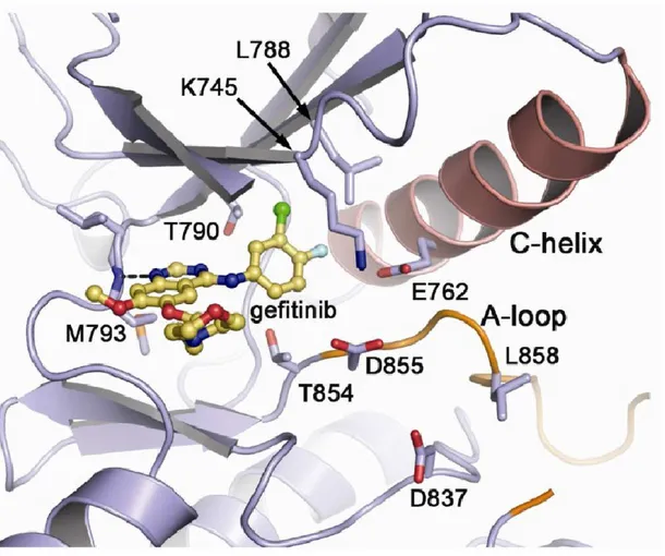
Figure 3. Crystalized structure of gefitinib binding the ATP-binding domain of the EGFR
The kinase domain of the EGFR is composed of a C-terminal-lobe and an N-terminal-lobe connected by a hinge region. This hinge region comprises part of the ATP-binding site. A hydrogen bond is formed between the Met793 residue of the EGFR and the quinazoline moiety of gefitinib, thus blocking ATP binding. Taken from: (Eck and Yun 2010).
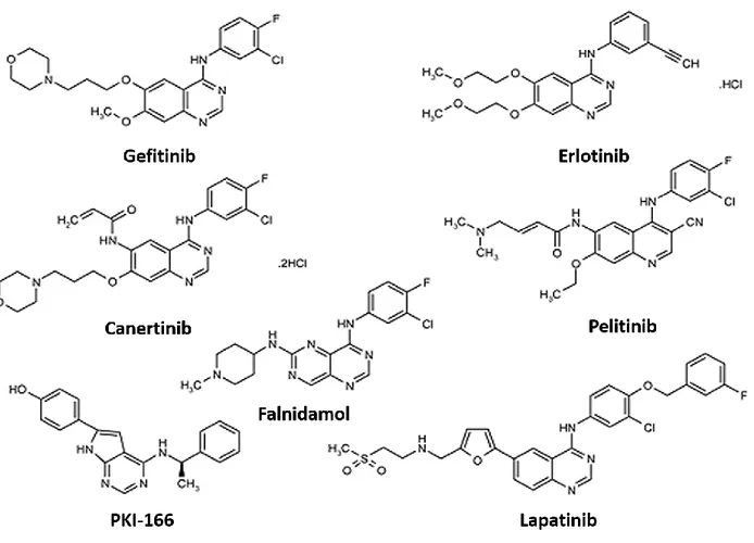
Figure 4. Chemical structures of EGFR tyrosine kinase inhibitors
EGFR tyrosine kinase inhibitors are members of the class of compounds known as the 4-anilinoquinazolines that target the ATP-binding pocket of the kinase domain of the EGFR. The quinazaline core is essential for its inhibitory effects on the EGFR. Furthermore, modifications to this core have been essential in the development of both second generation reversible and irreversible inhibitors. Adapted from: (van Montfort and Workman 2009; Hamed, Abou El Ella et al. 2013)
I.4 EGFR inhibitor resistance
Even though the majority of TNBCs overexpress the EGFR, attempts at targeting this receptor in patients has shown limited success. One reason these patients lack a response to EGFRTKis stems from the development of drug resistance. This resistance can be innate to the patient’s cancer (i.e. mutations in the EGFR or downstream signaling mediators) known as intrinsic resistance or can be developed by the patient throughout the treatment regiment, known as acquired resistance. Since EGFRTKis are clinically approved for the treatment of lung cancer, it is important to note that the majority of studies in the literature examine EGFRTKi resistance in this cancer. However, key studies evaluating TKi resistance in breast cancer will be highlighted throughout this thesis.
I.4.1 Intrinsic resistance
Cancer cells have innate characteristics that make them resistant to the currently used therapeutics. In fact, approximately 50% of all cancer patients are resistant to chemotherapy before treatment (Lippert, Ruoff et al. 2008). This is known as intrinsic or primary resistance. The most common mechanisms of intrinsic EGFRTKi resistance include: EGFR mutations characterized by a loss in sensitivity to EGFRTKi treatment such as exon 20 insertions or duplications (Greulich, Chen et al. 2005; Yasuda, Kobayashi et al. 2012), the amplification of another RTK, cMET (Engelman, Zejnullahu et al. 2007) and altered survival (PIK3CA mutations)(Cizkova, Susini et al. 2012) and apoptotic (Bim expression) (Faber, Corcoran et al. 2011) pathways. As our research focuses primarily on the role of ARF1 in the development of acquired resistance to EGFRTKis, the remainder of this thesis will be dedicated to the description of important mechanisms of acquired resistance.
I.4.2 Acquired resistance
Acquired resistance or secondary resistance (Figure 5), unlike intrinsic resistance, occurs in patients that were previously responsive to therapy and is clinically defined as: a systematic progression of the disease after a complete or partial response or following a period of 6 months of stable disease in patients treated with a targeted therapy (Jackman, Pao et al. 2010). It is generally divided into two subgroups: 1- Genetic alterations in the primary oncogene that lead to increased downstream signaling. This normally occurs through either a secondary mutation in the target kinase (the EGFR) or amplification of the target kinase. Briefly, point mutations have been
identified in the EGFR that alter either the affinity of EGFRTKis for the receptor or enhance the activity of the receptor itself (Engelman and Janne 2008; Engelman and Settleman 2008; Sierra, Cepero et al. 2010). 2- Development of resistance that is independent of genetic changes in the EGFR, such as the activation of downstream signaling pathways or other receptors, changes in tumor histology, evasion of apoptosis and alterations in drug metabolism. Amplification of other members of the EGFR family as well as other RTKs such as cMET and AXL have all been implicated in the resistance to EGFRTKis. Additionally, up-regulation of signals through the Ras/MAPK and PI3K/AKT pathway also mediate resistance. Finally, epithelial-mesenchymal transformation (EMT), a process in which epithelial cancer cells transform into more invasive mesenchymal phenotype to evade the therapeutic effects of EGFRTKis (Engelman and Janne 2008; Engelman and Settleman 2008; Ellis and Hicklin 2009; Sierra, Cepero et al. 2010). The importance of altered RTK expression and signaling, as well as the activation of downstream signaling cascades in the regulation of acquired resistance will be further discussed throughout this thesis.
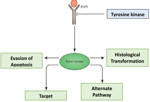
Figure 5. Mechanisms of EGFRTKi resistance
Acquired resistance plays an important role in the tumor response to EGFR inhibition. Four mechanisms of acquired resistance have been proposed: 1- Target modifications: Mutations in the EGFR itself enhance EGFR activation or block the binding of the inhibitor to the receptor, 2- Alternate pathway activation: The activation of other receptors (HER2, HER3, cMET, AXL) or signaling pathways (Ras/MAPK, PI3K/AKT) compensate for the loss of EGFR signals, 3- Evasion of apoptosis: modification in the tumor cell apoptotic machinery that prevents inhibitor-induced
Tyrosine kinase inhibitors Histological Transformation Evasion of Apoptosis Target Modifications Alternate Pathway Activation
cell death and 4- Histological transformation: processes such as epithelial-mesenchymal transformation that allow tumors cells to acquire new properties and decreased inhibitor sensitivity. Adapted from: (Chong and Janne 2013)
I.5. Membrane receptors
Membrane receptors act to relay signals between a cell and its environment. These receptors bind external stimuli such as peptides, hormones, growth factors and cytokines and relay their message to the nucleus through the activation of signaling pathways. Membrane receptors are generally divided into four families: 1- G-Protein-Coupled Receptors (GPCRs), 2- Catalytic Receptors 3- Channel-Linked Receptors, 4- Non-Catalytic Single Transmembrane Receptors. Briefly, GPCRs are a large superfamily of seven transmembrane receptors that relay their signals by binding guanine nucleotide-binding proteins (G-proteins). These G-proteins link the receptor to downstream effectors that regulate biological activities and cellular functions (Hamm 1998). Altered signaling through this receptor superfamily is associated with multiple diseases, including cancer. Secondly, the catalytic receptors are single transmembrane receptor that once bound to their ligand act directly as phosphorylating enzymes. In other words, these receptors possess enzymatic activity (Yarden and Ullrich 1988). This family is also implicated in the development and progression of cancer. As the EGFR is a member of this group of receptors, this family will be described in detail below. Next, the channel-linked receptors are generally hormone receptors that regulate the influx and efflux of ions through the cell membrane(Levitan 1988). Lastly, the non-catalytic receptors are best known to propagating signals downstream of the interleukins, peptides, hormones and neuronal cues. Like catalytic receptors, this family also consists of single transmembrane receptors. However, they do not possess catalytic activity and rely on interacting proteins to propagate its signals (Cooper and Qian 2008). In summary, membrane receptors play important roles in the communication between a cell and its environment and the de-regulation of these receptors has been linked to disease development.
I.5.1 Receptor tyrosine kinases
The RTK family of receptors are catalytic receptors that possess intrinsic kinase activity. Protein kinases are enzymes that are involved in the phosphorylation of tyrosine, serine or threonine residues (Tsai and Nussinov 2013). Families of protein phosphatases act to dephosphorylate proteins making phosphorylation a reversible process(Alonso, Sasin et al. 2004). Phosphorylation and dephosphorylation are very important in the regulation of cellular activities(Shah, Shah et al.
2013). Kinases are classified as protein-serine/threonine kinases (385 members), protein-tyrosine kinases (90 members) and tyrosine-kinase-like proteins (44 members). Of the 90 protein-tyrosine kinases, 58 are receptor-tyrosine kinases (RTKs) (Manning, Whyte et al. 2002). Alterations in RTK activity, receptor overexpression, chromosomal translocation, gene amplification, mutations and impaired receptor downregulation have all been associated with the development of cancer (Abella and Park 2009; Tsai and Nussinov 2013). In fact, 30 of the identified RTKs have been implicated in cancer(Weinstein 2000).
I.5.2 EGFR family
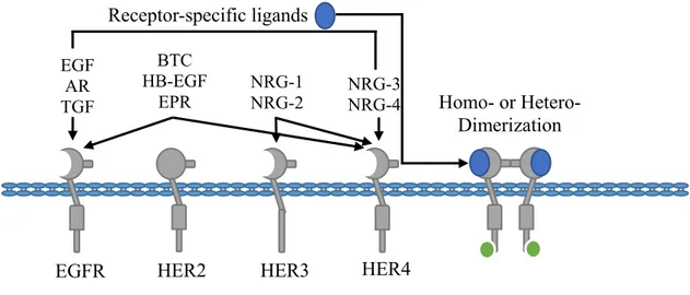
The EGFR family of RTKs, the most characterized members of the catalytic receptors, is composed of four members: EGFR, HER2, HER3 and HER4, also depicted ErbB1, ErbB2, ErbB3 and ErbB4 (Figure 6). These RTKs are ubiquitously expressed and play roles in the regulation of normal cell cycle progression, apoptosis, cell differentiation, development and gene transcription (Lemmon and Schlessinger 2010). The EGFRs are activated upon ligand binding. Presently, ten polypeptide growth factor ligands have been identified which include: EGF, amphiregulin (AR), transforming growth factor-(TGF), betacellulin (BTC), heparin-binding EGF-like growth factor (HB-EGF), epiregulin (EPR) and neuregulins 1-4 (NRG1-4), which include the heregulins (HRG) (Roskoski 2014). These ligands have been shown to activate specific EGFR family members and favor distinct receptor dimerization patterns. The EGFR members have been shown to mediate oncogenesis via several mechanisms that include receptor overexpression, mutations and ligand-independent signaling (Burtness 2007). In addition, the expression of these receptors has been associated with a poor prognosis in most cancers. Indeed, the activity of EGFRs has been shown to promote cancer cell proliferation, survival, migration and invasion (Herbst 2004; Roskoski 2014).
Figure 6. The EGFR family of receptor tyrosine kinases
The EGFR family of receptors is composed of four members: EGFR, HER2, HER3 and HER4. The activation of this family of receptors is governed by ligand-dependent homo- or heterodimerization. There are currently 11 identified ligands with specificity for certain EGFR family members, as illustrated above. HER2 differs from other members in that it has no ligand-binding domain and its activation is dependent on the formation of heterodimers. All EGFRs, except HER3, have intracellular kinase activity required for the initiation of downstream signaling cascades. Therefore, HER3 is also dependent on heterodimerization to potentiate its signals. Adapted from: (Itamochi 2010)
I.5.3 EGFR
The EGFR was the first receptor to be demonstrated to possess kinase activity and is the RTK that has been the best characterized (Carpenter and Cohen 1990). Its role in the regulation of proliferation, apoptosis and metastasis has been well defined in several cancer models, as well as in cancer patients (Herbst 2004; Roskoski 2014). Over 80% of TNBC patients have elevated EGFR expression levels and the activity of this receptor has been shown to play an important role in the oncogenic properties of this breast cancer subtype (Siziopikou, Ariga et al. 2006; Engebraaten, Vollan et al. 2013; Roskoski 2014). This makes the EGFR and its downstream effectors potential therapeutic targets in this patient subpopulation. However, attempts at targeting the EGFR have had limited success.
P
P
EGFR HER2 HER3 HER4
EGF AR TGF BTC HB-EGF EPR NRG-1 NRG-2 NRG-3 NRG-4 Receptor-specific ligands Homo- or Hetero- Dimerization
I.5.3.1 EGFR structure
The EGFR consists of an extracellular domain, a single transmembrane domain and an intracellular domain with protein kinase activity(Ullrich, Coussens et al. 1984) (Figure 7). The extracellular domain is glycosylated and consists of four domains: domain I through IV. Domains I and III mediate ligand binding, whereas, domains II and IV regulate receptor dimerization (Roskoski 2014). The intracellular domain consists of a juxtamembrane domain, a protein kinase domain and a carboxyl-terminal tail. The juxtamembrane domain has been shown to play an essential role in the tyrosine phosphorylation of the EGFR without regulating receptor dimerization and ligand binding(He and Hristova 2012). However, this domain plays an important role in the stabilization of receptor dimers and promotes receptor activation(Jura, Endres et al. 2009).The protein kinase domain is required for the activity of the receptor and promotes the activation of downstream signaling effectors(Roskoski 2014). Finally, the C-terminal tail contains essential phosphorylation sites that recruit signaling adaptors and effectors, thus promoting the activation of downstream signals(Yarden and Sliwkowski 2001). Furthermore, this tail has also been shown to function in a negative-feedback loop required for receptor inactivation(Gajiwala 2013).
Figure 7. EGFR structure
The EGFR is a single transmembrane receptor with an extracellular, transmembrane and intracellular domains. The extracellular domain mediates ligand binding through it’s I and III domains and receptor dimerization through its II and IV domains. The intracellular domain consists of a juxtamembrane domain that mediates receptor activation, dimerization and internalization, a kinase domain and c-terminal autophosphorylation domain that contains tyrosine residues that serve as recruitment sites for signaling mediators. Adapted from: (Bazley and Gullick 2005).
I III II IV Juxtamembrane domain Kinase domain Transmembrane Intracellular Extracellular Y845 Y1045 Y1068 Y1086 Y1101 Y1148 Y1173 C-terminal tail
I.5.3.2 EGFR activation mechanism
The EGFR receptor exists as a monomer at the plasma membrane. Upon ligand binding, a large conformational change occurs in the extracellular domain. This removes the dimerization autoinhibition in domain IV and promotes homo and heterodimerization of the EGFR. The juxtamembrane domain then promotes the phosphorylation of the activation segment of the protein kinase domain leading to kinase activation. The activated kinase domain tyrosine phosphorylation residues on the EGFR creating docking sites for the recruitment of signaling adaptors and effectors which initiate the activation of signaling cascades. The kinase domain can also directly phosphorylate and activate other siganling mediators to initiate downstream signals (Burgess, Cho et al. 2003; Nolen, Taylor et al. 2004; Lemmon and Schlessinger 2010).
I.5.3.3 EGFR in resistance
With expression levels of the EGFR being elevated in TNBC patients, it would be considered a good therapeutic target. However, the majority of these patients are unresponsive to EGFRTKis. The development of drug resistance is one explanation why TNBC patients do not respond to these therapies. While less common in breast cancer patients, mutations in the EGFR have been identified as key mediators of EGFRTKi treatment response in lung cancer patients. Indeed, a high proportion of lung cancer patients develop resistance through EGFR mutations such as EGFRT790M that increases the affinity of the kinase domain of the EGFR for ATP and in turn reduces the sensitivity of ATP-competitive reversible EGFRTKis(Yun, Mengwasser et al. 2008; Chong and Janne 2013). These mutations can be either acquired throughout the treatment or innate (Lee, Shin et al. 2013). However, in breast cancer patients, these mutations in the EGFR are very rare(Bhargava, Gerald et al. 2005) and downstream signaling mediators play a more important role in acquired resistance (Ferrer-Soler, Vazquez-Martin et al. 2007). Additionally, increase expression and the nuclear translocation of the EGFR have also been implicated in resistance (Brand, Iida et al. 2014; Lee, Seo et al. 2014). Indeed, the inhibition of the nuclear translocation of the EGFR significantly improved the response to EGFR inhibitors. Briefly, the endocytosis of the EGFR has been implicated in its translocation from the membrane into the nucleus. However, it remains unknown how the EGFR is transported from the vesicle into the nucleus. Once in the nucleus, EGFR acts as a transcriptional co-activator for various oncogenes implicated in resistance (Brand, Iida et al. 2014). Additionally, AKT, an importance mediator of resistance, has been shown
to Serine phosphorylation EGFR leading to its nuclear translocation (Huang, Chen et al. 2011). While alterations in EGFR-mediated signals influence patient response to EGFR inhibitors, other EGFR family members as well as other RTKs have also been implicated in the development of resistance. A description of these receptors can be found below.
I.5.4 HER2
This EGFR family member, like the EGFR, is also well characterized for its role in mediating breast oncogenesis. In fact, 20-30% of breast cancers have HER2 receptor overexpression or gene amplification. Additionally, HER2 expression is associated with a poor prognosis and increased cancer cell proliferation (Roskoski 2014). Unlike other EGFR family members, HER2 has no ligand and exerts its oncogenic properties through the heterodimerization with other EGFRs. While HER2 has been reported to dimerization with both EGFR and HER3, studies have shown that it favors HER3 heterodimer formation (Citri, Skaria et al. 2003). This would suggest that targeting HER2 could significantly reduce signals downstream of both the EGFR and HER3, making HER2 a favorable therapeutic target. However, HER2 monoclonal antibodies have been shown to be most effective at inhibiting signals downstream of HER2 homodimers and to have no effect on the ability of HER2 to heterodimerize with the EGFR and HER3 (Ghosh, Narasanna et al. 2011). Additionally, the effectiveness of HER2-targeted therapies is hindered by the development of resistance (Rexer and Arteaga 2012). Therefore, there is significant room for improvement in the treatment regiments of HER2-positive breast cancer patients.
Like EGFR, this receptor has also been implicated in the development of resistance to EGFRTKis. In fact, the amplification of HER2 is commonly found in EGFRTKi resistance lung cancer patients that do not develop mutations in the EGFR. Interestingly, 12% of EGFRTKi resistance lung tumors had amplified HER2 expression compared to only 1% in non-resistant tumors (Takezawa, Pirazzoli et al. 2012). Conversely, HER2 expression is linked to EGFRTKi sensitivity in breast cancer. However, over time, mutations in the ATP-binding pocket of the kinase domain of HER2 have been observed and this leads to the development of acquired resistance in breast cancer (Piechocki, Yoo et al. 2007). Additionally, prolonged inhibitor treatment has been shown to enhance and stabilize HER2 heterodimerization with both EGFR and HER3 and potentiate downstream signaling (Jain, Penuel et al. 2010; DeFazio-Eli, Strommen et al. 2011). Together,
these findings demonstrate that HER2 mediates breast oncogenesis and that targeting this receptor could be important in the sensitivity of breast cancer patients to EGFR inhibitors.
I.5.5 HER3
The influence of the third member of this receptor family, HER3, in breast cancer has been less documented. However, 20-30% of invasive breast cancers have shown to overexpress the HER3 receptor (Karamouzis, Badra et al. 2007). Also, HER3 has been linked to HER2 positivity and decreased incidence of metastasis. However, no true correlations between HER3 and patient survival have been made within the literature (Lemoine, Barnes et al. 1992; Gasparini, Gullick et al. 1994; Bieche, Onody et al. 2003). Like HER2, the oncogenic properties of HER3 is highly dependent on its heterodimerization with other EGFRs (Koutras, Fountzilas et al. 2010). In fact, HER2-HER3 heterodimerization has been associated with a high mitogenic potential (Citri, Skaria et al. 2003).
Additionally, HER3 has been implicated in EGFRTKi resistance. Indeed, increased HER3 expression is associated with decreased sensitivity to EGFRTKis(Stegeman, Kaanders et al. 2013; Nakata, Tanaka et al. 2014). Furthermore, increased HER3 activation, heterodimerization as well as decreased in activity of phosphatases targeting the HER3 have all been linked to acquired resistance to EGFR inhibitors(Koizumi, Shimoyama et al. 2005; Sergina, Rausch et al. 2007; Xia, Petricoin et al. 2013). Additionally, the pharmacological targeting of HER3 has been shown to overcome EGFRTKi resistance(Huang, Li et al. 2013). The increased activation and dimerization is explained by an altered localization of HER3 in lung cancer cells. Indeed, prolonged EGFR inhibition promoted the membrane localization of HER3 (Sergina, Rausch et al. 2007). Additionally, HER3 has been shown to promote resistance through the activation of the PI3K/AKT pathway. In fact, heterodimerization of HER3 with either mutant EGFR (T790M) or another RTK cMET have been shown to promote PI3K activation in lung cancer cells (Engelman, Mukohara et al. 2006; Engelman, Zejnullahu et al. 2007). While the functions of HER3 in breast cancer has yet to be fully elucidated, it has been demonstrated in the literature that HER3 exerts its oncogenic properties through it dimerization with HER2 and is an important mediator of EGFRTKi sensitivity.
I.5.6 HER4
Of the four EGFR family members, the functions of HER4 in breast cancer are the least discussed. Its expression is detectable in approximately 50% of diagnosed breast cancers. However, unlike other EGFR family members, HER4 is associated with the inhibition of cellular proliferation and the induction of apoptosis (Sartor, Zhou et al. 2001; Naresh, Long et al. 2006). Interestingly, HER4 has been demonstrate to localize to the mitochondria and enhance the release of cytochrome C to promote apoptosis (Naresh, Long et al. 2006). Additionally, HER4 expression has been correlated with a good prognosis, increased patient survival, ER positivity, decreased HER2 signaling and increased response to hormonal therapy (Knowlden, Gee et al. 1998; Tang, Concepcion et al. 1999; Bieche, Onody et al. 2003; Witton, Reeves et al. 2003; Barnes, Khavari et al. 2005; Naresh, Thor et al. 2008). Meanwhile, the role of HER4 in EGFRTKi resistance has yet to be evaluated within the literature.
Together, the EGFR family of receptors are important mediators of oncogenesis and their activity and downstream signals have been implicated in the development of drug resistance.
I.5.7 Other RTK family members in resistance
While members of the EGFR family play an important role in mediating EGFRTKi resistance, other RTK family members have also been shown to compensate for the loss of EGFR signaling. First, the amplification cMET receptor, an oncogene overexpressed in breast cancer and associated with a poor prognosis, has been shown to promote EGFRTKi resistance in lung, brain and breast cancer cells (Engelman, Zejnullahu et al. 2007; Stommel, Kimmelman et al. 2007; Gastaldi, Comoglio et al. 2010; Raghav, Wang et al. 2012; Sohn, Liu et al. 2014). This receptor was shown to activate the PI3K pathway through the transactivation of HER3. Moreover the inhibition of this HER3 transactivation re-sensitized EGFRTKi resistance cells to EGFR inhibition (Engelman, Zejnullahu et al. 2007). Additionally, another RTK overexpressed in breast cancer and associated with a poor prognosis, AXL, has also been shown to mediate EGFRTKi resistance in both lung and breast cancer (Vuoriluoto, Haugen et al. 2011; Zhang, Lee et al. 2012; Byers, Diao et al. 2013; Meyer, Miller et al. 2013). Recently, AXL was shown to promote resistance by enhancing signals downstream of the EGFR. Indeed, the pharmacological inhibition or depletion of AXL significantly hindered the activation of the EGFR and its downstream signals. Furthermore, other RTKs such as the insulin growth factor-1 receptor (IGF1R) and fibroblast growth factor receptor
1 (FGFR1) have also been shown to be activated in EGFRTKi resistant cancer (Cortot, Repellin et al. 2013; Azuma, Kawahara et al. 2014). Inhibition of IGF1R was shown to prevent the development of EGFRTKi resistant lung cancer cells.(Cortot, Repellin et al. 2013) Whereas, the inhibition or depletion of FGFR1 was shown to decrease the activation of both AKT and ERK1/2, two important mediators of EGFRTKi resistance (Azuma, Kawahara et al. 2014).
Altogether, mutations in the EGFR itself or increased signaling through other RTKs (HER2, HER3, cMET, AXL) promote resistance to EGFRTKis. Therefore, targeting signaling mediators downstream of these receptor could improve the response of patients to EGFR inhibition and delay the onset of resistance to these inhibitors.
I.6 Signaling adaptors
Activated RTKs are linked to their downstream signaling pathways through the recruitment of adaptor and effector proteins. Briefly, the autophosphorylation of the EGFR or the transphosphorylation of tyrosine residues by other kinases such as Src within the c-terminal tail of the EGFR create docking sites for adaptor proteins including Grb2 and Shc (Rozakis-Adcock, McGlade et al. 1992; Biscardi, Maa et al. 1999; Roskoski 2014). The recruitment of these adaptors has been shown to greatly increase the ability of the EGFR to phosphorylate and activate its downstream signaling mediators (Rojas, Yao et al. 1996; Migliaccio, Mele et al. 1997). Furthermore, adaptors assist in the assembly of spatially organized signaling cascades leading to the induction of important physiological responses such as cell proliferation, survival, migration and invasion (Hsieh, Yang et al. 2010). In this next section, we will discuss the importance of adaptor proteins as mediators of signals downstream of the EGFR and their influence in the development of breast cancer.
I.6.1 Grb2
The best characterized adaptor recruited to the EGFR is Grb2. It is classically known to link activated RTKs to the Ras/MAPK pathway. Grb2 is constitutively bound to the Ras guanine exchange factor (GEF) son of sevenless (SOS). Upon RTK activation, the Grb2/SOS complex is recruited to the receptor bringing SOS into close proximity with the GTPase Ras. This leads to Ras activation and the initiation of the Ras/MAPK pathway (van der Geer, Hunter et al. 1994; Kairouz and Daly 2000). As Grb2 has been previously reported to recruit a GEF to the EGFR
leading to the activation of the small GTPase Ras, we highlight the importance of Grb2 in the activation of other small GTPases, ARF1 and ARF6, in Chapter II. Grb2 has also been shown to interact with other important signaling mediators such as Grb2-associated binding protein-1 (Gab1) which recruits phosphatidylinositol-4, 5-biphosphate 3 kinases (PI3K) to RTKs leading to its activation (Ong, Hadari et al. 2001).
The role of Grb2 in breast cancer remains controversial. An increased protein and mRNA expression of Grb2, as well as an amplification of the GRB2 gene locus have been observed in breast cancer cells and primary breast tumors (Daly, Binder et al. 1994; Verbeek, Adriaansen-Slot et al. 1997; Yip, Crew et al. 2000). Additionally, depletion of Grb2 is associated with a decreased ERK1/2 activation in breast cancer cells and delayed onset of mammary tumors induced by the polyomavirus middle T antigen suggesting that Grb2 may play an important role in mammary oncogenesis (Gale, Kaplan et al. 1993; Suen, Bustelo et al. 1993). However, Grb2 has also been shown to negatively regulate signals downstream of the EGFR (Li, Couvillon et al. 2001; Haines, Minoo et al. 2009; Belov and Mohammadi 2012). Indeed, the tyrosine phosphorylation of Grb2 by the EGFR itself and the prolactin receptor, an important mediator of breast development and oncogenesis, has been shown to attenuate its interaction with SOS and thus block the activation Ras downstream of the EGFR (Li, Couvillon et al. 2001; Haines, Minoo et al. 2009). Additionally, Grb2 has been shown to recruit the ubiquitin ligase, Casitias B-lineage Lymphoma protein (Cbl), to the EGFR leading to receptor ubiquination and its down-regulation (Belov and Mohammadi 2012). On the same note, recent attempts at targeting Grb2 in cancer have had little to no therapeutic effects (Dharmawardana, Peruzzi et al. 2006).
Together, Grb2 has been shown in the literature to be recruited to activated RTKs and promote the activation of signaling cascades. However, its role in breast cancer is still controversial.
I.6.1.1 Grb2 structure
The adaptor Grb2 consists of a Src Homology 2 (SH2) domain flanked by two Src Homology 3 (SH3) domains (van der Geer, Hunter et al. 1994) (See Figure 8). Grb2 interacts with tyrosine phosphorylated residues such as those on the EGFR through its SH2 domain and proline-rich motifs such as those present on SOS through its SH3 domains(Lowenstein, Daly et al. 1992; van der Geer, Hunter et al. 1994; Kairouz and Daly 2000). Other proteins known to interact with the SH2 domain of Grb2 include: other RTKs and the adaptor Shc (Lowenstein, Daly et al. 1992;
Rozakis-Adcock, McGlade et al. 1992). Whereas, Grb2 binds proteins such as dynamin, a GTPase involved in endocytosis and Cbl, an E3 ubiquitin protein ligase involved in EGFR down-regulation through its SH3 domains(Lowenstein, Daly et al. 1992; Sparks, Rider et al. 1996; Yamazaki, Zaal et al. 2002).
Figure 8. Grb2 structural domains
The adaptor protein Grb2 contains a SH2 domain flanked by two SH3 domains. Classically, Grb2 is recruited to the activated EGFR through its SH2 domain. This recruits the exchange factor SOS, bound to its SH3 domain, leading to the activation of Ras/MAPK pathway. Other important signaling mediators such as the Gab1 and Shc family of adaptors as well as the ligase Cbl have all been shown to interact with Grb2. Adapted from: (Skolnik, Lee et al. 1993).
I.6.2 Shc family of adaptors
Another important family of adaptors characterize for their role downstream of RTKs is the Shc family of adaptors. It was first identified in a screen for novel SH2-containing proteins. Interestingly, Shc adaptors were discovered due to their high homology (~60%) with the tyrosine kinase, Src (Pelicci, Lanfrancone et al. 1992). They are best known for their role in mediating the activation of the Ras/MAPK and the PI3K/AKT pathways (Ravichandran 2001). Presently, four members of this family of adaptors have been identified and are designated: ShcA, ShcB, ShcC and ShcD(Wills and Jones 2012). While ShcB and ShcC are generally expressed within the central nervous system (CNS), ShcA is more ubiquitously expressed and highly expressed in epithelial cells(Pelicci, Lanfrancone et al. 1992; Nakamura, Sanokawa et al. 1996; O'Bryan, Songyang et al. 1996; Pelicci, Dente et al. 1996). Within the CNS, ShcB and ShcC have been shown to promote the activation of the Ras/MAPK pathway downstream of both the EGFR and Trk receptors, a family of neurotrophins important for the survival of neurons (Sakai, Henderson et al. 2000). This suggests that ShcB/C exert similar effects in CNS as ShcA does in both the CNS and epithelium (van der Geer, Hunter et al. 1994; Sakai, Henderson et al. 2000). As for ShcD, it has been shown
SH3
SH2
SH3
Grb2
to be expressed in the CNS, muscle, epithelia and bone precursors. However, its role in these tissues has yet to be thoroughly characterized(Hawley, Wills et al. 2011; Wills and Jones 2012). Collectively, the Shc family of adaptors are important regulators of signals downstream of RTKs in a variety of tissues within the human body. As our research focusses on the role of Shc adaptors in mediating ARF1 activity in breast cancer cells, the structure of the ShcA isoforms as well as their functions and role in breast cancer will be detailed below.
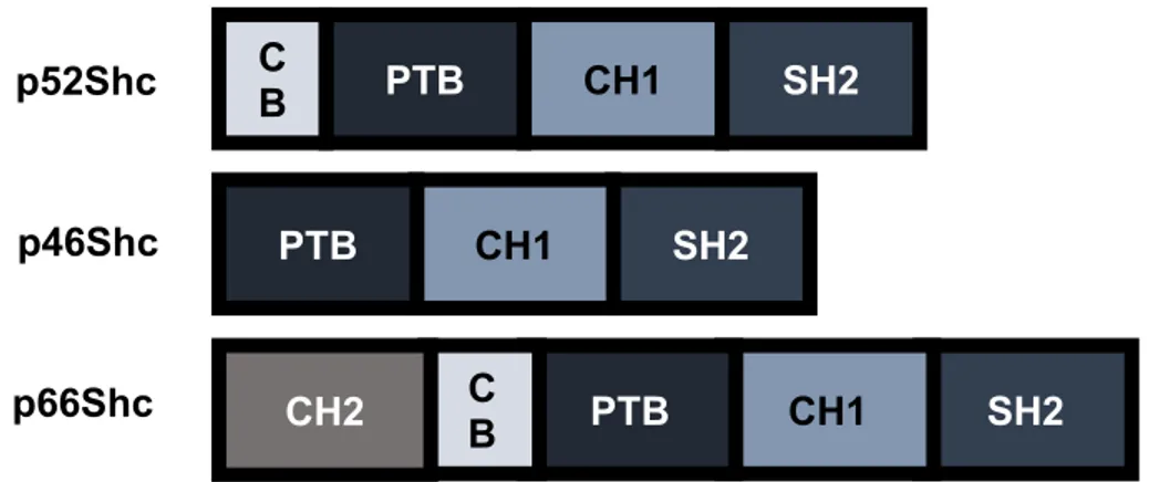
I.6.2.1 ShcA isoform structures
The ShcA family is composed of three isoforms: p66Shc, p52Shc and p46Shc. While the three isoforms originate from the same gene, their expression are governed by different transcriptional initiation sites as well as translational start sites (Luzi, Confalonieri et al. 2000). Structurally, the three isoforms high homology, with differences only present within their N-terminus, and are composed of a protein tyrosine binding domain (PTB), a collagen homology domain (CH1) and a SH2 domain (Migliaccio, Mele et al. 1997) (Figure 9). Both the PTB and SH2 domains of ShcA interact with phosphotyrosine residues and have been implicated in its receptor recruitment (Pelicci, Lanfrancone et al. 1992; van der Geer, Wiley et al. 1996; Ravichandran 2001). The PTB and SH2 domains are connected via a CH1 domain which is rich in proline motifs and known to interact with SH3-containing proteins such as Src(Migliaccio, Mele et al. 1997; Luzi, Confalonieri et al. 2000; Ravichandran 2001). Additionally, three tyrosine residues (Y239, Y240 and Y317 in p52Shc) are phosphorylated upon the engagement of ShcA to an activated RTK. Two of these residues (Y239 and Y240) are known to be phosphorylated by Src, whereas the kinase involved in Y317 phosphorylation has yet to identified (Ravichandran 2001). The phosphorylation of these residues has been implicated in the interaction between Grb2 and Shc(van der Geer, Wiley et al. 1996). Indeed, mutation of these residues to alanine blocked the interaction between these two adaptors.
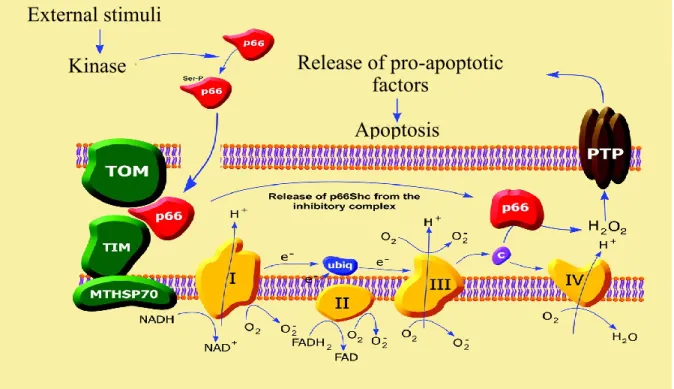
The expression and structure of the p66Shc isoform differs from the other two ShcA isoforms. While p52/46Shc has been reported to be ubiquitously expressed, p66Shc is specifically expressed within the epithelium (Pelicci, Lanfrancone et al. 1992). This isoform also differs from the other two isoforms in that it consists of an additional CH domain (CH2). The CH2 domain is best characterized for its functions in mediating oxidative stress upon the phosphorylation of serine 36 (Migliaccio, Giorgio et al. 1999). Oxidative stress, hydrogen peroxide treatment or irradiation,