HAL Id: hal-01601907
https://hal.archives-ouvertes.fr/hal-01601907
Submitted on 27 May 2019
HAL is a multi-disciplinary open access
archive for the deposit and dissemination of
sci-entific research documents, whether they are
pub-lished or not. The documents may come from
teaching and research institutions in France or
abroad, or from public or private research centers.
L’archive ouverte pluridisciplinaire HAL, est
destinée au dépôt et à la diffusion de documents
scientifiques de niveau recherche, publiés ou non,
émanant des établissements d’enseignement et de
recherche français ou étrangers, des laboratoires
publics ou privés.
Copyright
Characterization of the First α-(1
→3) Branching
Sucrases of the GH70 Family
Marlène Vuillemin, Marion Claverie, Yoann Brison, Etienne Séverac, Pauline
Bondy, Sandrine Morel, Pierre Monsan, Claire Moulis, Magali Remaud Simeon
To cite this version:
Marlène Vuillemin, Marion Claverie, Yoann Brison, Etienne Séverac, Pauline Bondy, et al..
Char-acterization of the First α-(1
→3) Branching Sucrases of the GH70 Family. Journal of Biological
Chemistry, American Society for Biochemistry and Molecular Biology, 2016, 291 (14), pp.7687-7702.
�10.1074/jbc.M115.688044�. �hal-01601907�
Characterization of the First
␣-(133) Branching Sucrases of
the GH70 Family
*
□SReceived for publication, August 26, 2015, and in revised form, January 11, 2016 Published, JBC Papers in Press, January 13, 2016, DOI 10.1074/jbc.M115.688044
Marle`ne Vuillemin, Marion Claverie, Yoann Brison, Etienne Se´verac, Pauline Bondy, Sandrine Morel, Pierre Monsan, Claire Moulis1, and Magali Remaud-Sime´on2
From the Universite´ de Toulouse, Institut National des Sciences Applique´es (INSA), Universite´ Paul Sabatie´ (UPS), Institut National Polytechnique (INP), Laboratoire d’Inge´nieries des Syste`mes Biologiques et des Proce´de´s (LISBP), 135 Avenue de Rangueil, F-31077 Toulouse, France, CNRS, UMR5504, F-31400 Toulouse, France, and Institut National de la Recherche Agronomique (INRA), UMR792 Inge´nierie des Syste`mes Biologiques et des Proce´de´s, F-3140 Toulouse, France
Leuconostoc citreum NRRL B-742 has been known for years to
produce a highly␣-(133)-branched dextran for which the syn-thesis had never been elucidated. In this work a gene coding for a putative␣-transglucosylase of the GH70 family was identified in the reported genome of this bacteria and functionally charac-terized. From sucrose alone, the corresponding recombinant protein, named BRS-B, mainly catalyzed sucrose hydrolysis and leucrose synthesis. However, in the presence of sucrose and a dextran acceptor, the enzyme efficiently transferred the gluco-syl residue from sucrose to linear␣-(136) dextrans through the specific formation of␣-(133) linkages. To date, BRS-B is the first reported ␣-(133) branching sucrase. Using a suitable sucrose/dextran ratio, a comb-like dextran with 50% of␣-(133) branching was synthesized, suggesting that BRS-B is likely involved in the comb-like dextran produced by L. citreum NRRL B-742. In addition, data mining based on the search for specific sequence motifs allowed the identification of two genes puta-tively coding for branching sucrases in the genome of
Leucono-stoc fallax KCTC3537 and Lactobacillus kunkeei EFB6.
Bio-chemical characterization of the corresponding recombinant enzymes confirmed their branching specificity, revealing that branching sucrases are not only found in L. citreum species. According to phylogenetic analyses, these enzymes are pro-posed to constitute a new subgroup of the GH70 family.
Numerous lactic acid bacteria from the Leuconostoc genus isolated from different habitats, such as sugar juice, fermenting vegetables, or dairy products, have long been known to produce slimes in sucrose solution (1, 2). These slimy compounds were rapidly characterized as dextrans, homopolymers of␣-D
-gluco-pyranosyl units mainly linked by␣-(136) linkages. Dextrans with a high content of␣-(136) linkages and a low degree of branching found their first industrial applications in the 1940s
as a source of synthetic blood volume expanders (3). These findings motivated Jeanes et al. (22) to investigate the diversity of polymers produced during sucrose fermentation by different strains of lactic acid bacteria. A total of 96 strains were screened, and their dextrans were structurally characterized. In this study one strain, Leuconostoc mesenteroides NRRL B-742, also known as L. mesenteroides ATCC 13146 and renamed
Leu-conostoc citreumNRRL B-742 (4), received particular attention. Indeed, this strain, first isolated from a can of spoiled tomatoes in 1927 (1), was shown to produce two types of dextrans that differed by their structures and degree of solubility from sucrose. The less soluble polymer contained 75% of␣-(136) linkages and 15% of␣-(134) linkages, whereas the more solu-ble polymer displayed an uncommon comb-like structure con-sisting of a linear backbone of␣-(136)-D-glucopyranosyl
resi-dues grafted with one␣-(133)-linked glucosyl unit on every glucosyl moiety (5–9). Concomitantly with those findings, dex-trans were shown to be synthesized by ␣-transglucosylases, commonly named glucansucrases, and today classified in the family 70 of glycoside hydrolases (GH70)3(10). The GH70 fam-ily belongs to the clan GH-H, together with families GH13 and GH77. To date, 279␣-transglucosylase sequences have been reported in the CAZy database, and only 25% has been func-tionally characterized (11). Furthermore, only four three-di-mensional structures of GH70 enzymes have been solved (12– 15). The enzymes are organized into five different structural domains, A, B, C, IV, and V. The A, B, and C domains are related to GH13 family enzymes, whereas domains IV and V are GH70-specific domains. The fold of these enzymes is unique, and except for domain C, all domains are formed by non-contigu-ous fragments of sequences that assemble around a U-turn (15). These enzymes are␣-retaining enzymes. They mainly catalyze ␣-transglucosylation reactions through a classical double dis-placement mechanism. The reaction starts with the formation of a-D-glucosyl-enzyme intermediate involving a nucleophilic
aspartate, an acid-base glutamic acid, and a third aspartate that stabilizes the covalent intermediate. In a second step the inter-mediate can be intercepted by a water molecule (hydrolysis reaction) or by the hydroxyl group of an acceptor
(transgluco-*This work was supported by the French National Research Agency (ANR-12-CDII-0005, Engel 2012-2015) and the Re´gion Midi-Pyre´ne´es (France). The authors declare that they have no conflict of interest with the contents of this article.
□S
This article containssupplemental Tables S1–S4 and Figs. S1–S3.
1To whom correspondence may be addressed: INSA, LISBP, 135 Avenue de
Rangueil, F-31077 Toulouse, France. Tel.: 446; Fax: 33-561-559-400; E-mail: moulis@insa-toulouse.fr.
2To whom correspondence may be addressed: INSA, LISBP, 135 Avenue de
Rangueil, F-31077 Toulouse, France. Tel.: 446; Fax: 33-561-559-400; E-mail: remaud@insa-toulouse.fr.
3The abbreviations used are: GH, glycoside hydrolase; HPAEC-PAD, high
per-formance anion exchange chromatography with pulsed amperometric detection; HPSEC, high performance size exclusion chromatography; con-tig, group of overlapping clones.
THE JOURNAL OF BIOLOGICAL CHEMISTRY VOL. 291, NO. 14, pp. 7687–7702, April 1, 2016 © 2016 by The American Society for Biochemistry and Molecular Biology, Inc. Published in the U.S.A.
sylation). The synthesis of the polymer occurs via successive transfers of glucosyl units onto the non-reducing extremity of the growing␣-glucan chain (Scheme 1) (16–18).
Among GH70 sucrose-active enzymes, four subgroups of glucansucrases can be distinguished: the dextransucrases, alternansucrases, mutansucrases, and reuteransucrases, which are all efficient polymerases (10). In addition, one branching sucrase (GBD-CD2) was engineered from L. citreum NRRL B-1299 DSR-E dextransucrase (19). This enzyme displays no polymerase activity but catalyzes␣-(136) dextran branching very efficiently with single ␣-(132)-linked glucosyl units (Scheme 1) (20). Recently, genome sequencing of L. citreum NRRL B-1299 allowed the identification of the first natural ␣-(132) branching sucrase, BRS-A, that shows the same spec-ificity as GBD-CD2. This enzyme was proposed to be involved in the formation of the native dextrans of L. citreum NRRL B-1299 that contain a high percentage of␣-(132) linkages (21, 22).
The formation of the regular hyper-branched dextran of
L. citreum NRRL B-742 is even more intriguing. Several attempts to isolate the GH70 enzymes of L. citreum NRRL B-742 were reported. However, although the enzyme responsi-ble for the production of dextran with both ␣-(136) and
␣-(134) linkages was isolated, the enzyme contributing to the comb-like structure was never identified (5). Of note, most of the GH70 enzyme activities were found strongly attached to the cell wall, making the enzymes difficult to purify (7). Crude enzyme extracts or sucrose fermentation broths were thus used to produce ␣-(133)-branched glucooligosaccharides from sucrose and maltose (7), and these compounds were shown to possess interesting functional properties. In particular, they stimulated in vitro growth of beneficial bacteria such as bifido-bacteria or lactobacilli and inhibited the development of patho-gens such as Salmonella sp. or Escherichia coli (23, 24). In 2000, Kim and Robyt (25) cloned a gene from the mutant strain L.
cit-reum NRRL B-742CB. The encoded protein, DSR-B-742, shared⬎95% amino acid identity with DSR-B dextransucrase from L. citreum NRRL B-1299 and synthesized a quasilinear dextran with⬎95% of␣-(136) linkages (26). Thus far the mode of synthesis of the comb-like dextran has remained unknown.
A genomic approach was undertaken to gain insight into the formation of this hyper-branched␣-glucan and to have access to enzymatic tools of interest for functional glucooligosaccha-ride synthesis. First, the genome of L. citreum NRRL B-742 was sequenced (27) and mined to determine its content in GH70 ␣-transglucosylase encoding genes. One putative GH70 gene SCHEME 1. Main reactions catalyzed by GH70 sucrose-active enzymes.
showing divergence from various consensus sequences of GH70 family enzymes was cloned for recombinant expression in E. coli. The corresponding protein, named BRS-B, is an ␣-(133) branching sucrase. A shorter version of BRS-B, named BRS-B-⌬1, was rationally designed and characterized. The specificity of BRS-B and its truncated form was investigated and revealed the interest of these enzymes for the production of ␣-(133)-branched dextrans with a controlled degree of branching. In addition, comparison of these enzymes with the recently discovered ␣-(132) branching sucrase (BRS-A) allowed the identification of two enzymes in public databases that were found to catalyze dextran branching and were clus-tered with previously characterized branching sucrases in the GH70 phylogenetic tree.
Experimental Procedures
Isolation of brsB Gene and Analysis of BRS-B Primary Structure
The brsB gene was identified in the L. citreum NRRL B-742 genome (27) (NCBI reference sequence: NZ_CCNG01000013.1) by performing a nucleotide BLAST against a homemade GH70 ␣-transglucosylaencoding gene database. The protein se-quence of BRS-B is deposited under the GenBankTMaccession number CDX65123.1. Protein sequence comparison was per-formed either using protein Blast (NCBI) against the non-re-dundant protein sequence database (nr) for local alignments or using Clustal Omega for global alignments with other GH70 enzymes. Protein sequence global alignment with GTF-180-⌬N glucansucrase (15) and ⌬N123-GBD-CD2 (12) was used to determine length and position of domains A, B, C, IV,
and V. The signal peptide cleavage was predicted using SignalP 4.1 server.
Isolation of Genomic DNA
L. citreumNRRL B-742 was grown on 1.5 ml of MRS medium (Sigma-Aldrich) at 30 °C overnight. Genomic DNA was iso-lated using the Wizard威 genomic DNA purification kit (Pro-mega, Madison, WI) according to the supplier’s recommenda-tions for Gram-positive bacteria.
Cloning of brsB and brsB⌬1 Genes
The gene brsB and a truncated form lacking the signal pep-tide and the last 575 amino acids at the C terminus of the pro-tein (brsB⌬1) were amplified by PCR from L. citreum NRRL B-742 genomic DNA template. The primers used are described in Table 1. The addition of the CACC sequence (underlined) to the 5⬘-forward primers allowed the correct insertion of the genes into the pENTR/D-TOPO威 vector (Life Technologies). From a positive entry clone, LR recombination (Gateway威 LR Clonase威 II enzyme mix, Life Technologies) was performed using the destination vectors described in Table 1. Expression clones were selected on LB agar plates supplemented with 100 g ml⫺1of ampicillin. Plasmids were extracted with the Sigma GenElute HP Plasmid Miniprep kit, verified by restriction anal-yses and sequencing (GATC Biotech, Constance, Germany).
Recombinant Production of BRS-B and BRS-B-⌬1
Starter Culture—E. coliBL21-AI and E. coli BL21 star DE3 cells (detailed in Table 1) were freshly transformed by pBAD-TABLE 1
49/brsB and pET-55/brsB⌬1, respectively. Thirty milliliters of LB medium, supplemented with ampicillin (100g ml⫺1), were inoculated with 200l of transformation mix and incubated overnight at 37 °C under agitation (200 rpm).
Erlenmeyer Flask Cultures—One liter of modified ZYM5052 medium (28) contained (i) 0% lactose, 0% glucose, 0.5% glycerol and 0.01%L-arabinose for BRS-B production or (ii) 0.1%
lac-tose, 0% glucose and 1% glycerol for BRS-B-⌬1 production. Both were supplemented with ampicillin (100 g ml⫺1) and were inoculated with the corresponding starter culture at an
A600 nmof 0.05. Cultures were incubated at 21 °C under agita-tion (150 rpm). After a 26-h incubaagita-tion, cells were harvested by centrifugation, dispersed in 50 mMsodium acetate buffer, pH
5.75, at a final A600 nm 80 and disrupted by sonication. The recombinant enzymes were recovered in the soluble fraction after centrifugation (15,000⫻ g, 30 min, 4 °C) of the crude cell extract.
Affinity Chromatography Purification of Enzymes
All purification assays were performed using the A¨ KTAxpress(GE Healthcare) at 12 °C. For His6tag affinity chromatography, cells were centrifuged and resuspended in binding buffer (20 mMphosphate sodium buffer, pH 7.4, 500 mMNaCl, 20 mM
imidazole, 2.5% (v/v) glycerol) at a final A600 nmof 200. After disruption by sonication, centrifugation (30,000⫻ g, 30 min, 4 °C), and filtration through a 0.22-m cartridge (Sartorius Ste-dim Biotech, Aubagne, France), lysates were applied onto a 1-ml HisTrap HP威 column (GE Healthcare) that had been equilibrated with the binding buffer. The proteins were eluted by an imidazole gradient from 10 to 500 mMover 25 min. Eluate
fractions of 3 ml were desalted onto a 10-DG column (Bio-Rad) with 50 mMsodium acetate buffer at pH 5.75 with 100 mM
NaCl, 0.05 g liter⫺1CaCl2, and 0.1% (v/v) Tween 80. In order to remove small contaminants, purified eluates were washed 3 times onto Vivaspin 2 with a molecular weight cut-off of 100 (Sartorius Stedim Biotech).
Enzymatic Activity Assay
One unit of BRS-B, BRS-B-⌬1, BRS-C, or BRS-D is defined as the amount of enzyme that catalyzes the production of 1mol of fructose/min at 30 °C in 50 mMsodium acetate buffer at pH
5.75 from 292 mMsucrose. The enzyme activities were
deter-mined by measuring the amount of reducing sugars using the dinitrosalicylic acid method (29).
pH and Temperature Optima; Effect of Divalent Ions
Optimal pH and temperature were determined in duplicate using 1 unit䡠ml⫺1of enzyme in the presence of 292 mMsucrose
and 119 mMdextran (68,400 g䡠mol⫺1) for both BRS-B (in
son-icated extract) and the pure BRS-B-⌬1. To evaluate the optimal pH, the assays were performed at 30 °C in 50 mMcitrate phos-phate buffer with pH values varying from 3.5 to 7.0 in incre-ments of 0.5 units. Optimal temperature was determined from assays carried out in 50 mMsodium acetate buffer at pH 5.75 at
temperatures of 23, 26, 30, 33, 37, 40, or 45 °C. The effect of salts on transferase activity of BRS-B-⌬1 was determined by incubat-ing the enzyme (1 unit䡠ml⫺1) with 1 mM EDTA to trap the
possible presence of any divalent ion in the catalytic core of the
enzyme or with 1 mM CaCl2, CuCl2, ZnCl2, or MgCl2 salts.
Activity assays were performed at 30 °C in 50 mMsodium
ace-tate buffer, pH 5.75, 292 mMsucrose, and 119 mMof dextran (68,400 g䡠mol⫺1). For all these experiments, the enzymatic activity was determined by measuring fructose release by high performance liquid chromatography (HPLC) on a Dionex sys-tem and using an Aminex威 HPX-87K carbohydrate analysis column (300⫻ 7.8 mm; Bio-Rad). Column oven temperature was set at 65 °C, and ultrapure water was used as eluent (0.6 ml min⫺1).
Determination of BRS-B-⌬1 Kinetic Parameters
Reaction with Sucrose—Kinetic parameters with sucrose as the sole substrate were determined in 50 mMsodium acetate
buffer at pH 5.75 supplemented with 250g ml⫺1bovine serum albumin, 23g ml⫺1of purified BRS-B-⌬1 at 30 °C and using initial sucrose concentrations ranging from 3 to 600 mM. All
initial velocities were measured by quantifying fructose release at the early stage of reaction (when ⬍5% of the substrate is consumed and when leucrose and other oligosaccharide pro-duction can be neglected). At regular time intervals samples were withdrawn and immediately heated at 95 °C for 5 min to stop the reaction. Fructose concentration was determined by HPLC as described above. Km, Ki, and Vmaxparameters were determined using SigmaPlot Kinectics software and fitted to a non-competitive substrate inhibition model.
␣-(133) Transglucosylation—Kinetic parameters for ␣-(13 3) transglucosylation were determined at 30 °C in 50 mM
sodium acetate buffer, pH 5.75, supplemented with 250 g ml⫺1BSA, 9.5 mg liter⫺1of enzyme, 300 mMsucrose, and
dex-tran of 68,400 g䡠mol⫺1at final concentrations ranging from 31 to 617 mM. For the sake of clarity, dextran concentrations will
be expressed as molar concentrations of anhydroglucosyl units within the dextran, as determined by dividing dextran mass concentrations by 162 g䡠mol⫺1. All initial velocities were mea-sured when⬍5% of the sucrose was consumed. Initial␣-(133) transglucosylation velocities were calculated by subtracting fructose (representing the total activity) by free glucose (repre-senting hydrolase activity) production rates. Glucose and fruc-tose concentrations were determined by HPLC using the same conditions as those described previously. Kmdextranand Vmax were estimated using SigmaPlot Kinetics software and fitted to a Michaelis-Menten model.
Synthesis of␣-(133)-branched Dextrans Using BRS-B and BRS-B-⌬1
Various␣-(136)-linked dextrans of average molar masses ranging from 1500 to 2⫻ 106g䡠mol⫺1(Sigma) were incubated at different concentrations with sucrose (9 – 438 mM) using 1
unit䡠ml⫺1BRS-B (or BRS-B-⌬1) at 30 °C in 50 mMsodium
ace-tate buffer at pH 5.75 for 8 –16 h. Reactions were stopped by a 5-min incubation at 95 °C. The various tested sucrose/dextran ratios are reported insupplemental Table S1.
HPAEC-PAD Analysis
To determine production yield and assay sucrose depletion, reaction media were analyzed by HPAEC-PAD (high perfor-mance anion exchange chromatography with pulsed
ampero-metric detection) using a CarboPacTMPA100 (4⫻ 250 mm) analytical column coupled with a CarboPacTMPA-100 guard (4 ⫻ 50 mm). Glucose, fructose, leucrose, and sucrose were separated using a 30-min sodium acetate gradient from 6 to 300 mMin 150 mMNaOH. The amount of each sugar was
deter-mined using standards of 5, 10, 15, and 20 mg kg⫺1. Samples were prepared in ultra-pure water and diluted to a final concen-tration of 10 –15 mg kg⫺1of total sugars for quantitative anal-yses and a final concentration of 250 mg䡠kg⫺1(total sugars) for qualitative analyses. The percentage of glucosyl units from sucrose incorporated into glucose (%Gglucose) or leucrose (%Gleucrose) was calculated as follows.
%Gglucose⫽ 共关glucose兴tf ⫺ 关glucose兴t0兲 ⫻ 342 共关sucrose兴t0 ⫺ 关sucrose兴tf 兲 ⫻ 180 ⫻100 (Eq. 1) %Gleucrose⫽ 共关leucrose兴tf ⫺ 关leucrose兴t0兲
共关sucrose兴t0 ⫺ 关sucrose兴tf 兲 ⫻100 (Eq. 2)
High Performance Size Exclusion Chromatography (HPSEC) Analysis
HPSEC analyses were carried out using Shodex OH-Pak SB-802.5 and SB-805 columns in series coupled with a Shodex OH-Pak SB-G guard column and placed in an oven at 70 °C. Elution was performed using 0.45MNaNO3and 1% ethylene glycol (v/v) as the eluent using a flow rate of 0.3 ml min⫺1. The samples were diluted at 10 g kg⫺1of total sugars and also in 0.45
MNaNO3and 1% ethylene glycol.
The percentage of glucosyl units from sucrose incorporated into␣-(133)-branched dextran was calculated as,
%Gdextran⫽
Areadextran⫻ 342 Areasucrose⫻ 162
(Eq. 3)
The percentage of glucosyl units from sucrose incorporated into small oligosaccharides was deduced as,
%Gsmall oligosaccharides⫽ 100% ⫺共%Gdextran⫹ %Gglucose
⫹ %Gleucrose兲 (Eq. 4) Here %Gdextran, %Gglucose, and %Gleucrosehave been determined from HPSEC and HPAEC-PAD analyses.
1H and13C NMR Analysis of Oligosaccharides and Polymers
To determine ␣-(133) linkage content, freeze-dried poly-mer samples were dispersed in deuterated water at a final con-centration of 20 mg ml⫺1. NMR spectra were recorded on an Advance 500 MHz spectrometer (Bruker) operating at 500 MHz for1H NMR and 125 MHz for13C using a 5-mm z-gradi-ent TBI probe. The data were processed using TopSpin 3 soft-ware. One-dimensional1H NMR spectra was acquired by using a zgpr pulse sequence (with water suppression). All measure-ments were performed at 298 K, and chemical shifts were ref-erenced to an internal reference sodium 2,2,3,3-tetradeutero-3-trimethylsilylpropanoate (1H⫽ 0 ppm) and acetone (13C⫽ 31.08 ppm). The percentages of␣(133) and ␣(136) linkages in
␣-glucans were calculated by integrating the corresponding anomeric proton signals (30).
Model Construction
Sequence alignment between BRS-B (from residues Ile-220 to Ile-1300) and⌬N123-GBD-CD2 was generated using MUS-CLE (31). This alignment was checked, locally corrected in vari-able loop regions, and submitted to SWISS-MODEL online ser-vice for protein structure prediction using the alignment mode (32). The same procedure was applied for the sequence of BRS-A (from residues Gly-223 to Val-1289). BRS-B and ⌬N123-GBD-CD2 share 53% sequence identity; BRS-A and ⌬N123-GBD-CD2 share 60% sequence identity. All drawings were realized using PyMOL software (DeLano Scientific).
Identification of New Branching Sucrases
Blast analysis (blastp and blastn) were performed to identify new putative branching sucrases in NCBI public databases. The putative proteins displaying similarities with characterized branching sucrases (i.e. BRS-A, BRS-B, and/or GBD-CD2) were selected considering that the sequence should display (i) one phenylalanine at subsite⫹1 (position 675, BRS-B numbering), (ii) a non-aromatic residue at subsite⫹1 (position 711, BRS-B numbering), (iii) one isoleucine at position 783 (BRS-B num-bering), (iv) one histidine at position 785 (BRS-B numnum-bering), (v) one lysine at position 789 (BRS-B numbering), and (vi) one valine at position 795 (BRS-B numbering).
Cloning of the brsC Gene
First, a synthetic brsC gene (contig GenBankTM accession number AEIZ01000002, positions 68,715– 63,391 on reverse strand) was designed to optimize its expression in E. coli (Gene-Cust, Dudelange, Luxembourg). The gene was then amplified by PCR from pUC57/brsC plasmid DNA template using the primers described in Table 1. The PCR product was then inserted into the pENTR/D-TOPO vector (Life Technologies).
From a positive entry clone, LR recombination (Gateway LR Clonase II enzyme mix; Life Technologies) was performed with the pET-55-DEST destination vector (Merck Millipore). Expression clones were selected on LB agar plates supple-mented with 100 g ml⫺1of ampicillin. Plasmids were then extracted using the GenElute HP Plasmid Miniprep kit (Sigma), verified by restriction analyses, and sequenced (GATC Bio-tech). E. coli TOP10 competent cells (Life Technologies) were used for all cloning experiments.
Recombinant Production of BRS-C and BRS-D Branching Sucrases
The construction pET28b/brD was provided by Biomatik (Cambridge, ON, Canada) where the brsD gene (GenBankTM acces-sion number AZBY01000038.1, locus tag LAKU_38c00010) was designed to optimize the heterologous expression in E. coli.
Starter Culture—E. coli BL21 star DE3 cells were freshly transformed by pET-55/brsC, pET28b/brD. 30 ml of LB medium supplemented with ampicillin (100 g ml⫺1) were inoculated with 200l of transformation mix and incubated overnight at 37 °C under agitation (200 rpm).
Erlenmeyer Flask Cultures—1 liter of modified ZYM5052 medium (28) (with the changes 0.1% lactose, 0% glucose, and 1% glycerol, supplemented with ampicillin (100g ml⫺1) was inoculated with the starter culture at an A600 nmof 0.05. Cul-tures were incubated at 21 °C under agitation (150 rpm). After 26 h of incubation, cells were harvested by centrifugation, resuspended in 50 mMsodium acetate buffer, pH 5.75, at a final
A600 nm of 80, and disrupted by sonication. Recombinant enzymes were recovered after centrifugation (15,000⫻ g, 30 min, 4 °C) in the soluble fraction of the crude cell extract.
Phylogenetic Analysis
A phylogenetic tree was constructed from 60 protein sequences, including the functionally characterized GH70 enzymes indexed in the CAZY database and the branching sucrases BRS-A, BRS-B, BRS-C, BRS-D, and GBD-CD2.Protein sequences were aligned using Clustal Omega. GBlock program was used to remove highly variable regions. A total of 1371 positions (42%) were then conserved. The tree was constructed using the Neighbor-joining method using the SeaView 4.5.4 software. The percentage of replicate trees in which the associ-ated taxa clustered together in the bootstrap test (100 repli-cates) is shown next to the branches. The tree is drawn to scale, with branch lengths in the same units as those of the ary distances used to infer the phylogenetic tree. The evolution-ary distances were computed using the Poisson correction method and are in the units of the number of amino acid sub-stitutions per site. The tree was displayed using FigTree v1.4.2 software.
Results and Discussion
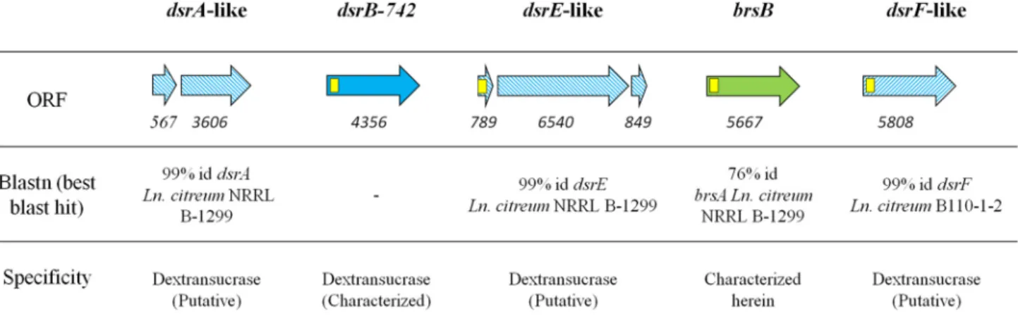
Characterization of a Novel GH70 Branching Sucrase from L. citreum NRRL B-742—The analysis of L. citreum NRRL B-742 genome (27) revealed the presence of five putative ␣-transglucosylase encoding genes: (i) the dsrB-742 gene pre-viously isolated by Kim et al. (26), (ii) three additional genes,
dsrA-like, dsrE-like, and dsrF-like, that share 99% identity with the characterized dextransucrase genes from L. citreum NRRL B-1299 and B110-1-2 (33–35), and (iii) one gene of 5667 bp, named brsB, putatively encoding a 1888-amino acid protein showing 76% of amino acid identity and 83% of similarity with BRS-A from L. citreum NRRL B-1299 (21) (GenBankTM acces-sion number WP_040177263.1, best Blast hit against the non-redundant (nr) protein database) (Fig. 1).
For BRS-B, a signal peptide with a cleavage site between posi-tions 39 and 40 was predicted using SignalP 4.1 server. Based on sequence alignment, the protein was predicted to belong to the GH70 enzyme family and to adopt the same structure as those displayed by other GH70 enzymes (12–14), which consist of five structural domains A, B, C, IV, and V, of which the limits were assigned by comparison with those of GTF180-⌬N and ⌬N123GBD-CD2 (12, 15) (Fig. 2). The three catalytically impor-tant amino acids involved in the formation of the-D
-glucosyl-enzyme intermediate are conserved in the BRS-B sequence and are located in catalytic domain A. By analogy with other GH70 ␣-transglucosylases, Asp-671, Glu-709, and Asp-788 residues are predicted to play the role of the nucleophile, the acid/base catalyst, and the transition state stabilizer, respectively. Do-main V is formed by two fragments, one at the N terminus and FIGURE 1. GH70 encoding genes in L. citreum NRRL B-742 genome. Yellow squares represent putative signal peptide encoding sequences. Hatched arrows indicate sequences showing⬎99% identity with previously characterized glucansucrases. The solid blue arrow indicates the gene previously characterized dsrB-742 (26). The green arrow stands for the brsB gene characterized in this study. Numbers below the arrows correspond to the size of the ORF in bp. The enzyme specificity was predicted according to sequence homology with other characterized GH70 enzymes.
FIGURE 2. Schematic representation of the structural organization of BRS-B. The five different structural domains were assigned by comparison with those of GTF180-⌬N and ⌬N123-GBD-CD2: (i) domain V in red), (ii) domain IV in yellow, (iii) domain B in green, (iv) domain A in blue, and (v) domain C in purple. The
catalytic residues (DED) are indicated in bold and white, YG repeats (36) with checkered red motifs and APY repeats (39) with white triangles. Signal peptide is represented by a blue circle. The red hatched regions correspond to zones for which no structural information is available.
the other at the C-terminal extremity of the protein, and is predicted to constitute both extremities of the U-shape fold (15). Like most of the domains V of GH70 enzymes, this domain is rich in YG repeats that were previously suggested to play a key role in interactions between glucansucrases and their polymers (36). Eight and two YG repeats that could be involved in ␣-glu-can binding were identified at the N- and C-terminal extremi-ties, respectively (Fig. 2). Additionally, at the C terminus, 6 APY repeats were identified and shared 99% of identity with those found at the C-terminal ends of the alternansucrase (ASR) from
L. citreum NRRL B-1355 and the branching sucrase BRS-A from L. citreum NRRL B-1299 (513 out of 518 amino acids) (37, 38). The APY repeats of alternansucrase (ASR) were previously deleted without causing any alteration of enzyme activity or specificity (39).
The brsB gene was cloned into pBAD-DEST49 vectors and expressed in E. coli BL21-AI host strain to characterize the cor-responding protein. The level of production reached 700 units䡠liter⫺1of culture. Western blot analyses confirmed that the full-length BRS-B was the most abundant protein, even if slightly degraded forms could also be observed (data not shown).
As the native E. coli BL21-AI does not produce any sucrose-active enzymes, the sonication extract was used to characterize the catalytic activities of BRS-B. The optimal temperature and pH were estimated at 30 –33 °C and pH 5.5– 6 (data not shown). All subsequent assays were thus performed at 30 °C and at pH 5.75. Enzymatic reactions were first carried out in the presence of 292 mM sucrose, the natural substrate for most of GH70
enzymes. The main reaction products formed were glucose and leucrose (O-␣-D-glucopyranosyl-(135)-D-fructopyranose). A
total of 51% of the glucosyl units from sucrose were incorpo-rated into leucrose, and 33% were transferred onto water (hy-drolysis reaction). The remaining part, representing 16% of the transferable glucosyl units, was incorporated into various small size glucooligosaccharides, which were not isolated and char-acterized due to their very low level of production ( supplemen-tal Fig. S1). No trace of high molar mass␣-glucans was detected using HPSEC-RI, indicating that BRS-B is not an efficient polymerase.
Acceptor reactions were carried out using 1500 g䡠mol⫺1 dex-tran as the acceptor (66.6 mM) and sucrose as the donor (292
mM). As seen on HPAEC-PAD chromatograms, the product
profile rapidly changed during the course of the reaction (Fig. 3a). The intensity of the peaks corresponding to linear iso-maltooligosaccharides decreased, whereas new signals were clearly visible. The products synthesized after total sucrose depletion were analyzed by NMR spectroscopy. The1H NMR spectrum showed two peaks assigned to anomeric resonances at 4.99 ppm and 5.33 ppm, which correspond to the anomeric proton of a glucosyl unit engaged in␣-(136) or ␣-(133) link-ages, respectively. Integration of the anomeric signals showed that the amount of␣-(133) linkages in dextran reached 39% that of the total osidic linkages (Fig. 3b). These results demon-strate that BRS-B catalyzes the transglucosylation reaction from sucrose onto dextran acceptors through the formation of ␣-(133) linkages.
Several dextrans of higher molar mass between 39,100 g䡠mol⫺1and 2⫻ 106g䡠mol⫺1were also tested as acceptors. All of them were efficiently branched by BRS-B, as evidenced by the percentage of␣-(133) linkages reaching a maximum of 50%, indicating that each glucosyl moiety of the␣-(136) backbone chain was substituted by an ␣-(133)-linked glucosyl residue (Fig. 4a).
The J-Mod spectrum of the dextran shows two anomeric13C chemical shifts at 98.63 ppm and 100.30 ppm corresponding to the1H anomeric signals at 4.98 ppm and 5.33 ppm of the ␣-(136) and ␣-(133)-linked glucosyl units, respectively. The presence of␣-(133)-linked branching was further attested by the typical signal at 81.37 ppm, which was assigned to C-3 of the 3,6-O-disubstituted glucopyranosyl units (9, 40). Only one type of resonance is observed at 100.30 ppm, indicating that the ␣-(133) linkages of the dextran are solely branching linkages. This is confirmed by the presence of only one chemical shift at 61.20 ppm attributed to free C6. At 66.33 ppm, the resonances are attributed to bound C6 of the 3,disubstituted and 6-O-disubstituted glucosyl units of the main chain (Fig. 4b).
To date, BRS-B is the first reported ␣-(133) branching sucrase. Its ability to branch high molar mass dextran up to 50% suggests that BRS-B is likely involved in the synthesis of L.
cit-reumNRRL B-742 comb-like dextran. Based on sequence align-ment, the other putative GH70 enzymes encoded by L. citreum NRRL B-742 are predicted to be dextransucrases synthesizing linear dextrans. Dextransucrase activities were previously iso-lated in the fermentation broth of L. citreum NRRL B-742 (5, 22, 26). It can thus be assumed that the synthesis of the hyper-branched dextran is due to the synergistic action of at least one dextransucrase and the branching enzyme, BRS-B. The dextransucrase would synthesize the␣-(136) backbone, and the ␣-(133) branching sucrase would be involved in branching via␣-(133) osidic bond formation. There are sev-eral questions that remain, including understanding which dex-transucrases of L. citreum NRRL B-742 work in tandem with BSR-B. Additionally, it is not known whether or not elongation and branching are concomitant. These pending questions require additional experiments, such as RT-quantitative PCR studies, secretome analysis at regular intervals of the culture, and knock-out of either polymerases or branching sucrase encoding genes combined with structural analyses of the pro-duced dextran.
Construction of BRS-B-⌬1, a Truncated Form of BRS-B—
Despite several attempts, the purification of BRS-B did not give satisfying results due to very weak protein binding onto nickel and cobalt affinity resins. This was attributed to an inadequate exposure of the tags, which could prevent correct interaction with the resin. To facilitate the purification process, a shorter form deleted of the signal peptide and all APY motifs at the C-terminal end, was constructed (Fig. 2). Notably, 8 of the 10 identified YG repeats were conserved. The position of the trun-cations at the C- and N terminus ends were determined from sequence alignment with the␣-(132) branching sucrase ⌬N123 GBD-CD2. The production of the truncated protein BRS-B-⌬1 was optimized to a level of 1200 units䡠liter⫺1using pET-55-DEST and BL21 Star DE3 E. coli strain. Western blot analysis showed that the protein was not degraded at the N-terminal
extremity. The C-terminal His6tag was then used to purify the enzyme onto Ni-NTA affinity resin. The purification yield reached 85%, with a corresponding purification factor of 17. BRS-B-⌬1-specific activity on sucrose (292 mM) was estimated at 24 units䡠mg⫺1of protein. The optimal temperature and pH were determined to be between 33 and 40 °C and 5–5.75, respectively (Fig. 5, a and b) and are in the same range as those observed for the non-purified enzyme BRS-B. A slight improve-ment of transferase activity (near 6%) was observed with the addition of 1 mMCa2⫹(Fig. 5c), as previously described for the
branching sucrase⌬N123-GBD-CD2 (increase of 13% (12)). In
contrast, Zn2⫹ ions had the most deleterious effect on BRS-B-⌬1 activity, which decreased by 23% in the presence of Zn2⫹ ions (Fig. 5c). From a sucrose substrate, BRS-B-⌬1 showed a slightly higher hydrolytic activity than BRS-B and produced 42% glucose, 48% leucrose, and 10% of small oligosaccharides. The profiles of products obtained with dextran acceptors were similar to those obtained with the full-length protein (data not shown). Of note, the deletion of the APY repeats at the C ter-minus extremity of the protein did not affect the enzyme spec-ificity and efficiency, as previously observed with the alternan-sucrase and inuloalternan-sucrase (39, 41).
FIGURE 3. Analysis of the products synthesized with BRS-B enzyme from sucrose (292 mM) and dextran 1500 g䡠molⴚ1(66.6 mM). a, HPAEC-PAD
chromatograms of enzymatic reaction stopped after 1 min (1) and enzymatic reaction stopped after 8 h (2). Peaks corresponding to glucose (G), fructose (F), leucrose (L), sucrose (S), and isomaltooligosaccharides with a degree of polymerization (DP) of x. b,1H NMR spectrum (in the anomeric region) of the products
FIGURE 4.1H NMR (a) and13C NMR (b) analyses of the purified branched dextran of 2ⴛ 106g䡠molⴚ1synthesized by BRS-B.13C chemical shifts are given
The kinetic parameters of the recombinant protein BRS-B-⌬1 were determined with and without dextran acceptor. Without the acceptor, sucrose hydrolysis was inhibited by ⬎150 mMsucrose, and the kinetic data fitted a non-competitive
substrate inhibition modeled by Equation 1 with a Km, Ki, and
kcatvalues estimated at 7.0⫾ 0.7 mM, 595⫾ 88 mM, and at 81.4 s⫺1, respectively. An initial rate of transglucosylation was deter-mined at a fixed sucrose concentration (300 mM) and using
dextran acceptor (68,400 g䡠mol⫺1) concentrations varying from 31 to 617 mM(in anhydroglucosyl unit equivalents). A classical
Michaelis-Menten profile was obtained with Kmdextran and
kcat values of 0.27 ⫾ 0.08 mM and 949 s⫺1, respectively. A
12-fold increase of the catalytic constant was observed com-pared with the reaction without dextran (Table 2). The forma-tion of␣-(133) branches onto dextran molecules is very effi-cient (kcat/Kmdextran ⫽ 3514 s⫺1mM⫺1), and the kcatvalue is close to that reported for␣-(132) branching formation cata-lyzed by the␣-(132) branching sucrase GBD-CD2 (20) (Table 2). Such values range among the highest kcatvalues reported for GH70 members.
Synthesis of Dextrans with Controlled Amounts of␣-(133) Linkages—The opportunity to synthesize different ␣-glucan structures with a controlled degree of branching using BRS-B-⌬1 was investigated in more details. To this end, low and high molar mass dextrans (1500 and 2⫻ 106g䡠mol⫺1, respectively) were tested as acceptors at different sucrose:acceptor ratios. When sucrose was in large excess, the production yields of ␣-(133)-branched dextrans decreased in favor of the forma-tion of side products (mainly glucose and leucrose, producforma-tion of small oligosaccharides being negligible). In contrast, when the dextran concentration increased, glucose and leucrose pro-duction was reduced (Fig. 6a). The percentage of␣-(133)
link-ages in branched dextrans was tightly controlled and evolved from 5 to 50% by adjusting the sucrose:acceptor ratio (Fig. 6b). This parameter was also shown to be critical for controlling the percentage of ␣-(132) branching catalyzed by the ␣-(132) branching sucrase, GBD-CD2 (20). An initial sucrose/dextran ratio higher than three has to be used to obtain a highly branched dextran. By controlling the sucrose:acceptor ratio and the size of the dextran acceptor, a large array of products can be produced, opening the possibility of new applications for these kinds of molecules.
Comparison of BRS-B with Other Branching Sucrases and Glucansucrases—The four functional sequence signatures (motifs I-IV), usually well conserved in the GH70 family and also found in related the GH13 family (10), were aligned with the corresponding motifs found in the characterized GH70 enzymes from other organisms (Table 3). Interestingly, BRS-B exhibits numerous sequence similarities with GBD-CD2 and BRS-A from L. citreum NRRL B-1299, two branching sucrases (19, 21). In particular, Phe-675 of motif II and Ile-783, His-785, Lys-789, and Val-795 of motif IV are only conserved in the branching sucrases. They are not found in other GH70 con-served motifs and could thus be suggested to be important for branching activity. Other amino acids are uniquely found in BSR-B. They include residues 673-IS of motif II, 711-PKGE of motif 796-IH of motif IV, and F1184 of motif I (Table 3).
The three-dimensional model of BRS-B was constructed by homology modeling using⌬N123-GBD-CD2 branching sucrase as template (Fig. 7). The model was compared with the 3D-structures of⌬N123-GBD-CD2 (PDB: 3TTQ) and with the inactive mutant of GTF-180-⌬N in complex with sucrose sub-strate (PDB: 3HZ3). According to the model, BRS-B also adopts a U-shape fold organized in five domains. As shown in Fig. 7, FIGURE 5. Effect of temperature (a), pH (b), and the addition of divalent ions (c) on BRS-B-⌬1 transferase activity. Results are given as the means ⫾ S.D.
FIGURE 6. Synthesis of␣-(133)-branched dextrans using BRS-B-⌬1 and varying sucrose:acceptor initial ratio (mass concentrations). a, determination of the percentage of glucosyl moieties from sucrose incorporated into free glucose (black), leucrose (light gray), and␣-(133) dextran (dark gray). The produc-tion of other oligosaccharides was negligible. b, control of the amount of␣-(133) linkages in branched dextrans of 1500 (Œ) and 2 ⫻ 106g䡠mol⫺1(E) as the
function of the sucrose:acceptor initial ratio. TABLE 2
Kinetic parameters of truncated variant BRS-B-⌬1 for hydrolysis reaction (in the presence of sucrose) and for␣-(133) transglucosylation (in the presence of sucrose and dextran 68.4 kg. molⴚ1)
Comparison with the kinetic parameters of GBD-CD2 and⌬N123-GBD-CD2 is shown.
Enzyme Km sucrose Km dex Km dex Vmax kcat
mM mMof anhydroglucosyl units mM mol mg⫺1min⫺1 s⫺1
Sucrose
BRS-B⫺⌬1 7.0⫾ 0.7 34.5⫾ 1.1 81.4
⌬N123-GBD-CD2 7.5⫾ 1.0 36.3⫾ 0.6 76
GBD-CD2 10.8⫾ 0.8 34.6⫾ 0.5 109
Sucrose and dextran (68.4 kg䡠molⴚ1)
BRS-B⫺⌬1 112⫾ 33 0.27⫾ 0.08 402⫾ 36 949
⌬N123-GBD-CD2 206⫾ 34 125⫾ 21 0.3⫾ 0.05 462⫾ 45 947
GBD-CD2 42⫾ 2 75⫾ 3 0.174 303⫾ 5 970
TABLE 3
Sequence alignment of the conserved motifs (I–IV) of BRS-C and BBRS-D catalytic core with other characterized GH70 enzymes
1
Spe, Specificity. Enzymes are grouped together according to their linkage specificity: M for mutansucrase, R for reuteransucrase, A for alternansucrase, 4,6 GT for␣-4,6 glucanotransferase, D for dextransucrase, BS for branching sucrase.
2
The two catalytic residues are indicated in bold red the nucleophile (D) and the acid/base catalyst (E). The transition state stabilizer (D) is represented in bold blue. The residues in bold green and in bold orange correspond to residues of substrate binding subsites⫺1 and ⫹1, respectively, as originally identified in GTF180-⌬N: sucrose complex (PDB entry 3HZ3) (15).
3
the BRS-B model displays loops in the vicinity of the catalytic gorge, which differs in length and amino acid composition in comparison with⌬N123-GBD-CD2. Of note, the-hairpin loop partly covering the domain A of⌬N123-GBD-CD2 is absent in BRS-B. Near the subsite ⫹1, two loops that belong to the domain B are shorter than in⌬N123-GBD-CD2 (G2130-W2135 and Q2158-F2163). In addition, in the catalytic gorge, the two loops connecting strand4 to helix ␣5 and strand 7 to helix ␣8 of the (/␣)8barrel are longer in BRS-B.
Then, we compared the active site residues delineating the subsites⫺1 and ⫹1 of BRS-B with those of GTF-180-⌬N and ⌬N123-GBD-CD2. As shown in Fig. 8, the residues of subsite⫺1 are all conserved. In subsite⫹1, BRS-B and ⌬N123-GBD-CD2 only differ by one residue: P711 is replaced by G2250 in⌬N123
-GBD-CD2. In contrast, only a few residues constituting subsite ⫹1 of GTF-180-⌬N are conserved in BRS-B (i.e. Q792, D788 and E709, BRS-B numbering). In particular, N1029 and W1065 of GTF-180-⌬N are not conserved in BRS-B. N1029 is replaced by Phe-675 (Fig. 8a). Notably, one phenylalanine was also found in⌬N123-GBD-CD2, and was shown to be critical for dextran branching (12). Moreover, comparison of BRS-B and GTF-180 backbone, also revealed a rearrangement around residues A710 and P711that was observed in ⌬N123-GBD-CD2. This rear-rangement was previously suggested to be responsible for a weaker binding of the fructosyl ring in branching sucrases (12). The identification of the determinants for␣-(133) branching specificity remains difficult in the absence of three-dimensional structures of BRS-B in apo form or in complex with sucrose or FIGURE 7. Surface representation of BRS-B model and⌬N123-GBD-CD2 crystal structure (PDB code 3TTQ). Domains A, B, C, IV, and V are colored in light
blue, green, pink, yellow, and red, respectively. Variable loop regions are shown in bright colors. Sucrose is shown in the active site as yellow and red sticks for
carbon and oxygen atoms, respectively.
FIGURE 8. Comparison of the active sites of BRS-B,⌬N123-GBD-CD2 (PDB code 3TTQ) and GTF-180-⌬N glucansucrase (PDB code 3HZ3). a,
superimpo-sition of BRS-B with GTF-180-⌬N in complex with sucrose (PDB code 3HZ3). Side chains forming the subsites ⫺1 and ⫹1 are shown as sticks. Sucrose is depicted as yellow and red sticks. GTF-180-⌬N is represented in gray, whereas domains A and B of BRS-B are represented in blue and green, respectively. b, superimpo-sition of BRS-B with⌬N123-GBD-CD2 structure (PDB code 3TTQ).⌬N123-GBD-CD2 is represented in gray. Domain A and domain B of BRS-B are represented in
dextran. However, according to the structural comparison of branching enzymes and polymerases, amino acids can be pointed out that constitute interesting targets for mutation to assess their role in the␣-(133) branching specificity.
Searching for New Branching Sucrases in Public Databases—
The emergence of natural branching activities through the iso-lation and characterization of BRS-A (21) and BRS-B in L.
cit-reumraised the question of the representation of such type of activity in lactic acid bacteria clade. To shed some light on this issue, a blast search against public sequence databases were carried out to ascertain which sequences contain the residues uniquely conserved in branching enzyme catalytic cores. Two new putative branching sucrases (renamed BRS-C and BRS-D) were identified, one in Leuconostoc fallax KCTC3537 and the other in Lactobacillus kunkeei EFB6. The percentage of amino
acid identity and similarity with BRS-A and BRS-B is detailed in supporting information (supplemental Table S2), and the align-ment of the conserved motifs is presented in Table 3. Based on the sequence alignment, the two putative branching sucrases are predicted to possess the catalytic triad DED and the five structural domains, A, B, C, IV, and V, specific to the GH70 family enzymes (supplemental Fig. S2). Similar to the charac-terized BRS-A (21) and BRS-B, YG repeats (36) were mainly identified at the N-terminal extremity of the protein. All newly identified genes encoding putative branching sucrases are located on the reverse strand of their genome, as previously observed for brsA in L. citreum NRRL B-1299 genome (21) and
brsBin L. citreum NRRL B-742 genome. All of them are orga-nized in tandem with another putative glucansucrase ( supple-mental Fig. S3).
FIGURE 9. Analysis of the products synthesized using BRS-C and BRS-D enzymes from sucrose (292 mM) and dextran 1500 g䡠molⴚ1(66.6 mM). a,
comparison of BRS-B and BRS-C HPAEC-PAD chromatograms. (1) Enzymatic reaction at the initial time (t⫽ 0 min), (2) enzymatic reaction using BRS-B stopped after 8 h, and (3) enzymatic reaction using BRS-C stopped after 8 h. nC, nanocoulombs. b, comparison of BRS-A and BRS-D HPAEC-PAD chromatograms. (1) Enzymatic reaction at the initial time (t⫽ 0 min), (2) enzymatic reaction using BRS-B stopped after 8 h, and (3) enzymatic reaction using BRS-C stopped after 8 h. G, peak corresponding to glucose fructose (F), leucrose (L), sucrose (S), and isomaltooligosaccharides with a degree of polymerization (DP) of x. c,1H NMR spectrum of the products synthesized by BRS-B (1) and BRS-C (2) at 500 MHz in D
2O leucrose (Leu),␣-D-glucose
(␣-D-Glcp),-D-glucose (-D-Glcp),␣-(136)-linked glucosyl residues (␣-(136)), and ␣-(133)-linked glucosyl residues (␣-(133)). d,1H NMR spectrum of
the products synthesized by BRS-A (1) and BRS-D (2) at 500 MHz in D2O. The
1H NMR spectra of the BRS-A modified dextran displayed all the chemical
shift characteristics of␣-(132)-branched dextran. The anomeric region of the spectrum contained three main resonances corresponding, respectively, to anomeric resonance of␣-(136)-linkedD-Glcp residues of the main linear chain with free carbon 2 (4.98 ppm, peak C), to anomeric resonance from ␣-(136)-linkedD-Glcp residues of the main linear chain, with the carbon 2 involved in an␣-(132) linkage with a branched glucosyl unit (5.18 ppm, peak
Characterization of Two New Putative Branching Sucrases and Phylogenetic Analysis—The genes encoding the putative branching sucrases were expressed in E. coli. The production levels of the two new proteins are reported in supplemental Table S3. As already observed for BRS-A, BRS-B, and GBD-CD2, the putative BRS-C and BRS-D mainly produced glucose, leucrose, and a low proportion of small oligosaccharides from sucrose substrate (for detailed production yields, see supple-mental Table S4). Acceptor reactions were then performed using sucrose and a small dextran of 1500 g䡠mol⫺1as an accep-tor. At the end of the reaction, HPAEC-PAD and H1 NMR analyses demonstrated that dextran was modified by BRS-C and BRS-D (Fig. 9). BRS-C from L. fallax KCTC 3537 catalyzed ␣-(133) transglucosylation from sucrose to dextrans, whereas BRS-D from L. kunkeei EFB6 was specific for␣-(132) transglu-cosylation. These findings reveal for the first time that the branching specificity is not restricted to L. citreum sp. but is also present in L. fallax as well as in L. kunkeei. Interestingly, the phylogenetic analyses show that the branching sucrases cluster together in a distinct branch (Fig. 10). In addition, the branching sucrase subtree is divided into two subgroups,
one corresponding to enzymes showing an␣-(132) linkage specificity and the other to enzymes with␣-(133) linkage specificity.
The discovery of the new branching sucrases expands the natural repertoire of the characterized GH70 ␣-transglucosy-lases. For years the GH70 family has been mainly known to house glucansucrases. Recently, a new subgroup was disclosed that contains 4,6-␣ glucanotransferases, a type of ␣-transgluco-sylases that uses␣-(134)-linked glucans as a donor instead of sucrose (42, 43). According to our biochemical and phyloge-netic analyses, branching sucrases could also constitute a new GH70 subgroup. Five enzymes of this group have now been characterized, including the engineered ␣-(132) branching sucrase GBD-CD2 (19, 20), BRS-A (21), BRS-B, BRS-C, and BRS-D.
The route is now opened to promising structure-function relationship studies that will aim at identifying the structural determinants responsible for polymerization or branching abil-ity and linkage specificabil-ity. Despite common structural motifs with GH70 glucansucrases, the branching sucrases also display clear distinctive features. According to sequence analyses, dif-FIGURE 10. Phylogenetic tree of GH70 enzymes. Each enzyme is labeled with its GenBankTMaccession number and its origin. The species of microorganism are
indicated with Ln. for Leuconostoc, Lb. for Lactobacillus, S. for Streptococcus, and W. for Weissella. Glucansucrase enzymes are highlighted in green,␣-4,6 glucanotrans-ferases are in pink, and branching sucrases are in bold and purple. The scale bar corresponds to a genetic distance of 0.05 substitutions per position.
ferences have been highlighted in the substrate binding subsite ⫹1, which can be used to rapidly sort out branching sucrases from genomic data. Mutational and structural studies will now be necessary to better define the signature of the branching specificity and to provide further insight into the branching mechanism. Finally, a broad variety of branched dextrans varying in terms of size, osidic linkage and branching degree can be easily synthesized using these enzymes, emphasizing the potential of GH70 enzymes to access new structures of biotechnological interest.
Author Contributions—M. V., P. M., M. C., and M. R.-S. planned the experiments. M. V., M. C., Y. B., E. S., P. B., and S. M. performed the experiments. M. V., Y. B., S. M., C. M., and M. R.-S. analyzed the data. M. V., Y. B., C. M., and M. R.-S. wrote the paper. All authors reviewed the results and approved the final version of the manuscript.
Acknowledgments—We are grateful to MetaSys, the Metabolomics and Fluxomics Center at the Laboratory for engineering of Biological Systems and Processes (Toulouse, France) for NMR experiments. We thank the ICEO facility, which is dedicated to enzyme screening and discovery and part of the Integrated Screening Platform of Toulouse (PICT, IBiSA) for providing access to HPLC equipment and protein purification systems.
References
1. Hucker, G., and Perderson, C. (1930) Studies on the coccaceae. XVI. The genus Leuconostoc. Tech. Bull. N. Y. Agric Exp. Stn. 167
2. Van Tieghem, P. (1878) On sugar-mill gum. Ann. Sci. Nat. Bot. Biol.
Ve´-ge´tales. 7,180 –203
3. De Belder, A. (2003) Dextran, AA Editions, Handbook Amersham Biosciences
4. Bounaix, M.-S., Gabriel, V., Morel, S., Robert, H., Rabier, P., Remaud-Sime´on, M., Gabriel, B., and Fontagne´-Faucher, C. (2009) Biodiversity of exopolysaccharides produced from sucrose by sourdough lactic acid bac-teria. J. Agric. Food Chem. 57, 10889 –10897
5. Coˆte´, G. L., and Robyt, J. F. (1983) The formation of␣-D-(133) branch linkages by an exocellular glucansucrase from Leuconostoc mesenteroides NRRL B-742. Carbohydr. Res. 119, 141–156
6. Jeanes, A., and Seymour, F. R. (1979) The␣-D-glucopyranosidic linkages of dextrans: comparison of percentages from structural analysis by periodate oxidation and by methylation. Carbohydr. Res. 74, 31– 40
7. Remaud, M., Paul, F., Monsan, P., Lopez-Munguia, A., and Vignon, M. (1992) Characterization of␣-(133) branched oligosaccharides synthe-sized by acceptor reaction with the extracellular glucosyltransferases from
L. mesenteroidesNRRL B742. J. Carbohydr. Chem. 11, 359 –378 8. Robyt, J. F. (1985) Encyclopedia of Polymer Science, (Kroschwitz, J. I., ed)
Wiley VCH, New York
9. Seymour, F. K., Knapp, R. D., Chen, E. C. M., Bishop, S. H., and Jeanes, A. (1979) Structural analysis of Leuconostoc dextrans containing 3-O-␣-D -glucosylated␣-D-glucosyl residues in both linear-chain and branch-point positions, or only in branch-point positions, by methylation and by13C
NMR spectroscopy. Carbohydr. Res. 74, 41– 62
10. Leemhuis, H., Pijning, T., Dobruchowska, J. M., van Leeuwen, S. S., Kralj, S., Dijkstra, B. W., and Dijkhuizen, L. (2013) Glucansucrases: three-dimensional structures, reactions, mechanism,␣-glucan analysis, and their implications in biotechnology and food applications. J. Biotechnol. 163, 250 –272
11. Lombard, V., Golaconda Ramulu, H., Drula, E., Coutinho, P. M., and Hen-rissat, B. (2014) The carbohydrate-active enzymes database (CAZy) in 2013. Nucleic Acids Res. 42, 490 – 495
12. Brison, Y., Pijning, T., Malbert, Y., Fabre, E´., Mourey, L., Morel, S., Po-tocki-Ve´rone`se, G., Monsan, P., Tranier, S., Remaud-Sime´on, M., and Dijkstra, B. W. (2012) Functional and structural characterization of a-1,2 branching sucrase derived from DSR-E glucansucrase. J. Biol. Chem. 287,
7915–7924
13. Ito, K., Ito, S., Shimamura, T., Weyand, S., Kawarasaki, Y., Misaka, T., Abe, K., Kobayashi, T., Cameron, A. D., and Iwata, S. (2011) Crystal structure of glucansucrase from the dental caries pathogen Streptococcus mutans. J.
Mol. Biol. 408,177–186
14. Pijning, T., Vujicˇic´-Zˇ agar, A., Kralj, S., Dijkhuizen, L., and Dijkstra, B. W. (2012) Structure of the␣-1,6/␣-1,4-specific glucansucrase GTFA from
Lactobacillus reuteri121. Acta Crystallogr. Sect. F Struct. Biol. Cryst.
Com-mun. 68,1448 –1454
15. Vujicic-Zagar, A., Pijning, T., Kralj, S., Lo´pez, C. A., Eeuwema, W., Di-jkhuizen, L., and Dijkstra, B. W. (2010) Crystal structure of a 117-kDa glucansucrase fragment provides insight into evolution and product spec-ificity of GH70 enzymes. Proc. Natl. Acad. Sci. 107, 21406 –21411 16. Moulis, C., Joucla, G., Harrison, D., Fabre, E., Potocki-Veronese, G.,
Mon-san, P., and Remaud-Simeon, M. (2006) Understanding the polymeriza-tion mechanism of glycoside-hydrolase family 70 Glucansucrases. J. Biol.
Chem. 281,31254 –31267
17. Mooser, G., Hefta, S. A., Paxton, R. J., Shively, J. E., and Lee, T. D. (1991) Isolation and sequence of an active-site peptide containing a catalytic aspartic acid from two Streptococcus sobrinus ␣-glucosyltransferases.
J. Biol. Chem. 266,8916 – 8922
18. MacGregor, E. A., Jespersen, H. M., and Svensson, B. (1996) A circularly permuted ␣-amylase-type ␣/-barrel structure in glucan-synthesizing glucosyltransferases. FEBS Lett. 378, 263–266
19. Fabre, E., Bozonnet, S., Arcache, A., Willemot, R.-M., Vignon, M., Mon-san, P., and Remaud-Simeon, M. (2005) Role of the two catalytic domains of DSR-E dextransucrase and their involvement in the formation of highly ␣-1,2 branched dextran. J. Bacteriol. 187, 296–303
20. Brison, Y., Fabre, E., Moulis, C., Portais, J.-C., Monsan, P., and Remaud-Si-me´on, M. (2010) Synthesis of dextrans with controlled amounts of␣-1,2 link-ages using the transglucosidase GBD-CD2. Appl. Microbiol. Biotechnol. 86, 545–554
21. Passerini, D., Vuillemin, M., Ufarte´, L., Morel, S., Loux, V., Fontagne´-Faucher, C., Monsan, P., Remaud-Sime´on, M., and Moulis, C. (2015) Inventory of the GH70 enzymes encoded by Leuconostoc citreum NRRL B-1299: identification of three novel␣-transglucosylases. FEBS J. 282, 2115–2130
22. Jeanes, A., Haynes, W. C., Wilham, C. A., Rankin, J. C., Melvin, E. H., Austin, M. J., Cluskey, J. E., Fisher, B. E., Tsuchiya, H. M., and Rist, C. E. (1954) Characterization and classification of dextrans from ninety-six strains of bacteria1b. J. Am. Chem. Soc. 76, 5041–5052
23. Chung, C. H., and Day, D. F. (2004) Efficacy of Leuconostoc mesenteroides (ATCC 13146) isomaltooligosaccharides as a poultry prebiotic. Poult. Sci.
83,1302–1306
24. Chung, C.-H., and Day, D. F. (2002) Glucooligosaccharides from
Leucono-stoc mesenteroidesB-742 (ATCC 13146): a potential prebiotic. J. Ind.
Mi-crobiol. Biotechnol. 29,196 –199
25. Kim, D., and Robyt, J. F. (1995) Production, selection, and characteristics of mutants of Leuconostoc mesenteroides B-742 constitutive for dextran-sucrases. Enzyme Microb. Technol. 17, 689 – 695
26. Kim, H., Kim, D., Ryu, H. J., and Robyt, J. F. (2000) Cloning and sequencing of the ␣-1-⬎ 6 dextransucrase gene from Leuconostoc mesenteroides B-742CB. J. Microbiol. Biotechnol. 10, 559 –563
27. Passerini, D., Vuillemin, M., Laguerre, S., Amari, M., Loux, V., Gabriel, V., Robert, H., Morel, S., Monsan, P., Gabriel, B., Fontagne´-Faucher, C., Re-maud-Simeon, M., and Moulis, C. (2014) Complete genome sequence of
Leuconostoc citreumstrain NRRL B- 742. Genome Announc. 2, e01179 –14 28. Studier, F. W. (2005) Protein production by auto-induction in
high-den-sity shaking cultures. Protein Expr. Purif. 41, 207–234
29. Miller, G. L. (1959) Use of dinitrosalicylic acid reagent for determination of reducing sugar. Anal. Chem. 31, 426 – 428
30. Irague, R., Massou, S., Moulis, C., Saurel, O., Milon, A., Monsan, P., Re-maud-Sime´on, M., Portais, J.-C., and Potocki-Ve´rone`se, G. (2011) NMR-based structural glycomics for high-throughput screening of carbohy-drate-active enzyme specificity. Anal. Chem. 83, 1202–1206
31. Edgar, R. C. (2004) MUSCLE: multiple sequence alignment with high accuracy and high throughput. Nucleic Acids Res. 32, 1792–1797 32. Biasini, M., Bienert, S., Waterhouse, A., Arnold, K., Studer, G., Schmidt,
(2014) SWISS-MODEL: modelling protein tertiary and quaternary struc-ture using evolutionary information. Nucleic Acids Res. 42, W252–W258 33. Fraga Vidal, R., Moulis, C., Escalier, P., Remaud-Sime´on, M., and Monsan, P. (2011) Isolation of a gene from Leuconostoc citreum B/110 –1-2 encod-ing a novel dextransucrase Enzyme. Curr. Microbiol. 62, 1260 –1266 34. Monchois, V., Willemot, R.-M., Remaud-Simeon, M., Croux, C., and
Monsan, P. (1996) Cloning and sequencing of a gene coding for a novel dextransucrase from Leuconostoc mesenteroides NRRL B-1299 synthe-sizing only␣(1–6) and ␣(1–3) linkages. Gene 182, 23–32
35. Bozonnet, S., Dols-Laffargue, M., Fabre, E., Pizzut, S., Remaud-Simeon, M., Monsan, P., and Willemot, R.-M. (2002) Molecular characterization of DSR-E, an␣-1,2 linkage-synthesizing dextransucrase with two catalytic domains. J. Bacteriol. 184, 5753–5761
36. Giffard, P. M., and Jacques, N. A. (1994) Definition of a fundamental repeating unit in streptococcal glucosyltransferase glucan-binding regions and related sequences. J. Dent. Res. 73, 1133–1141
37. Argu¨ello-Morales, M. A., Remaud-Simeon, M., Pizzut, S., Sarc¸abal, P., Willemot, R., and Monsan, P. (2000) Sequence analysis of the gene encod-ing alternansucrase, a sucrose glucosyltransferase from Leuconostoc
mes-enteroidesNRRL B-1355. FEMS Microbiol. Lett. 182, 81– 85
38. Janecek, S., Svensson, B., and Russell, R. R. (2000) Location of repeat
ele-ments in glucansucrases of Leuconostoc and Streptococcus species. FEMS
Microbiol. Lett. 192,53–57
39. Joucla, G., Pizzut, S., Monsan, P., and Remaud-Simeon, M. (2006) Con-struction of a fully active truncated alternansucrase partially deleted of its carboxyl-terminal domain. FEBS Lett. 580, 763–768
40. Maina, N. H., Tenkanen, M., Maaheimo, H., Juvonen, R., and Virkki, L. (2008) NMR spectroscopic analysis of exopolysaccharides produced by Leuconostoc
citreumand Weissella confusa. Carbohydr. Res. 343, 1446–1455
41. Olivares-Illana, V., Lo´pez-Munguía, A., and Olvera, C. (2003) Molecular characterization of inulosucrase from Leuconostoc citreum: a fructosyl-transferase within a glucosylfructosyl-transferase. J. Bacteriol. 185, 3606 –3612 42. Kralj, S., Grijpstra, P., van Leeuwen, S. S., Leemhuis, H., Dobruchowska, J. M.,
van der Kaaij, R. M., Malik, A., Oetari, A., Kamerling, J. P., and Dijkhuizen, L. (2011) 4,6-␣-Glucanotransferase, a novel enzyme that structurally and func-tionally provides an evolutionary link between glycoside hydrolase enzyme families 13 and 70. Appl. Environ. Microbiol. 77, 8154–8163
43. Leemhuis, H., Dijkman, W. P., Dobruchowska, J. M., Pijning, T., Grijpstra, P., Kralj, S., Kamerling, J. P., and Dijkhuizen, L. (2013) 4,6- ␣-Glucano-transferase activity occurs more widespread in Lactobacillus strains and constitutes a separate GH70 subfamily. Appl. Microbiol. Biotechnol. 97, 181–193