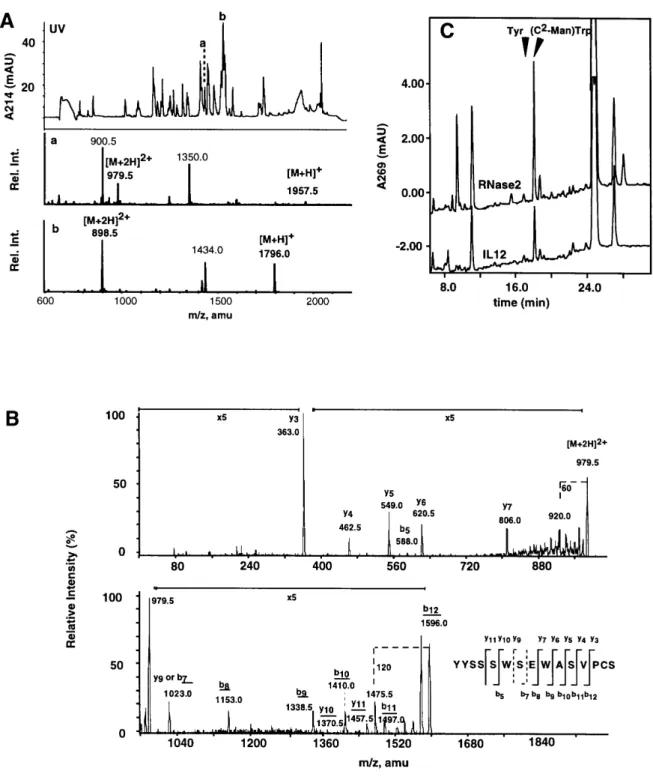
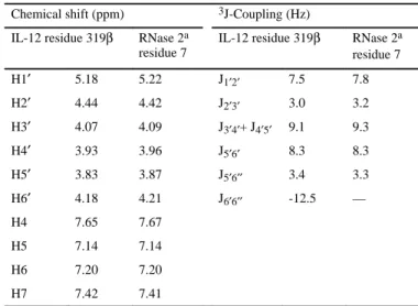
Recombinant human interleukin-12 is the second example of a C-mannosylated protein
7
0
0
Texte intégral
Figure
![Fig. 1. Structure of C 2 -α-mannosyltryptophan [(C 2 -Man-)Trp].](https://thumb-eu.123doks.com/thumbv2/123doknet/14884857.646233/1.892.475.837.255.566/fig-structure-c-α-mannosyltryptophan-c-man-trp.webp)
+2
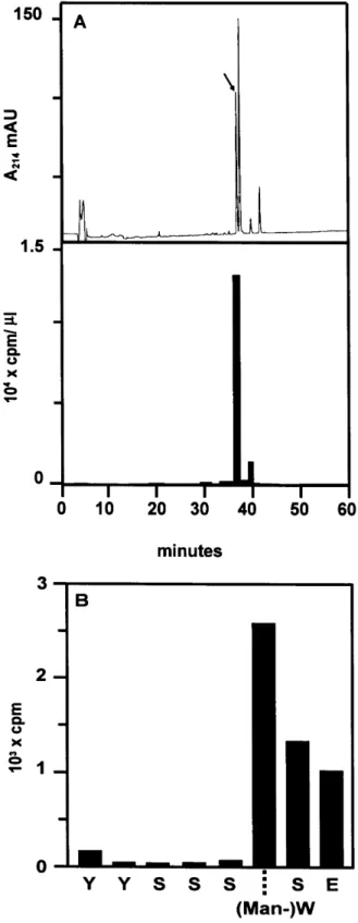
![Fig. 5. C-Mannosyltransferase activity in IL-12 producing cells. (A) The presence of C-mannosyltransferase activity in microsomes from NC-37 cells was determined by incubation of acceptor peptide (0.9 mM) with 0.9 µM Dol-P-[2– 3 H]Man (5.61 Ci/mmol) and NC](https://thumb-eu.123doks.com/thumbv2/123doknet/14884857.646233/5.892.124.390.63.790/mannosyltransferase-activity-producing-presence-mannosyltransferase-microsomes-determined-incubation.webp)
Documents relatifs