Myocardial infarction does not affect circulating
haematopoietic stem and progenitor cell self-renewal
ability in a rat model
J. M. Kr¨opfl
1, C. M. Spengler
1,2, A. Frobert
3, G. Ajalbert
3and M. N. Giraud
31Exercise Physiology Lab, Institute of Human Movement Sciences and Sport, ETH Zurich, Zurich, Switzerland 2Zurich Center for Integrative Human Physiology (ZIHP), University of Zurich, Zurich, Switzerland 3Cardiology, University of Fribourg, Fribourg, Switzerland
Edited by: Kenneth MacLeod
New Findings
r What is the central question of this study?
Although peripheral blood haematopoietic stem and progenitor cells are potentially important in regeneration after acute myocardial infarction, their self-renewal ability in the post-acute phase has not yet been addressed.
r What is the main finding and its importance?
In rat peripheral blood, we show that myocardial infarction does not negatively affect circulating haematopoietic stem and progenitor cell self-renewal ability 2 weeks after acute infarction, which suggests a constant regenerative potential in the myocardial infarction post-acute phase.
Given the importance of peripheral blood haematopoietic stem and progenitor cells (HPCs) in post-acute regeneration after acute myocardial infarction (MI), the aim of the present study was to investigate the number and secondary replating capacity/self-renewal ability of HPCs in peripheral blood before and 2 weeks after MI. In female Lewis inbred rats (n= 9), MI was induced by ligation of the left coronary artery, and another nine underwent sham surgery, without ligation, for control purposes. Myocardial infarction was confirmed by troponin I concentrations 24 h after surgery. Peripheral blood was withdrawn and fractional shortening and ejection fraction of the left ventricle were assessed before (day 0) and 14 days after MI or sham surgery (day 14). After mononuclear cell isolation, primary and secondary functional colony-forming unit granulocyte–macrophage (CFU-GM) assays were performed in order to detect the kinetics of functional HPC colony counts and cell self-renewal ability in vitro. The CFU-GM counts and cell self-renewal ability remained unchanged (P> 0.05) in both groups at day 14, without interaction between groups. In the intervention group, higher day 0 CFU-GM counts showed a relationship to lower fractional shortening on day 14 (ρ = −0.82; P < 0.01). Myocardial infarction did not negatively affect circulating HPC self-renewal ability, which suggests a constant regenerative potential in the post-acute phase. A relationship of cardiac contractile function 14 days after MI with circulating CFU-GM counts on day 0 might imply functional colony count as a predictive factor for outcome after infarction.
Corresponding author C. M. Spengler: Exercise Physiology Laboratory, Institute of Human Movement Sciences and Sport, ETH Zurich, Winterthurerstrasse 190, 8057 Zurich, Switzerland. Email: [email protected]
http://doc.rero.ch
Published in "Experimental Physiology 103(1): 1–8, 2018"
which should be cited to refer to this work.
Introduction
Acute myocardial infarction (MI) is characterized by a generalized inflammatory reaction that triggers the rapid mobilization of bone marrow stem and progenitor cells, which are important for acute and post-acute regeneration processes. The contribution of circulating cells to myocardial repair, however, is not yet fully understood, although experimental studies have seen the potential in bone-marrow-derived precursor cells for cardiac regeneration after MI (Wojakowski et al. 2012). The possible therapeutic effect of circulating haematopoietic stem and progenitor cells (HPCs) on damaged heart tissue is currently the subject of intense debate (Anversa
et al. 2013). The current predominant view is that
these cells secrete a variety of cytokines that activate endogenous progenitors in the heart muscle, which are responsible for the repair process and the improvement in, for example, ventricular function (Anversa et al. 2013). Therefore, the body’s own regenerative ability [to counteract pathological heart remodelling and improve ejection fraction (EF)/systolic function] depends, among other factors, on the circulating number and secondary replating capacity/self-renewal ability of these cells after the infarction. Although some human and animal studies have documented the kinetics of HPCs in the first week after infarction (Paczkowska et al. 2005; Assmus et al. 2012; Wojakowski et al. 2012), the post-acute phase (e.g. 14 days after MI) has had little attention so far, whereas long-term studies mainly assessed endothelial progenitor cell kinetics (Regueiro et al. 2015). Acute MI was shown to increase the number of immune cells (Tsujioka et al. 2009; Gentek & Hoeffel, 2017), bone marrow (Assmus et al. 2012) and peripheral blood (Leone
et al. 2005; Assmus et al. 2012) HPCs 1 week after MI,
with bone marrow functional HPC colony numbers being elevated after MI, possibly as a result of an increased bone marrow activity. Peripheral blood HPCs remained elevated 2 weeks after MI in a rat model (Abdelmonem
et al. 2015), but the self-renewal ability of peripheral
blood HPCs was not assessed, although this would provide important information regarding self-regeneration during this post-acute phase. In addition, controversy exists regarding whether the initial increase in HPC numbers would have declined towards baseline by day 14 as extrapolated from literature results (Shintani et al. 2001). The aim of the present study, therefore, was to investigate the following: (i) the number of functional colonies and self-renewal ability of circulating HPCs in the peripheral blood and their association with heart function; and (ii) the MI-induced change of risk-associated blood cell counts, e.g. neutrophil-to-lymphocyte ratio, in a rat model.
Methods
Ethical approval. All animals received care in compliance with the European Convention on Animal Care. The surgical procedures were performed in accordance with the Swiss Animal Protection Law after obtaining permission from the State Veterinary Office, Fribourg, and approval from the Swiss Federal Veterinary Office, Switzerland (ethics approval no. 2013 09E FR). All procedures used conform to the principles and regulations as described by given guidelines (Grundy, 2015).
Animals, invasive procedures and postoperative care.
Eighteen female Lewis inbred rats (weight 215.2± 11.3 g;
from Janvier, France) were divided into an intervention
group (IG; n = 9) and a control group (CG; n = 9).
Food (normal diet/pellets) and fresh water were provided
ad libitum. One rat of the CG unexpectedly died before
day 14.
As previously described (Frobert et al. 2014), animals were placed in an induction chamber with 5% isoflurane in oxygen for 5–7 min. A toe and tail pinch confirmed sedation. Animals were then placed on a warming pad at 37°C and intubated. Anaesthesia was sustained with 2.5%
isoflurane in oxygen at a minute ventilation of 2.5 l min−1.
The depth of anaesthesia was continually monitored by assessment of the tail-pinch reflex and respiratory rhythm. Following a left thoracotomy between the fourth and fifth interstitial space, the pericardium was opened to access the heart, and the left anterior descending coronary artery was ligated in animals of the IG, whereas animals of the CG were closed without ligation. The investigators took all possible steps to minimize animals’ pain and suffering.
One-third of the dose of buprenorphine (0.1 mg kg−1)
was injectedS.C. 20–30 min before the beginning of the
anaesthesia and two-thirds immediately before the end of the surgery as postoperative analgesia. A second dose was administered 6 h after surgery, and buprenorphine was added to the water during the night. Further doses were administered after 24 and 48 h by supplementation of the water with buprenorphine during the night. Between 24 and 48 h, further doses of analgesics were administered depending on the score during postoperative pain evaluation. Welfare monitoring of animals via behavioural observation was performed every day. After surgery, the animals were kept separately in a cage warmed with a heat lamp until they had fully recovered from anaesthesia. They were then put back together, five rats per
cage (1800 cm2), according to the Eurostandard type III.
Food (pellets) and fresh water were provided ad libitum. A 12 h–12 h light–dark cycle and a constant temperature of
24°C were maintained during the entire experimentation
time. Blood sampling was performed under general
anaesthesia maintained with 2.5% isoflurane in 2.5 l min−1 oxygen with a face mask. At the end of the study, animals were humanely killed. Exsanguination was performed
under general anaesthesia (isoflurane 2.5% in 2.5 l min−1
oxygen), the thorax was opened, blood withdrawn from the vena cava and the heart harvested.
To verify infarct development (IG only), a blood sample was collected from the caudal tail artery at 24 h after left anterior descending coronary artery
ligation. Plasma was stored at −80°C. Troponin I
quantification was performed as previously described (Frobert et al. 2015) using the AccuTNI3+ immunoassay (Beckman Coulter, Nyon, Switzerland). Heart function was assessed under light anaesthesia (2% isoflurane) using a Vevo3100 (FUJIFILM VisualSonics, Toronto, Canada) high-resolution ultrasound imaging system. Pre-MI (day 0), 24 h (day 1), 1 week (day 7) and 2 weeks (day 14) post-MI, the EF was determined in B-mode and the fractional shortening (FS) in M-mode on a parasternal long-axis view.
Mononuclear cell isolation. At baseline and 14 days after MI induction or sham surgery, peripheral blood
was withdrawn from the caudal vein (700–1000 μl)
into lithium-heparinized tubes. 100 μl of whole blood
was kept for haematological analysis and the remaining volume was subjected to a standard Ficoll gradient
centrifugation (Histopaque; Sigma-Aldrich, Buchs,
Switzerland) according to the manufacturer’s instructions in order to isolate peripheral blood mononuclear cells for haematopoietic stem and progenitor cell functionality tests, such as the number of functional colonies and secondary replating capacity/self-renewal ability (primary and secondary colony-forming unit assays, respectively).
Analysis of blood cell counts. Blood cell counts were analysed at baseline (day 0) and 14 days after MI or control
surgery (day 14) using 100μl of whole blood. Analysis was
performed with a general rat program of a haematology analyser (dilution 1:3, 1:6 or 1:12; ADVIA 2120i; Siemens, Zurich, Switzerland). Lithium-heparin anti-coagulated blood has some limitations for blood cell analysis (e.g. platelet clumping; Guzman et al. 2008), which was taken into account for the presentation of final results.
Primary and secondary colony-forming unit assays.
Primary and secondary haematopoietic colony-forming unit granulocyte–macrophage (CFU-GM) assays were performed as published (Stelzer et al. 2010), with slight modification of the procedure for peripheral blood. Peripheral blood mononuclear cells were plated at a
concentration of 200,000 cells ml−1 in methylcellulose
culture medium for rats (Methocult GF R3774; StemCell Technologies, Vancouver, BC, Canada) in 12-well
flat-bottomed plates and incubated at 37°C (5% CO2,
>95% humidity). On day 6 of incubation, colonies
consisting of ࣙ50 cells were scored, providing the
number of functional haematopoietic progenitor colonies (primary CFU-GMs) of each animal. As a next step, ࣘ60 primary CFU-GM colonies were individually plucked from the methylcellulose culture medium and transferred to separate wells of a 48-well flat-bottomed plate and thoroughly mixed with methylcellulose culture medium to produce a single-cell suspension. After another 8 days of incubation, each well was scored for
the number of CFU-GM colonies consisting of ࣙ50
cells (secondary CFU-GMs). The secondary replating capacity/self-renewal ability is known to be associated with the proliferative capacity of myeloid progenitor cells (Gordon et al. 1998; Withey et al. 2005). The original protocol for human cells was modified for rat peripheral blood by transferring fewer colonies (ࣘ60 instead of 90), because rat cells have a higher CFU-GM replating efficiency than human cells with respect to the number of clonogenic primary colonies and the produced number of secondary colonies per primary replated CFU-GM (Alenzi et al. 2002).
Analysis of secondary CFU-GM assays. For analysis of secondary CFU-GM assays, the number of secondary CFU-GMs produced by each primary CFU-GM was used
as raw data. Counts>100 were truncated. The secondary
replating capacity of an individual was defined to be the mean of the log 2 of the number of colonies plus one for the following reasons. The log 2 scale is natural, as the distribution of the number of secondary colony-forming cells is skewed to the right. One was added because the log 2 of zero cannot be calculated. In addition, it is a continuous measure of the number of duplications of a primary CFU-GM. This measure has properties similar to the measure used by Gordon et al. (1998), because counts of zero are adequately taken into account and the log scale reduces skewness.
Statistics. Data are represented as means ± SD unless otherwise stated. A priori power analysis (Student’s paired
t test) was used to calculate the necessary sample size. Based
on published differences (Assmus et al. 2012) in CFU-GM
bone-marrow colony numbers before (45± 15) and 7 days
after MI (69± 18) in mice, an α error of 0.05, a 1 − β error
of 0.8 and an effect size of 1.437, a sample size of n= 6
was determined. To consider possible drop-outs, a total
sample size of n= 9 was chosen. A two-way ANOVA was
carried out in order to compare the time effects between day 0 and day 14 and possible interactions among groups.
Post hoc tests were performed when appropriate. Pearson’s
correlation analysis was used to determine the relationship between variables.
Results
Confirmation of infarction. Cardiac troponin I was
significantly increased 24 h after MI (26.01± 6.08 ng ml−1)
in the IG. Fractional shortening and EF were significantly reduced already after 24 h and stayed low until 2 weeks post-MI (Fig. 1A and B).
Numbers of progenitor cells and secondary replating capacity/ability for self-renewal. At baseline, groups
were comparable (P > 0.05) regarding both CFU-GM
numbers and cell self-renewal ability. Two weeks after MI, on day 14, both CFU-GM numbers and the self-renewal ability of HPCs did not differ from respective
base-line values (P > 0.05; Fig. 2A and B). There was no
statistical interaction between groups (P > 0.05). Also,
individual changes in CFU-GM numbers were not related to respective changes in self-renewal ability. In the IG, CFU-GM numbers on day 0 and FS on day 14 were
significantly associated (ρ = −0.82; P < 0.01; Fig. 3).
Cell self-renewal ability on day 0 was not associated with
cardiac function on day 14 (P> 0.05).
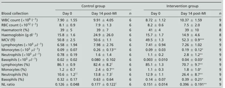
Blood cell counts. Time effects for blood cell counts (Table 1) of the IG showed significant increases of
mean corpuscular volume (P< 0.001), monocytes (P <
0.05), neutrophils (P < 0.01), basophils (P < 0.05),
the percentage of lymphocytes (P < 0.01), percentage
of monocytes (P < 0.05), percentage of neutrophils
(P < 0.01), percentage of basophils (P < 0.05) and
neutrophil/lymphocyte ratio (P < 0.01) 2 weeks after
MI. In the CG, only monocytes and the percentage of
monocytes (both P< 0.01) were significantly elevated at
day 14 after surgery. Only the percentage of lymphocytes,
percentage of neutrophils and neutrophil/lymphocyte ratio showed a significant interaction between groups (P< 0.05).
Post hoc tests between groups revealed that the CG and
the IG were comparable at baseline for all variables, except for the IG having a higher percentage of neutrophils (both
P< 0.01). At day 14 the groups differed in the percentage
of neutrophils, percentage of lymphocytes (both P< 0.05)
and the neutrophil/lymphocyte ratio (P < 0.05), with
the percentage of neutrophils and neutrophil/lymphocyte ratio being higher and the percentage of lymphocytes lower in the IG.
Day 0 FS was significantly correlated with day 0 mean
corpuscular volume (r = −0.709, P < 0.05), whereas
day 14 FS was significantly correlated with the percentage
of monocytes (r= 0.738, P < 0.05).
Discussion. In this study, we investigated the regenerative potential of circulating HPCs in the post-acute phase after MI in a rat model. Interestingly, no significant change in the mean number of functional progenitor cells (CFU-GM count) and cells’ self-renewal ability was present in the IG or the CG 14 days after induction of MI or control surgery, although the EF and FS were significantly reduced (Figs 1 and 2) in the IG.
Our results extend the current knowledge, because we assessed the functional count of HPCs as CFU-GMs
in cell culture rather than the number of CD34+ cells.
Wojakowski et al. (2004), for example, reported circulating
numbers of CD34+cells being elevated 1 week after MI
in comparison to healthy control subjects (Wojakowski
et al. 2004). A similar finding was reported in a rat model
with induced MI, where numbers of CD34+cells 1 week
100 80 60 40 20 0
Day-0 Day-1 Day-7 Day-14 post MI Day-0 Day-1 Day-7 Day-14 post MI
time points time points *** *** *** *** A B *** *** FS (%) EF (%) 100 80 60 40 20 0
Figure 1. Fractional shortening (FS; A) and ejection fraction (EF; B) at baseline (day 0), 24 h (day 1), 1 week (day 7) and 2 weeks (day 14) after induction of acute myocardial infarction (MI) in rats
Values are means± SD, n = 9. It is clearly apparent that both parameters show a highly significant decrease after 1 day, indicating a huge impairment of systolic heart function attributable to MI.
after MI were compared with baseline before MI (Lehrke
et al. 2006). It was also shown that the CD34+cell count was highest 7 days after MI in humans and was lower on day 14, reaching values comparable to the control group (Shintani et al. 2001). This is consistent with our results in the rat model. In addition, the literature states that circulating progenitor cells should be back to baseline levels by 24 h after an operative procedure (Choi et al. 2010).
We also showed that the self-renewal ability of HPCs in the post-acute phase after MI on average was not reduced compared with baseline and showed the same behaviour as in the CG. Our results therefore imply that HPCs circulating in the post-acute phase after MI are as functionally competent as before MI and still have the potency to form colonies. Unfortunately, previous studies reporting increased bone marrow and peripheral blood HPCs/CFU-GMs up to 1 or 2 weeks after MI (Leone et al. 2005; Assmus et al. 2012; Abdelmonem et al. 2015) did not investigate the self-renewal ability of peripheral blood HPCs, such that our results cannot be compared with these (Xin et al. 2008).
Looking at Fig. 2B more in detail, it is apparent that three animals of the IG showed an increased self-renewal ability, whereas the other six animals had constant to decreasing values. This is an interesting observation, because these three could resemble the picture of progenitor mobilization associated with a higher
ex vivo expansion of CD34+cells (Ivanovic et al. 2010) after MI, showing survival of only the ‘fittest’ progenitor cells with the highest self-renewal ability.
The presence of an on average constant regenerative potential of HPCs in the post-acute phase supports the body’s active self-regenerative potential, although the IG in comparison to the CG showed signs of MI-induced inflammation, such as increased percentage of neutrophils (associated with increased long-term mortality in acute MI patients; Gentek & Hoeffel, 2017) and neutrophil/lymphocyte ratio (Caimi et al. 2016) 2 weeks after MI onset. The decreased percentage of lymphocytes at day 14 in the IG might be attributable to the post-infarction cardiac impairment that triggered important autonomic reflexes (e.g. sympathetic overdrive) and could also impact the physiology of lymphocytes in a non-classical fashion (Nunes-Silva
et al. 2017). Within groups, the significant increase in
numbers of mature neutrophils and monocytes, but unchanged differentiation of immature HPCs (numbers of CFU-GMs) could imply disease- (Gentek & Hoeffel, 2017) or non-disease-related inflammation (Selig & Nothdurft, 1995). Furthermore, it is important to consider that an automatic haematology analysis system, such as ADVIA 2120i, distinguishes monocytes and neutrophils only according to their morphology, e.g. levels of peroxidase activity (Canovi & Campioli, 2016), but not based on their functionality. Immature cells being functionally competent to form colonies in in vitro conditions might not show the same dynamics as the different mature myeloid subgroups or total white blood cell counts found with flow cytometry.
The day 0 functional CFU-GM count was negatively correlated with FS 14 days after MI (Fig. 3), which might
25 n.s. n.s. n.s. n.s. no interaction no interaction A B 20 15 10 5 0 0 2 4 6 8
Day-0 Day-14 post MI
Intervention Group, n = 9 Control Group, n = 7 Intervention Group, n = 7 Control Group, n = 6
Day-0 Day-14 post MI
time points
Day-0 Day-14 post MI Day-0 Day-14 post MI
time points Self-r ene w al abilit y (A UC) CFU-GM (per 2∙1 0 5 MNC/ml )
Figure 2. Colony-forming unit granulocyte–macrophage (CFU-GM) count per totally plated mononuclear cells (MNC; A) and self-renewal ability of blood haematopoietic stem and progenitor cells (B) at baseline (day 0) and 2 weeks (day 14) after acute myocardial infarction (MI)
Parameters did not show any significant change at day 14 nor interaction between groups, which suggests a constant regenerative potential in the post-acute phase after MI. Abbreviation: AUC, area under the curve.
Table 1. Blood cell counts before and two weeks after MI
Control group Intervention group
Blood collection Day 0 Day 14 post-MI n Day 0 Day 14 post-MI n
WBC count (×109l−1) 7.90± 1.55 9.91 ± 4.05 6 8.72 ± 1.12 10.37 ± 1.59 9 RBC count (×1012l−1) 8.1± 0.9 7.9± 1.3 6 8.2± 0.6 7.5± 2.0 8 Haematocrit (%) 39± 5 39 ± 7 6 41 ± 4 39 ± 10 8 Haemoglobin (g dl−1) 15.8± 1.6 24.9 ± 26.0 6 15.7 ± 1.7 14.9 ± 4.6 8 MCV (fl) 50.8± 2.5 50.6 ± 1.1 6 49.5 ± 1.3 52.3 ± 0.9∗∗∗ 9 Lymphocytes (×103μl−1) 6.58± 1.94 7.98 ± 2.76 6 7.41 ± 0.94 7.26 ± 1.02 9 Monocytes (×103μl−1) 0.09± 0.07 0.26 ± 0.13∗∗ 6 0.09 ± 0.03 0.19 ± 0.12∗ 9 Neutrophils (×103μl−1) 0.78± 0.19 1.6± 1.4 6 1.1± 0.2 2.8± 1.2∗∗ 9 Basophils (×103μl−1) 0.02± 0.02 0.080± 0.102 6 0.003± 0.010 0.04 ± 0.03∗ 9 Lymphocytes (%) 86.1± 0.9 82.4 ± 8.2† 6 85.1 ± 1.3 70.7 ± 9.7∗∗ 9 Monocytes (%) 1.2± 0.7 2.4± 0.7∗∗ 6 1.1± 0.3 1.8± 1.0∗ 9 Neutrophils (%) 10.6± 1.2†† 13.8 ± 7.3† 6 12.9 ± 1.1 26.4 ± 8.7∗∗ 9 Basophils (%) 0.32± 0.17 0.63 ± 0.64 6 0.14 ± 0.07 0.39 ± 0.21∗ 9 NL ratio 0.126± 0.048 0.177± 0.122† 6 0.151± 0.014 0.396 ± 0.191∗∗ 9 Values are means± SD. Abbreviations: MCV, mean corpuscular volume; MI, myocardial infarction; NL ratio, neutrophil-to-lymphocyte ratio; RBC, red blood cell; and WBC, white blood cell. Significant differences between time points are indicated as follows:∗P< 0.05,
∗∗P< 0.01 and∗∗∗P< 0.001. Significant differences from the intervention group for the same time points are indicated as follows:
†P< 0.05 and††P< 0.01.
indicate a predictive potential of the baseline colony number for contractile function in the post-acute phase after MI. A higher number of angiogenic colony forming units (CFU-As) was already found to be significantly associated with cardiovascular disease risk (Mavromatis
et al. 2012), but no study has yet reported the possible
prediction of MI outcome by the pre-MI number of CFU-GM colonies. The results of our study extend these findings of Mavromatis et al. (2012), where a higher circulating proangiogenic cell activity by CFU-As was associated with worse clinical outcome in those with
25 20 15 10 5 0 0 3 6
Day-0 CFU-GM count (per 2×10⁵ MNC/ml)
D ay -1 4 FS (%) 9 12 15 18 n = 9 P < 0.01ρ = −0.82
Figure 3. Colony-forming unit granulocyte–macrophage (CFU-GM) numbers at baseline (day 0) and fractional shortening (FS) at 2 weeks (day 14) post-acute myocardial infarction (MI)
Parameters were negatively correlated (n= 9), which might indicate a predictive potential of baseline colony numbers for contractile function in the post-acute phase after MI.
cardiovascular disease. The functional CFU-GM count of circulating HPCs can be suggested to have a predictive potential in a pre-diseased state, as it had already been
suggested for the CD34+ count and the prediction of
future metabolic deterioration in healthy individuals (Fadini et al. 2015). This would mean that constant tissue regeneration and substitution in the healthy are accompanied by a comparatively low circulating number of CFU-GMs and a ‘better’ outcome after a deteriorating cardiovascular incident, such as MI.
Interestingly, besides significantly increased cardiac troponin I concentrations 24 h after left anterior descending coronary artery ligation, our high-resolution ultrasound imaging system showed a significant reduction of the heart function parameters FS (M-mode) and EF (B-mode). Cardiac troponin I concentrations provide an excellent quality control of infarct size and may be used as a prognostic marker (Frobert et al. 2015). High-resolution microimaging has already been shown to be a useful method for the accurate assessment of cardiac function in mice (Okajima et al. 2007), and with the present study was first proved to be adaptable to a rat model. The reduction in heart function for FS and EF remained low until day 14 post-MI.
In our study, FS was related to the percentage of monocytes on day 14. Distinct monocyte subsets have already been suggested to predict cardiovascular events in patients with heart disease (Rogacev et al. 2012), but the relationship between monocytes and cardiac function 2 weeks post-MI has not yet been determined. Our results could indicate that the relative amount of circulating monocytes is a possible indicator for cardiac contractile function.
In conclusion, myocardial infarction did not negatively affect circulating HPC self-renewal ability on day 14 after MI, which suggests a constant regenerative potential in the MI post-acute phase. Possibly, the day 0 circulating CFU-GM count might have the potential to predict the outcome after infarction, which would stress the importance of interventions able to support life-long regeneration in the (still) healthy, such as regular physical exercise.
Limitations. One limitation could be seen in the base-line difference of percentage neutrophil counts. This difference might be attributable to IG and CG being from the same strain of rats but coming from different litters. To minimize bias, the same animals were used at baseline and after 2 weeks. Therefore, peripheral blood was investigated instead of bone marrow. Ideally, cell proliferation would have been measured by Ki-67/PI staining (Kim & Sederstrom, 2015) using flow cytometry in parallel to the cell culture experiment. This, however, was not possible owing to the limited sample material available.
References
Abdelmonem M, Kassem SH, Gabr H, Shaheen AA & Aboushousha T (2015). Avemar and Echinacea extracts enhance mobilization and homing of CD34+stem cells in rats with acute myocardial infarction. Stem Cell Res Ther 6, 172.
Alenzi FQ, Marley SB, Lewis JL, Chandrashekran A, Warrens AN, Goldman JM & Gordon MY (2002). A role for the Fas/Fas ligand apoptotic pathway in regulating myeloid progenitor cell kinetics. Exp Hematol 30, 1428–1435. Anversa P, Kajstura J, Rota M & Leri A (2013). Regenerating
new heart with stem cells. J Clin Invest 123, 62–70. Assmus B, Iwasaki M, Sch¨achinger V, Roexe T, Koyanagi M,
Iekushi K, Xu Q, Tonn T, Seifried E, Liebner S, Kranert WT, Gr¨unwald F, Dimmeler S & Zeiher AM (2012). Acute myocardial infarction activates progenitor cells and increases Wnt signalling in the bone marrow. Eur Heart J 33,
1911–1919.
Caimi G, Lo Presti R, Canino B, Ferrera E & Hopps E (2016). Behaviour of the neutrophil to lymphocyte ratio in young subjects with acute myocardial infarction. Clin Hemorheol Microcirc 62, 239–247.
Canovi S & Campioli D (2016). ‘Complete blood counts and automated leucocyte differential results obtained by Siemens ADVIA 2120i in 145 cases of acute leukaemia in adults: insights into cellular pathophysiology and differential diagnosis’. Int J Lab Hematol 38, e107–e110.
Choi YH, Neef K, Reher M, Liakopoulos OJ, Zeriouh M, Wittwer T, Stamm C, Madershahian N, Teschendorf P & Wahlers T (2010). The influence of pre-operative risk on the number of circulating endothelial progenitor cells during cardiopulmonary bypass. Cytotherapy 12, 79–87.
Fadini GP, Bonora BM, Marcuzzo G, Marescotti MC, Cappellari R, Pantano G, Sanzari MC, Duran X, Vendrell J, Plebani M & Avogaro A (2015). Circulating stem cells associate with adiposity and future metabolic deterioration in healthy subjects. J Clin Endocrinol Metab 100, 4570–4578. Frobert A, Valentin J, Cook S, Lopes-Vicente J & Giraud MN
(2014). Cell-based therapy for heart failure in rat: double thoracotomy for myocardial infarction and epicardial implantation of cells and biomatrix. J Vis Exp 91, 51390.
Frobert A, Valentin J, Magnin JL, Riedo E, Cook S & Giraud MN (2015). Prognostic value of troponin I for infarct size to improve preclinical myocardial infarction small animal models. Front Physiol 6, 353.
Gentek R & Hoeffel G (2017). The innate immune response in myocardial infarction, repair, and regeneration. Adv Exp Med Biol 1003, 251–272.
Gordon MY, Marley SB, Lewis JL, Davidson RJ, Nguyen DX, Grand FH, Amos TA & Goldman JM (1998). Treatment with interferon-alpha preferentially reduces the capacity for amplification of granulocyte-macrophage progenitors (CFU-GM) from patients with chronic myeloid leukemia but spares normal CFU-GM. J Clin Invest 102, 710–715. Grundy D (2015). Principles and standards for reporting
animal experiments in The Journal of Physiology and Experimental Physiology. Exp Physiol 100, 755–758.
Guzman DS, Mitchell MA, Gaunt SD, Beaufrere H & Tully TN Jr (2008). Comparison of hematologic values in blood samples with lithium heparin or dipotassium ethylenediaminetetraacetic acid anticoagulants in Hispaniolan Amazon parrots (Amazona ventralis). J Avian Med Surg 22, 108–113.
Ivanovic Z, Kovacevic-Filipovic M, Jeanne M, Ardilouze L, Bertot A, Szyporta M, Hermitte F, Lafarge X, Duchez P, Vlaski M, Milpied N, Pavlovic M, Praloran V & Boiron JM (2010). CD34+ cells obtained from “good mobilizers” are more activated and exhibit lower ex vivo expansion efficiency than their counterparts from “poor mobilizers”. Transfusion 50, 120–127.
Kim KH & Sederstrom JM (2015). Assaying cell cycle status using flow cytometry. Curr Protoc Mol Biol 111, 28.26.21-11. Lehrke S, Mazhari R, Durand DJ, Zheng M, Bedja D, Zimmet
JM, Schuleri KH, Chi AS, Gabrielson KL & Hare JM (2006). Aging impairs the beneficial effect of granulocyte
colony-stimulating factor and stem cell factor on
post-myocardial infarction remodeling. Circ Res 99, 553–560. Leone AM, Rutella S, Bonanno G, Abbate A, Rebuzzi AG,
Giovannini S, Lombardi M, Galiuto L, Liuzzo G, Andreotti F, Lanza GA, Contemi AM, Leone G & Crea F (2005). Mobilization of bone marrow-derived stem cells after myocardial infarction and left ventricular function. Eur Heart J 26, 1196–1204.
Mavromatis K, Aznaouridis K, Al Mheid I, Veledar E, Dhawan S, Murrow JR, Forghani Z, Sutcliffe DJ, Ghasemzadeh N, Alexander RW, Taylor WR & Quyyumi AA (2012). Circulating proangiogenic cell activity is associated with cardiovascular disease risk. J Biomol Screen 17, 1163–1170. Nunes-Silva V, Frantz S & Ramos GC (2017). Lymphocytes at
the heart of wound healing. Adv Exp Med Biol 1003, 225–250.
Okajima K, Abe Y, Fujimoto K, Fujikura K, Girard EE, Asai T, Kwon SH, Jin Z, Nakamura Y, Yoshiyama M & Homma S (2007). Comparative study of high-resolution microimaging with 30-MHz scanner for evaluating cardiac function in mice. J Am Soc Echocardiogr 20, 1203–1210.
Paczkowska E, Larysz B, Rzeuski R, Karbicka A, Jalowinski R, Kornacewicz-Jach Z, Ratajczak MZ & Machalinski B (2005). Human hematopoietic stem/progenitor-enriched CD34+ cells are mobilized into peripheral blood during stress related to ischemic stroke or acute myocardial infarction. Eur J Haematol 75, 461–467.
Regueiro A, Cuadrado-Godia E, Bueno-Beti C, Diaz-Ricart M, Oliveras A, Novella S, Gen´e GG, Jung C, Subirana I, Ortiz-P´erez JT, Roqu´e M, Freixa X, N ´u˜nez J, Escolar G, Marrugat J, Hermenegildo C, Valverde MA, Roquer J, Sanchis J & Heras M (2015). Mobilization of endothelial progenitor cells in acute cardiovascular events in the PROCELL study: time-course after acute myocardial infarction and stroke. J Mol Cell Cardiol 80, 146–155. Rogacev KS, Cremers B, Zawada AM, Seiler S, Binder N, Ege P,
Große-Dunker G, Heisel I, Hornof F, Jeken J, Rebling NM, Ulrich C, Scheller B, Bohm M, Fliser D & Heine GH (2012). CD14++CD16+ monocytes independently predict
cardiovascular events: a cohort study of 951 patients referred for elective coronary angiography. J Am Coll Cardiol 60, 1512–1520.
Selig C & Nothdurft W (1995). Cytokines and progenitor cells of granulocytopoiesis in peripheral blood of patients with bacterial infections. Infect Immun 63, 104–109.
Shintani S, Murohara T, Ikeda H, Ueno T, Honma T, Katoh A, Sasaki K, Shimada T, Oike Y & Imaizumi T (2001).
Mobilization of endothelial progenitor cells in patients with acute myocardial infarction. Circulation 103, 2776–2779. Stelzer I, Fuchs R, Schraml E, Quan P, Hansalik M,
Pietschmann P, Quehenberger F, Skalicky M, Viidik A & Schauenstein K (2010). Decline of bone marrow-derived hematopoietic progenitor cell quality during aging in the rat. Exp Aging Res 36, 359–370.
Tsujioka H, Imanishi T, Ikejima H, Kuroi A, Takarada S, Tanimoto T, Kitabata H, Okochi K, Arita Y, Ishibashi K, Komukai K, Kataiwa H, Nakamura N, Hirata K, Tanaka A & Akasaka T (2009). Impact of heterogeneity of human peripheral blood monocyte subsets on myocardial salvage in patients with primary acute myocardial infarction. J Am Coll Cardiol 54, 130–138.
Withey JME, Marley SB, Kaeda J, Harvey AJ, Crompton MR & Gordon MY (2005). Targeting primary human leukaemia cells with RNA interference: Bcr-Abl targeting inhibits myeloid progenitor self-renewal in chronic myeloid leukaemia cells. Br J Haematol 129, 377–380.
Wojakowski W, Landmesser U, Bachowski R, Jadczyk T & Tendera M (2012). Mobilization of stem and progenitor cells in cardiovascular diseases. Leukemia 26, 23–33.
Wojakowski W, Tendera M, Michalowska A, Majka M, Kucia M, Maslankiewicz K, Wyderka R, Ochala A & Ratajczak MZ (2004). Mobilization of CD34/CXCR4+, CD34/CD117+, c-met+stem cells, and mononuclear cells expressing early cardiac, muscle, and endothelial markers into peripheral blood in patients with acute myocardial infarction. Circulation 110, 3213–3220.
Xin Z, Meng W, Ya-Ping H & Wei Z (2008). Different biological properties of circulating and bone marrow endothelial progenitor cells in acute myocardial infarction rats. Thorac Cardiovasc Surg 56, 441–448.
Additional information
Competing interests None declared.
Author contributions
The experiments were performed partly in the laboratory space of the Cardiology Group (University of Fribourg, Switzerland) and the Division of Hematology (University Hospital Zurich, Switzerland). J.M.K., C.M.S., A.F. and M.N.G. designed the work, took part in acquisition, analysis or interpretation of data and drafted the manuscript or critically revised it for important intellectual content. G.A. took part in data acquisition and analysis and critically revised the manuscript for important intellectual content. All authors approved the final version of the manuscript and agree to be accountable for all aspects of the work in ensuring that questions related to the accuracy or integrity of any part of the work are appropriately investigated and resolved. All persons designated as authors qualify for authorship, and all those who qualify for authorship are listed.
Funding
The study was supported by the Swiss National Science Foundation (SNF 310030-149986 to M.N.G.), the University of Fribourg and ETH Zurich.
Acknowledgements
We would like to thank Drs Ingeborg Stelzer and Franz Quehenberger for their help with calculation of the areas under the curves. We would also like to acknowledge Professor Dr Markus Manz, Division of Hematology, University Hospital Zurich, for providing the laboratory space for cell culture analyses.