AVIS
Ce document a été numérisé par la Division de la gestion des documents et des archives de l’Université de Montréal.
L’auteur a autorisé l’Université de Montréal à reproduire et diffuser, en totalité ou en partie, par quelque moyen que ce soit et sur quelque support que ce soit, et exclusivement à des fins non lucratives d’enseignement et de recherche, des copies de ce mémoire ou de cette thèse.
L’auteur et les coauteurs le cas échéant conservent la propriété du droit d’auteur et des droits moraux qui protègent ce document. Ni la thèse ou le mémoire, ni des extraits substantiels de ce document, ne doivent être imprimés ou autrement reproduits sans l’autorisation de l’auteur.
Afin de se conformer à la Loi canadienne sur la protection des renseignements personnels, quelques formulaires secondaires, coordonnées ou signatures intégrées au texte ont pu être enlevés de ce document. Bien que cela ait pu affecter la pagination, il n’y a aucun contenu manquant.
NOTICE
This document was digitized by the Records Management & Archives Division of Université de Montréal.
The author of this thesis or dissertation has granted a nonexclusive license allowing Université de Montréal to reproduce and publish the document, in part or in whole, and in any format, solely for noncommercial educational and research purposes.
The author and co-authors if applicable retain copyright ownership and moral rights in this document. Neither the whole thesis or dissertation, nor substantial extracts from it, may be printed or otherwise reproduced without the author’s permission.
In compliance with the Canadian Privacy Act some supporting forms, contact information or signatures may have been removed from the document. While this may affect the document page count, it does not represent any loss of content from the document.
Modulation cholinergique à long terme des potentiels évoqués visuels
dans le cortex visuel chez le rat
Par
Jun
Il
Kang
École d'optométrie
Mémoire présenté à la Faculté des étues supérieures
en vue de l'obtention du grade de Maîtrise. (M.Sc.)
en sciences de la vision
option sciences fondamentales et appliquées
décembre 2007
200BJUIN 05
Faculté des études supérieures
Ce mémoire intitulé:
Modulation cholinergique
à
long terme des potentiels évoqués visuels
dans le cortex visuel chez le rat
Présenté par :
Jun Il Kang
A été évalué( e) par un jury composé des personnes suivantes:
Maurice Ptito
Président-rapporteurElvire Vaucher
Directeur de rechercheJean-François Bouchard
Membre du juryRÉsuMÉ
Plusieurs études récentes suggèrent un rôle de l'acétylcholine (ACh) dans la plasticité synaptique
corticale induite par stimulation sensorielle. En particulier, certaines de nos expériences
précédentes ont prouvé que les lésions spécifiques des neurones cholinergiques projetant au
cortex visuel primaire (VI) réduisent les capacités d'apprentissage visuel des rats dans un
( .
labyrinthe, le visual water maze. Dans l'étude présente, les potentiels évoqués visuels (PEV).ont
été analysés en présence de différents composés pharmacologiques. Le but était de déterminer si
ce rôle modulateur du système cholinergique était (1) dû à une facilitation à long terme des réponses thalamo-corticales et (2) à l'interaction avec les voies glutamatergiques via les récepteurs NMDA généralement impliqués dans les phénomènes de potentiation à long terme.
44 rats males pigmentés anesthésiés ont été utilisés pour cette étude. Une électrode de tungstène
d'enregistrement et un guide de canule «push-pull» pour l'infusion locale de drogues ont été
implantés dans VI deux jours avant l'expérience. Les PEVs ont été évoqués par un réseau
sinusoïdal pendant 7 cycles (10 minutes de stimulation et 20 minutes de repos, 3h). Ces
injections de drogues ou de véhicule ont été effectuées I !lI/min pendant la troisième stimulation
visuelle, après deux enregistrements du niveau de base du PEV. Sept groupes expérimentaux ont
été analysés: groupe de témoin (n=1]), injection scopolamine (antagoniste du récepteur
muscarinique) Lp. (n=5), injection carbachol (analogue d'ACh) Lc. (n=6), injection aCSF i.e.
+
carbachol Le. (n=6), injection CPP (antagoniste du récepteur NMDA) i.e.+ carbachol i.e. (n=6),
injection mecamylamine (antagoniste du récepteur nicotinique) i.c.+ carbachol Lc. (n=5), et
injection scopolamine Lp. + carbachol i.c. (n=5).L'amplitude du PEY enregistré dans le groupe témoin ou scopolamine i.p. n'a pas augmenté à
long terme durant les 3 heures d'expérience. Cependant, le carbachol a augmenté de manière
significative l'amplitude des PEVs (50%). Cette augmentation était soutenue au moins 2h. Un
traitement préliminaire avec la scopolamine, CPP ou mecamylarnine, a supprimé cette
potentialisation à long terme.
Ces résultats suggèrent que l'administration unitaire de carbachol jumelée avec une stimulation
visuelle induit une augmentation à long terme de la réponse corticale aux stimulations visuelles
subséquentes est dépendante des récepteurs nicotiniques et muscari nique d'acétylcholine. Ce
mécanisme implique la transmission glutarnatergique par les récepteurs NMDA.
Cette étude suggère que le système cholinergique peut favoriser le traitement cortical de certains
Acétylcholine
Électrophysiologie
Potentiel évoqué visuel
Transmission thalamocorticale
Modulation cholinergique
Récepteur NMDA
Plasticité corticale
Potentialisation à long terme
SUMMARY
A growing body of evidence suggests a role of acetylcholine (ACh) in the cortical synaptic
plasticity induced by sens ory stimulation. Partieularly, sorne of our previous experiments have
shown that specific lesions of the cholinergic neurons projecting to the primary visual cortex
(VI) reduced the visualleaming capacities of the rats in a visual water maze. In the present study,
visual evoked potentials (VEP) were analyzed in different experimental procedures to detennine
whether .this cholinergic modulatory role was due (1) to a long-tenn facilitation of
thalamo-cortical responses and (2) to the interaction of cholinergie and long tenn potentiation pathways,
which involves NMDA transmission.
44 anesthetized male rats were used for this study. A recording tungsten electrode and a
push-pull canula guide for local drug infusion were implanted in VI two days before the experiment.
The VEP were evoked by sinusoidal gratings during 7 cycles (10 min of stimulation and 20 min
of rest, 3h). Drugs or vehic\e injections (1 ~l Imin) were perfonned during the third visual stimulation, after recording two baseline VEPs. Seven experimental groups were analyzed;
control group (n=II), scopolamine (muscarinic receptor antagonist) i.p. injection (n=5),
carbachol (an acetylcholine analog) Lc. injection (n=6), aCSF i.c. + carbachol i.c. injection (n=6), CPP (NMDA receptor antagonist) i.c.
+ carbachol i.c. injection (n=6), mecamylamine (nicotinic
receptor antagonist) i.c. ;1- carbachol i.c. injection (n=5), and scopolamine Lp.+ carbachol i.c.
injection (n=5) were analyzed.The amplitude of the VEP recorded in control or scopolamine Lp. injection groups did not show
any spontaneous long-tenn enhancement throughout the 3h experiment. However, carbachol
Pre-treatment with scopolamine, CPP or mecamylamine abolished this long term enhancement effect
of carbachol.
These results showed that a single activation of cholinergie system paired with visual stimulation
induced long term enhancement of the cortical response. Moreover this enhancement of
thalamocortical signal~, involves nicotinic and muscarinic acetylcholine receptors as weIl as glutamatergic NMDA transmission.
This study suggests that cholinergie system could facilitate the visual processing of certain visual
KEYWORDS
Acetylcholine
Electrophysiology
Visual evoked potential
Thalamocortical transmission Cholinergie modulation NMDA receptor Cortical plasticity Long-tenn potentiation V~sual cortex
T ABLE DES MATIÈRES
RÉSLTMÉ FRANÇAIS ... iii
MOTS CLÉS FRANÇAIS ... v
SUMMARY ... , ... vi
KEYWORDS ... viii
TABLE DES MATIÈRES ... , ... ix
LISTE DES FIGURES ET DES T ABLEAUX ... ; ... xi
LISTE DES ABRÉVIA TIONS ... xii
REMERCIEMENTS ... xiii
INTRODUCTION... ... ... ... ... 1
1. Prologue •... ~ ... 1
2. Visual system in the brain ... 1
2.1 Primate ... 1 2.2 Rat ... 7 3. Plasticity ... 10 3.1 Synaptic plasticity ... Il 3.2 Cortical Plasticity ... 20 4. Acetylcholine ... 23
4.1 Cholinergie pathways in the brain ... 24
4.2 Nicotinic system ... 24
4.3 Muscarinic system ... 26
4.4 Cholinergie modulation of cortical plasticity ... 27
5.
PURPOSE OF THIS STUDy ... 30METHODS ... 32 1. Animal preparation ... 32 2. Implantation ... 32 3. Visual stimulation ... 34 4. Recording procedure ... : ... ,. 36 5. Drug infusion ... 38 6. Histology ... 38 7. Data analysis ... 39 RESUL TS •... , ... 40
1. Repetition of 0.03Hz pattern visual stimulation do es ... 40 not affect the amplitude of the VEPs after 4 hours
2. Carbachol induces a long tenu augmentation ofVEP ... 40
amplitude 3. Effects of muscarinic, nicotinic and NMDA receptor ... 44
antagonist on the augmentation of the amplitude of the VEPs DISCUSSION ... 50
1. Technical aspects ... ~ ... 50
2. Acetylcholine modulates cortical responses in adult ... : ... 52
visual cortex 3. Carbachol modification throughmuscarinic and nicotinic ... 54
Receptors 4. NMDA receptor dependent modulation mechanism ... 56
5. Attentional process through cholinergie system in VI ... 57
6. Clinical significance and perspective application ... 59
LISTE DES FIGURES ET DES TABLEAUX
FlOURES
Figure l. Cross section of the retina ... , ... " ... 3
Figure 2. Optic tract ... ... ... ... ... ... ... ... ... 4
Figure 3. Visual pathway through visual cortex ... 6
Figure 4. Optic tract in LON ... ... ... 8
Figure 5. Structure of a neuron ... 12
Figure 6. Synapse and action potential ... 13
Figure 7. Long-terrn potentiation ... 16
Figure 8. Long-terrn depression ... 18
Figure 9. Rat cholinergie central pathway ... 25
Figure 10. Experimental procedure ... ; ... 35
Figure Il. Cresyl violet staining ... :... 37
Figure 12. Oraphical representation of the changes in the amplitude of the VEP ... 41
(control, carbachol, scopolamine, scopolamine and carbachol) Figure 13. Representative example of carbachol injected rat VEP ... 43
Figure 14. Graphical representation of the changes in the amplitude oftheVEP ... 45
(aCSF and carbachol, CPP and carbachol, mecamylamine and carbachol) Figure 15. Representative example ofthe effect of mecamylamine, CPP and ... 47
scopolamine on the VEP at t=0 and t= 180 Figure 16. Hypothetical model of cholinergie function pathway ... 53
TABLEAUX
Table 1. Injection procedure ... 33 Table 1. Control, carbachol, scopolamine, scopolamine and carbachol ... 38 Table 2. ACSF and carbachol, scopolamine and carbachol, mecamylamine ... 45
and carbachol
LISTES DES ABRÉVIATIONS
ACh: Acetylcholine
aCSF: Artificial cerebro-spinal fluid
AMPA: a-amino-3-hydroxy-5-methylisoxazole-4- propionic acid)
CaMKII: Calciumlcalmodulin kinase Il
CNS: Central nervous system
CPP: 3-(2-Carboxypiperazin-4-yl)-propyl-l-phosphonic acid
GABA: Gamma-amino-butyric acid
LGN: Lateral Geniculate Nucleus
L TP: Long-term potentiation
nAChR: Nicotinic cholinergie receptor
NMDA: N-methyl-D-aspartate
NMDAR: NMDA receptor
PI<;A: Prote in kinase A
VEP; Visual evoked potential
REMERCIEMENTS
Most of anyone 1 would like to thank you my director, Dr Elvire Vaucher. Especially for
her support during my master degree, providing a pleasant environment ofstudy, giving
abundant advices and encouraging me to enlarge my knowledge in neuroscience. And after aIl, 1
also would like to thank you for accepting me as a member of laboratory that gave me the reason
to fly aIl over from Korea to here Montreal, which is such a beautiful place.
1 also thank you to the members of my laboratory. 1 especially thank you Florence for aIl
ofher technical advices and motivating counsel. 1 also thank you to Alexandre, Mylène and
Ziwei for sharing their knowledge and experiences. Brian, who is not in my laboratory but with
his help 1 could understand electrophysiology much more easily.
1 want to say thank you to ail the members of my church. 1 know that they ail morally
supported me. Especially pastor Lee and preacher Jung who are always praying for me.
Thank you to my parents and my sister, who keep encouraging me during ail along my
study and taking care of me .
. 1 thank you to "réseau vision du FRSQ" and "École d'optométrie" for granting me
•
scholarship during my master degree.
And finally 1 thank you to heavenly ûod who created me and provided wisdom, power
I~TRODUCTION
1. Prologue
With a philosophical ideology, Descartes expressed process of vision in two
distinguishable states: a mechanical state which reflects light transmitted instantaneously into our
senses and a perceptual state which represents signs interpreted by our innate ideas and cognition.
Previous work by many neuroscientists had revealed in details visual mechanisms in the
mechanical state.
Until few decades, since the study of the brain was technically unavailable, Descartes'
perceptual state was considered as mind work and yields its study to psychologists. However,
with the development of techniques, electrophysiology being in the forefront, neuroscientists
were able to observe the activation of brain during its action. This revolutionary ability allows
the study of the perceptual state, i.e. study of the interpretation ofvisual information in the brain.
Descartes' two states are now converging.
2. VlSual system in the brain
Vision is provided through highly complex and organized interconnected process among
various parties of the brain. And this mechanism, which is still not completely revealed, differs
from species to species. Even though in our experiment we only used rat as test subject, to afford
a better comprehension primate's visual function is introduced first.
2.1 Primate
Light reflected by objects reaches our retina where two different photoreceptor classes
the afferent light which allow color vision. In order to reach the photoreceptors light must go
through other layers in the retina: ganglion cell layer, inner plexiform layer, inner nuclear layer,
outer plexiform layer, and outer nuclear layer (Figure 1). Visual information adapted by
photoreceptors is transformed into electrical signaIs and transferred to the retinal ganglion cells.
Axons from retinal ganglion cells project to optic chiasm where they are distributed to three
major subcortical targets: the superior colliculus, the pretectum and the Lateral Geniculate
Nucleus (LGN). Visual information from retina reaching LGN via optic nerves and optic tracts,
are delivered mainly to the cortex in the occipital area which is called primary visual cortex (VI)
(Figure 2). Flow of visual information after VI is illustrated in Figure 3. Although superior
colliculus and pretectum have an essential role in visual ability by controlling saccade movement
and pupillary reflexes since our studies are performed in visual cortex here we will focus on the
flow from LGN.,
Visual signaIs are generally processed in two pathways: the dorsal and the ventral. The
dorsal pathway starts in the retina with ganglion cells of M type (M for Magnus, meaning large
because of the large receptive field of these cells). These cells respond transiently to sustained
illumination. M cells projections passing through magnocellular layers (1 and II) of LGN (Figure
4) reach the layer IV in the VI. Layer IV is divided into three sublayers designated IV A, IV B,
and IV C. Layer IV C is itself subdivided into IV Ca and IV C~, and axons in magnocellular layers ofLGN project into IV Ca. The majority ofneurons in the layer IV are spiny stellate cells
whose axons pass information on to the dendrites of the pyramidal cells which are excitatory and
use glutamate as their transmitters, in layers IV B and III. From VI, information is conveyed in
(A) Section of retina
(B)Û
Light
\ 1~
li
To optie nerve
'1
=
--
=
=
~
==7
)(
0
li
Light
Pigment
epitheliw11
Photo-reeeptor
outer
segments
~
}
~~~:îf
olayer
c;::}
Outer
plexiform
}
;~~~
layer
}
Inner
plexifon11
layer
Ganglion
ce Il layer
Nerve 6ber
layer
NEUROSCIENCf. Thire! Edilion. Figure 10.4 ~) 20()4 Slnuu", AS$(JC>ales lne
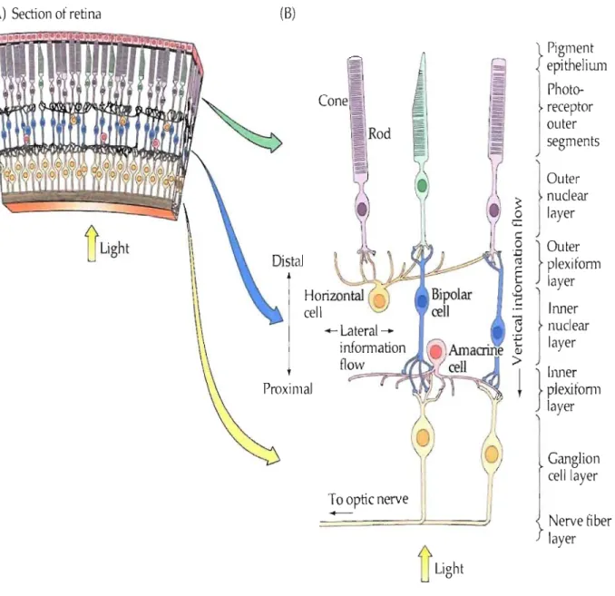
Figure 1: Cross section of the retina. Photoreceptors (rod and cone) are situated
in
the deep
layer. Light must pass through ganglion celllayer, inner plexiform layer, inner nuclear layer,
outer plexiforrn layer and outer nuclear layer to reach photoreceptors.
Optic
nerve
LGN
Optic tract
Hypothalamus
Optic
nerv
~
~
Ciliary gangliOn,
,
Preganglionic parasympathetic
fiber in cranial nerve III
Edinger-Westphal nucleus
Pretectum
Superior Colliculus
- -
-=---
--..J...
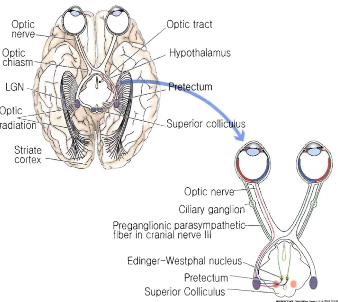
Figure 2: Optic tract.
Optic nerves from the gaglion cells in the nasal half of each retina cross
each other in optic chiasm and reach the contralateral part of the pnmary visuaJ cortex. On the
contrary axons from temporal vi suai field do not cross. (LGN: Lateral geniculate nucleus)
posterior parietal cortex (Figure 3). Relatively, neurons in this area response hardly to colour or to stationary objects analyse (Corbetta et al., 1991).
On the other hand, ventral pathway starts with P cells (for parvi, or small) and via parvocellular layers in LGN (3 to 6) projects to IV C~ and IV A layer ofVl. VI projects to V4 linked with inferior temporal cortex where neurons are sensitive to the outline of images or orientation, colour and shape. It is suggested that dorsal· pathway is concemed with "where" objects are, and ventral pathway with "what" the objects are (Mishkin et al., 1983). CompelIing evidence suggest that different systems are specialized for different visual functions (Zeki, 1978; DeYoe and van Essen, 1988; Livingstone and Hubei, 1988; Zeki and Shipp, 1988). Visual information sent in different area is used differently i.e. visuospatial recognition for dorsal pathway and recognition of complex objects for ventral pathway (Figure 3).
In primate visual cortex specifie features of the organization of V 1 are orientation colurnns, blobs and ocular dominance columns. Discovered by HubeI' and Wiesel, using tangential penetrations with microelectrodes, neurons in the same orientàtion colurnn usually respond to same oriented light bars (HubeI and Wiesel, 1968). Each colurnn contains cells in layer IVC and permits cortical cells to produce Iinear receptive field properties from the information generated by cells of LGN. Blobs, mostly situated in layer II and III, respond to different color stimuli but have no preferred orientation. Transferring monocular visual information optic nerves cross each other in optic chiasm imd most of them (-75%) reach contralateral visual cortex. In the result according to their input source those two separate tracts compose ocular dominance coluinns.
Recognition
Action
Parietal
visual
areas
Figure 3
:
Visual pathway through
visu al cortex. Visual infonnation from LON reaches VI. VI
2.2 Rat
Comparing to' primate, rat's visual system has sorne distinctive features. First, laterally
placed eyes provide a large panoramic visual field lacking binocular overlap. Moreover only a
small proportion (-5%) of retinal ganglion cells project ipsilaterally. Secondly, rat's
photoreceptors are also different from those of primate. Not only has the density of co ne in rat' s
retina appeared to be lesser than other mammals but also photoreceptor cones had shown to be
sensitive at ultraviolet Iight (Jacobs et al., 1991).
Although inrat's system visual pathway also inc1udes LGN, superior colliculus and
pretectum since in our study the main. interest is on LGN afferent pathway physiological
description will be focused on the stmcture of LGN. Precisely, retinal ganglion inputs reach the
dorsal area of LGN (dorsal LGN or dLGN). Despite the lack of lamination in dLGN of the rat
many studies have discriminated various regions in the distribution of cells ofdifferent sizes, in
the composition of afferent axons and different patterns of degeneration after lesions (Land,
1987; Martin, 1986; Reese, 1988). According to Reese (1988) discerning caudodorsally located
nucleus as "outer shell" and "inner core" for ventromedial located nucleus, regions of dLGN
were observed to be innervated by different classes of retinal ganglion cells. Variety in the
regional distribution of inputs signifies that different cell types within dLGN are located in
different broàd regions. Cells in the outer shell mostly project to VI ànd to cortical area Oc2L
(occipital cortex; cytoarchitectonic area 18a) Even though the homology between Oc2L with V2
region of primate is still controversial.
In the visual cortex, visual information reaches mostly the layer IV of VI through dLGN.
But its projection was also observed in lower layer III and layer VI. It was observed that geniculocortical axons form asymmetrical synapses in layer IV (sparsely spined stellate cells,
A.
B.
Il IIIIV
VVI
L
LGN
LP
L
sc~J
(A) Simplified visual information flow in rodent; Y2 is used for Oc2L to compare with primate. JC, inferior colluculus; L, lateral nucleus of the thalamus; PT, pretectum; LP, secondary visual (Iateral posterior) thalamic nucleus; Ym, medial visual area (B) Features of thalamocortical and intracortical connections in Y 1. Cortical layers are indicated by Roman numerals on the left. B, bipolar cel!; C, chandelier cel!; M, multipolar celI; NSS, nonspiny (smooth) stellate cell; P, pyramidal cel!; Pr, bitufted projection cel! with a myelinated axon; SpS, spiny geniculocortical. Pyramidal cells are usually glutamatergic. Bipolar, chandelier, multipolar, and smooth stellate cells are mostly GABAergic. Projection between layers is detai led in the text.
spiny nonpyramidal cells with perikarya and dendritic spines of pyramidal cells) and the lower
part of layer III (dendritic spines of basal dendrites of pyramid~l cells) (Feldman and Peters, 1978; Peters et al., 1976; Peters and Feldman, 1976, 1977). Layer IV cells project prominently in
a precise manner to lower layers II1II1 while weaker projection extends laterally and diffusely in
layer II/III. A vertical projection also exists heading to layers V and VI. Vertical intracortical
conneétions conveying information to layers above and below in the visual cortex provide
binocular or receptive field properties (Gilbert, 1983). Comparing to cells in layer IV, cells in
layer II/III compose a widespread intralaminar connections projecting to lamina V. Pyramidal
neurons in layer V receive projections from layer II/III and from layer VI. Whereas lower layer
V make clustered projections in a diffuse manner to layer l, the bottom of layers II1III, and the
top of layer IV and V. Finally neurons in layer VI make clustered projections to the boarder of
layer III and IV. Additional projections were also observed from layer VI to the boarder of layer
V and VI and layer IIII (Burkhalter, 1989).
Mainly nonpyramidal neurons which are about 15% of the entire neuronal population in
rat's visual cortex are GABAergic. Three distinct families of GABAergic neurons are
distinguished according to their immunoreractivity: parvalbumin (PV), calretinin (CR), and
somatostatin (SOM). PV-immunoreactive neurons are present in ail layers except layer 1 and
constitute about 51 % of GABAergic neurons. SOM-immunoreactive neurons are also absent in
layer 1 but mainly located in infragranular layers V and VI. Finally CR coexpressing neurons
account for 17% of GABAergic neurons and they are abundant in layer 1 (Gonchar and
Burkhalter, 1997). Function of GABAergic neurons during visual stimulation will be discussed
Although the functional organization of rodent is similar to that of primate, in the rat' s
visual cortex there was no evidence of orientation colurnn (Girman et al., 1999). However interestingly, most neurons show a sharp adjusted selectivity about the direction of stimuli
presented with a tendency for horizontal stimuli (Bume et al., 1984; Girman et al., 1999). This implies a distinctive mechanism for neurons in the visual cortex of the rat toward orientation
selectivity .
3. Plasticity
Adaptation was the essential ability to survive, expand habitats and propagate during
evolution even for bacteria. In MacMillan English dictionary it is described as "abilities that
make it possible for organisms to deal with their environment". For mammals, adaptation was
readily observable in the nervous - system especially in the central nervous system (CNS)
inc1uding the brain. One of the brain's striking adapting ability is usually referred as 'Ieaming'.
Learoing is determined as the acquisition and development of memories and behaviours
produced by experience. With many studies in CNS, various kinds of adaptation which
corresponds in neuroscience to a reorganization of the neuronal circuits, termed "plasticity" had
been revealed. Plasticity is also considered as one of the mechanism du ring leaming process in
brain. Although plasticity is not an event persisting for millions of years as evolution had
occurred, plasticity is not a response against a spontaneous change. Adaptation as a result of long
and continuous modification requires long term to launch such function. PI asti city however
reflects a long term modification: Two kind of plasticity are studied: synaptic plasticity and
3.1 Synaptic plasticity
Nervous system is composed of two types of different cells: neurons and glia. Although
recently their functioning importance has been revised, glia's main role is contributing to brain
function by supporting and nourishing neurons. On the other hand, neurons are responsible of
sensing the changes, deliver it to other neurons and command the body's responses to these
senses. Neuron receives outer signal through dendrite and transfer it to neighbouring neuron by
axon (Figure 5). Junction between neurons are called synapses, generally synapses are
established between axons and dendrites, axons and axons or cell bodies. Nervous sense is
transported under form of electrical signal and at the end of the axon it is transferred by releasing
chemical molecules (neurotransmitters) delivered by synaptic vesicles. At the dendritic
membrane of neuron receiving neurotransmitters there are proteins (receptor) which bind
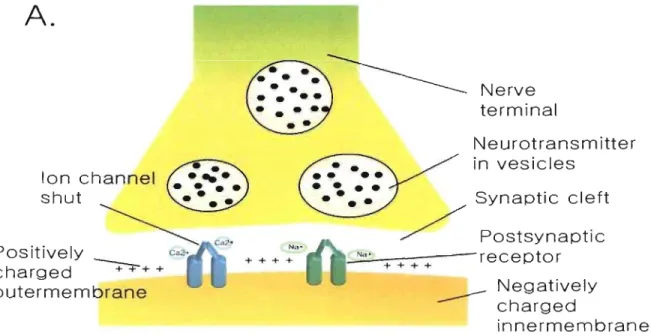
specifically to the releaseà neurotransmitter. Outer-membrane of neuron is positively charged
and near synapse inner.;membrane is negatively charged. Binding of neurotransmitter induces
conforrnational change of ionic channel and allows positive ions enter in the neuron. Entrance of
positive ions changes membrane polarity and this potential change is propagated to the next
neuron (Figure 6). Neurotransmitter releasing neuron is called presynaptic neuron and receiving
neuron is called postsynaptic neuron. Neurotransmitter receptor perrnitting positive ion (Na+,
Ca2+) entrance is called excitatory (e.g. glutamate) and negative ion (Cr) admitting receptor is
called inhibitory e.g. GABA: y-amino-butyric-acid. Synaptic plasticity is the ability of a synapse
between presynaptic and postsynaptic neurons to change its strength by changing the efficacy of
Node of Ranvier
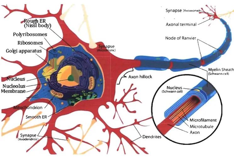
Figure 5: Structure ofa neuron. Neuron is consisted ofaxon, dendrite and soma (cell body).
Myelin sheath is a phospholipid layer surrounding axon.
It is an outgrowth of glial cells.
A.
Ion channel shut Positively charged~+
+ outermembrane Na-+ Na-+ Na-+ Na-+ Nerve terminal l\Jeurotransm itter in vesicles Synaptic cleft Postsynaptic ~N'~'+ _ _ ~---receptor + + + +---
Negatively charged innermembraneResting Synapse
B.
+ + + +1. Action potential reaches
presynaptic terminal
2. Depolarization induces Ca2~ entrance 3. Ca2+ triggers release of neurotransmitter • binds 10 receptor site • • on postsynaptic membrane
~
4. Neurotransmitter• • • • • and open ion channels
Na+ . • • •
+ + + +
Ne"
5. Opening of ionic channels
causes entrance of positive ions
and change in postsynaplic
membrane potential
6. Action potential propagates through next cell
Active Synapse
Figure 6: Synapse and action potential.
CA)
Action potential is propagated through synapse and
delivered to next cell. Outer membrane of neuron is positively charged due to the sodium and
calcium ion.
(B)When ionic channel is opened those ions enter in the neuron and convert the
polarity. This signal is transmitted through the cell.
as the one of Eric Kandel in aplysia had revealed part of the molecular mechanisms for synaptic
plasticity (Castelluci et al., 1978).
Presynaptic facilitation is likely to be a result of Ca2+ influx that activate
ca1ciumlcalmodulin-dependent protein kinases II (CaMKII). These kinases prominently
phosphorylate synaptic vesic1e associated proteins, synapsin and detach them from cytoskeleton.
Increase of Ca2+ level can be induced directly from voltage-gated Ca2+ channels or indirectly
from the modulation of presynaptic K
+ channels. This facilitation can occur autonomously by
homosynaptical transmitter release from the terminal itself or heterosynaptically by a modulatoryneuron at axo-axonic synapses.
The most common mechanism of postsynaptic plasticity results from the direct
}
phosphorylation of an ionotropic receptor by serine/threonine or tyrosine protein kinases.
Typically when modification of existing synaptic proteins, mostly protein kinases (i.e. PKA,
PKC), is involved, it alters the synaptic function (Shi et al., 1999). However a second long lasting mechanism which is triggered by protein phosphorylation depends on second messenger
neurotransmitters and involves changes in the levels of key protein as weil as gene transcription
(Kaang et al., 1993). This second mechanism provides the mechanism for long-lasting memory storage.
3.1.1 Long-term potentiation
A representative effect of changes in the efficacy of synaptic connection is observed
during the phenomenon called long-term potentiation (L TP) (Figure 7). Discovered in the rabbit
hippocampus (Andersen et al., 1966), LTP is the long-lasting enhancement of communication and results when postsynaptic neuron shows a persistent increase in synaptic strength following
high frequency stimulation of a chemical synapse. Possessing common featùres with long-term
memory L TP has been the most attractive candidate for cellular mechanism of learning.
L TP is usually induced with a short tetanic stimulation (l00 Hz in 1 sec) in presynaptic
area and observed as an increase of excitatory postsynaptic potential (EPSP) in postsynaptic area
lasting more than an ho ur (Hùang and Kandel, 1994). Non-tetanic stimulus induced in
presynaptic area causes release ofneurotransmitter glutamate which binds to AMPA
(a-amino-3-hydroxy-5-methylisoxazole-4- propionic acid) receptors embedded in the postsynaptic
membrane. This binding opens the sodium channels and ion influx cause a short EPSP. This
depolarization however, when repeated stimuli at high frequency are given to the presynaptic
fibre, causes the postsynaptic neuron to depolarize progressively. When such a train of stimuli
was applied it expresses st ronger and prolonged EPSP which will remove magnesium ion
blocking the NMDA (N-methyl-D-aspartate) receptors. This opening allows calcium influx wh en
glutamate is bound. The ri se in intracellular Ca2+
con~entration
triggers the activation of severalprotein ki~ase enzymes, enzyme serving to transfer phosphate to donor molecule. For example, calciumlcalmodulin-dependent protein kinase II (CaMKII), prote in kinase C (PKC) and
cAMP-dependent protein kinase A (PKA) (Sweatt, 1999) are activated by calcium entrance (Figure 7 A).
During L TP, CaMKII and PKC become independent on calcium and autonomously
active. Consequently CaMKII and PKC will activate AMPA receptors (AMPAR) by
phosphory lation to increase their activity and induce the insertion of additional AMP AR into the
postsynaptic membrane (Malenka and Bear, 2004). It is suggested that this AMPAR insertion do es not involve protein synthesis. During non-stimulated state AMP AR are generally
internalized inside the synapse and with L TP inducting stimulation under the influence of protein
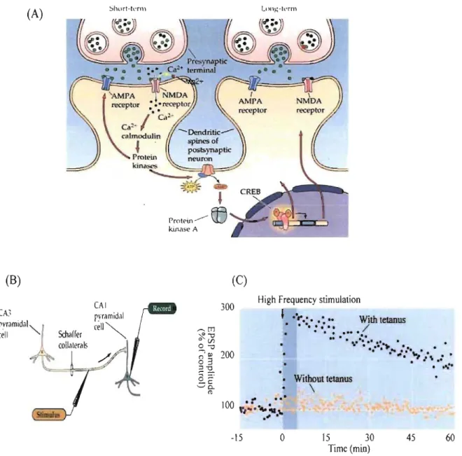
(A)
Short-tefll1(B)
CAl CA~ pyr~mid~1 œil " Schaffer"'-rrot
"
in
~
eo
kinase A(C)
300 ---.[TJ ~." o CFJ 0 ."High Frequency stimulation
' ... : •• :.. With tetanus
.
':
..
': ....
/.. .
.e.
.... ~.I .... '. ....,~8
3
~"2.. pvramidal"
1
céll ""-~ COllalleralS .../,:.
~
200.: • "'.e. .:. '.'
- •• "" 1 \ . : •. .
....
..,_
.
o~ _1: '-' 0-It • Without tetanus \...
100 .. ,...
.: •• "'-:.. \0 .".
-15o
15 30 Time (min) 45Figure 7: Long-term potentiation. A) LTP mechanism in hippocampus. Sustained stimulation of postsynaptic cell with sufficient strength removes Mg2+ ion and allows calcium entrance. B) Recording procedure ofLTP in hippocampus. C) After tetanic stimulation synaptic response increases 200% comparing to baseline and last for an hour.
NEUROSCIENCE, Third Edition © 2004 Sinauer Associates, Inc.
Protein synthesis appears during the late phase of LTP, called L-LTP. Persistent
activation of protein kinases such as MAPK, especially extracellular signal-regulated kinase
(ERK), can induce L-LTP (Sweatt, 1999; Lynch, 2004; Kelleher et al., 2004) followed by
. activating transcription factors like protein kinase MÇ.
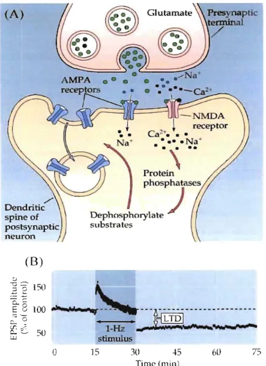
3.1.2 Long-term de pression
Another fonn of synaptic plasticity is long-tenn depression (L TD). As the name imply
unlike L TP, L TD shows a depression of EPSP amplitude after delivering a prolonged trains of
presynaptic stimulation at low (0.5-10 Hz) frequency (Dudek and Bear, 1992). Although low
frequency stimulation (LFS) is a standard method to induce a homosynaptic LTD in
hippocarnpal neuron, NMDAR antagonists were able to block this process. Application of
NMDA showed that just like the LTP, an appropriate activation of postsynaptic NMDARs is
sufficient to induce L TD (Lee et al., 1998; Kamal et al., 1999). And another result demonstrate
that rlse in postsynaptic calcium ion concentration through NMDAR was the critical variables
not the stimulation frequency itself (Neveu and Zucker, 1996) (Figure 8).
With an elevation of Ca2+ concentrationCaMKII is able to activate Calcineurin, which in
turn is able to activate a postsynaptic substrate PPI. The activity of PPI is persistently increased
while L TD inducing stimulation (Thiels et al., 1998). L TD is associated with dephosphorylation
of the GluRl subunit of the AMPAR. GluRl contains serine 831 that can be phosphorylated by .
CaMKII and PKC while serine-845 is phosphorylated by PKA (Roche et al., 1996; Barria et al.,
1997). The PKA site shows higher basal phosphorylation thari the CaMKII-PKC. LFS causes
dephosphorylation of the PKA site and LTD. On the contrary, TBS causes phosphorylation of
(
A)
. , /Dendritic
spine of
p
o
s
t
s
yn
a
pti
c
n
e
uron
(8)
r
ec
e
pt
o
r
Ca-T • • •• ••• • N
a
-prot:
J
..
pho
s
phatases
D
e
PhOSPhOrylat
e
)
substTates--
...
-
,....Q..-E
8
100
...
w.~~
~
i
___
' n _ _ _ n n n _ _ _ _ _ n _ _ _ _-
-..,~ \
~
•
•
i
~
~~
r.r: ;,;,';: ~::-.. 50 l.LJI
-
Hz
__
.~...
~s
tim
ul
us
o
15 30 45 60 75Time
(min)Figure 8:
Long-term depression.
(A) Dephosphorylation mechanism induces intemalization ofAMP AR. (B) Low frequency stimulation generates long-term depression.
reversing previously induced LTD showed that dephosphorylation of AMPARs is a mechanism
for L TD in hippocampal CA 1.
Besides the phosphorylation regulation, several lines of evidence suggest that AMPAR
expression in the postsynaptic membrane is subject to mechanism in LTD; (1) Prior saturation of
L TD yields the AMPARs at the synapse and insensitive to inhibitors of NSF -GluR2 interaction
(Luthi et al., 1999). (2) A presynaptic stimulation at 5 Hz induced an NMDAR dependent depression of miniature excitatory postsynaptic current amplitude and
a
decrease of GluRl expressed in surface (Carroll et al., 1999). Altogether these results suggest that AMPAR internalization is an expression mechanism for L TD. It was proposed that subtype of AMP AR GluR2 binds with N-ethylmaleimide-sensitive factor (NSF), which is an important protein duringmembrane fusion events (Nishimune et al., 1998). Blocking this interaction causes the process of rapid internalization of receptors and decrease of AMP AR currents. Since LFS had no effect
after receptor internalization it is estimated that LTD requires the pool of NSF-regulated
AMP ARs (Lus cher et al., 1999; Luthi et al., 1999).
3.1.3 Plasticity of plasticity: Metaplasticity
Continuous reinforcement or weakening of synapse will cause destabilization of neuronal
networks over time by driving neurons towards maximal and/or minimal action potential firing
frequency ranges. However, other forms of plasticity than L TP or L TD provide the negative
feedback; scaling and metaplasticity. To maintain synaptic strength and plasticity within a
functional dynamic range synaptic scaling lowers amplitudes of small EPSP in response to
continuai excitation and raise them after prolonged blockage (Turrigiano et al., 1998; Watt et al.,
the synapse (Watt et al., 2000). Metaplasticity, introduced by W.C. Abraham and M.F. Bear
(Abraham and Bear, 1996), refers to the plasticity of synaptic plasticity. Principle meaning is that
previous activation history of synapse determines the present plasticity: Depending on current
synaptic state the level of synaptic inhibition or the activity of modulatory afIerents (or
neurotransmitters, e.g. Acetylcholine) can influence the efIect of plasticity overtime (Abraham
and Tate, 1997). For example, if a neuron was already exposed to many plasticity, metaplasticity
drops the future plasticity efficacy by changing NMDAR subunits and lowerthe concentration of
Ca2
+ influx (Flint et al., 1997; Carmignoto and Vicini, 1992; Phil pot et al., 2001).
3.2 Cortical plasticity
While synaptic plasticity occurs between two neurons, cortical plasticity refers to the
changes occurring in the organization of the cortex according to the experience. Brain activity
transferring from the given function to a difIerent location results of normal experience or brain
damage is the remarkable consequence of cortical plasticity.
Few decades ago when neocortex was considered unalterable, Hubei and Wiesel had
de~onstrated that during development before a specifie period, called the critical period, ocular dominance column in VI was highly plastic (Wiesel and Hubei, 1963). Depriving monocular
vision during development results in an expansion of the columns serving the open eye and those
which were responding to the deprived eye become reduced in size and afIerent complexity.
Following the result of Hubei and Wiesel, cortical plasticity had beenshown to somatosensory
cortex (Van der Loos and Woolsey, 1973), auditory cortex (Moore, 1985) and in diverse
Cortical synaptic plasticity has many features that are similar to those of hippocampal
synaptic plasticity. For example when a rat's whisker of postnatal day 12-14 was stimtilated it
drove a recombinant AMP AR subunits into synapses in the somatosensory cortex (Takahashi et al., 2003). Aiso in the visual cortex, similarly with LTD, 24h monocular deprived rat showed various changes in the GluRI phosphorylation level (Heynen et al., 2003). Another important common mechanism is NMDAR dependency. Blocking NMDARs in developing visual cortex
blocks the effects of monocular deprivation suggests the involvement of NMDAR (Bear et al., 1990) for a long tenn effect. Confinning previous experiment and without affecting visual
responses, suppression ofNMDAR subunit (NR1) expression shows that NMDAR is involved in
visual cortex plasticity (Roberts et al., 1998). Involvement of NMDAR signifies Ca2+ entrance
after its opening. Similarly with synaptic plasticity numerous results indicate three essential
kinases during monocular deprivation; PKA (Beaver et al., 2001), extracellular-signal-regulated kinase (ERK; Di Cristo et al., 2001) and aCaMKII (Taha et al., 2002). In the cytoplasm those kinases are responsible to phosphorylate substrates like synapsin (Hosaka et al., 1999), AMPAR (Barria et al., 1997; Benke et al., 1998), GABAR (Brandon et al., 2003), or actin (Matus, 2000). Those are molecules that have crucial role in synaptic transmission, neuronal excitability and
morphological stabilization. Addhionally, kinase activity during ocular-dominance plasticity
drives to activation ofCREB (cAMP response element binding) (Mower et al., 2002; Liao et al., 2002). CREB proteins, being transcription factors, bind to certain DNA sequences called cAMP
response elements (CRE) has been in concem since its implication during long-tenn synaptic
facilitation (Martin and Kandel, 1996). Starting by cellsurface receptor activation, production of
a second mess enger such as cAMP or Ca2+ activates in tum protein kinase which induces CREB
regulates certain transcription factors such as c-fos, c-jun or egr-l (Boutillier et al., 1992;
Masquilier, and Sassone-Corsi, 1992). Gene transcription synthesizes new proteins, a pro cess
essential for both ocular dominance plasticity (Taha et al., 2002) and long-term changes in synaptic strength (Silva ~t al., 1998).
Although cortical plasticity shares numerous consequences with synaptic plasticity there
is no consistent correlation founded between the monocular deprivation effect in vivo and the
ability to induce homosynaptic plasticity in vitro (Renger et al., 2002; Bartoletti et al., 2002).
ContinuaI induction of L TP in synapse does not induce facilitation of cortical plasticity (Hensch,
2003). This disassociation suggests sorne more conditions have impact on cortical plasticity.
Neurotrophin, depending of visual experience, modulates electrical activity and synaptic
transmission by increasing transmitter release or depolarisation of neuron (Sala et al., 1998; Kafitz et al., 1999). Development ofGABA-mediated inhibition, known as triggering the critical period (Fagiolini and Hensch, 2000), was accelerated in BDNF (brain derived neurotrophic
factor)-overexpressing mice (Huang et al., 1999). Neurotransmitters, other than GABA, also have modulator effect during cortical plasticity. Lesion in the basal forebrain, the main source of
ACh in the neocortex, accompanied with destruction of cortical adrenergic innervations retarded
ocular dominance plasticity (Bear and Singer, 1986). In vitro study in prefrontalcortex have
demonstrated that dopamine facilitates LTD of glutamatergic transmission (Otani et al., 1998), and serotonergic axons destructed kitten showed no ocular dominance shi ft after monocular
deprivation (Gu and Singer, 1995). Those supplementary experiments imply that cortical
plasticity can be induced through numerous variables e.g. injection of neuromodulator.
Cortical plasticity was not only found in juvenile cortex but also in adult cortex after
motility (ijoltmaat et al., 2005) status are unfavourable comparing those of young cortex, this phenomenon was confirmed through behavioural observation (Kami and Sagi, 1991),
electrophysiology (Heynen et al., 2001) and tMRI (Furmanski et al., 2004).
In the visual cortex, cortical plasticity was mainly observed by ocular dominance shift.
However, recently, more experimentsdemonstrate that modulatory ability in cortex level can be
reflected through various experiments. Teyler et al showed that with visual tetanic stimulation it
is possible to induce an LTP-Iike increased cortical response in human (Teyler et al., 2005). Supplementing experiences showing about cortical modification, for example orientation tuning
(Fregnac et al., 1988) or contextual modulation (Cri st et al., 2001) indicate thilt ocular dominance plasticity is not the only representative model of cortical plasticity.
4. Acetylcholine
As stated above the neurotransmitter ACh has a functional effect during cortical
modulation and consequently on cortical plasticity. Influence of ACh in various regions of the
brain is weil demonstrated in cognitive functions like attention, consciousness, learning, memory
and sleep (Nobili and Sannita, 1997; Baxter and Chiba, 1999; Hasselmo, 1999; Sarter and Bruno,
2000; Hobson and Pace-Schott, 2002).
Many animal experiments have consistently shown that by blocking muscarinic
receptors, subjects had deficiency of learning acquisition ability (Torres et al., 1994; Baxter and GaHagher, 1996; Doman et'al., 1997; Davidson and Marrocco, 2000). Moreover, excitotoxic lesions of neurons in nucleus basalis induced a severe impairment in memory (Dekker et al., 1991). It was suggested that cholinergie activity promote the cortical processing of thalamic inputs and at the same time inhibit the intracortieal associations (McCormiek et al., 1993; Tang
et al., 1997). Especially during attentional function sorne special cortical cholinergic inputs are found to be enhanced (Conner et al., 2003).
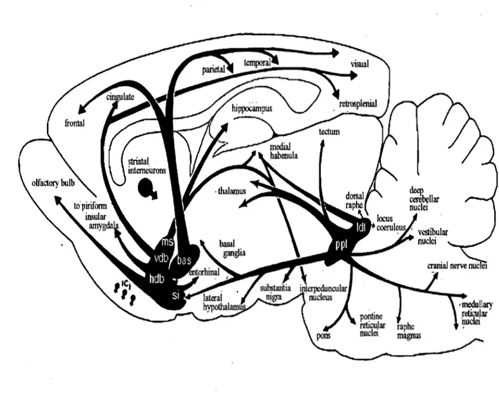
4.1 Cholinergie pathways in the brain
Cholinergic forebrain projections are generally divided into six distribution pathways;
Chl-Ch6 (Mesulam et al., 1983) (Figure 9). Cholinergie nuclei from the medial septum (Chi), the vertical and horizontallimb of the diagonal band (Ch2 and Ch3), project to the hippocampus
and prefrontal and occipital cortex, the nucleus basalis of Meynert (Ch4) project to the entire
cerebral cortex, and the cholinergic neurons in the pedunculopontine tegmental nucleus (ChS)
and laterodorsal tegmental nucleus (Ch6) project to superior colliculus, thalamus, basal forebrain
and substantia nigra. According to their activating agoni st cholinergic receptors are categorized
as muscarinie (activated by muscarine) and nicotinic (activated by nicotine) receptors.
4.2 Nicotinic system
,
Nicotinic cholinergic receptors (nAChR) 'are ionotropic and are found both in peripheral
and central nervous system (PNS and CNS). It is assumed that neuronal nAChR structure is pentameric and composed of two subunit typesgenerally 2a subunits for 3~ subunits. However diversity of nicotinic receptors through various combinations between subunits (ni ne a: a2 to al0, three ~: ~2 to ~4, 1728 possible receptors; Steinlein, 1998)' allows difference in their selectivity for and sensitivity to nicotinic agonists and antagonists which results in a difference in
Figure 9: Rat cholinergic central pathway. Diagonal band of Broca (vdb& hdb) is the main ACh
distributor in the occipital cortex (Gaykema et al., 1990; Zaborsky et al., 1997). Abbreviation;
ms: medial septum, vdb: vertical diagonal band of broca, hdb: horizontal diagonal band of Broca,
bas: nucleus basalis, si: substantia innominata, ppt: pedunculopontine tegmentum, ldt:
Activation ofnicotinic receptors opens Na+, K+ and Ca2+ channels. Comparing to muscle
nAChRs which are more permeable to Na+ ion, neuronal nAChR are highly permeable to Ca2+.
A general consent is that neuronal nAChRs are located in a presynaptic element (Wonnacott,
1997; Dani, 2001) to modulate neurotransmitter, for example glutamate (Radcliffe and Dani,
1998). The Ca2+ permeabili~ ratio of nAChR demonstrated that increase of Ca2+ concentration
induced by nAChR activation was high enough to influence intracellular Ca2+ dependent
mechanisms and may act in synaptic plasticity (McGehee, 2002). In central nervous system a4p2
and a7 are dominant subtypes of nAChR. Those receptors possess sorne distinct properties.
While a4p2 excites neuron by increasing sodium and potassium permeability a7 acts through
calcium channel. In hippocampus a4p2 and a7 are both widely distributed but it is observed that
a4p2 is dominantly involved in the modulation of GABAergic inhibition to the human cerebral
cortical interneurons compared to a7 (Alkondon et al., 2000). Furthermore it is known that
supplemental -injection of nicotine enhance the attention in human and rodents (Levin et al.,
1998; Mirza and Stolerman, 1998). Deletion of a7 nicotinic receptor showed impairing of
attention in a 5-choice seriai reaction-time task.
4.3 Muscarinic system
Muscarinic receptors are known to be metabotropic. Hammer et al distinguished strong
pirenzepine affinity receptors as MI and intermediate or low affinity receptors as M2 (Hammer
et al., 1980). Later on five types of subunit genes are characterized from ml to m5 and their
exprèssion types are named MI to MS. Thefamily of MI receptors comprising MI, M3 and MS,
pathway (Nathanson, 2000). On the contraI)' the M2-like receptors (M2 and M4) inhibit voltage-. gated Ca2+ channel by deactivating adenylate cyclase via G-protein (GÙ (Egan and North, \986).
A preliminary study demonstrated that Ml subtype is widely spread in ail corticallayers while M2 and M4 are less abundant (Levey et al., 1991). Stimulation of postsynaptic muscarinic receptors induces neuron depolarization by inhibiting K
+
efllux, usually Ca2+ independent(McCormick and Prince, 1986). M21M4 receptors are traditionally considered to be situated at presynaptic area for an autoreceptor as a negative feedback implication (Douglas et al., 2001).
4.4 Cholinergie modulation of cortical plasticity
It has been proposed that modulation in receptive field properties contribute to the memol)' coding of a stimulus referring its importance (Weinberger, 2003). A study demonstrating that basal forebrain cholinergic lesions inhibit the learning process but not the performance suggests that cholinergic activity is required to mediate learning associated
expansion and retuning of cortical receptivefields (Conner et al., 2003).
Relation between vision and ACh has yet many undiscovered mysteries. Direct application of ACh to visual cortex modifies neuronal responses showing increase of spontaneous activity, facilitation of evoked responses, or suppression of evoked responses. Assumed to be action of arousal state or attention it has been demonstrated that the visual response cao be enhanced by stîmulating the mesencephalic reticular nuclei (Singer 1977). Another observation is that during sensol)' stimulation ACh is released in the sensol)' cortex (Inglis and Fibiger, 1995; Kilgard and Merzenich, 1998; Verdier and Dykes, 2001; Laplante et al., 2005). As mentioned previously, when kitten cortex has impaired innervation from cholinergie basal forebrain and dorsal noradrenaline bundle, its ocular dominance shift was
blocked despite the monocular eyelid suture (Bear and Singer, 1986). Following experiments by
blocking muscarinic but not nicotinic receptors, and muscarinic MI but not M2 demonstrated
that it can prevent the ocular dominance shift in kitten visual cortex (Gu and Singer, 1989, 1993).
Studies with selective cholinergic immunolesion with cholinergic fibre toxin 192 IgG saporin
confirmed that MI and M2 lesioned juvenile mouse the synaptic plasticity was gravely affected
(Kuczewski et al., 2005). Those indicate that muscarinic Ml receptors may have a critical role
during cortical plasticity.
Cholinergic modulation effect is also shown in orientation dominance column shift.
Normally neurons do not altemate its preferred orientation simply by continuai exposure to
another. However, when this repeated visual stimulus of sub-optimal orientation is paired with
application of ACh, responses of neurons become stronger at the expense of diminishing
response against the previous optimal orientation and remained long lasting (Greuel et al., 1988).
Cortical plasticity induced by ACh is also found in different location. For example in
rodent somatosensory cortex unilateral delete of a digit (e.g. a whisker) followed by
neighbouring digit stimulation results an expansion of the adjacent digit responding neurons.
However with cholinergic deficiency caused by basal forebrain damage no propagation of
receptive field was observed (Juliano et al., 1991). Aiso stimulation of basal forebrain paired
with whisker showed a long-term enhanced somatosensory response (Verdier and Dykes, 2001).
Additionally in the auditory cortex, combining nucleus basalis stimulation and tone emission it
induced changes of receptive field (Ma and Suga, 2003). Ori the contrary this effect was not
shown when muscarinic receptors were blocked with antagonist (Miasnikov et al., 2001).
Although the exact meèhanism of how ACh application can induce increase of cortical
directly interfere with intracellular second messenger. It has been shown that Ml receptors
stimulation leads to an increase of inositol 1,4, 5-triphosphate (Hamilton and Nathanson, 2001)
and this change results in augmentation of intracellular Ca2+ level (Yamamoto et al., 2000) which will promote the plasticity in the visual cortex (Kato et al., 2000). This pre-increased Cjl2+ level can activate intracellular protein kinases (Hamilton and Nathanson, 2001) which may
facilitate responses induced by NMDA receptor (Aramakis et al., 1999). Another possibility of cholinergie contribution is to reduce membrane K + conductance. While activation of muscarinic receptor increasing the depolarization of cortical pyramidal cells associated with NMDA
receptor-gated conductance is supporting result (Kirkwood et al., 1999), this effect will facilitate depolarization in response to visual input which is transmitted through glutamatergic neurons.
These cholinergic actions enhance the opening of NMDAR dependent synaptie transmission.
With a direct contact in visual cortex ACh can regulate GABAergic neuronal inhibition (Xiang et al., 1998; Erisiret al., 2001) and since GABAergic interneurons have crucial role in cortical plasticity (Fagiolini and Hensch, 2000) ACh can influence modification threshold of cortical
PURPOSE OF THIS STUDY
Even though plasticity in adult cortex has now received much recognition, since detailed mechanisms are different from those of critical period there still remain much to understand. Although other neurotransmitters (e.g. norepinephrine or serotonin: reviewed by Gu, 2002) also participated in synaptic plasticity, ACh seems to have a crucial role in modulatory effect by facilitating and/or by accelerating the plasticity mechanism.
In a previous study, we have demonstrated that in the VI of an anaesthetized rat, ACh was released via a repetitive stimulation of sinusoidal grating (Laplante et al., 2005). Here, during this experiment we tried to observe if this endogenous distribution of ACh could affect the cortical response and be examined by change of visual evoked potential (VEP). Aiso by decomposing the activating pathway of cholinergie modification we tried to clarify the underlying mechanism of cortical plasticity in adult visual cortex and observe whether this effect was valuable for long term. Since several studies suggest the role of nicotinic (Rosato-Siri et al., 2006; Kawai et al., 2007) and muscarinic receptor (Origlia et al., 2006; McCoy and Mc Mahon, 2007) during synaptic plasticity we blocked both receptors altematively and observe the change of cortical response through VEP. Aiso based on studies showing in vitro that ACh or muscarinic receptor activation can induce LTP in slices of VI mediated by an enhancemtmt effect ofNMDA receptor conductance (Brocher et al., 1992; Kirkwood et al., 1999; Kojic et al., 2001) we tested whether blocking NMDA receptor affected ACh induced facilitating effect.
In the result we demonstrate that during pattemed visual stimulation paired with cholinergic activation could enhance thalamocortical L TP which is mediated through both muscarinic and nicotinic receptors in vivo increasing the amplitude of VEP. Thus synaptic
mechanisms involving ACh promote plasticity by increasing the input thalamic signais that will launch translational cascades indicating long term enhancing effect on synaptic strength.
METHODS
1. Animal preparation
Guidelines set out by the Canadian Council for the Protection of Animais were ~ollowed for ail procedures. 44 Long-Evans rats (250-300g) were obtained from Charles River Canada
(St-Constant, Quebec, Canada) and maintained in a 12h lightJdark cycle with free access of food
during both pre- and post-implantation period. During ail experiments every effort to reduce both
the suffering and number of animais used was made. Experiments were performed during 210
minutes (from tO to t7). Experimental groups were control (n=II), carbachol(n=6) injected
(injection time=90 min: t2) group, scopolamine (n=5) injected group (t2), CPP (n=6) (t2) +
carbachol (t=120 min: t4) injected group, aCSF (n=6) (t2) + carbachol (t4) injected group,
scopolamine (n=5) (t=60 min: tl) + carbachol (t2) injected group, and mecamylamine (n=5) (t2)
+carbachol (t4) injected group.
2. Implantation
Two days before recording, animais were placed in a Plexiglas box and anaesthetized
with a gaseous mixture of isoflurane (5%) along with oxygen and air. After transferring the
animal to the stereotaxic apparatus, anaesthesia (isoflurane 1.5%) was administered through a
mask. The rectal temperature was monitored and maintained at 37°C with a thermostatically
controlled heating pad (FHC, Bowdoinham, ME, USA). A dental drill was used to make an ho le
(-3.0x 3.5 mm) in the skull above the left visual cortex and a push-pull cannula guide (plastics 1 ,
Roanoke, VA) and the electrode guide (polyurethane tubing) were implanted. The dura mater was left intact except the cannula guide inserted location where a small incision was made with
Tt (0 min) Tl (30 min) Tl (60 min) T4 (90 min) TS (120 min)' T6 (150 min) T7 (180 min) Carbachol (n=6) aCSF+CCh (n=6) Sco+CCh (n=5) Injection aCSF injection Sco CCh injection ,injection CCh injection
Table 1: Injection procedure. Recording result from Tl was used as baseline for each group.
Carbachol, aCSF, CPP and mecamylamine was injected intracortical. Only scopolamine was injected intraperitoneal.
30 gauge needle. The cannula guide was inserted at the VI (mm from Bregma: AP -7.5, L +3.6,
V -0.7 mm) with an angle 0000 and the tubing was placed perpendicularly on the dura mater 0.4
mm left lateral (Figure 10A). Final recording location was decided by the optimum response
generating area after multiple recording tests. Two nylon screws (Small parts, Miami Lakes, FL,
USA) were also screwed in the skull (3.0 mm anterior and 4.0 mm right lateral of the hole) and
the guides were secured with dental cement after covering the exposed region with agarose gel to
prevent dryness and direct contact with dental cement solvent. After suturing incised skins and
applying a local anaesthetic (Xylocaine) to sutured point, animais were returned to the cage.
3. V;sual stimulation
The visual stimulation during 10 minutes was provided with a patterned sinusoidal
grating displayed on a computer screen every 30 minutes. The computer monitor (30x25 cm,
Thanium; Apple Computer Inc., Cupertino, CA, USA) was placed 30 cm unilaterally parallel to
the midline of the rat. A horizontal sinusoidal grating (contrast 100%,0.12 cyc/deg, 0.033 Hz)
was produced by Vpixx software (v 8.5; Sentinel Medical Research Corp., Quebec, Canada) and
displayed on the computer monitor (Figure IOA). Each visual stimulus (100 p1s) was displayed
20 times and evoked potential changes in the visual cortex were recorded during that period.
Selected orientation and spatial frequency of the grating were based on published values that
have shown to induce an optimal response in VI of the rat, Girman (Girman et al., 1997) and
Aspiration
(A)
Druo infusiont
- ~ - - - -- ---~
(B)
1
1 1 1 1 1 1 1 1 1 1 1 1 1 1Stimulat
i
on
10min
20
min
(Break)
tO(Omin) t1
(
aOIDiD)
lOmin
100ms