1
Université de Montréal
Neurotensinergic modulation
of
glutamatergic neurotransmission in VTA neurons.
par Poulomee Bose
Département de Psychaitrie
Faculté de Médecine
Thèse présentée à la Faculté de Médecine
en vue de l’obtention du grade de Philosophie Doctor (Ph.D) en Sciences Biomedicale.
Juillet 2015
II
Université de Montréal
Faculté des études supérieures et postdoctorales
Cette thèse intitulée:
Neurotensinergic modulation
ofglutamatergic neurotransmission in VTA neurons
Présentée par: Poulomee Bose
a été évaluée par un jury composé des personnes suivantes :
Jean Seguin, président-rapporteur Richard A Warren, directeur de recherche
Valerie Mongrain, membre du jury Andrew Chapman, examinateur externe
I
Résumé
L’aire tegmentaire ventrale (VTA) contient une forte densité de terminaisons neurotensinergiques ainsi que des récepteurs à la surface des neurones dopaminergiques et non-dopaminergiques. Le VTA a été impliqué dans des maladies comme la schizophrénie, les psychoses et l’abus de substance. Les drogues d’abus sont connues pour induire le phénomène de sensibilisation - un processus de facilitation par lequel l’exposition à un stimulus produit une réponse augmentée lors de l’exposition subséquente au même stimulus. La sensibilisation se développe dans le VTA et implique mécanismes dopaminergiques et glutamatergiques. Il a été montré que les antagonistes neurotensinergiques bloquaient le développement de la sensibilisation et certains mécanismes de récompense et ces effets pourraient être médiés indirectement par une modulation de la neurotransmission glutamatergique. Cependant, on connaît peu les mécanismes de modulation de la transmission glutamatergique par la neurotensine (NT) dans le VTA.
Le but de la présente thèse était d’étudier la modulation neurotensinergique de la neurotransmission glutamatergique dans les neurones dopaminergiques et non-dopaminergiques du VTA. Pour ce faire, nous avons utilisé la technique du patch clamp dans la cellule entière dans des tranches horizontales du VTA pour étudier les effets de différents agonistes et
antagonistes neurotensinergiques. Les neurones ont été identifié comme Ih+ (présumés
dopaminergiques) ou Ih- (présumés non-dopaminergiques) selon qu’ils exprimaient ou non un
courant cationique activé par l’hyperpolarisation (Ih). Des techniques d’immunocytochimie ont
II
Dans une première étude nous avons trouvé que la neurotensine indigène (NT1-13) ou son fragment C-terminal, NT8-13, induisait une augmentation comparable des courants
postsynaptiques excitateurs glutamatergiques (CPSEs) dans les neurones Ih+ ou Ih- du VTA.
L'augmentation induite dans les neurones Ih+ par la NT8-13 a été bloquée par le SR48692, un
antagoniste des récepteurs NTS1, et par le SR142948A, un antagoniste des récepteurs NTS1 et NTS2, suggérant que l'augmentation était médiée par l’activation des récepteurs NTS1. Dans
les neurones Ih- l'augmentation n’a été bloquée que par le SR142948A indiquant une implication
des récepteurs NTS2.
Dans une deuxième étude, nous avons testé les effets de la D-Tyr[11]NT (un analogue neurotensinergique ayant différentes affinités de liaison pour les sous-types de récepteurs
neurotensinergiques) sur les CPSEs glutamatergiques dans les neurones Ih+ et Ih- en parallèle
avec une série d’expériences comportementales utilisant un paradigme de préférence de place conditionnée (PPC) menée dans le laboratoire de Pierre-Paul Rompré. Nous avons constaté que
la D-Tyr[11]NT induisaient une inhibition dépendante de la dose dans les neurones Ih+ médiée
par l'activation de récepteurs NTS2. En revanche, la D-Tyr[11]NT a produit une augmentation
des CPSEs glutamatergiques médiée par des récepteurs NTS1 dans les neurones Ih-. Les résultats
des expériences comportementales ont montré que des microinjections bilatérales de D-Tyr[11]NT dans le VTA induisait une PPC bloquée uniquement par la co-injection de SR142948A et SR48692, indiquant un rôle pour les deux types de récepteurs, NTS1 et NTS2. Cette étude nous a permis de conclure que i) la D-Tyr[11]NT agit dans le VTA via des récepteurs NTS1 et NTS2 pour induire un effet de récompense et ii) que cet effet est dû, au moins en partie, à une augmentation de la neurotransmission glutamatergique dans les neurones non-dopaminergiques (Ih-).
III
Dans une troisième étude nous nous sommes intéressés aux effets de la D-Tyr[11]NT sur les réponses isolées médiées par les récepteurs N-méthyl-D-aspartate (NMDA) et acide α-amino-3- hydroxy-5-méthyl-4-isoxazolepropionique (AMPA) dans les neurones du VTA. Nous avons
constaté que dans les neurones Ih+ l’amplitude des CPSEs NMDA et AMPA étaient atténuées
de la même manière par la D-Tyr[11] NT. Cette modulation des réponses était médiée par les
récepteurs NTS1 et NTS2. Au contraire, dans les neurones Ih-, l’amplitude des réponses NMDA
et AMPA étaient augmentées en présence de D-Tyr[11]NT et ces effets dépendaient de l’activation des récepteurs NTS1 localisés sur les terminaisons glutamatergiques. Ces résultats fournissent une preuve supplémentaire que le NT exerce une modulation bidirectionnelle sur la neurotransmission glutamatergique dans les neurones du VTA et met en évidence un nouveau type de modulation peptidergique des neurones non-dopaminergiques qui pourrait être impliqué dans la sensibilisation.
En conclusion, la modulation neurotensinergique de la neurotransmission glutamatergique dans les neurones dopaminergiques et non-dopaminergiques du VTA se fait en sens opposé soit, respectivement, par une inhibition ou par une excitation. De plus, ces effets sont médiés par différents types de récepteurs neurotensinergiques. En outre, nos études mettent en évidence une modulation peptidergique de la neurotransmission glutamatergique dans le VTA qui pourrait jouer un rôle important dans les mécanismes de lutte contre la toxicomanie.
Mots-clés : dopamine, aire tegmentaire ventrale, glutamate, neurotensine, courant postsynaptique excitateur, patch-clamp.
IV
Abstract
The ventral tegmental area (VTA) contains a high density of neurotensin (NT) terminals and receptors that are expressed on dopaminergic (DA) and non-DA neurons. This area of the brain is strongly implicated in disorders like schizophrenia, psychosis and drug abuse. Drugs of abuse induce behavioural sensitization- a facilitatory process whereby exposure to a stimulus results in an enhanced response to a subsequent exposure of the same stimulus. Sensitization develops in the VTA and involves glutamatergic neuroadaptations in VTA DA neurons. NT antagonists prevent the development of sensitization and reward mechanisms and this could be mediated through a modulation of glutamatergic neurotransmission in the VTA. However, how NT modulates glutamatergic neurotransmission in VTA neurons remains unclear.
The present thesis was aimed at investigating the NTergic modulation of glutamatergic neurotransmission in VTA DA and non-DA neurons. Whole cell patch clamp electrophysiology in acute VTA horizontal slices was used to study the effects of different
NTergic agonists on VTA neurons. Neurons were classified as either Ih+ (putative
dopaminergic neurons) or Ih- (putative non-dopaminergic neurons) based on the presence or
absence of a hyperpolarisation activated cationic current (Ih) respectively.
Immunohistochemical techniques were routinely used to label neurons and confirm their location in the medial VTA.
In the first study we report that native neurotensin (NT1-13) or its C-terminal fragment, NT8-13 induced comparable increases in the amplitude of glutamatergic excitatory post-synaptic
V
blocked by SR48692 (NTS1 antagonist) and SR142948A (NTS1/NTS2 antagonist),
suggesting that the augmentation effect was mediated by NTS1 receptors. In Ih- neurons,
however, only SR142948A blocked the increase in the EPSC amplitude, indicating the involvement of NTS2.
In the second study we tested the effects of D-Tyr[11]NT (an active NT analog with differential
binding affinities for NT receptor subtypes) on glutamatergic EPSCs in Ih+ and Ih- neurons and
conducted (by Romrpe’s Lab) a parallel series of behavioral experiments using a conditioned place preference (CPP) paradigm. We found that D-Tyr[11]NT induced a dose dependent
inhibition of EPSCs in Ih+ neurons that was mediated by the activation of NTS2 receptor. In
contrast, D-Tyr[11]NT dose dependently enhanced glutamatergic EPSCs through an NTS1
receptor involvement in Ih- neurons. Results from behavioural experiments show that bilateral
VTA microinjections of D-Tyr[11]NT induced a CPP that was blocked only by co-injection of
SR142948A and SR48692, indicating a role for NTS1. This study allowed us to conclude that i) NT acts on VTA NTS1 receptors to induce a rewarding effect and ii) that this effect is due, at
least in part, to an enhancement of glutamatergic inputs to non-dopamine (Ih-) neurons.
The third study entailed investigating the effects of D-Tyr[11]NT on isolated n-methyl-D-aspartate (NMDA) and α-amino-3-hydroxy-5-methyl-4-isoxazolepropionic acid (AMPA)
receptor mediated EPSCs in VTA neurons. We found that in Ih+ neurons both NMDA and
AMPA EPSC amplitudes were attenuated by D-Tyr[11]NT. This attenuation appeared to be
mediated by both NTS1 and NTS2 receptors. In Contrast, in Ih- neurons both NMDA and
AMPA EPSC amplitudes were enhanced by an NTS1 dependent mechanism. Additionally, the enhancement effect resulted from a presynaptic potentiation of glutamatergic inputs. These results provide additional evidence that NT exerts a bidirectional modulation on glutamatergic
VI
neurotransmission in VTA neurons and highlights a novel peptidergic modulation of non-DA neurons that might be implicated in sensitization mechanisms. Altogether, our studies allowed us to conclude that the NTergic modulation of glutamatergic neurotransmission in VTA DA and non-DA neurons is oppositely regulated by NTS2 and NTS1 receptors respectively. Additionally it highlights a peptidergic modulation of glutamatergic inputs to VTA non-DA neurons that might be crucial for addiction mechanisms.
VII
Table of Contents
Résumé ... i
Abstract ... iv
Table of Contents ... vii
List of Tables ... ix
List of Figures ... x
List of Abbreviations: ...xii
Acknowledgements ... xviii
Introduction ... 1
Chapter 1: VTA anatomy, neuronal composition and projections. ... 3
1.1 VTA Anatomy: ... 3
1.2 Neuronal composition of the VTA ... 4
1.2.1 DA neurons: ... 4
1.2.2 GABA neurons: ... 8
1.2.3 Glutamatergic neurons: ... 10
1.2.4 Neurotensinergic neurons: ... 11
1.3 VTA projections and functions: ... 11
1.3.1 VTA Projections ... 11
1.3.2 Functions of the VTA: ... 15
Chapter 2: Neurotensin synthesis, receptors and effects. ... 18
2.1.1 Discovery and synthesis:... 18
2.1.2 NT as a neuropeptide neurotransmitter: ... 19 2.1.3 NT receptors: ... 19 NTS1 receptors: ... 20 NTS2 receptors: ... 21 NTS3 receptors: ... 22 2.1.4 NT analogs: ... 24 2.2 NT in the Midbrain: ... 28 2.3 Modulation of DA neurotransmission by NT. ... 30
VIII
2. Effects of NT receptor activation. ... 31
2.4 Effects of NT administration in the VTA. ... 34
2.5 Effect of NT on glutamatergic neurotransmission. ... 36
2.6 Relevance of a role for NT in behavioural disorders. ... 39
Chapter 3. Questions and Hypothesis ... 41
Results ... 46 Contribution of co-authors ... 47 Article 1 ... 49 Article 2 ... 85 Article 3 ... 137 4. Discussion ... 182
4.1 Summary of the results ... 182
4.2 NT exerts a complex effect on glutamatergic neurotransmission in VTA neurons. ... 186
4.2.1 Bidirectional effects of NT analogs on glutamatergic EPSCs in Ih+ neurons. ... 186
4.2.2 NT analogs increase glutamatergic neurotransmission in Ih- neurons. ... 190
4.3 Conclusion ... 193
4.4. Technical considerations:... 194
4.4.1 Identification of DA neurons in the VTA ... 194
4.4.2 Recording technique: ... 197
4.5 Implications in behavioural disorders and future directions. ... 198
IX
List of Tables
Table 1 Summary of the characteristics of NT receptor subtypes, locations, agonists and antagonists. 23
X
List of Figures
Figure 1 Properties of in vitro recorded VTA DA neurons. 7
Figure 2 Schematic of the principal brain regions that innervate the VTA. 14
Figure 3 NT distribution and NTergic projections of the rat brain. 28
Figure 4 Mechanism of action of NT on DA cells. 32
Figure 5 Summary of the electrophysiological and neurochemical effects of
NT administered in the VTA.
34
Figure 6 Schematic of the proposed model. 43
Figure 7 A
&B Direct and Indirect pathway of Specific hypothesis 2. 44
A1 Figure 1 Effect of NT1-13 and NT8-13 on glutamatergic EPSCs. 70
A1 Figure2 Effect of NT receptor antagonists on glutamatergic EPSCs. 72
A2 Figure 1 Induction of a CPP by [D-Tyr11]NT . 105
A2 Figure 2 Mean preference score of [D-Tyr11]NT injected animals. 107
A2 Figure3 Effects of SR142948 and SR48692 on [D-Tyr11]NT-induced CPP. 109
A2Figure 4 Effect of [D-Tyr11]NT and antagonists on Ih+ neurons 111
A2 Figure 5 Effects of [D-Tyr11]NT on glutamatergic EPSCs in Ih+ and Ih
-neurons.
113
A2 Figure 6 Effect of [D-Tyr11]NT and antagonists on Ih- neurons. 115
A2 Figure 7 Effect of SR142948 and SR48692 on glutamatergic EPSCs in Ih+ and
Ih- neurons
117
A3 Figure 1 Characteristics of the glutamatergic EPSCs. 149
A3 Figure 2 Effects of D-Tyr[11]NT on NMDA EPSCs. 151
XI
A3 Figure4 Figure 4: Locus of neurotensinergic modulation of evoked AMPA
EPSCs in Ih- neurons.
XII
List of Abbreviations:
ACSF: Artificial cerebrospinal fluid
AHP: Afterhyperpolarisation
AM251: N-(Piperidin-1-yl)-5-(4-iodophenyl)-1-(2,
4-dichlorophenyl)-4-methyl-1H-pyrazole-3-carboxamide
AMPA: α-amino-3-hydroxy-5-methyl-4-isoxazolepropionic acid
Amyg: Amygdala
ANOVA: Analysis of Variance
AP: Action Potential
APDs: Antipsychotic drugs
APV: (2R)-amino-5-phosphonovaleric acid
ATP: Adenosine triphosphate
BLA: Basolateral amygdala
Bmax: Maximum specific binding
BMI: Bicuculline methiodide
BNST: Bed Nucleus of Stria Terminalis
XIII
cGMP : Cyclic guanosine monophosphate
CHO: Chinese Hamster Ovary cell lines
CNQX: 6-cyano-7-nitroquinoxaline-2,3-dione
CNS: Central nervous system
CPP: Conditioned Place Preference
D2R: Dopamine receptor subtype 2
DA: Dopamine
DAG: Diacylglycerol
DAT: Dopamine transporter
DMSO: Dimethyl Sulfoxide
DOPAC: 3,4-dihydroxyphenylacetic acid
DR: Dorsal Raphe
EC: Entorhinal cortex
EGPF: Enhanced green fluorescent protein
EGTA: Ethylene glycol tetraacetic acid
EPSC: Excitatory Post Synaptic Current
GABA: ɣ aminobutyric acid
XIV
GIRK: G-protein coupled inwardly rectifying potassium channels
GLT1: Glutamate Transporter subtype1
GTP: Guanosine triphosphate
HEPES: (4-(2-hydroxyethyl)-1-piperazineethanesulfonic acid)
HVA: Homovanillic acid
i.c.v : intracerboventricular
Ih : Hyperpolarisation activated cationic current
IP3: Inositol triphosphate
KA: Kainic acid
Kd: Dissociation constant
KOH: Potassium hydroxide
LDTg : Laterodorsal tegmentum
LH: Lateral hypothalamus
LS: Lateral Septum
LTD: Long term depression
MAPK: Mitogen activated protein kinases
mGluR: metabotropic Glutamate receptor
XV
nAcb: Nucleus accumbens
NMDA: n-methyl-D-aspartate
NN: neuromedin N
NR1: NMDA receptor subtype1
NT: Neurotensin
NTS: NT receptor subtype
NTS1: Neurotensin receptor subtype1
NTS2: Neurotensin receptor subtype2
NTS3: Neurotensin receptor subtype3
PAG: Periaqueductal gray
PBP: Parabrachial pigmented area
PFC: Prefrontal cortex
PFR: Para fasciculus retroflexus area
PKC: Protein Kinase C
PLC: Phospholipase C
PN: Paranigral nucleus
PPR: Paired pulse ratio
XVI
QX314: N-Ethyllidocaine
RMTg: Rostromedial tegmental nucleus
SK: Small conductance calcium activated potassium channels
SN: Substantia nigra
SNC: Substantia nigra pars compacta
SorLA: Sortilin
STN: Sub thalamic nucleus
TH: Tyrosine hydroxylase
VGluT: Vesicular glutamate transporter
vHipp: Ventral hippocampus
VP: Ventral pallidum
VTA: Ventral tegmental area
XVII
Pour
Mes parents pour faire mon rêve leur.
Shubho- de croire en moi.
Dedicated to
My parents for making my dream – theirs.
Shubho- for believing in me.
XVIII
Acknowledgements
It is almost impossible to complete a PhD program alone. Every aspiring PhD student is steered by a team of people who constantly motivate and push the student to do his/her best. I express my heartfelt gratitude to my supervisor, Dr. Richard A Warren for having me under his tutelage for my doctoral studies. He has always been there, leading me to think
scientifically, patiently listening to my questions, supporting me during the most difficult times, and always motivating me to achieve more. Without Dr. Richard, this surely would not have been possible.
I sincerely thank all the jury members for having taken out time from their busy schedule to read and revise the manuscript. I express my sincere gratitude to Dr. Pierre Paul Rompre, who has constantly picked me up when I stumbled upon a difficulty and lent me priceless advice on being focused. A special note of thanks to Dr. Valerie Mongrain and Dr. Daniel Levesque for being a part of the comité de parrainage for my PhD. Their comments and inputs have indeed added perspective and direction throughout my studies at the Université de Montreal.
I would like to thank all my friends- colleagues and members of other labs, with who I have had the pleasure of working. A special mention to Claude Bouchard for his ever willing help and Alexandra Gallo, for always saying “You will “. I also take the opportunity to thank my brother, Jaideep Mallick for his relentless inspiration and support.
Finally, a huge thank you to my parents, Pulak Kumar Bose and Swapna Bose for making my dreams theirs. Their faith in me has always instilled the confidence to strive for excellence and without them I would not be where I am. Last but not the least, I thank my husband,
XIX
Shubhashish Dhole, for his unconditional support and sacrifice. He has always believed in me and instilled the courage to persist, work hard and achieve.
1
Introduction
This thesis focuses on the modulation of glutamatergic neurotransmission by neurotensin (NT) in the ventral tegmental area (VTA). NT is a thirteen amino acid endogenous neuropeptide that modulates neurotransmission in several brain regions like the VTA, the prefrontal cortex (PFC) and the nucleus accumbens (nAcb)(Binder et al. 2001a). The VTA is a midbrain dopamine (DA) rich region and plays a role in sensitization, reward, motivation cognition and processes information about emotion(Nestler 2013). The neurotensinergic projections from surrounding brain areas (eg. PFC, lateral hypothalamus(LH),) densely innervate the VTA and 80-90%percent of the VTA neurons express NT receptors, the activation of which is known to modulate DA cell activity and consequently DA dependent behaviors(Rompre et al. 1998). The VTA DA neurons also receive glutamatergic inputs containing NT terminals and receptors from limbic brain regions such as the PFC(Vezina and Queen 2000a).
Drugs of abuse elicit motor stimulant effects that enhance with repeated drug administration, and this sensitized behavioral response can endure for months after the last repeated drug administration(Robinson and Berridge 1993). The sensitized behavioural response is a bipartite phenomenon, consisting of initiation and expression phases. The VTA serves as the key anatomical substrate for the initiation of sensitization. Furthermore, the glutamatergic inputs to the VTA are essential for the development of drug induced synaptic plasticity on DA neurons (Bellone and Luscher 2006;Ungless et al. 2001). It was found that blockade of
ionotropic glutamate receptors disrupted the development of psychostimulant induced behavioural sensitization. Interestingly, it was also found that blockade of NT receptors
2
disrupted the development of sensitization. Since the VTA is heavily innervated by NT receptors and terminals and often these terminals are found on glutamatergic efferents from limbic regions such as the PFC, it is possible that NT modulates these inputs to the DA neurons of the VTA and thereby play a role in the development of sensitization. However, how glutamatergic neurotransmission is modulated by NT in the VTA at the cellular level remains elusive. This study was aimed at characterising the effects of NT on glutamatergic neurotransmission in VTA DA and non-DA neurons using electrophysiological and
immunohistochemical techniques.
In my attempt to create a background for my readers, in the subsequent sections, I will draw an overview of the VTA and its projections that are relevant to this study ; its neuronal
composition Following this, I will focus on the properties of NT as a neuropeptide
neurotransmitter, the distribution of NT neurons and terminals in the midbrain, the mechanism of action of NT ,effects of NT administration in the VTA, effects of NT on glutamatergic neurotransmission and the relevance of a role for NT in reward mechanisms and
3
Chapter 1: VTA anatomy, neuronal composition and
projections.
The VTA serves as a key anatomical substrate of neuroadaptive changes that result in reward and addiction mechanisms and is also crucial for the development of sensitization. Drugs of abuse induce synaptic plasticity on VTA DA neurons by modulating glutamatergic inputs to the VTA. The present study involves characterizing the neurotensinergic modulation of glutamatergic inputs to VTA neurons. This section therefore focusses on the anatomical organisation of the VTA, its neuronal composition, projections and the functions.
1.1 VTA Anatomy
:
In 1984, Lindvall and Bjorklund (Lindvall et al., 1984) grouped the DA containing nuclei of the midbrain and named them A1-A17. The VTA or the A10 group of cells was further
divided into four subzones that were called the paranigral nucleus (PN), the parabrachial
pigmented area (PBP), the parafasciculus retroflexus area (PFR), and the ventral tegmental tail (VTT). The cell density in these sub-regions is estimated by TH (tyrosine hydroxylase- rate limiting enzyme for DA synthesis) immunocytochemistry (Kohler et al. 1983). The PFR and VTT, border the VTA rostrally and caudally respectively and contain a low density of
dopaminergic cell bodies that are small in size. Laterally the VTA is marked by the PN. Both PN and PBP are rich in dopaminergic cells, and comprise mainly of medium to large sized TH-positive cell bodies (Binder et al. 2001a).
4
1.2 Neuronal composition of the VTA
The VTA is majorly comprised of dopaminergic, glutamatergic and GABAergic neurons. Among other peptidergic neurons that are found in the VTA, the neurotensinergic neurons are particularly relevant in the present study. While there is a controversy over the exact number of each neuronal sub-population in the VTA, there seems to be a general agreement in most studies that in the VTA DA neurons constitute about 60-65%, the ɣ-aminobutyirc acid (GABA) neurons account for 30-35% and the glutamatergic neurons constitute about 5 % of the total cell population (Margolis et al. 2006a;Nair-Roberts et al. 2008;Yamaguchi et al. 2011). About 1-2% of the VTA neurons are neurotensinergic (Binder et al. 2001a). The following section describes the characteristics of each of these subpopulations of VTA neurons.
1.2.1 DA neurons:
Using TH immunohistochemistry Margolis and group, morphologically classified VTA TH-positive DA neurons as fusiform, round, multipolar or elliptical. The fusiform and elliptical dopaminergic neurons, are approximately equal in number and each population constitute about 32% of the VTA DA neurons. Both fusiform and elliptical DA neurons have an elliptical cell body with the exception that fusiform neurons have two dendrites at opposite ends of the major axis whereas elliptical dopaminergic neurons lack a readily identifiable number of dendrites. DA cells with round or multipolar soma constitute 20% and 17% of the VTA dopaminergic neurons. The size of TH positive dopaminergic neurons varied between
5
of VTA DA neurons branch out in forks or tufts and these have 2-3 fine processes which sometimes wrap around another terminal. The VTA dendrites are also possessed with irregular spinule like projections which can possibly serve as dendritic DA release sites. In vivo
recordings of VTA DA neurons display two modes of spike firing: tonic single spike activity and burst spike firing (Goto et al. 2007;Grace and Bunney 1983a;Grace and Bunney
1983b;Grace and Onn 1989;Koyama and Appel 2006). However, when recorded in vitro, in slices, VTA DA neurons display only the tonic pacemaker activity of 0-5Hz (as shown in figure 1B) while the bursts are absent due to the transection of afferent synaptic inputs
required to promote a DA neuron to the burst firing mode (Mao et al. 2011;Ungless and Grace 2012).
The resting membrane potential in DA neurons recorded in vitro in VTA slices from the rat vary between -44mV and -47mV and the action potential (AP) threshold lies between -24 to-28mV (Margolis et al. 2006a). Recent studies nominate the AP width as a criteria to identify DA neurons. In vitro recordings of VTA DA neurons display a broader AP width (than non-DA neurons) of >2 ms as shown in Figure 1C (Mao et al. 2011;Ungless and Grace 2012). VTA DA neurons are characterized by the presence of a hyperpolarisation activated cationic
current (Ih) (Ferrario et al. 2005;Kempadoo et al. 2013a;Margolis et al. 2006a). The Ih current
results from an activation of non-specific cationic conductance at hyperpolarised potentials, when the voltage of the neuron is stepped from -40mV to -120 mV and the inward current reflects as a long sag at the most hyperpolarised potential. Figure 1D shows an example of an
Ih current in a DA neuron recorded in-vitro from a rat VTA slice preparation.
Based on the presence or absence of Ih and TH immunohistochemistry, VTA dopaminergic
6
recorded neuron with TH immunohistochemistry (left panel shows: recorded neuron; middle
panel: TH positive neuron; right panel: merged). The Ih+TH+ cell group defines the
dopaminergic cell type whereas the Ih+TH- represent either the glutamate or GABA neurons
(Lammel et al. 2014;Li et al. 2013;Margolis et al. 2006a;Margolis et al. 2012a;Ungless and
Grace 2012). However there is no ambiguity about Ih- cells representing the non-DA cell type,
the exact neurotransmitter identity of which is still elusive (Lammel et al. 2014;Margolis et al. 2006a).
In addition to the firing pattern of DA neurons, Johnson and North (Johnson and North 1992a)
included hyperpolarisation of DA neurons by dopamine D2 receptor (dopamine receptor
subtype 2 which mediates inhibitory actions by inhibiting adenylyl cyclase activity) agonist, quinpirole as a property typical to DA neurons. However recent studies suggest that only 55%
of TH+ DA neurons are hyperpolarised by quinpirole (Margolis et al. 2006a). Additionally,
about 20% of TH+ DA neurons in the VTA are insensitive to quinpirole application. DA
neurons are also characterised by the presence of DA transporter (DAT), D2 autoreceptors and
G protein regulated inward rectifier potassium channel subtype 2 (GIRK2) however none of the characteristics unequivocally characterize DA neurons (Bellone and Luscher 2006;Saal et al. 2003;Wanat et al. 2008a).
The use of different parameters for characterizing a DA neuron in different studies, has led to the understanding that the VTA DA neurons are a heterogeneous group of neurons and there exist controversies in the identification and classification of this neuronal population (Ungless and Grace 2012).This issue is addressed in detail in the discussion section of the thesis. Figure 1 shows the properties of in vitro recorded DA neurons in the VTA.
7
Figure 1: Properties of in vitro recorded VTA DA neurons. A, Immunohistochemistry of a VTA neuron filled with biocytin via the recording electrode. Left, Biocytin staining; middle, TH staining; right, co-labelling of biocytin and TH. B, Representative whole-cell recording of regular spontaneous action potentials (tonic pacemaker activity). C, Representative trace of an action potential from a DA neuron. DA neurons were associated with action potential duration
of >2 ms. D, A representative trace of Ih induced by hyperpolarizing steps from −40 to −55
mV through −115 mV in 10 mV increments. Scale bar: A, 10 μm. Calibration: B, 350 ms, 6.25 mV; C, 1.0 ms, 10 mV; D, 80 ms, 80 pA. Adapted from Mao D et al., 2011.
8
1.2.2 GABA neurons:
GABAergic neurons constitute 30-35% of the total neuronal population in the VTA, and their main function in the VTA was assigned as providing inhibitory input to dopaminergic cells by Johnson and group(Johnson and North 1992a). GABA neurons have been studied using glutamic acid decarboxylase (GAD67) immunohistochemistry and like DA neurons, GABA neurons exist in fusiform (44%), round (11%), multipolar (38%) and elliptical shapes (7%). GABA neurons possess fewer dendritic processes and are usually larger than DA neurons with a diameter greater than 30µm. DA neurons were reported to have an approximate diameter of 24-26µm(Grace and Onn 1989;Margolis et al. 2006a).
GABA neurons are grouped into two sub populations based on their firing rates; one neuronal population is characterized by a relatively high frequency firing (≈ 8 Hz), while the second cluster consists of slow-firing cells (≈ 0.7 Hz) (Korotkova et al. 2002;Korotkova et al. 2004). However, a later study reported a spontaneous firing rate of around 5 Hz for GAD67 identified GABA neurons in the VTA (Margolis et al. 2006a). It is important to note that in the former study, the GABA neurons were identified by the lack of TH, smaller AP width and a smaller
amplitude of Ih. Recent studies suggest that a small or no Ih and the lack of TH signal also
qualify as characteristics for glutamatergic neurons (Hnasko et al. 2012). Therefore it is possible that the two population of GABA neurons also contain a subset of glutamatergic neurons.
The resting membrane potential of GABA neurons recorded in vitro is similar to that of DA neurons and range between -44 and -46mV. The AP threshold for GABA neurons is more hyperpolarised than DA neurons, and vary between -30 and -32 mV. However when compared
9
between the two groups of GABA neurons (as identified by difference in firing rates) the spike threshold is more negative for the fast firing cells (Korotkova et al. 2004). The AP duration for
in vitro recorded GABAergic neurons of the VTA is smaller than DA neurons and the narrow
AP width of <3 ms is often used to identify a GABAergic neuron (Margolis et al.
2012a;Ungless and Grace 2012). Both subpopulations of GABA neurons have little or no Ih
current. The amplitude of Ih GABA neurons is smaller than that of DA neurons and range
between 5-100pA(Margolis et al. 2012a).
Although initially GABA neurons were differentiated from DA neurons by their sensitivity to opioid peptides and insensitivity to quinpirole (Johnson and North 1992a), recent evidences suggest that GAD67 positive GABA neurons do not unequivocally adhere to these
characteristics(Margolis et al. 2012a). In this latter study the authors report that a significant proportion of GABA neurons (7 out of 13) were not hyperpolarised by opioid peptides. Most
of these neurons confirmed by GAD67 immunohistochemistry, were found to be Ih+ and only a
small proportion to be Ih-(4 out of 31 cells). Furthermore, about 30% of GABA neurons are
inhibited by quinpirole. However, Korotkova et al.,(Korotkova et al. 2004) report that GABA
neurons (as identified by a difference in their firing rates) have little or no Ih, therefore a subset
of neurons n the two populations is truly GABAergic as Margolis et al.,(Margolis et al. 2012a)
reports that a small proportion of GABA neurons were also Ih-. Therefore it appears that at
least some GABA neurons and some glutamatergic neurons are Ih- and it is reliable to
designate a non-dopaminergic identity to these neurons. In summary, although some Ih
-neurons are GABA, whether GABA -neurons are exclusively Ih- is still debatable as there are
reports of contradiction as explained above. However, it is important to note that within the
10
Nonetheless categorizing neurons as Ih+ or Ih- does enable us in characterizing effects induced
by NT (discussed in Section 4.4).
1.2.3 Glutamatergic neurons:
The identification of glutamatergic neurons in the VTA is comparatively recent and glutamateric neurons are located primarily to the medial aspects of the VTA (Kawano et al. 2006;Lammel et al. 2014). The glutamatergic neurons represent 2-4% of the total VTA neuronal population and share resemblance in terms of electrophysiological and
morphological properties to DA neurons of the same area. The glutamatergic neurons of
medial VTA are said to have little or no Ih current and have reduced D2 receptor sensitivity
(Root et al. 2014). Since in the present study the location of cell recordings was restricted to the medial VTA, it is possible that a subset of our neurons are glutamatergic. Glutamatergic neurons are characterized by the presence of vesicular glutamate transporters (VGluT- that pack glutamate into synaptic vesicles). All glutamatergic neurons in the VTA contain VGluT2. VGluT2 neurons sometimes colocalise TH and these neurons may co-release dopamine and glutamate from their projection terminals (Hnasko et al. 2012) and are mostly found in the medial VTA (Sanchez-Catalan et al. 2014).
VgluT2 neurons form local synapses on VTA DA and non-DA neurons (Dobi et al. 2010). Furthermore, VGluT2 neurons project to the mPFC (Yamaguchi et al. 2011), which indicates that this population of cells possibly modulate dopaminergic activity not only through direct synaptic contacts but also affect dopaminergic activity indirectly by modulating PFC neurons
11
that project back to the VTA and provide feedback to dopaminergic neurons (Dobi et al. 2010).
1.2.4 Neurotensinergic neurons:
NT positive cell bodies are located in the dorsolateral aspect of the VTA and there are few NT only neurons in the VTA. Most VTA neurotensinergic neurons co-localise TH and
cholecystokinin (CCK) and are significant in number when compared to the substantia nigra pars compacta(SNc) (where NT positive cell bodies do not co-localise TH) (Jennes et al. 1982a;Uhl et al. 1979). The VTA is densely innervated by NT terminals from surrounding brain regions and NT receptors are abundantly found on the cell bodies, dendrites of VTA neurons (Binder et al. 2001a). However NT terminals rarely form direct synaptic contacts on VTA neurons and less than 10 % of these synaptic contacts are with DA neurons.
Nevertheless, about 60% of the NTergic terminals are in close vicinity of DA cells (within5µm) thus enabling NT to act on DA cells by volume or paracrine transmission (Woulfe and Beaudet 1989;Woulfe and Beaudet 1992).
1.3 VTA projections and functions:
1.3.1 VTA Projections
The dopaminergic projections from the VTA innervate the PFC, ventral pallidum (VP), nAcb, amygdala, thalamus and the hippocampus and activation of dopaminergic neurons in the VTA lead to DA release in these terminal fields (Oades and Halliday 1987;Sotty et al. 2000a). The dopaminergic projections are implicated in reward, reinforcement, memory
12
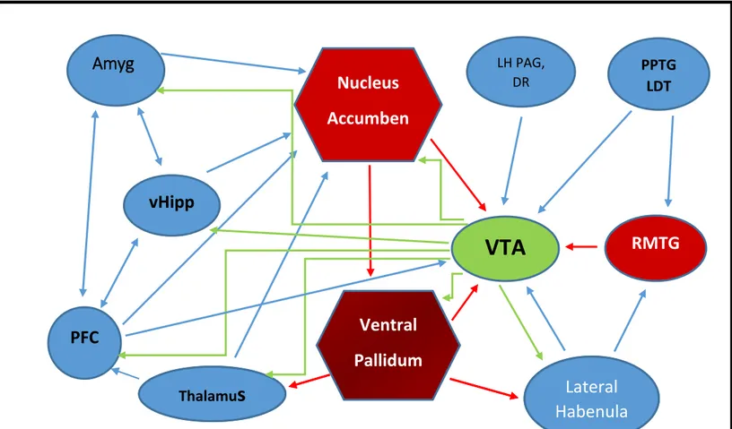
formation, processing information about emotion and drug seeking behaviour (Britt and Bonci 2013;Russo and Nestler 2013). The dopaminergic projections of the VTA are shown in green in Figure 2.
The dopaminergic neurons of the VTA send out projections to different brain regions and often these regions send back projections, thus connecting reciprocally and forming loops (Watabe-Uchida et al. 2012). The concerted activity of such projections provide a positive feedback mechanism that make the VTA a control centre for certain behaviours. For example, psychostimulant induced DA release from VTA DA neurons cause an increase of NT
expression in the nAcb shell on neurons that selectively project back to the VTA (Geisler and Zahm 2006). Since NT increase the excitability of DA neurons (Binder et al. 2001a)(discussed in chapter 2), the enhanced release of NT in the VTA, causes DA release in the nAcb which results in enhanced NT release in the VTA. This leads to a positive feedback mechanism, which may contribute to facilitate or lock in neuroadaptive changes associated with psychostimulant drug addiction (Geisler and Wise 2008).
The glutamatergic projections to the VTA come from the PFC, thalamus, amygdala, lateral habenula, hypothalamus, pedunculopontine and laterodorsal tegmentum (PPTG/LDT) and the dorsal raphe (DR). The glutamatergic projections to the VTA from these anatomical substrates are shown in blue in Figure 2. The glutamatergic afferents modulate DA cell firing and
activation of glutamatergic neurons in the efferent areas increase the rate of DA cell firing and eventually takes the DA neurons to a burst firing mode that leads to DA release and DA dependent behaviour (Geisler and Wise 2008). For example, PFC glutamatergic neurons
13
project to DA neurons, that send back efferents to the PFC. Also the PFC projections possess NT receptors at its terminals which may modulate glutamatergic neurotransmission in the VTA and this is particularly crucial to the development of sensitization induced by the psychostimulant –amphetamine (Cador et al. 1999a;Kim and Vezina 1998).
There is also evidence for peptidergic innervation of the VTA. For example, the lateral hypothalamus in addition to sending glutamatergic inputs to the VTA also send orexinergic,
and neurotensinergic projections to the VTA (Aston-Jones et al. 2010).The lateral
hypothalamus send an abundant source of peptidergic input to the VTA and is particularly relevant for its implication in reward (Tyhon et al. 2008). Recent studies show that lateral hypothalamic NTergic projections to VTA promote reward by modulating glutamatergic neurotransmission in VTA DA neurons (Kempadoo et al. 2013a) (discussed in detail in Chapter 2).
Local interneurons provide the major inhibitory inputs to DA neurons (Tan et al. 2012). The other inhibitory afferents to the VTA arise from the nAcb and the VP (Britt and Bonci 2013;Xia et al. 2011). However, recent studies suggest that the tail of the VTA or the rostromedial tegmental nucleus (RMTg) sends GABAergic projections to the VTA DA neurons and this inhibitory input serves as the “ master brake” for VTA dopaminergic pathways (Barrot et al. 2012).The inhibitory inputs to the VTA are shown in red in Figure 2.
14
Figure 2: Schematic of the principal brain regions that innervate the VTA. Green:
Dopaminergic projections; Blue: excitatory projections; Red: inhibitory projections (Modified from Britt and Bonci 2013). Abbreviations: Amyg, amygdala; vHipp, ventral hippocampus; LH, lateral hypothalamus; PAG, periaqueductal gray; DR, dorsal raphe; PPTG/LDT, pedunculopontine and
Laterodorsal tegmentum; RMTG, rostromedial tegmental nucleus.
Thalamu
s
Lateral
Habenula
Amyg
PFC
vHipp
Thalamus
Nucleus
Accumben
s
Ventral
Pallidum
VTA
LH PAG, DR PPTG LDTRMTG
Lateral
Habenula
15
1.3.2 Functions of the VTA:
Being one of the major substrates of the limbic system, the VTA is implicated in reward, working memory formation, cognition, motivation and drug addiction. The functional role of VTA depends on its afferent inputs and efferent outputs. Since majority of the VTA neuronal population is accounted for by DA neurons, most of its functions also involve these neurons.
The dopaminergic projection from the VTA to the nucleus accumbens is vital for the development of drug addiction ,sensitization and reward mechanisms (Britt and Bonci 2013) (Grueter et al., 2012; Britt et al., 2013). In an elegant study by Deisseroth and colleagues (insert citation) it was shown that optogenentic stimulation of VTA DA neurons induced intracranial self-stimulation in rats, promoted conditioned place preference to
psychostimulants whereas stimulation of GABAergic interneurons in the VTA disrupted reward and promoted conditioned place aversion(Tan et al. 2012;van et al. 2012;Witten et al. 2011). Enhanced DA cell firing in the VTA, leads to release of DA in the terminal fields and this release augments with repeated activation of DA neurons induced by the action of psychostimulants.
The dopaminergic projection to the PFC is critical for working memory formation and
learning (D'Ardenne et al. 2012). Normal cognitive functions, motivated behaviour, emotions as well as pathological manifestations for e.g. schizophrenia and ADHD (attention deficit hyperactivity disorder) have also been implicated in abnormal functioning of these projections (Lammel et al. 2008;Volkow et al. 2011). Prefrontal cortical dopamine tone is characterized by an inverted U shaped dose response curve, suggesting that too much or too little DA perturbs working memory formation and other PFC functions (Arnsten 2009;Arnsten and Li
16
2005). This suggests that functionally distinct sets of DA neurons projecting to different cortical layers are activated at either ends of the inverted U curve, thereby mediating the dual roles. It is possible for example, that DA neurons that have a role in working memory project selectively to cortical layers associated with primary sensory cortices, thereby keeping the continuum of representation of a stimulus even in its absence(Chandler et al. 2014).
On the other hand, afferents from the LHb to the VTA synapse on DA neurons that project to the medial PFC and induce aversion (Lammel et al. 2012).Thus it is possible that these DA neurons selectively project to PFC regions associated with limbic structures and not sensory structures.
Lateral hypothalamic projections to the VTA and NAcc have been associated with pain modulation. Stimulation of LH efferents (orexinergic) to the VTA can induce antinociception, thus suggesting the involvement of VTA DA neurons. Recent studies highlight a preferential role for D2 receptors in the NAcc over VTA D2 receptors in antinociception. However, a role for D1 receptors in the VTA has been attributed to antinociception suggesting the involvement of a concerted D1-D2 mechanism in antinociception (Moradi et al. 2015a;Moradi et al.
2015b).
LH VTA projections are also implicated in reward. Activation of NT neurons in the LH that project to the VTA were found to augment locomotor activity , induce prolonged dopamine efflux in the ventral striatum and transient increase in VTA NT levels. Intra VTA injections of NT antagonist, attenuated DA efflux in the NAcc, suggesting that lateral hypothalamic
afferent induced transient NT release in the VTA links LH signalling to prolonged DA release
17
Recent studies report that selective activation of VTA DA neurons rescue depression like symptoms in mice subjected to chronic mild stress (Chaudhury et al. 2013). Owing to the heterogeneity of DA neurons in the VTA, VTA functions emerges as a direct readout of functioning of distinct neuronal populations within the VTA.
18
Chapter 2: Neurotensin synthesis, receptors and effects.
An action of NT in modulating glutamatergic neurotransmission in the ventral midbrain is required for the development of amphetamine sensitization. Since NTergic innervation is dense in the VTA and VTA neurons possess NT receptors, NT may modulate DA neural activity either directly or by modulating inputs to VTA neurons. This section aims to discuss NT as a neuropeptide neurotransmitter, the distribution of NT in the midbrain, the mechanism of action of NT, effects of NT administration in the VTA, effects of NT on glutamatergic neurotransmission and the relevance of a role for NT in reward mechanisms and
schizophrenia.
2.1.1 Discovery and synthesis:
NT is a tridecapeptide that was originally isolated and sequenced from the bovine
hypothalamus in 1973(Carraway and Leeman 1973). The NT gene encodes a 170-amino acid
precursor protein that contain both the tridecapeptide NT and a closely related hexapeptide, neuromedin N (NN). In the brain, NT and NN are produced by the action of the prohormone convertase PC2(Kitabgi 2010).
In neurons, NT is stored in dense core vesicles and is released in a traditional calcium dependent manner. NT transmission is arrested by the cleavage of NT by endopeptidases.
19
2.1.2 NT as a neuropeptide neurotransmitter:
NT is an endogenous neuropeptide that serves as a neuromodulator and neurotransmitter in the central nervous system(CNS) (Vincent et al. 1999). In the CNS, NT is known for its role in reward mechanisms, pain modulation and regulation of body temperature (Kleczkowska and Lipkowski 2013) . The neuromodulating role of NT in dopaminergic and glutamatergic systems is implicated in diseases like Parkinson’s disease and schizophrenia (Binder et al. 2001a;Tanganelli et al. 2012). NT is believed to act both as a psychostimulant and a neuroleptic in the CNS as NT administration produced similar dopamine dependent
behaviours in animals that receive exposure to psychostimulants (Dobner et al. 2003;Fadel et al. 2006). On the other hand NT also increases glutamate levels in the thalamocortical system (projections from the thalamic nucleus to the prefrontal cortex) that is hypothesized to
ameliorate negative symptoms such as cognitive deficits associated with schizophrenia (Borroto-Escuela et al. 2013).
2.1.3 NT receptors:
NT has three well characterised receptors, NT receptor subtype (NTS) 1 to 3. NTS1 and 2 belong to the G protein coupled receptor family, with 7 transmembrane domain. NTS 3 belong to the single transmembrane domain receptor type 1 and is mainly located intracellularly (Mazella et al. 1998;St-Gelais et al. 2006a)
20
The possibility of the existence of a fourth NT receptor , SorLA/LR11, has been proposed, which like NTS 3 receptors belong to the single transmembrane domain receptor type 1(St-Gelais et al. 2006a).
NTS1 receptors:
NTS1 receptor was first cloned in rat and is the high affinity receptor for NT (Kd =
0.1-0.3nM). NTS1 receptor is expressed in dendrites, cell bodies and terminals in the VTA. NTS1 is the predominant receptor subtype in DA cells of the VTA and exist at presynaptic as well as postsynaptic sites to VTA neurons (Binder et al. 2001a). Functionally, NTS1 is coupled to phospholipase C (PLC), inositol triphosphate (IP3), mitogen activated protein kinases
(MAPKs) and the production of diacylglycerol (DAG). These signalling cascades are linked to an elevated level of intracellular calcium and suggest induction of excitatory effects to
depolarise the neuron (St-Gelais et al. 2006a;Trudeau 2000). NTS1 receptor activation is also linked with an enhanced formation of cyclic guanosine monophosphate (cGMP) and
production of arachidonic acid (Binder et al. 2001a;Binder et al. 2001c). The properties of NTS1 receptors in terms of its size, location, receptor type, agonists and antagonists have been summarized in Table 1.
Once NT binds to NTS1, the receptor ligand complex is internalized into neurons that express NTS1 and the NTS1 receptors reach the lyzosomes for degradation (Beaudet et al. 1994;Vandenbulcke et al. 2000). Recent studies suggest that prolonged exposure to NT
21
receptors to the cell surface and this process is mediated by NTS2 receptors(Perron et al. 2006).
NTS2 receptors:
NTS2 receptor is a low affinity NT receptors (Kd =3-10nM)(Binder et al. 2001a). Table 1
summarizes the properties of NTS2 receptors in terms of its location, size, receptor
classification, agonists and antagonists. While the NTS2 receptor share sequence homology with NTS1 receptor, these receptors functionally coupled to different downstream signalling cascades. NTS2 receptor does not stimulate cytosolic calcium mobilization or IP3
accumulation but is linked to mitogen activated protein kinases (MAPK) and are suggested to elicit inhibitory effects when cloned human NTS2 receptors are expressed on Chinese
Hamster Ovary cell lines(CHO) cell lines (Sarret et al. 2002). However, when the human NTS2 receptors were transfected in COS cells, IP3 production was reported to be constitutive. This constitutive activity was enhanced by almost 50% by an NTS1 antagonist (SR48692), not affected by NT concentrations of up to 10µM and decreased below constitutive levels by levocabastine (a histaminergic antagonist which is known to bind to NTS2 receptors, thus suggesting a weak partial inverse agonist activity. Additionally, NT, concentration
dependently reversed the effect of SR48692 back to constitutive levels, suggesting that it acts like a neutral antagonist. Therefore, whether NT acts as an agonist, inverse agonist or neutral antagonist for NTS2 receptors is undetermined (Richard et al. 2001).
In contrast to NTS1, NTS2 receptors once sequestered into the cell as NT-NTS2 complex, preferentially reaches the recycling complex and efficiently recycles back to the cell surface (Botto et al. 1998).
22
NTS3 receptors:
NTS3 receptors are also called gp95/ sortilin owing to its 100% homology with the previously cloned gp95 protein that is involved in receptor sorting (Mazella et al. 1998;Vincent et al. 1999). NTS3 receptors are located in glia, neurons and adipocytes and only 5-10% of these receptors are found on the cell surface (Mazella et al. 1998). Table 1 summarizes the
characteristics of NTS3 receptors in terms of its size, receptor classification, location, agonists and antagonists. These receptors recognize NT only after it is translocate to the plasma
membrane and acts like a scavenger protein to sequester extracellular NT. This receptor is predominantly associated with the Golgi apparatus and the endoplasmic reticulum (Mazella and Vincent 2006b) but is known to heteromize with NTS1 after translocation to the cell surface and modulate NTS1 activity in terms of activating MAPK (Sarret et al. 2003b).
23
Table 1 summarizes the characteristics of the different NT receptor subtypes, their locations, agonist and antagonists.
NT receptor subtype
Size Receptor
classification
Location Agonist Antagonists
NTS1 50-60 kDa G protein coupled- 7 transmembrane spanning regions Neurons Glia (astrocytes) • NT (Kd=0.1-0.3nM) • NN • Xenopsin • SR48692 • SR142948A NTS2 45 kDa G protein coupled- 7 transmembrane spanning regions Neurons Glia (astrocytes) • NT(Kd=3-10nM) • NN • Xenopsin • SR48692 (expressed in oocytes) • SR142948A (expressed in oocytes or CHO cells) • Levocabastine (expressed in oocytes or CHO cells) • NT(express ed in oocytes or CHO cells) • SR142948A • Levocabasti ne
NTS3 100 kDa Type I amino acid receptor single transmembrane spanning region Neurons Glia (astrocytes) Intracellular vesicles containing GluT4 glucose transporter NT triggers insertion of the receptor into the membrane. • NT (Kd=0.10.3nM) • Receptor associated protein(40kDa endoplasmic reticulum associated protein) • Cleaved sortilin propeptide Uncleaved sortilin propeptide functional antagonist blocks agonist binding until cleaved from receptor.
24
2.1.4 NT analogs:
Neurotensin analogs are derived from cleavage of the native NT peptide and since 1975 when Carraway and Leeman(Carraway and Leeman 1973) confirmed the importance of the carboxy terminal domain in conferring the biological activity and binding of NT, the first few analogs that were synthesized were variants of the C-terminal domain. Interestingly, NT analogs respond differently in the same anatomical substrates or the same analog behaves differently in various regions of the brain (Sotty et al. 2000a). Since, the binding affinity of these analogs to NT receptors have different orders of potency as does it depend on the dose of the analog used, the physiological effects that translate from these binding events also vary. In the present study, three NT analogs have been used to evaluate their effects on glutamatergic transmission in DA and non-DA neurons of the VTA. NT1-13(the native peptide), NT8-13 (the C terminal hexapeptide) and D-Tyr [11] NT1-13 (the native peptide substituted at the 11 th residue by a D-tyrosine which renders the peptide more resistant to cleavage by endopeptidases). The following section aims to describe the properties of each of these analogs, and the similarity and differences in the effects mediated by them.
NT 1-13 and NT8-13: NT1-13 is the 13 amino acid, native neurotensin peptide which is endogenous in the brain, mostly found in the dopamine rich regions (for example the VTA) and most effects mediated by this peptide are through its C terminal region. When applied exogenously, NT1-13 fails to cross the blood brain barrier and is not resistant to peptide
degradation. AT the rat NTS 1 receptor, NT1-13 has a Kd of 1.97nM while NT 8-13 has a Kd
25
receptors. However, at the NTS 2 receptors, NT1-13 has a similar potency as that of NT8-13(Kitabgi et al. 1980a;Labbe-Jullie et al. 1994).
NT1-13 when locally applied increases dopamine cell firing in the VTA (Seutin et al.
1989;Shi and Bunney 1991b), stimulates dopamine metabolism and consequentially dopamine release in the terminal fields of DAergic projections (Cador et al. 1995;Kalivas and Taylor 1985a) and that these effects are similarly mimicked by NT8-13.
NT8-13 or the hexapeptide C terminal fragment produces most of the known effects of NT and therefore most peptide agonists for NT receptors are analogs of this hexapeptide. There are discrepancies on the similarity in effectiveness of NT1-13 and NT 8-13. For example, a study by Rompre et al., (Rompre and Boye 1993) suggests, both these peptides are equally effective in operant responding for brain stimulation reward paradigms. This observation is further supported by the similarity of both these peptides in terms of binding affinities and receptor activation(Kitabgi et al. 1980a). Interestingly, there are reports of differential actions of these two peptides too. For example, ventromesencephalic tegmental microinjections of NT1-13 but not NT81-13 induce conditioned place preference (an experimental paradigm that reflects sensitization)(Glimcher et al. 1984a). Another, evidence comes from the evaluation of dopamine efflux using electrochemical methods in different brain regions upon different NT analog administration. In the rostral nucleus accumbens, the effect of NT and D-Tyr [11] NT 1-13 (another NT analog, described in detail in the following paragraphs) were found to be similar, whereas NT8-13 was less potent (Sotty et al. 2000a). In glial cells, NT 8-13 was
reported to cause an increase in both internal and external Ca2+ levels that implicated both
26
dependent on inositol triphosphate (IP3)(Trudeau 2000). Another study in the VTA of guinea pigs, revealed two types of responses, a fast and short duration and a long and slow duration inward current that were induced on application of NT. While both the responses could be induced, by NT 1-13 only the fast/ short inward current could be induced by NT8-13. Additionally, NT was found to be more potent in reducing the DA induced inhibition in the VTA (Nalivaiko et al. 1998a). Therefore, although a general consideration might be made that NT8-13 is more or equally potent compared to NT in exerting its effects, there are studies that report otherwise, which suggests, that there are multiple receptor types that are activated by each of these peptides and that this activation is concentration sensitive. In the present studies, NT1-13 and NT8-13 have been used to test their effectiveness in modulating glutamatergic responses in VTA neurons and NT antagonists have been used in identifying the receptors involved in mediating these responses.
D-Tyr [11]NT1-13 is a neurotensin analog that has the 13 amino acids of the peptide intact
but the 11th position is substituted by a D-tyrosine residue that makes it more resistant to
cleavage by endopeptidases(Checler et al. 1983a). In fact, after an intracerebroventricular injection of NT, 98% of the NT was cleared and degraded in brain tissues during a 30 min period after the injection. Under the same conditions,33% of D-Tyr [11]NT1-13 was retained, thereby suggesting a half-life 1.5 times greater than that of NT(Checler et al. 1983a). D-Tyr [11]NT1-13 is known to stimulate DA release in vitro and in vivo , because of the close interplay between interacting DA and NTergic systems majorly in the limbic system(Steinberg et al. 1995). This peptide is known to sensitize to the locomotor effects of amphetamine
sensitization, when injected in the VTA and has similarity in effectiveness when compared to NT but has a greater potency, thus possibly explaining its property of being resistance to
27
peptidase degradation(Rompre 1997a). A similar sensitization effect to cocaine, possibly mediated by NMDA receptors in the VTA has also been reported(Rompre and Bauco 2006a). In a more recent study, a role for D-Tyr [11]NT1-13 in induction of both context dependent and independent , amphetamine sensitization has been elucidated(Rouibi and Rompre 2014).In comparison to NT and NT8-13, D-Tyr [11]NT1-13 has a more stable metabolic profile and that in the presence of thiorphan, a peptidase inhibitor D-Tyr [11]NT1-13 induces larger locomotor effects caused by elevated levels of extracellular dopamine concentrations, than NT alone(Steinberg et al. 1995) . However, it is important to note that thiorphan by itself increases extracellular dopamine concentration, and therefore the evaluation of effects metabolically unstable NT analogs, in terms of increasing dopamine concentration is not possible (Labbe-Jullie et al. 1994). However, the failure of D-Tyr [11] NT1-13 in stimulating dopamine release in the PFC when injected in the VTA cannot be explained by the
metabolically stable profile whereas both NT8-13 and NT in similar range of concentrations have been effective in causing dopamine release. Interestingly, in the same study, differential effects of D-Tyr [11] NT1-13 are reported in the rostral and caudal aspects of the nucleus accumbens. For example, in the caudal aspects, NT8-13 and NT were more potent than D-Tyr [11] NT1-13, whereas, in the rostral aspects NT8-13 was less potent than D-Tyr [11] NT1-13 and NT. Therefore the effects of these three peptides in limbic regions specially seem to be varied and dependent on (i) the concentration in which it is applied, (ii) the binding affinity of NT analogues at multiple NT receptors and their consequent activation (Sotty et al. 2000a).
28
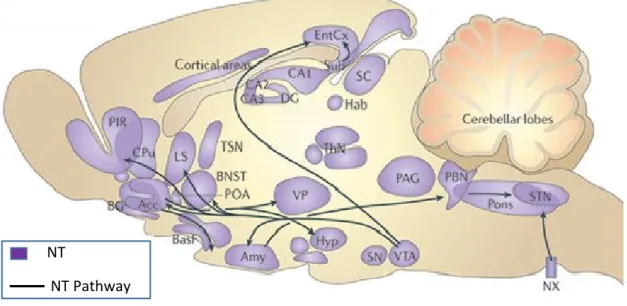
2.2 NT in the Midbrain:
In the VTA NT colocalizes with TH and these mixed NT/DA neurons project to the PFC, Entorhinal cortex(EC), nAcb, BLA and lateral septum (LS) (Fallon 1988). The incoming NT afferents to the VTA do not colocalize TH suggesting their non-dopaminergic origin. In rats, these fibers were reported to originate from the bed nucleus of the stria terminalis (BNST), lateral hypothalamus (LH), LS and the preoptic area (Zahm et al. 2001). Neurotensinergic efferents from the VTA project to the EC, amygdala, nAcb, piriform cortices, LS and pre-optic area. Figure 3 shows the neurotensinergic projections and NT rich regions of the rat brain. Evidence for local direct synaptic connections between NT axon terminals from adjacent brain regions and TH positive cells and dendrites within the VTA, indicate a possible presynaptic mode of action of NT owing to the vast majority of NT positive terminals as evidenced by electron microscopic autoradiography (Woulfe and Beaudet 1992).
Eighty to ninety percent of midbrain NT receptors are located on DA neurons of the VTA and these are predominantly NTS1 receptors. The remaining NT receptors are found on projection neurons (glutamatergic or GABAergic), non-Dopaminergic axon terminals and glial cells (Fassio et al. 2000;Nicot et al. 1994;Szigethy and Beaudet 1989). There are also reports of NTS2 mRNA in the midbrain, however their exact cellular location has not been verified (Lepee-Lorgeoux et al. 1999;Walker et al. 1998).
29
Figure 3: NT distribution and NTergic projections of the rat brain. Acc, accumbens nucleus; Amy, amygdala; BasF, basal forebrain; BG, basal ganglia; BNST, bed nucleus of the stria terminalis; CA1, hippocampal field CA1; CPu, caudate putamen; DG, dentate gyrus; EntCx, entorhinal cortex; Hab, medial habenula; Hyp, hypothalamic nucleus; LS, lateral septum; MPA, medial proptic area; NX, dorsal motor nucleus of the vagus; PAG, periaqueductal grey; PBN, parabrachial nucleus; PIR, piriform area; POA, preoptic area; Sub, subiculum; SN, substantia nigra; STN, solitary tract nucleus; ThN, thalamic nucleus; VP, ventral pallidum; VTA, ventral tegmental area. Adapted from (Griebel and Holsboer 2012).
NT
30
2.3 Modulation of DA neurotransmission by NT:
The localization of NT receptors on DA neurons in the VTA raises the possibility of a
functional interaction between these two neurotransmitter systems (Binder et al. 2001a). Once NT binds to NT receptors on DA cells, an NT –NTS complex is formed and NT receptors are activated. The section below describes these events and their consequent effect on DA cells.
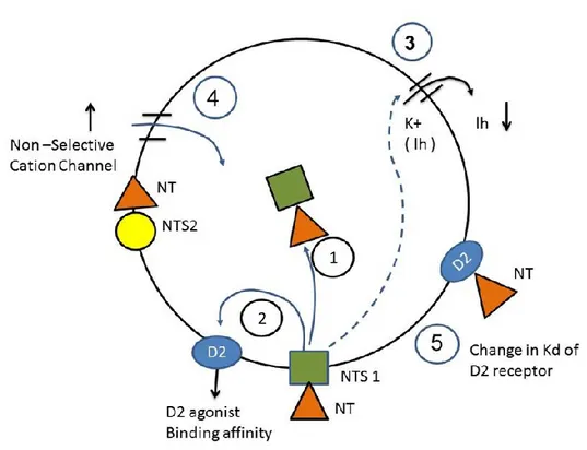
1. Effects of NT-NTS complex formation
Once NT binds to NT receptors, the NT-NTS complex is internalized and depending on the receptor type that was activated, the receptor is either recycled to the cell surface or degraded in the lysosome (Mazella et al. 1998;Mazella and Vincent 2006b). The internalized NT ligand, eventually moves to surround the nucleus and has been reported to increase TH gene
expression in DA neurons (Burgevin et al. 1992). Step1 of Figure 4 shows the formation of the NT-NTS complex.
The NT-NTS complex decreases the agonist binding affinity of D2 dopamine receptors for DA
and DA agonists as shown in Step2 of Figure 4. This leads to a decrease in DA autoinhibition
(binding of DA to D2 receptors increase K+ conductance and hyperpolarise the neuron) and
shifts the activity of post synaptic cells to D1(dopamine receptor subtype 1 , that depolarises the neuron and increase firing) mediated transmission. The mechanism of this decrease is a concerted effect of direct receptor interactions between NTS1 and D2; and activation of second messenger pathways. At the receptor level, NT via allosteric receptor/receptor interactions and second messenger dependent pathways decreases the dissociation constant
31
that the dynamics of the receptors are altered, leaving the density of functional receptors unaltered (Fuxe et al. 1992;Tanganelli et al. 1993). Although, NTS1, has been the only well characterised receptor known to modulate D2 function, the potency of NT analogs in
decreasing D2 receptor agonist binding affinity is incongruent with their binding affinities for
NTS1. NT is more potent than NT8-13 in decreasing agonist binding affinity at D2 receptors,
however the binding affinity at NTS1 is higher for NT8-13 than NT. This indicates the
possible involvement of another NTR, in addition to NTS1 in mediating this effect (Kitabgi et al. 1980a;Li et al. 1993a;Li et al. 1993b).
2. Effects of NT receptor activation
Activation of NTS1 by intracerebroventricular (i.c.v) injections of NT depolarised and increased the firing rate of midbrain DA neurons as evidenced from extracellular singe unit field recordings. This depolarisation and increase in firing rate culminate in an increase in the number of active DA neurons (Kobayashi et al. 1977;Pinnock 1985;Shi and Bunney 1991a;Shi and Bunney 1991c).
NT-NTS1 binding decreases the binding affinity of DA agonists for D2 receptors (Figure 4 step1), thus removing the inhibitory effect of D2 receptors that consequentially led to DA cell firing. Using patch clamp recordings of rat midbrain DA neurons that were identified with TH
immunohistochemistry it was reported that NT and D2 receptors oppositely regulate the same
K+ conductance. While NT decrease the K+ conductance resulting in depolarisation and
enhance the firing rate of the neuron, D2 receptors enhance it to hyperpolarize the neuron
32
channel conductance, as in extracellular recordings of firing of DA neurons, NT attenuated the
inhibitory effect of the D2R agonist, at concentrations that were insufficient to promote
augmentation in firing rates (Werkman et al. 2000a). NT induced attenuation of D2 inhibition is not a result of antagonizing general excitation as glutamate (neurotransmitter that increases DA cell firing) failed to mimic this effect (Shi and Bunney 1990).
The cell depolarisation induced in step 2 leads to a two component inward current. While the first component is a fast excitation, the second component is a slow excitation. The fast component comprised an increase in the non-selective cationic conductance mediated by the
activation of Gαq and Gα11 (G protein subtypes) and IP3 and involved NTS2 as shown in Step4
of Figure 4. The slow component is comprised of a decrease in an inwardly rectifying K+
channel conductance (Ih) and involves PKC activation and activation of NTS1 (Cathala and
Paupardin-Tritsch 1997;Chien et al. 1996;Farkas et al. 1996;Nalivaiko et al. 1998a). This
reflects as a decrease in Ih current. Step 3 of figure 4 shows the decrease of Ih. Additionally,
only NT8-13 was able to induce the fast response, however NT8-13 has a higher affinity for NTS1 than NTS2 (Kitabgi et al. 1980a). The NT initiated inward current was blocked by the
NTS1 antagonist SR48692, was equally permeable to Na+ and K+ ions and blocked externally
by Ca2+ and Mg2+ ions. (Farkas et al. 1996;Nalivaiko et al. 1998a)
As described earlier, it is important to note that most of the work on the mechanism of NT action on DA cells have focussed on NTS1, however in the VTA NTS2 exist on the cell body, dendrites of DA neurons and terminals from afferent areas, and therefore NTS2 receptor activation will also have consequences on DA cell activity (Jennes et al. 1982a;Woulfe et al. 1992;Woulfe and Beaudet 1992). However little is known about the effects of NTS2 receptor
33
activation in neurons. In other expression systems activation of NTS2 have resulted in inhibitory effects (Sarret et al. 2002).For details see section on NTS2.
Figure 4 is a schematic that shows the effects of NT on DA cells as described in the section above.
Figure 4: Mechanism of action of NT on DA cells. 2: once NT binds to the NT1receptor, the
NT-NT1 complex is rapidly internalized. 1: the NT-NT1complex decreases the agonist binding
affinity of the DA D2receptor. 3: binding of NT to NT1 decreases Ih. 4: NT binding to the
NTR increases the conductance of a nonselective cation channel, transduced by activation of
Gαq and/or Gα-11 G-protein subtypes and IP3. 5: NT interacts with the extracellular portion of
2