Contrasting roles for parvalbumin-expressing inhibitory
neurons in two forms of adult visual cortical plasticity
The MIT Faculty has made this article openly available. Please share
how this access benefits you. Your story matters.
Citation
Kaplan, Eitan S, Sam F Cooke, Robert W Komorowski, Alexander A
Chubykin, Aurore Thomazeau, Lena A Khibnik, Jeffrey P Gavornik,
and Mark F Bear. “Contrasting Roles for Parvalbumin-Expressing
Inhibitory Neurons in Two Forms of Adult Visual Cortical Plasticity.”
eLife 5 (March 4, 2016).
As Published
http://dx.doi.org/10.7554/eLife.11450
Publisher
eLife Sciences Publications, Ltd.
Version
Final published version
Citable link
http://hdl.handle.net/1721.1/102620
Terms of Use
Creative Commons Attribution
*For correspondence: mbear@ mit.edu
†These authors contributed
equally to this work Competing interests: The authors declare that no competing interests exist. Funding:See page 24
Received: 08 September 2015 Accepted: 03 February 2016 Published: 04 March 2016 Reviewing editor: Thomas D Mrsic-Flogel, University of Basel, Switzerland
Copyright Kaplan et al. This article is distributed under the terms of theCreative Commons Attribution License,which permits unrestricted use and redistribution provided that the original author and source are credited.
Contrasting roles for
parvalbumin-expressing inhibitory neurons in two
forms of adult visual cortical plasticity
Eitan S Kaplan
1†, Sam F Cooke
1†, Robert W Komorowski
1,
Alexander A Chubykin
2, Aurore Thomazeau
1, Lena A Khibnik
3,
Jeffrey P Gavornik
4, Mark F Bear
1*
1
Picower Institute for Learning and Memory, Massachusetts Institute of Technology,
Cambridge, United States;
2Department of Biological Sciences, Purdue University,
West Lafayette, United States;
3Department of Neurology, Sanford Health, Fargo,
United States;
4Department of Biology, Boston University, Boston, United States
Abstract
The roles played by cortical inhibitory neurons in experience-dependent plasticity are not well understood. Here we evaluate the participation of parvalbumin-expressing (PV+)GABAergic neurons in two forms of experience-dependent modification of primary visual cortex (V1) in adult mice: ocular dominance (OD) plasticity resulting from monocular deprivation and stimulus-selective response potentiation (SRP) resulting from enriched visual experience. These two forms of plasticity are triggered by different events but lead to a similar increase in visual cortical response. Both also require the NMDA class of glutamate receptor (NMDAR). However, we find that PV+ inhibitory neurons in V1 play a critical role in the expression of SRP and its behavioral correlate of familiarity recognition, but not in the expression of OD plasticity. Furthermore, NMDARs expressed within PV+ cells, reversibly inhibited by the psychotomimetic drug ketamine, play a critical role in SRP, but not in the induction or expression of adult OD plasticity.
DOI: 10.7554/eLife.11450.001
Introduction
Understanding how brain synapses, cells, and circuits are persistently modified by experience to store information represents one of the great challenges in neuroscience. Mouse visual cortex has proven to be an excellent model system in which to examine experience-dependent neural response modification. One robust type of visual response plasticity is reliably elicited in adult (> P60) mice by simply closing one eyelid. Over the course of 5–7 days of monocular deprivation (MD), the responses in visual cortex evoked by stimulation of the non-deprived eye progressively increase (Sawtell et al., 2003;Sato and Stryker, 2008). This deprivation-enabled response potentiation is driven by visual experience through the non-deprived eye, as only the responses through one eye are potentiated (i.
e., it is input specific) and it fails to occur if both eyelids are closed or if the animals are kept in a
dark room (Blais et al., 2008). There is evidence that the response potentiation is mediated in part by “Hebbian” strengthening of excitatory synaptic transmission in visual cortex, as induction requires cortical NMDA receptor (NMDAR) activation (Sawtell et al., 2003) (Sato and Stryker, 2008) and a-calcium/calmodulin-dependent protein kinase II (aCAMKII) expression in principal cells (Ranson et al., 2012). This form of ocular dominance (OD) plasticity is likely responsible for the increase in visual acuity that occurs through the non-deprived eye following adult monocular depri-vation (Iny et al., 2006), and is of particular interest in the context of recovery of brain function after deprivation, disease, or damage (Cho and Bear, 2010).
Another robust form of visual response plasticity is induced by exposure of awake mice to ori-ented visual grating stimuli. Brief daily presentation of a phase-reversing grating of a single orienta-tion causes a large and persistent increase in the peak cortical response to this orientaorienta-tion, as measured by visual evoked potentials (VEPs) or unit recordings (Frenkel et al., 2006;Cooke et al., 2015). This phenomenon is termed stimulus-selective response potentiation (SRP) because only responses to the experienced orientation are increased. Abundant evidence suggests that SRP is also mediated by “Hebbian” mechanisms, particularly those revealed by the study of long-term syn-aptic potentiation (Cooke and Bear, 2010;2014). The mechanisms utilized for SRP within V1 have also been shown to mediate a fundamental form of long-term visual recognition memory, manifested behaviorally as orientation-selective habituation (OSH) (Cooke and Bear, 2015;Cooke et al., 2015). Both monocular deprivation and selective visual experience trigger input-specific increases in the short latency VEP measured in layer 4 of mouse visual cortex and, as reviewed above, both OD plas-ticity and SRP share some molecular requirements (e.g., NMDAR activation). Thus, it came as a sur-prise that response potentiation after monocular deprivation and SRP do not occlude one another (Frenkel and Bear, 2004), suggesting that they employ different mechanisms or are expressed by different synapses. We became interested in the possibility of differential involvement of cortical inhibition mediated by the parvalbumin-expressing (PV+) fast spiking neurons. PV+ fast-spiking inhibitory neurons comprise the most numerous sub-class of GABAergic cortical neurons (Xu et al., 2010) and receive substantial feed-forward glutamatergic input from the thalamus
eLife digest
What we see or fail to see through our eyes leaves a lasting impression bychanging the strength of connections between neurons in a part of the brain called the visual cortex. These changes are referred to as synaptic plasticity.
One example of synaptic plasticity results in the visual cortex becoming more responsive to the stimulation of one eye when the other eye is patched for about a week. This phenomenon is known as “ocular dominance plasticity”. Another example is the increase in responsiveness that occurs in the visual cortex when animals repeatedly view stripes of a single orientation. This phenomenon is known as “stimulus-selective response potentiation”. Some previous studies had suggested that both kinds of plasticity might be induced in the same way. However, both forms of plasticity can happen at the same time, suggesting that distinct mechanisms may be involved.
To tease out how these two kinds of plasticity work, Kaplan, Cooke et al. inactivated one particular type of neuron that is thought to be involved in triggering ocular dominance plasticity and is found in the visual cortex. These inhibitory neurons produce a molecular marker called
parvalbumin and are therefore referred to as “parvalbumin-expressing neurons”.
The experiments showed that ocular dominance plasticity could still be seen in adult mice when these parvalbumin-expressing neurons were inactivated. However, when these same neurons were inactivated, the visual cortex no longer responded differently to lines with familiar or new
orientations. This was the case even when the mice had seen lines in a given orientation for long periods of time. Similarly, observations of the behavior of the mice also showed that their ability to distinguish new from familiar stimuli was lost when the parvalbumin-expressing neurons were inactivated locally within part of the visual cortex.
The connections between neurons that bring information from the eyes to the visual cortex release a chemical neurotransmitter called glutamate. One important protein that detects
glutamate, called an NMDA receptor, is required for ocular dominance plasticity. Further
experiments showed that NMDA receptors within the parvalbumin-expressing neurons are needed for stimulus-selective response potentiation, but not for ocular dominance plasticity. This indicates that although the two forms of plasticity may share molecular requirements, different connections and cells types are likely involved.
Changes in parvalbumin-expressing neurons and NMDA receptors have been implicated in disorders such as autism and schizophrenia. To improve treatments for these disorders, it is crucially important to better understand the links between these neurons and the retrieval of memories.
(Cruikshank et al., 2007). These interneurons synapse preferentially on the somata and initial axon segments of principal cells, and therefore are in a position to strongly and precisely modulate even short latency visually evoked responses (Cristo et al., 2004; Markram et al., 2004; Kepecs and Fishell, 2014). Moreover, previous studies have suggested that fast-spiking interneurons participate in the expression of cortical eye dominance and OD plasticity in juvenile mice ( Yazaki-Sugiyama et al., 2009;Smith and Bear, 2010). The contribution of this class of neuron to cortical plasticity is also of particular interest given the evidence for cortical PV+ neuron dysfunction in vari-ous psychiatric disorders characterized by impaired cognitive function (Gogolla et al., 2009).
In the current study we used a variety of approaches to understand the contribution of PV+ neu-rons to adult OD plasticity and SRP. We find that neither the pharmacogenetic silencing of PV+ cells nor the specific deletion of NMDARs within them affect the relative eye dominance or the expression of deprivation-enabled potentiation of VEPs in adult mice. In contrast, and quite unexpectedly, we discovered that the expression of SRP is dependent on PV+ cell activity, and that perturbations of PV+ neuron function, most notably cell-type specific ablation of NMDARs, disrupt the expression of both SRP and its behavioral correlate, familiarity recognition.
Results
Ocular dominance is maintained in the absence of PV+ neuron activity
In order to understand the role of PV+ neurons in the expression of experience-dependent visual cortical plasticity, we selectively inactivated these cells using a Dreadd (Designer Receptors Exclu-sively Activated by Designer Drugs) pharmacogenetic system: Specifically, we expressed a re-engi-neered G-protein coupled receptor, hM4D(Gi), which is activated exclusively by an otherwise inert small molecule, clozapine-N-oxide (CNO) (Nichols and Roth, 2009). The binding of CNO to the hM4D(Gi) receptor activates intracellular Gi-mediated signaling and subsequent hyperpolarization of
the cell in which the receptors are expressed. We locally expressed hM4D(Gi) receptors in PV+ cells of binocular V1 using an adeno-associated viral vector containing a construct for Cre-selective expression (AAV9-hSyn-DIO-HA-hM4D(Gi)-IRES-mCitrine) in mice that express Cre recombinase only in PV+ cells (B6;129P2-Pvalbtm1(cre)Arbr/J, PV-Cre). We confirmed the selective expression of hM4D (Gi) receptors in PV+ neurons in binocular V1 using immunohistochemistry (Figure 1A–E). In order to ensure that PV+ neurons could be inactivated by this method, we took slices of V1 for ex vivo intracellular recordings. Bath application of CNO drastically inhibited current evoked action potential firing of PV+ neurons in Layer 4 (2-way repeated measures ANOVA, interaction of CNO x current injection, F(8,72) = 6.227, P < 0.001, n = 10 cells, significant at data points above 100pA: q(8) =
3.716, P = 0.014) but had no effect on neighboring non-expressing cells (Figure 1F–G). Layer 4 recordings in vivo demonstrated that systemic administration of CNO resulted in the elevation of visually-evoked potential (VEP) magnitude, and an increase in the firing rate of excitatory single units (Figure 1H–I), which are expected consequences of inactivating PV+ inhibitory neurons in binocular V1.
Mouse binocular V1 is ~2–3 times more strongly activated by the contralateral (contra) eye than the ipsilateral (ipsi) eye, an inherent bias known as ocular dominance (OD). We probed the involve-ment of PV+ neuron activity in maintaining baseline OD. PV-Cre mice were infected with AAV virus to deliver hM4D(Gi) Dreadd receptors to PV+ neurons in binocular V1 (Figure 2A). Electrodes were simultaneously implanted into layer 4 of V1 for VEP recordings. VEPs elicited through just contralat-eral or ipsilatcontralat-eral eyes were acquired consecutively before and after CNO administration (Figure 2A–B). After CNO injection, VEPs driven through each eye increased significantly in magni-tude (2-way repeated measures ANOVA, effect of CNO, F(7)= 75.986, P < 0.001; contralateral eye:
222.18 ± 14.79 mV baseline vs. 679.81 ± 64.12 mV CNO, Student-Newman-Keuls (SNK) post hoc test, q(7)= 13.925, n = 8 mice, P < 0.001; ipsilateral eye: 105.56 ± 6.78 mV baseline vs. 358.63 ± 35.79 mV
CNO, SNK post hoc test, q(7)= 7.711, n = 8 mice, P < 0.001, (Figure 2C), consistent with the
occur-rence of cortical disinhibition. However, the OD of visual responses in V1 (contra/ipsi ratio) was not significantly altered by CNO injection (2.13 ± 0.12 baseline vs. 1.96 ± 0.18 CNO, student’s paired two-tailed t-test, t(7)= 0.859, n = 8 mice, P = 0.42,Figure 2D). This result suggests that the inherent
Figure 1. The hM4D(Gi) DREADD system locally inactivates parvalbumin+ neurons in binocular primary visual cortex (V1). (A) An example of V1 expression of hM4D(Gi) in a parvalbumin (PV)-Cre recombinase (Cre) mouse infected locally in binocular V1 with AAV9-hSyn-DIO-hM4D(Gi)-mCitrine. (B) A DAPI stain for cell nuclei is shown in blue. (C) Infected cells expressing hM4D(Gi) are labeled in green. (D) Immuno-labeled PV+ cells are shown in red. (E) The merged image reveals that hM4D(Gi)-expressing cells are also PV+. (F) Intracellular current clamp recordings of hM4D(Gi)-infected PV+ layer 4 neurons in ex vivo slices of V1 reveal that green-labeled infected cells exhibit a non-adapting fast-spiking phenotype typical of fast-spiking inhibitory neurons (black). These cells do not fire action potentials in the presence of CNO (red), the exogenous ligand for hM4D(Gi) receptors, despite depolarizing current injection. In contrast, neighboring cells that are not mCitrine+ show no impact of CNO application. (G) HM4D(Gi)-mediated inactivation of putative fast-spiking PV+ inhibitory neurons is here summarized as the number of action potentials resulting from a given current injection before (black) and after CNO application (red). (H) The effects of hM4D(Gi)-mediated inactivation of putative PV+ fast-spiking inhibitory neurons in vivo are apparent from electrophysiological recordings from V1 of awake, head-fixed mice viewing phase-reversing sinusoidal grating stimuli. Averaged Visually Evoked Potential (VEP) recordings recorded in layer 4 reveal increased VEP magnitude in the presence of CNO (red), relative to pre-CNO (baseline) recordings (black), indicative of reduced inhibition. (I) Phase reversal-evoked action potentials recorded from neurons in layer 4 also exhibit a similar effect with elevated firing rates in the presence of CNO (red) relative to the baseline recording (black). Labeled scale bars are presented throughout. Error bars are standard error of the mean (S.E.M.).
DOI: 10.7554/eLife.11450.003
The following source data is available for figure 1:
Source data 1. Action potential number for current injection.
Expression of adult ocular dominance (OD) plasticity does not require
the activity of PV+ neurons
The normal OD ratio is altered in the adult mouse by MD of the contralateral eye over 7 days, result-ing in an ocular dominance shift that features potentiation of response through the open ipsilateral eye (Sawtell et al., 2003;Sato and Stryker, 2008). In a new group of mice we assayed the effect of PV+ neuron inactivation on the expression of the shift in OD caused by MD. We recorded VEPs eli-cited through each eye from PV-Cre mice expressing hM4D(Gi) receptors in binocular V1 (Figure 2E). After baseline recordings the mice underwent contralateral eyelid suture and 7 days of monocular deprivation. Subsequently, the contralateral (deprived) eye was opened and VEPs were re-recorded in order to reveal the expression of an OD shift. There was a significant potentiation of VEP magnitude driven through the ipsilateral eye following 7 days of MD (69.5 ± 5.81 mV pre MD vs. 139.5 ± 9.33 mV post MD, n = 10 mice, student’s paired one-tailed t-test, t(9)= -10.184, P < 0.001,
Figure 2F). This potentiation of response through the ipsilateral (non-deprived) eye resulted in a sig-nificant shift in the OD ratio (contra/ipsi ratio, 3.08 ± 0.26 pre MD vs. 1.33 ± 0.10 post MD, 1-way repeated measures ANOVA, SNK post hoc test, q(2)= 11.77, P < 0.001,Figure 2G). Mice were then
injected with CNO and re-recorded to assess whether the OD shift would persist in the absence of PV+ neuron activity. CNO injection resulted in an increase in VEP magnitudes (Figure 2F), but importantly the shift in the contra/ipsi ratio was maintained (1.33 ± 0.10 post MD vs. 1.35 ± 0.18 post MD CNO, SNK post hoc test, q(2)= 0.127, P = 0.93,Figure 2G). Therefore, the expression of
the adult OD shift induced by 7 days of MD persists after a strong reduction in PV+ neuron inhibition.
Inactivation of PV+ neurons disrupts expression of stimulus-selective
response potentiation (SRP)
We next assayed the role of PV+ neurons in the expression mechanism underlying stimulus-selective response potentiation (SRP), a second form of experience-dependent visual cortical plasticity that also manifests as an increase in V1 responses (Frenkel et al., 2006;Cooke and Bear, 2010). Binocu-lar VEPs were recorded from awake, head-fixed adult mice viewing phase-reversing gratings of a particular orientation (Xostimulus) on each of 6 consecutive days (Figure 3A and B). On the 7th day
mice viewed blocks of the now familiar visual stimulus interleaved with blocks of a novel oriented stimulus (X +/- 60
˚
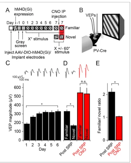
). To address whether PV+ neuron activity is required for the expression of SRP, familiar and novel oriented grating stimuli were presented before and after mice received CNO (Figure 3A).As is characteristic of SRP, there was a significant increase in the magnitude of the average VEP evoked by the familiar oriented grating stimulus over days (Friedman 1-way repeated measures ANOVA on ranks, n = 10 mice, X2(5)= 40.40, P < 0.001,Figure 3C). This potentiation was evident
by day 2 (263.3 ± 19.96 mV) in comparison with day 1 (184 ± 17.13 mV, SNK post hoc test, q(9) =
4.472, P < 0.05). The stimulus selectivity of VEP magnitude potentiation was apparent on day 7 and significantly affected by inactivating PV+ inhibitory neurons in V1 (2-way repeated measures ANOVA, interaction of treatment x stimulus, F = 78.927(1,9), P < 0.001,Figure 3D): Prior to delivery
of CNO the average VEP magnitude driven by the familiar stimulus (324.7 ± 22.18 mV) was signifi-cantly greater than that driven by a novel oriented stimulus (162.9 ± 16.31 mV, SNK post hoc test, q(9)= 10.709, P < 0.001). Following injection of CNO to inactivate hM4D(Gi)-infected neurons in V1,
there was no longer a significant difference in the magnitude of VEPs driven by the familiar stimulus (537.2 ± 66.69 mV) compared with a novel stimulus (525.1 ± 68.30 mV, SNK post hoc test, q(9) =
0.804, P = 0.58). The selectivity of the potentiation to a familiar stimulus can be summarized by plot-ting the ratio of VEP magnitudes driven by familiar and novel stimuli (Figure 3E). Mice expressed a significantly larger familiar/novel ratio before CNO injection (2.11 ± 0.17), corresponding to a greater response to the familiar stimulus, compared to after CNO injection (1.03 ± 0.04, Mann Whit-ney rank sum test, U = 0.00, P < 0.001). CNO injection did not affect the stimulus selectivity of SRP in wild type animals infected with virus (Figure 3—figure supplement 1A–B). These animals under-went SRP (Figure 3—figure supplement 1A) and on test day showed significantly larger VEPs to the familiar stimulus, both prior to CNO injection (familiar VEP: 361.06 ± 27.97 mV, novel VEP: 219.25 ± 17.9 mV, 2-way repeated measures ANOVA, n = 8 mice, SNK post hoc test, q(7)= 13.618, P < 0.001)
Figure 2. Inactivation of parvalbumin+ neurons has no impact on expression of ocular dominance (OD) or the ocular dominance shift as a result of monocular deprivation (MD) in the adult mouse. (A) For experiments described in this figure mice were infected across all cortical depths bilaterally in binocular V1 (green). During the same surgical implantation procedure VEP recording electrodes were also positioned in layer 4 and chronically fixed. (B) Mice (P45-60) were infected and implanted with electrodes and then left for 3 weeks for the AAV9 viral vector to reach maximal expression, after which they underwent habituation to head-fixation and a gray screen for two consecutive days. Following this, on experimental day 0, mice were presented with an Xophase-reversing sinusoidal grating stimulus separately to the left and right eye. VEPs were recorded from each hemisphere in
order to determine ocular dominance in binocular V1. Mice were then removed from the recording apparatus and, CNO was delivered systemically half an hour prior to undertaking the same recording procedure, this time using an orthogonal X + 90
˚
visual stimulus. (C) As is well documented, responses in V1 to stimuli viewed through the contralateral eye (blue) were greater in magnitude than those elicited through the ipsilateral eye (yellow), as measured here by VEP magnitude. After application of CNO (red outlines), VEP magnitude dramatically increased. (D) This increase was scaled such that the ratio of Contralateral:Ipsilateral VEP magnitude was maintained before (white) and after CNO (red). (E) In a second group of mice, a similar experimental protocol was observed prior to measuring ocular dominance by recording VEPs in each hemisphere elicited by an Xostimulus througheach eye. One hemisphere was then selected and the contralateral eye was sutured closed. After 7 days of monocular deprivation, the eye was opened and VEPs driven through either eye were again recorded, this time elicited by an X + 60
˚
stimulus. Mice were then systemically injected with CNO and, half an hour later, VEPs driven through either eye were recorded, this time elicited by an X - 60˚
stimulus. (F) After the adult mice underwent 7 days of MD, there was a significant potentiation of the V1 response to visual input through the ipsilateral eye (yellow). After application of CNO (red outlines), VEPs driven through each eye were elevated in magnitude, but again the increase in VEP magnitude was scaled. (G) As a result of open eyepotentiation after MD, the OD ratio shifted dramatically from the contralateral bias of pre MD (white) to an almost equal cortical response through contralateral and ipsilateral eyes (black). This shifted ratio was unaffected by hM4Di-mediated inactivation of putative fast-spiking inhibitory neurons during CNO application (red), indicating that the expression of OD and its shift as a result of MD in adult mice do not require fast-spiking inhibition. Significant comparisons are labeled with an asterisk and non-significant comparisons with n.s. throughout. Error bars are standard error of the mean (S. E.M.).
DOI: 10.7554/eLife.11450.005
The following source data is available for figure 2:
Source data 1. Ocular dominance and deprivation effects are maintained after PV neuron inactivation.
post hoc test, q(7)= 10.779, P < 0.001,Figure 3—figure supplement 1B). These results show that
the inactivation of PV+ neurons in binocular V1 disrupts the expression of SRP.
Disruption of SRP by PV+ neuronal inactivation is not due to saturation
of responses
Cortical neurons respond within a dynamic range, and it is possible that discrimination of familiar and novel stimuli would be lost as responses approach saturation. Our observation that OD is main-tained after PV+ neuron inactivation (Figure 2) indicates that V1 can still respond selectively to a strong (contralateral eye) and weak (ipsilateral eye) input. However, to address the possibility that a “ceiling effect” contributes to the disruption of SRP expression during PV+ cell inactivation, we con-ducted an additional experiment in which mice viewed sinusoidal grating stimuli across a range of contrast values (5, 10, 25, 50, 100%). VEPs were progressively greater in magnitude the greater the contrast of the viewed stimulus (2-way repeated measures ANOVA, n = 4 mice, effect of contrast, F(4,12)= 25.908, P < 0.001) both before and during PV+ neuronal activation using the hM4D(Gi)
sys-tem (SNK post hoc test, 5 vs. 100 percent contrast, baseline; q(4)= 5.178, P = 0.013; CNO; q(4)=
17.595, P < 0.001,Figure 4A). Preservation of the approximately linear relationship of contrast and response during PV+ neuron inactivation indicates that responses have not exceeded their dynamic range.
We then induced SRP to different orientations in a different set of mice, one stimulus at 50% con-trast and the other stimulus at 100% concon-trast. After 6 days of SRP at 50%, there was a modest but significant difference in VEP magnitude for familiar (188.81 ± 11.80 mV) and novel orientations (145.06 ± 8.11 mV, 2-way repeated measures ANOVA, n = 8 mice, SNK post hoc test, q(7)= 3.608, P
= 0.023,Figure 4B), just as there was for the familiar (259.81 ± 17.66 mV) and novel orientations (192.81 ± 17.27 mV, SNK post hoc test, q(7)= 3.793, P = 0.018, Figure 4C) at 100% contrast. SRP
expression was abolished by PV+ neuronal inactivation using the hM4D(Gi) system at 50% contrast as VEPs elicited by familiar (404.69 ± 59.17 mV) and novel orientations (376.94 ± 56.79 mV) were no longer significantly different (SNK post hoc test, q(7)= 2.289, P = 0.128,Figure 4B). The same was
true at 100% contrast as VEPs evoked by the familiar (532.44 ± 63.82 mV) and novel orientations (514.88 ± 40.38 mV) were also no longer significantly different (SNK post hoc test, q(7)= 0.994, P =
0.494,Figure 4C). Again, the blockade of SRP expression was also clearly observed in the familiar/ novel ratio, which dropped significantly from (1.31 ± 0.05) to (1.08 ± 0.08) with CNO application at 50% contrast (student’s paired two-tailed t-test, t(7)= 2.983, P = 0.02,Figure 4D) and from (1.40 ±
0.10) to (1.02 ± 0.05) with CNO application at 100% contrast (student’s paired two-tailed t-test, t(7)=
2.955, P = 0.021,Figure 4E). Thus, SRP could still be abolished through selective loss of PV+ neuron activity even at reduced contrasts eliciting submaximal responses. The disruption of SRP expression by inactivation of PV+ neurons is not a trivial consequence of a “ceiling effect”.
Activation of PV+ neurons also disrupts expression of SRP
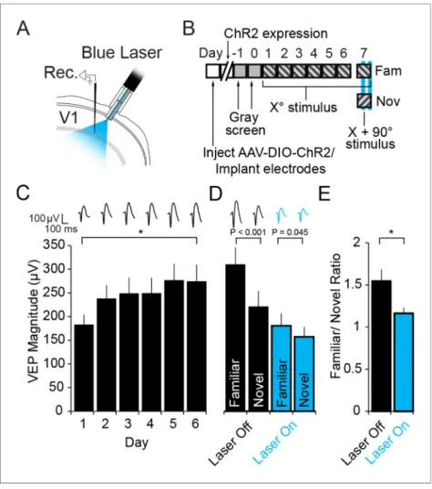
PV+ neurons have been implicated in the sharpening of orientation selectivity in V1 (Runyan et al., 2010;Adesnik et al., 2012;Atallah et al., 2012;Lee et al., 2012;Wilson et al., 2012). If orienta-tion selectivity were completely abolished by inactivaorienta-tion of PV+ neurons then the loss of SRP expression could be attributed to a failure of orientation discrimination rather than familiarity. A pre-vious experiment has indicated that activation of PV+ neurons in V1 actually mildly enhances orienta-tion-selectivity and visual discrimination (Lee et al., 2012). Therefore, we used optogenetics to activate PV+ neurons while mice were presented with familiar and novel stimuli to test whether SRP expression would be enhanced, unaffected or disrupted. PV+ neurons in binocular V1 of PV-Cre mice expressed Channel-rhodopsin 2 (ChR2) as a result of infection with AAV5-EF1a-DIO-hChR2 (H134R)-eYFP. During the same surgery VEP recording electrodes were implanted and optic fibers chronically implanted to deliver light to the recording site (Figure 5A). After stable expression 4 weeks after infection, mice were habituated to head-fixation on each of 2 days before undergoing a standard SRP experiment over 6 days (Figure 5B,C). Mice showed a significant increase in the mag-nitude of the average VEP evoked by the familiar oriented grating stimulus over days (Friedman repeated measures ANOVA on ranks, n = 11 mice, X2(5)= 24.818, P < 0.001,Figure 5C). This
poten-tiation was evident by day 2 (237.55 ± 29.01 mV) in comparison with day 1 (182.59 ± 21.97 mV, SNK post hoc test, q(10) = 7.675, P < 0.05). On day 7, mice were presented with interleaved blocks of
familiar and novel stimuli. Also interleaved were blocks of familiar and novel stimuli in which blue light (473 nm) was continuously delivered via the optic fiber to the recording site in binocular V1. VEPs elicited by the novel X + 90
˚
stimulus were significantly lower in magnitude (221.34 ± 33.55 mV) than those elicited by the familiar stimulus prior to optogenetic activation of PV+ neurons (310.70 ± Figure 3. The expression of Stimulus-selective Response Potentiation (SRP) requires activity in PV+ neurons in V1. (A) Mice expressing hM4D(Gi) receptors in PV+ cells underwent a standard SRP induction protocol. On day 7, mice viewed a novel oriented stimulus in addition to the familiar stimulus; before and after CNO injection. (B) We acquired binocular VEPs from awake, head-fixed mice elicited by the same full-field oriented sinusoidal grating stimulus over several days. (C) As a result of multiple days of experience cortical response was dramatically potentiated such that the familiar stimulus evoked VEPs of significantly greater magnitude than the novel stimulus (black bars). (D) After application of CNO, VEPs underwent a notable increase in magnitude as a result of disinhibition (red bars). Most notably, CNO rendered response to familiar and novel stimuli equivalent inmagnitude. (E) This lost discrimination of familiar and novel stimuli is reflected as a drop in the ratio of response to familiar and novel stimuli from approximately 2:1 (black) to approximately 1:1 (red). Significant comparisons are labeled with an asterisk and non-significant comparisons with n.s. throughout. Error bars are standard error of the mean (S.E.M.).
DOI: 10.7554/eLife.11450.007
The following source data and figure supplements are available for figure 3: Source data 1. PV Dreadds SRP induction and expression.
DOI: 10.7554/eLife.11450.008
Figure supplement 1. CNO has no impact on SRP expression in WT mice.
DOI: 10.7554/eLife.11450.009
Figure supplement 1—source data 1. CNO has no effect on SRP expression in WT mice.
36.70 mV, 2-way repeated measures ANOVA, n = 11 mice, SNK post hoc test, q(10)= 11.799, P <
0.001,Figure 5D). Optogenetic activation of PV+ neurons significantly diminished the selectivity of SRP, as VEPs elicited by the familiar stimulus (181.23 ± 26.43 mV) and novel stimulus (157.80 ± 21.05 mV) were more similar in magnitude (interaction of stimulus x laser, F(1,10)= 46.606, P < 0.001;
famil-iar vs. novel SNK post hoc test, q(10)= 3.094, P = 0.045,Figure 5D). The reduction of SRP selectivity
is most clearly observed in the significant difference in the familiar/novel ratio without (1.55 ± 0.14) and with optogenetic stimulation (1.16 ± 0.07, student’s paired two-tailed t-test, t(10)= 4.423, P =
0.001,Figure 5E). Laser stimulation did not affect the stimulus selectivity of SRP in wild type animals infected with virus (Figure 5—figure supplement 1), as there was no significant interaction between stimulus and laser (2-way repeated measures ANOVA, F(1,8)= 0.677, P = 0.434). These mice
under-went SRP (Figure 5—figure supplement 1A) and on test day showed significantly larger VEPs to the familiar stimulus, both with the laser off (familiar VEP: 273.69 ± 28.25 mV, novel VEP: 200.19 ± 19.53 mV, 2-way repeated measures ANOVA, n = 9 mice, SNK post hoc test, q(8)= 5.234, P = 0.005) and
the laser on (familiar VEP: 277.08 ± 29.80 mV, novel VEP: 194 ± 18.39 mV, q(8)= 5.916, P = 0.002,
Fig-ure 5—figFig-ure supplement 1B). Thus, SRP expression was disrupted with activation of PV+ neurons, just as it was with inactivation (Figure 3D–E). This result implies that PV+ neurons play a specific role in SRP expression beyond any role in enhancing orientation tuning.
Full expression of SRP requires NMDA receptors (NMDARs) expressed
in PV+ neurons
Multiple lines of evidence indicate that SRP is dependent upon the NMDA class of glutamate recep-tors (Frenkel et al., 2006;Cooke et al., 2015). Given the additional clear requirement for normal
Figure 4. Expression of SRP to two separate contrast values is blocked by hM4D(Gi)-mediated PV+ neuron inactivation, but differential response to contrast is maintained. (A) CNO delivery to PV-Cre mice that had been infected with AAV9-hSyn-DIO-hM4D(Gi)-mCitrine impacted VEP magnitude (red) significantly across a range of contrast compared to baseline (black). (B) SRP was induced to two differently oriented stimuli, each at different contrast values: 50% (gray) and 100% (black). Modest but significant SRP was expressed at 50% contrast prior to CNO application. After CNO application (red outlines), VEPs increased significantly in magnitude but were no longer significantly different for familiar and a second novel orientation. (C) SRP was also expressed for a different orientation at 100% contrast and, again, VEP magnitude was increased and SRP blocked by delivery of CNO. (D) The blockade of SRP expression by CNO at 50% contrast was apparent in the reduction in the familiar/novel ratio for VEP magnitude. (E) The blockade of SRP expression at 100% contrast was also observed as a significant drop in familiar/novel ratio of VEP magnitude after CNO delivery. Significant comparisons are marked with an asterisk throughout while non-significant comparisons are marked with n.s. Error bars are standard error of the mean (S.E.M.).
DOI: 10.7554/eLife.11450.011
The following source data is available for figure 4:
Source data 1. PV-neuron inactivation does not produce ceiling effect.
PV+ inhibitory cell function in SRP (Figure 3D,E) we selectively ablated the NMDAR from just PV+ cells by crossing the PV-Cre line of mice with a line in which the mandatory GluN1 subunit of NMDAR is excised by Cre recombinase activity (B6.129S4-Grin1tm2Stl/J, GluN1 fl/fl). The progeny of
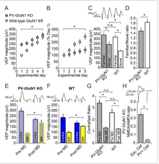
this cross, in which both alleles of Grin1 (the gene encoding GluN1) were floxed, are henceforth described as either PV-GluN1 KO or Wildtype (WT)-GluN1 fl/fl depending on whether, respectively, Cre was expressed or not. We implanted PV-GluN1 KO (n = 14) and littermate WT-GluN1 fl/fl mice (n = 17) with VEP recording electrodes in layer 4, binocular V1. After recovery and 2 daily sessions of habituation we recorded VEPs elicited by an Xooriented grating stimulus. Immediately apparent was the significantly greater basal magnitude of VEPs recorded in the PV-GluN1 mice (242.45 ± 20.20 mV) relative to their littermate controls (130.88 ± 9.69 mV, student’s two-tailed t-test, t(29)= -5.269, P
< 0.001), consistent with the occurrence of disinhibition as a result of reduced glutamatergic drive on cortical PV+ inhibitory cells (Figure 6A). We then presented the same stimulus to these mice over several consecutive days and observed a significant difference in SRP across genotypes (2-way repeated measures ANOVA, interaction of genotype x day, F(4,116)= 3.835, P = 0.006,Figure 6A).
Beyond day 3, VEP magnitudes were no longer significantly different between the PV-GluN1 KO mice (289.15 ± 24.57 mV) and WT-GluN1 fl/fl littermates (239.94 ± 20.72 mV, SNK post hoc test, q(29)
= 2.242, P = 0.119), suggesting an occlusion of SRP by the already elevated basal VEP magnitude in the PV-GluN1 KO mice. The significant deficit in SRP is clearly apparent when the data are normal-ized to day 1 values (2-way repeated measures ANOVA, interaction of genotype x day, F(4,116) =
12.326, P < 0.001,Figure 6B). Again, a significant deficit in SRP emerged by day 3 in the PV-GluN1 KO mice (119.26 ± 10.13% day 1) compared with WT-GluN1 fl/fl littermates (183.33 ± 15.83% day 1, SNK post hoc test, q(29)= 4.727, P = 0.002), demonstrating that SRP is compromised by a loss of
NMDAR expressed in PV+ neurons.
We also tested for the stimulus-selectivity of SRP expression by presenting both groups of ani-mals with interleaved blocks of the familiar Xostimulus and a novel X + 90
˚
stimulus (Figure 6C). Sig-nificant stimulus selectivity was present in both genotypes (2-way repeated measures ANOVA, stimulus, F(1) = 67.397, P < 0.001, interaction of genotype x stimulus, F(1,29) = 2.359, P = 0.135,Figure 6C): Although PV-GluN1 KO mice showed significant differences in VEP magnitude for famil-iar (340.24 ± 27.96 mV) and novel orientations (242.54 ± 24.40 mV, SNK post hoc test, q(13)= 6.372, P
< 0.001) the difference was more pronounced in the WT-GluN1 fl/fl mice (SNK post hoc test, q(16)=
10.254, P < 0.001), in which the familiar stimulus elicited VEPs 318.74 ± 25.19 mV in magnitude and the novel stimulus elicited VEPs 176.06 ± 11.55 mV in magnitude. The familiar stimulus evoked VEPs that were not significantly different in the WT-GluN1 fl/fl mice (318.74 ± 25.19 mV) and the PV-GluN1 KO mice (340.24 ± 27.96 mV, SNK post hoc test, q(29)= 0.947, P = 0.507) but those evoked in the
WT-GluN1 fl/fl by the novel stimulus (176.06 ± 11.55 mV) were significantly lower in magnitude than in the PV-GluN1 KO mice (242.54 ± 24.40 mV, SNK post hoc test, q(29)= 2.926, P = 0.045). The
signif-icant deficit in SRP expression in the PV-GluN1 KO mice is most apparent when the familiar/novel ratio of VEP magnitude (1.48 ± 0.13) is compared with the WT-GluN1 fl/fl littermates (1.83 ± 0.13, Mann Whitney rank sum test, U = 60.000, P = 0.020, Figure 6D). Thus, loss of NMDAR function selectively within PV+ neurons impairs the full expression of SRP.
Adult OD plasticity does not require NMDARs expressed in PV+
neurons
We also examined if loss of NMDAR from PV+ neurons has an effect on OD plasticity. We implanted VEP recording electrodes in a second cohort of 11 PV–GluN1 KO and 7 WT-GluN1 fl/fl mice. After recovery and habituation over 2 days, monocular VEPs were acquired through each eye. After 7 days of MD, PV-GluN1 KO mice exhibited a significant potentiation of response through the open ipsilateral eye (187.91 ± 25.52 mV) compared with day 0 (90.64 ± 10.69 mV, 2-way repeated measures ANOVA, SNK post hoc test, q(16)= 8.849, P < 0.001,Figure 6E). Similarly, WT-GluN1 fl/fl mice also
showed a significant potentiation of the open ipsilateral eye-response (154.57 ± 7.60 mV) after 7 days of MD compared with responses measured on day 0 (97.57 ± 15.85 mV, SNK post hoc test, q(16)
= 4.137, P = 0.01,Figure 6F). Comparisons of OD ratios prior to MD in the adult PV-GluN1 KO (3.40 ± 0.29) and WT-GluN1 fl/fl mice (2.57 ± 0.33) did not reveal any significant difference (2-way repeated measures ANOVA, effect of genotype, F(1)= 3.424, P = 0.083). Significant shifts in the OD
ratio occurred as a result of 7 days of MD in the adult PV-GluN1 KO (1.28 ± 0.33, SNK post hoc test, q(16)= 10.6, P < 0.001) and WT-GluN1 fl/fl mice (1.19 ± 0.09, SNK post hoc test, q(16)= 5.517, P =
Figure 5. Optogenetic stimulation of PV+ inhibitory neurons prevents SRP expression. (A) Blue light was delivered locally into V1 via optic fibers chronically implanted at a 45
˚
angle to target the VEP recording site in layer 4 of binocular V1 of PV-Cre mice infected with AAV5-EF1a-DIO-hChR2(H134R)-eYFP. (B) Experimental timeline showing that after viral infection, electrode implantation, and ChR2 expression; mice were accustomed to head-fixation and gray screen viewing. Subsequently, they underwent a standard SRP induction protocol over 6 days. On day 7, mice viewed a novel oriented stimulus in addition to the familiar stimulus and, on 50% of presentations of each stimulus, blue light (473 nm) was delivered to cortex to optogenetically activate PV+ cells. (C) Significant SRP was induced over 6 days as VEPs underwent a typical potentiation. (D) On day 7, SRP was expressed through significantly larger VEP magnitude in response to the familiar Xoorientation than a novel X + 90˚
stimulus when blue light was not delivered (Black bars). In the presence of blue light (blue bars), VEPs were suppressed, and there was a significant reduction in the differential magnitude of VEPs driven by familiar and novel stimuli. (E) The ratio of VEP magnitude elicited by familiar/novel stimuli was significantly reduced by optogenetic activation of PV+ neurons, reflecting a decrement in SRP expression. Significant comparisons are marked with an asterisk and post hoc test p values are reported in D to emphasize the impact of laser stimulation on SRP selectivity. Error bars are standard error of the mean (S.E.M.).DOI: 10.7554/eLife.11450.013
The following source data and figure supplements are available for figure 5: Source data 1. PV-neuronal activation.
DOI: 10.7554/eLife.11450.014
Figure supplement 1. Blue light has no impact on SRP expression.
DOI: 10.7554/eLife.11450.015
Figure supplement 1—source data 1. Laser does not effect VEPs in WT animals.
0.001). The shifted OD ratio also did not differ significantly across genotype (2-way repeated meas-ures ANOVA, interaction of genotype and MD, F(1,16)= 2.638, P = 0.124,Figure 6G). Thus, the loss
of NMDAR function from PV+ neurons did not have a significant impact on either the induction or the expression of adult OD plasticity, in contrast to SRP.
In order to confirm the removal of NMDARs selectively from PV+ cells, the PV-GluN1 KO mice were injected with an AAV5 vector to express GFP in a Cre-dependent fashion in PV+ cells only. After 1 month, fluorescence-guided intracellular recordings were performed from ex vivo slices of visual cortex. NMDAR-mediated synaptic transmission was normal in excitatory control cells but abolished in PV+ neurons (Figure 6H). This is expressed as significantly reduced NMDAR EPSC/ AMPAR EPSC ratio in PV+ cells (0.040 ± 0.011, n = 8 cells from 5 mice) compared to neighboring non-fluorescent excitatory control cells (0.345 ± 0.039, n = 10 cells from 4 mice, student’s one-tailed t-test, t(16)= -6.819, P < 0.001).
Acute ketamine treatment reversibly eliminates SRP expression
A group of non-competitive, open-channel NMDAR blockers, including ketamine, PCP and MK801 are known to have the paradoxical impact of increasing net neuronal activity in the brain. It is thought that this apparent disinhibition arises from the preferential impact of these molecules on fast-spiking neurons, due to the tonic activation and the increased open-time of NMDAR expressed within these cells (Homayoun and Moghaddam, 2007;Seamans, 2008). Interestingly, these com-pounds are also psychotomimetic and can reproduce, at high sub-anesthetic doses, most of the symptoms of schizophrenia (Krystal et al., 1994). Here we tested the possibility that a single acute dose of one of these substances, ketamine, would have an impact on the expression of SRP due to its action on NMDAR expressed in PV+ fast spiking inhibitory neurons.
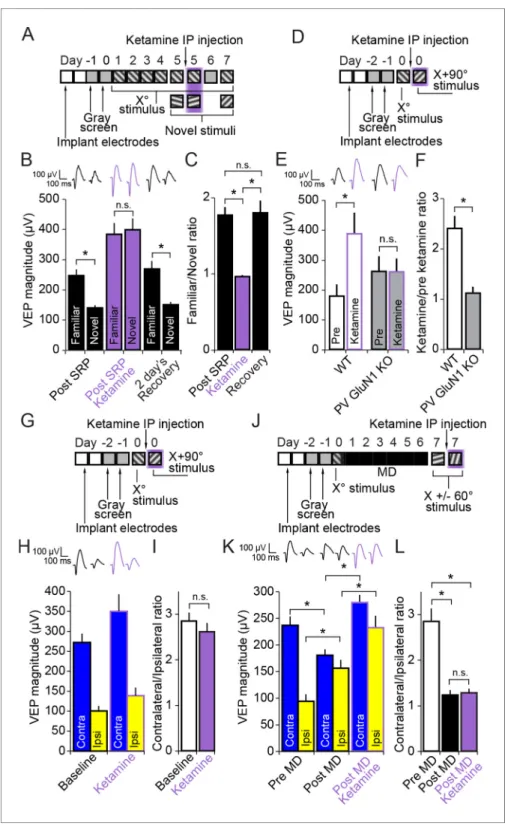
We implanted VEP recording electrodes in layer 4 of binocular V1 in a group of 10 C57BL/6 mice. After recovery and a standard SRP protocol we recorded VEP magnitudes driven by familiar and novel stimuli before, during and 2 days after recovery from systemic injection (i.p.) of a high but sub-anesthetic dose of ketamine (50 mg/kg) (Figure 7A). Ketamine had a significant effect on SRP expression (2-way repeated measures ANOVA, interaction of treatment x stimulus, F(2,18)= 29.479,
P < 0.001,Figure 7B). After SRP but prior to ketamine delivery the familiar Xostimulus elicited VEPs of significantly greater magnitude (245.47 ± 21.00 mV) than a novel X + 60
˚
stimulus (138.86 ± 9.32 mV, SNK post hoc test, q(9)= 9.228, P < 0.001) in these mice. After an hour of respite fromhead-fixa-tion, mice were injected with ketamine and, 15 min later, they were returned to head-fixation and VEP magnitudes were again recorded. Under the influence of ketamine, the familiar Xostimulus eli-cited VEPs of significantly increased magnitude (381.90 ± 37.25 mV) relative to pre ketamine (245.47 ± 21.00 mV, SNK post hoc test, q(9)= 7.223, P < 0.001). However, VEPs elicited by a second novel X
-60
˚
stimulus were increased relative to pre-ketamine by an even greater extent (397.53 ± 37.62 mV) relative to pre ketamine (138.86 ± 9.32 mV, SNK post hoc test, q(9)= 13.695, P < 0.001), such thatVEPs were no longer significantly different in magnitude in response to familiar and novel stimuli (SNK post hoc test, q(9)= 1.353, P = 0.349). After 2 day’s rest, allowing for complete recovery from
drug effects, the mice were returned to the head-fixation apparatus and exposed to the familiar Xo stimulus and a third novel stimulus, X + 90
˚
. Just as observed prior to the ketamine injection, the Xo stimulus evoked VEPs of significantly greater magnitude (267.46 ± 26.13 mV) than the novel X + 90˚
stimulus (149.40 ± 9.85 mV, q(9)= 10.219, P < 0.001) (Figure 7A and B).This significant effect of ketamine on the discrimination of familiar and novel stimuli is summarized by the ratio of VEP magnitude driven by the familiar/novel stimulus (1-way repeated measures ANOVA, F(2,18)= 24.683, P < 0.001,Figure 7C). This familiar/novel ratio dropped significantly from
1.76 ± 0.10 prior to ketamine to 0.96 ± 0.02 after ketamine (SNK post hoc test, q(9) = 8.443, P <
0.001). Upon recovery, the familiar/novel ratio significantly recovered (1.79 ± 0.16, SNK post hoc test, q(9)= 8.759, P < 0.001) and was not significantly different from the first test after SRP but prior
to ketamine injection (SNK post hoc test, q(9) = 0.316, P = 0.826). Thus, the psychotomimetic
non-competitive NMDAR antagonist ketamine disrupts SRP. The fact that this is an acute effect on already established SRP that recovers after drug washout indicates that the role for NMDAR in PV+ fast-spiking GABAergic neurons may be in memory retrieval rather than learning.
Figure 6. Loss of NMDA receptors selectively from parvalbumin+ cells impacts SRP but not adult OD plasticity. (A) VEPs recorded from mice in which the mandatory GluN1 subunit of the NMDA receptor was genetically ablated from PV+ cells using Cre recombinase technology (PV GluN1 KO, gray) were significantly greater in magnitude than those recorded from WT littermates (black), suggesting disinhibition of the visual response. In these same PV GluN1 KO mice, SRP was also significantly impacted as there is was significantly less gain in magnitude over days of repeated presentation of an X
˚
stimulus than observed in WT littermates. (B) This significant reduction in the magnitude of SRP was most clearly observed if VEP magnitude was normalized to the magnitude on day 1. (C) After SRP, both PV GluN1 KO mice and their WT littermates exhibited a significantly greater VEP magnitude elicited by the now familiar stimulus than interleaved presentations of a novel oriented stimulus. However, consistent with the observed difference in magnitude on day 1, VEPs elicited by a novel X + 90˚
stimulus in PV GluN1 KO mice were significantly greater in magnitude than those in WT littermate mice. No significant difference was observed for VEPs elicited by the familiar stimulus. (D) A significant difference in the ratio of VEP magnitude elicited by the familiar and novel stimuli reveals a deficit in SRP expression in PV GluN1 KO mice. (E) In contrast, PV GluN1 KO mice exhibited a normal adult OD shift after 7 days of MD, resulting from open eye potentiation (yellow outlines). (F) Wild-type (WT) littermates exhibited the same significant open eye potentiation (yellow bars) after 7 days of MD. (G) A comparison of the degree of OD shift as a result of 7 days of MD reveals a significant shift in OD ratio in both genotypes but no difference between genotypes, indicating that NMDA receptors in PV+ cells are not required for induction or expression of the OD shift. (H) To confirm genetic ablation of NMDARs selectively from parvalbumin (PV+) neurons in the PV-GluN1 KO; mice were injected with an AAV5 vector to express GFP in a Cre-dependent fashion in PV+ cells only. After 1 month, fluorescence-guided intracellular recordings were performed from ex vivo slices of visual cortex. NMDAR-mediated synaptic transmission was normal in excitatory control cells but abolished in PV+ cells. This is expressed here as the NMDAR EPSC/AMPAR EPSC ratio in excitatory cells (black outline) and PV+ cells (green outline). Sample EPSC traces mediated by the AMPAR (downward) and NMDAR (upward) are shown at the top of the panel. Significant comparisons are marked with an asterisk throughout while non-significant comparisons are marked with n.s. Error bars are standard error of the mean (S.E.M.).DOI: 10.7554/eLife.11450.017
The following source data is available for figure 6: Source data 1. PV-GluN1 KO
Ketamine affects V1 responses through NMDARs expressed in PV+
neurons
Ketamine blocks NMDAR expressed in all cell types throughout the CNS and is also known to have targets other than the NMDAR (Chen et al., 2009). In order to determine if ketamine has its effect on V1 responses specifically through NMDAR expressed in PV+ cells, we tested if there was a differ-ential effect of ketamine on VEP magnitude in PV-GluN1 KO mice and WT-GluN1 fl/fl mice. After a typical implantation and habituation protocol (Figure 7D) we tested VEP magnitudes elicited by a novel oriented Xostimulus in WT-GluN1 fl/fl mice (178.11 ± 38.41 mV, n = 8) and PV-GluN1 KO mice (260.79 ± 50.75 mV, n = 8) (Figure 7E). We then removed mice from head-fixation and allowed them to recover in their home-cage before delivering 50 mg/kg ketamine (i.p.). After 15 min, the mice were returned to head-fixation and we observed the impact of ketamine on VEPs elicited by a novel X + 90
˚
oriented stimulus, which was significantly different in its effect on the two genotypes (2-way repeated measures ANOVA, interaction of genotype x treatment, F(1,14)= 18.454, P < 0.001). In thecontrol WT-GluN1 fl/fl mice there was a significant potentiation of VEP magnitude as a result of keta-mine application (386.65 ± 70.75 mV) in comparison to pre treatment (178.11 ± 38.41 mV, SNK post hoc test, q(7)= 8.505, P < 0.001,Figure 7E), replicating our previous finding (Figure 7B). However,
in the PV-GluN1 KO mice, ketamine had no ostensible significant impact (258.67 ± 44.86 mV) in com-parison to pre treatment (260.79 ± 50.75 mV, SNK post hoc test, q(7)= 0.087, P = 0.952). A ratio of
VEP magnitudes pre and post ketamine treatment reveals the significant difference between ket-amine’s action on WT-GluN1 fl/fl mice (2.40 ± 0.25) and PV-GluN1 KO mice (1.12 ± 0.13, student’s two-tailed t-test, t(14) = 4.610, P < 0.001) (Figure 7F). Thus, while ketamine has a wide range of
effects in the CNS, it exerts itself on the response of V1 to visual input selectively through NMDAR expressed in PV+ cells.
Ketamine has no effect on the expression of the adult OD shift
We then tested whether or not ketamine would disrupt either the OD ratio or the shift induced by 7 days of MD in the adult mouse. Again, C57BL/6 mice (n = 8) were implanted with VEP electrodes and taken through a standard surgery recovery and habituation protocol before measuring the OD ratio prior to 50 mg/kg ketamine and 15 min after ketamine delivery (Figure 7G and H). The normal contra/ipsi OD ratio exhibiting contralateral eye dominance prior to ketamine (2.84 ± 0.18) was not significantly altered by ketamine (2.61 ± 0.19, student’s paired two-tailed t-test, t(7) = 0.871, P =
0.413) (Figure 7I). A separate group of mice (n = 11) then underwent a standard 7 day MD protocol. VEPs elicited by an Xo oriented stimulus were recorded at baseline, followed by the deprivation period. After eye opening VEP magnitudes were re-tested with a novel X + 60
˚
stimulus (the standard protocol to avoid contamination of the OD shift by SRP [Frenkel and Bear, 2004]). These mice were then tested again with a second novel X - 60˚
stimulus after 1 hr rest and an additional 15 min after systemic 50 mg/kg ketamine administration (Figure 7J). As expected, VEPs elicited through the open ipsilateral eye (93.73 ± 12.85 mV) were significantly potentiated by 7 days of MD in the adult mouse (155.73 ± 15.86 mV, 2-way repeated measures ANOVA, SNK post hoc test, n = 11 mice, q(10)= 7.102, P < 0.001), reflecting the well-documented OD shift. These same ipsilateral VEPs were then further potentiated by ketamine administration (232.03 ± 22.19 mV, SNK post hoc test, q(10)= 8.741,
P < 0.001). However, ketamine had a similar significant potentiating effect on the contralateral VEP (279.07 ± 14.53 mV), relative to pre-ketamine (180.14 ± 11.22 mV, SNK post hoc test, q(10)= 11.333,
P < 0.001,Figure 7K), suggesting a uniform scaling of response through the two eyes. This observa-tion is confirmed by the fact that the OD ratio, significantly shifted from (2.84 ± 0.29) to (1.23 ± 0.11, Friedman repeated measures ANOVA on ranks, n = 11 mice, X2(2)= 16.545, p<0.001, SNK post hoc
test, q(10)= 5.126, P < 0.05) by 7 days of MD, was not further significantly affected by delivery of
ketamine (1.28 ± 0.09, SNK post hoc test, q(10)= 0.426, P > 0.05,Figure 7L). Thus, while ketamine
prevents SRP expression through action on NMDAR expressed in PV+ cells, it does not significantly affect the adult OD shift after 7 days of MD, consistent once again with PV+ cells contributing to the expression of SRP but not adult OD plasticity.
Visual novelty detection requires PV+ neuron activity within V1
Given the clear involvement of PV+ neurons in the expression of SRP we wanted to determine if loss of PV+ neuronal function local to V1 would have any behavioral impact. Head-restrained mice
Figure 7. Ketamine prevents expression of SRP through blockade of NMDA receptors expressed in parvalbumin+ neurons but does not impact expression of the adult OD shift. (A) Mice were bilaterally implanted with VEP recording electrodes in layer 4 of binocular V1. After habituation to head-fixation and a gray screen for 2 days, SRP was induced over 4 days by repeatedly presenting sessions of an X
˚
stimulus. On day 5, SRP expression was tested by presenting interleaved blocks of the familiar X˚
stimulus and a novel X + 60˚
stimulus. In order to test the acute impact of blocking NMDA receptors on SRP expression, 50 mg/kg of ketamine was then delivered systemically 15 min before re-acquiring VEPs elicited by the familiar X˚
stimulus and interleaved presentations of a second novel X - 60˚
stimulus. Mice were then allowed 2 days recovery and a complete washout of ketamine before re-testing SRP expression by again testing VEP magnitude in response to the familiar X˚
stimulus and a third novel X + 90˚
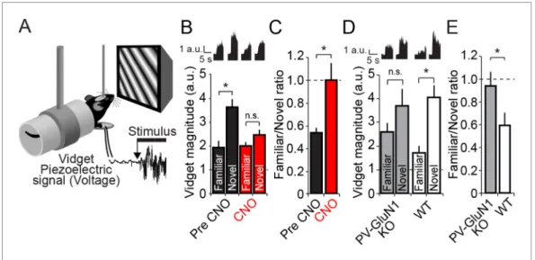
Figure 7 continued on next pageviewing a phase reversing visual grating stimulus are known to exhibit a stereotyped motor response called a visually-induced fidget, or vidget (Cooke et al., 2015). Vidgets can be measured via a piezo-electric sensor located beneath the forepaws of the mouse (Figure 8A). Importantly, it has been shown that the magnitude of the vidget response is inversely correlated to the familiarity of the visual stimulus. That is, a visual stimulus that is very familiar to the animal will on average evoke a rel-atively weak vidget behavioral response. In contrast, the presentation of a novel stimulus will on average evoke a vidget of significantly greater magnitude. Therefore, this behavioral response reflects the ability of the animal to discriminate and respond to a novel visual stimulus in its environ-ment. Importantly, genetic and pharmacological manipulations local to V1 that inhibit SRP also dis-rupt the behavioral discrimination of familiar and novel stimuli (Cooke et al., 2015), demonstrating that this differential vidget response to familiar and novel stimuli is dependent on the plasticity in visual cortex. Since PV+ neuron inactivation disrupted the expression of SRP, we tested whether this manipulation would likewise disrupt visual novelty detection.
A group of PV-Cre mice expressing hM4D(Gi) receptors in PV+ cells (n = 19), underwent a SRP protocol similar to the previous experiment (Figure 3A) in which mice viewed phase-reversing gra-tings of a particular orientation (Xostimulus) each day for 6 days. On the 7th day mice viewed blocks of the now familiar visual stimulus interleaved with blocks of a novel oriented stimulus (X + 60
˚
). On day 7, vidget behavioral responses were acquired via a piezoelectric device situated underneath the forepaws of the mice (Figure 8A), in order to measure the animal’s discrimination of familiar and novel stimuli. On day 8, PV+ cells in V1 were then inactivated by systemic delivery of CNO (i.p.) and vidget responses were acquired to the familiar Xostimulus and a second X - 60˚
novel stimulus. Inac-tivation of PV+ neurons in V1 significantly affected stimulus discrimination (2-way repeated measures ANOVA, interaction of treatment x stimulus, F(1,18) = 13.644, P = 0.002,Figure 8B): Prior to CNO,mice exhibited significantly larger vidget responses to the novel visual stimulus (3.64 ± 0.32 a.u.) than the familiar stimulus (1.95 ± 0.26 a.u., SNK post-hoc test, q(18)= 9.237, P < 0.001). During PV+
cell inactivation in V1, behavioral responses to the familiar (2.01 ± 0.16 a.u.) and novel visual stimuli (2.46 ± 0.22 a.u.) were no longer significantly different (SNK post-hoc test, q(18)= 2.503, P = 0.086).
Figure 7 continued
stimulus on day 7. (B) Significant SRP was expressed on experimental day 5 prior to ketamine delivery as the familiar X
˚
stimulus elicited VEPs of greater magnitude than the novel stimulus. Delivery of 50 mg/kg ketamine (purple) had two notable impacts on the VEP: First, the overall magnitude of the VEP increased. Second, and most importantly, the significant difference in magnitude of VEPs elicited by familiar and novel stimuli was no longer present. This effect was acute, as SRP expression was again significantly apparent 2 days later. (C) The ratio of VEP magnitude elicited by the familiar stimulus over the novel. This ratio was close to 2 and not significantly different prior to or after recovery from ketamine administration but dropped significantly to approximately 1 during ketamine exposure. (D) We tested whether ketamine had a differential impact on VEP magnitude in PV GluN1 KO mice and WT littermate mice. (E) In the WT littermate mice (white bars) 50 mg/kg ketamine had a significant potentiating effect on VEP magnitude, consistent with our previous observation. In contrast, ketamine had no significant impact on VEP magnitude in the PV GluN1 KO mice (gray bars). (F) The selectivity of ketamine’s impact on the WT mice is observed by comparing the ratio of VEP magnitude during ketamine over baseline, which was significantly greater for WT mice than the PV GluN1 KO mice, in which the ratio was approximately 1. (G) In a separate group of mice, a similar protocol was then used to determine whether the OD ratio is affected by ketamine. (H) Ketamine impacted both the VEPs driven through the contralateral eye (blue) and ipsilateral eye (yellow) equally. (I) This scaled effect is demonstrated by a lack of significant difference between OD ratios prior to (white) and during 50 mg/kg ketamine (purple). (J) We next tested whether ketamine has any impact on the expression of adult OD plasticity by recording VEP magnitudes through either eye in a new group of adult mice before taking them through a standard 7 day MD protocol. (K) As anticipated, 7 days of contralateral eye MD induced a significant ipsilateral eye potentiation (yellow) and ketamine then further potentiated VEPs elicited through both contralateral (blue) and ipsilateral eyes. (L) The OD ratio shifts significantly from a ratio heavily biased towards the contralateral eye, to a less biased ratio. Ketamine administration did not significantly affect the magnitude of the OD shift. Significant comparisons are marked with an asterisk throughout while non-significant comparisons are marked with n.s. Error bars are standard error of the mean (S.E.M.).DOI: 10.7554/eLife.11450.019
The following source data is available for figure 7: Source data 1. Ketamine impact on cortical plasticity.
The deleterious effect of PV+ cell inactivation in V1 on the animal’s ability to discriminate familiar and novel stimuli is most obvious when a ratio of response to familiar/novel visual stimuli is calcu-lated. Prior to CNO delivery this ratio was significantly lower (0.54 ± 0.04) than during CNO treat-ment (1.00 ± 0.15, Wilcoxon signed rank test, W = 138.000, Z = 2.777, P = 0.004,Figure 8C). These findings indicate that PV+ neuron activity is required not only for the expression of SRP, but also for visual novelty detection.
Discrimination of familiar and novel stimuli requires NMDAR within PV+
neurons
Finally, given the observation that the discrimination of familiar and novel visual stimuli is diminished by inactivation of PV+ neurons in V1, we tested whether a similar failure would occur in the PV-GluN1 KO mice (n = 17) compared with WT-PV-GluN1 fl/fl littermates (n = 15, 2-way repeated measures Figure 8. Discrimination of familiar and novel oriented stimuli involves PV+ neurons in V1 and NMDAR expressed within PV+ neurons. (A) Using the same protocol as described inFigure 3B, mice were progressively familiarized with a specific oriented stimulus. On the test day, as mice viewed familiar and novel stimuli, vidget behavioral responses were measured via a piezoelectric sensor located beneath the forepaws of the head-fixed mouse. (B) After a standard SRP protocol, mice expressing hM4D(Gi) receptors selectively within PV+ cells of binocular V1 were exposed to both familiar and novel stimuli. Prior to application of CNO, mice exhibited significant behavioral evidence of discriminating this familiar orientation from interleaved presentations of a novel oriented stimulus (black bars). After application of CNO, there was no longer successful discrimination of familiar and novel stimuli (red bars). Averaged vidget responses are displayed at the top of this panel. (C) A significant difference was observed in the ratio of response to familiar and novel stimuli from pre CNO (black) to post CNO (red). (D) A deficit in OSH was also apparent in PV GluN1 KO mice as vidget recordings demonstrated a failure to significantly discriminate familiar from novel orientations (gray bars). WT littermates exhibited significantly greater vidget magnitudes for novel than familiar stimuli, indicating unimpaired discrimination of familiarity from novelty (white bars). Averaged behavioral responses are displayed above with accompanying scale bars. (E) The significant deficit of PV GluN1 KO mice in discriminating familiar from novel stimuli is apparent in the ratio of behavior elicited by the familiar over the novel stimulus in comparison to WT littermates. Significant comparisons are marked with an asterisk throughout while non-significant comparisons are marked with n.s. Error bars are standard error of the mean (S.E.M.).
DOI: 10.7554/eLife.11450.021
The following source data and figure supplements are available for figure 8: Source data 1. Novelty detection after PV+ neuronal disruption.
DOI: 10.7554/eLife.11450.022
Figure supplement 1. Cumulative distributions of average vidget behavioral response to familiar and novel stimuli for each individual animal included in analysis presented inFigure 8DandFigure 8E.
DOI: 10.7554/eLife.11450.023
Figure supplement 1—source data 1. Per animal plots of Novelty detection after PV+ neuronal disruption.
ANOVA, stimulus, F(1)= 14.632, P < 0.001,Figure 8D). In the PV-GluN1 KO mice, the familiar
stimu-lus produced less behavioral response (2.75 ± 0.42 a.u.) than a novel stimustimu-lus (3.67 ± 0.79 a.u.). How-ever, the difference was not significant (SNK post hoc test, q(16)= 2.570, P = 0.079). In comparison,
the concurrently tested WT-GluN1 fl/fl littermate mice showed a suppression of behavioral response to the familiar stimulus (1.76 ± 0.26 a.u.) relative to the novel stimulus (4.08 ± 0.51 a.u.) that was sig-nificant (SNK post hoc test, q(14)= 5.008, P = 0.001). This observation was reinforced by a
compari-son of the ratio of behavior produced by familiar and novel stimuli (Figure 8E), in which there was a significant difference between the PV-GluN1 KO mice (0.59 ± 0.11) and their WT-GluN1 fl/fl litter-mates (0.94 ± 0.14, student’s one-tailed t-test, t(30)= -1.992, P = 0.028), reflective of the deficit in
discrimination of familiar and novel stimuli as a result of lost NMDAR function in PV+ cells. Individual animals’ average vidget responses to familiar and novel stimuli (Figure 8—figure supplement 1) reveal significantly decreased stimulus selectivity subsequent to CNO administration (Figure 8—fig-ure supplement 1A–B), and in the PV GluN1 KO mice as compared to WT-GluN1 fl/fl littermate con-trols (Figure 8—figure supplement 1C–D). Thus, not only do NMDAR in PV+ cells contribute to SRP expression but they are also involved in its behavioral correlate.
Discussion
Our experiments reveal a surprising role for PV+ inhibitory neuron activity in the expression of SRP but not of eye dominance or deprivation-enabled potentiation of the non-deprived eye after MD in the adult mouse. The data show that NMDARs in PV+ neurons are critically important for the expres-sion of SRP, and that familiarity recognition and novelty detection measured behaviorally are com-promised both by loss of PV+ neuron activity within V1 and loss of NMDAR from PV+ neurons.
PV+ interneurons and OD plasticity in V1
There has been a long-standing interest in the possible roles of PV+ neurons in juvenile OD plastic-ity. These include modulation of the sensitivity of visual cortex to the effects of MD (Fagiolini et al., 1994;Hanover et al., 1999;Huang et al., 1999;Chattopadhyaya et al., 2004) and the functional expression of the OD shift (Maffei et al., 2006;Yazaki-Sugiyama et al., 2009;Smith and Bear, 2010). Our findings indicate that expression of OD plasticity in adult mice does not require partici-pation of the PV+ neurons. In interpreting the current findings, it is important to recognize how juve-nile and adult OD plasticity differ. In juvejuve-nile mice (< ~P35), a rapid and reliable consequence of MD is depression in cortex of responses mediated by the deprived eye. This is followed by a progressive compensatory increase in responses through the non-deprived eye (Frenkel and Bear, 2004). In adult mice maintained under standard laboratory conditions, depression of deprived-eye responses can be a weak and variable consequence of MD, but potentiation of the non-deprived eye still occurs reliably (Sawtell et al., 2003;Sato and Stryker, 2008). This deprivation-enabled response potentiation is not mediated by homeostatic synaptic scaling because it requires visual experience through the non-deprived eye (Blais et al., 2008), activation of cortical NMDA receptors (Sawtell et al., 2003;Sato and Stryker, 2008), and is unaffected (in adults) by genetic deletion of TNFa that abolishes scaling (Ranson et al., 2012). These features are consistent with an alternative hypothesis that the adult OD shift occurs because deprivation of the dominant contralateral eye causes a metaplastic shift in the LTP threshold, enabling visual experience through the weaker ipsi-lateral eye to drive “Hebbian” synaptic strengthening (Cooper and Bear, 2012). This interpretation is supported by evidence that light deprivation promotes LTP in visual cortex (Kirkwood et al., 1996;Philpot et al., 2001;2003) and that aCaMKII mutants that lack LTP also lack adult OD plastic-ity (Ranson et al., 2012), and it is compatible with our finding that expression of the adult OD shift is not dependent on the activity of PV+ inhibitory neurons.
Saiepour and colleagues also recently observed that ocular dominance index (ODI) values of V1 neurons are not altered by PV+ interneuron suppression in monocularly deprived adult mice (Saiepour et al., 2015). Using single unit recordings they first showed that monocular deprivation caused a shift in ODI values towards the non-deprived eye, as expected. They then tested the dependence of this shift on PV+ cell activity by measuring ODI values of V1 neurons during optoge-netic suppression of PV+ cells. This optogeoptoge-netic-suppression did not alter the deprivation-induced shift in ODI values. Our results using VEP recordings and PV+ suppression via hM4Di Dreadd recep-tors are in agreement with these results from Saiepour and colleagues, demonstrating that