HAL Id: tel-03229524
https://tel.archives-ouvertes.fr/tel-03229524
Submitted on 19 May 2021HAL is a multi-disciplinary open access archive for the deposit and dissemination of sci-entific research documents, whether they are pub-lished or not. The documents may come from teaching and research institutions in France or abroad, or from public or private research centers.
L’archive ouverte pluridisciplinaire HAL, est destinée au dépôt et à la diffusion de documents scientifiques de niveau recherche, publiés ou non, émanant des établissements d’enseignement et de recherche français ou étrangers, des laboratoires publics ou privés.
Role of vSNAREs in post-synaptic AMPAR trafficking,
glutamatergic transmisson and plasticity
May Bakr
To cite this version:
May Bakr. Role of vSNAREs in post-synaptic AMPAR trafficking, glutamatergic transmisson and plasticity. Neurons and Cognition [q-bio.NC]. Université de Bordeaux, 2020. English. �NNT : 2020BORD0323�. �tel-03229524�
THÈSE PRÉSENTÉE
POUR OBTENIR LE GRADE DE
DOCTEUR DE
L’UNIVERSITÉ DE BORDEAUX
ÉCOLE DOCTORALE DES SCIENCES DE LA VIE ET DE LA SANTÉ
SPÉCIALITÉ NEUROSCIENCES
Par May BAKR
Role of vSNAREs in post-synaptic AMPAR trafficking,
glutamatergic transmission and plasticity
Sous la direction de : Dr. David PERRAIS
Soutenue le 15 Décembre 2020
Membres du jury
Dr. Stéphane OLIET Directeur de Recherche CNRS Président Dr. Gunnar GOURAS Professeur, Lund University, Suède Examinateur Dr. Sabine LEVI Directrice de Recherche CNRS Rapportrice Dr. Marie-Claude POTIER Directrice de Recherche CNRS Rapportrice
3
Interdisciplinary Institute for NeuroSciences (IINS)
CNRS UMR 5297
Université de Bordeaux Centre Broca Nouvelle-Aquitaine
146 Rue Léo Saignat
5
Résumé
La plasticité synaptique, c'est-à-dire la modification de la force synaptique en fonction de l'activité, est une caractéristique remarquable du système nerveux et a longtemps été considérée comme la base cellulaire de l'apprentissage et de la mémoire. Une forme bien caractérisée de plasticité synaptique est la potentialisation à long terme (PLT) de la transmission synaptique excitatrice dans les neurones pyramidaux de la région CA1 de l'hippocampe. La PLT nécessite le recrutement et la stabilisation rapides des récepteurs α-amino-3-hydroxy-5-méthyl-4-isoxazolepropionate (AMPAR) sur les sites postsynaptiques par le biais du trafic réglulé et de l'exocytose des endosomes de recyclage (RE). L'exocytose est médiée par une famille de protéines appelées récepteurs de la protéine soluble d'attachement à la NSF (N-ethylmaleimide-sensitive fusion protein) ou SNARE. Ces protéines servent de médiateurs à la fusion membranaire en formant un complexe composé d'une R-SNARE, généralement sur une membrane, et de deux ou trois Q-SNARE, généralement sur l'autre membrane. La formation du complexe SNARE fournit une spécificité pour une fusion contrôlable comme celle proposée pour la première fois par Rothman et al en 1993. Les protéines SNARE ont été bien caractérisées pour leur fonction dans la fusion des vésicules présynaptiques lors de la libération des neurotransmetteurs. Cependant, leur rôle dans le trafic membranaire post-synaptique dépendant de l'activité, et en particulier le trafic des AMPAR, est resté peu clair jusqu'à récemment. Étant donné l'importance du recyclage somato-dendritique dans la physiologie neuronale, notre objectif était d'identifier les principaux acteurs de l'exocytose des RE dendritiques. Dans cette étude, nous identifions VAMP4 comme la principale protéine vésiculaire SNARE qui intervient dans la majorité des cas d'exocytose des RE dans les dendritiques. En revanche, VAMP2 ne joue qu'un rôle mineur, même si elle a été précédemment identifiée comme critique pour l'expression post-synaptique de la PLT. Le knockdown (KD) de VAMP4 réduit la fréquence d'exocytose du récepteur de la transferrine (TfR), un marqueur des ERs et un marqueur de substitution des voies de trafic de l'AMPAR. Étonnamment, l'expression de la neurotoxine tétanique (TeNT), qui clive VAMP2, n’affecte pas l'exocytose du TfR. De plus, VAMP4 KD augmente la fraction d'AMPAR à la surface de la cellule et son recyclage. Conformément à ce résultat, dans les tranches organotypiques d’hippocampe, le VAMP4 KD augmente l'amplitude des courants excitateurs post-synaptiques (EPSC) médiés par les AMPAR sans affecter les EPSC médiés par les NMDAR dans les neurones pyramidaux CA1. Enfin, VAMP4 KD réduit la PLT alors que TeNT la bloque totalement. Nos données suggèrent un modèle dans lequel l’absence de VAMP4 conduit à un mauvais tri des AMPAR à l'état basal vers la membrane plasmique, ce qui affecte le PLT, vraisemblablement par un mécanisme d'occlusion. De plus, les changements opposés des niveaux de TfR et d'AMPAR à la surface des cellules sur la KD du VAMP4 suggèrent que ces récepteurs peuvent être triés et faire l'objet d'un trafic indépendamment. Nous proposons donc que VAMP4 et VAMP2 servent de médiateurs à des voies de trafic fonctionnellement distinctes et complémentaires qui modulent la force et la plasticité synaptiques.
Mots clés : synapse, plasticité synaptique récepteur AMPA, exocytose, SNARE, endosome de
7
Abstract
Synaptic plasticity, the activity-dependent modifications in synaptic strength, is a remarkable feature of the nervous system and has long been postulated as the cellular basis of learning and memory. A well-characterized form of synaptic plasticity is long-term potentiation (LTP) of excitatory synaptic transmission in CA1 hippocampal pyramidal neurons. LTP requires the fast recruitment and stabilization of α-amino-3-hydroxy-5-methyl-4-isoxazolepropionate receptors (AMPARs) at postsynaptic sites via the regulated trafficking and exocytosis of recycling endosomes (REs). Exocytosis is mediated by a family of proteins called soluble NSF (N-ethylmaleimide-sensitive fusion protein) attachment protein receptors or SNAREs. These proteins mediate membrane fusion by forming a complex composed of one R-SNARE, usually on one membrane, and two or three Q-SNAREs, usually on the other membrane. The formation of the SNARE complex provides specificity for a controllable fusion as first proposed by Rothman et al in 1993. SNARE proteins have been well characterized for their function in presynaptic vesicle fusion during neurotransmitter release. However, their role in activity-dependent post-synaptic membrane trafficking, and particularly AMPAR trafficking, remained elusive until recently. Given the importance of somato-dendritic recycling in neuronal physiology, our goal was to identify major players of dendritic RE exocytosis. In this study, we identify VAMP4 as the key vesicular SNARE protein that mediates the majority of RE exocytosis in dendrites. In contrast, VAMP2 plays only a minor role even though it was previously identified as critical for the post-synaptic expression of LTP. The knockdown (KD) of VAMP4 reduces the exocytosis frequency of transferrin receptor (TfR), a marker of REs, and a surrogate marker of AMPAR trafficking pathways. Surprisingly, the expression of tetanus neurotoxin (TeNT), which cleaves VAMP2, does not affect TfR exocytosis. Moreover, VAMP4 KD enhances the fraction of AMPARs at the cell surface and its recycling. Consistent with this result, in organotypic hippocampal slices, VAMP4 KD increases the amplitude of AMPAR mediated excitatory post-synaptic currents (EPSCs) without affecting NMDAR mediated EPSCs in CA1 pyramidal neurons. Finally, VAMP4 KD reduces LTP while TeNT totally blocks it. Our data suggest a model where the depletion of VAMP4 leads to a basal state missorting of AMPARs to the plasma membrane, which consequently impairs LTP possibly via an occlusion mechanism. Additionally, the opposing changes in the levels of both TfR and AMPAR on cell surface upon VAMP4 KD suggest that these receptors maybe sorted and trafficked independently. We therefore propose that VAMP4 and VAMP2 mediate functionally distinct and complementary trafficking pathways modulating synaptic strength and plasticity.
Key words: synapse, synaptic plasticity, AMPA receptor, exocytosis, SNARE, recycling
9
ACKNOWLEDGEMENTS AND DEDICATION
First and foremost, I thank the Almighty God for guiding me to this path, for reminding me that I may perhaps dislike a thing though it is good for me, for placing so many beautiful people in my life who have held my hand every step of the way and made this journey a little easier for me. I feel extremely grateful for those people who have given me the strength and tenacity to complete this work, but more importantly, have given me a home away from home.
I hereby dedicate this manuscript to whom I consider my life’s greatest blessing: my family. I also dedicate this work to my dear Venezuelan mother and friend, Virginia. Ich widme diese Arbeit auch meiner wunderbaren Venezolanische Mutter und Freundin, Virginia. Lastly, to my master thesis advisor: Dr. Inseon Song, for her kind words which I carry with me, always and endlessly.
I would like to express my deepest gratitude to my thesis supervisor, Dr. David Perrais for accepting me to his group and giving me the opportunity to work on this project. His invaluable guidance, dynamism, and discussions have inspired me to push my limits throughout the process of this research. I place on record, my sincere thank you to the IINS director: Dr. Daniel Choquet for his enthusiastic spirit, empathy, and knowledgeable scrutiny. Not only is he a phenomenal scientist, but he throws the best parties too! I feel extremely fortunate to start this PhD as part of Daniel’s team and finish it in our new “TraMS” team, so I can be the team’s official first graduate! It was indeed a great privilege and honor to work under both of their guidance.
I extend my heartfelt gratitude to the kindest heart of Magalie Martineau for her warm welcome during my first days in Bordeaux, for sharing her expertise, her attentive support and precious words of motivation, and above all her beautiful friendship without which the completion of this thesis would not have been possible. I am very much thankful as well to Lea Claverie for the great times we have shared in this lab, for always pushing me forward, and helping out in difficult times when the leaves were brown and the sky was grey i.e. my experiments were failing. Les mots ne suffisent pas pour exprimer ma profonde gratitude.
My sincere thanks similarly go to all my new DP team and office mates: the best PhD tutor of all times: Etienne Herzog, but also, Silvia Sposini, Marlene Pfeffer, Zehra Kazmi, Julie Angibaud, Paul Lapios, Vincent Paget-Blanc, and especially Lou Bouit for contributing to the experimental work of this thesis. Thank you all for your valued critique, constant help, and
10
support throughout this work and the thesis writing process. It has been a pleasure to do something I love alongside such remarkable team members.
A big thank you to my ex-colleague, but forever little sister, Florelle Domart for our fun cycling and city wandering days, for inviting me as the sole outlier to her CENBG team outings, and for forcing me to speak so much French, Merci Beaucoup Flo! I believe that I have become a little more French now. I would also like to thank Francesco Porcaro for his great friendship and all the good times the three of us had spent skating along the Garonne.
Needless to mention, the bright side of my PhD life, my first office friends/family members: my twin in rivalry: Valeria Pecoraro, ma deuxième Marmotte: Virgina Puente, my rich companion: Diogo Neto, Caroline Bonnet, Emeline Verdier, Angela Getz, and Charlotte Rimbault. Also, our beautiful neighbor: Hannah Zieger. I cannot express how amazing it has been going to work every day to such incredible colleagues, I really can’t thank you enough for all the wonderful times. Our office slowly turned into a family living room filled with photos, a 6-language educational board, our country’s flags, and so much food. It was sometimes impossible to work, but at least we could eat. Thank you for not removing my Egyptian flag after my parting. A special thank you to our cell culture Queen, Emeline Verdier, not only for her hippocampal neurons, but for being the sunshine of this office.
My beautiful sisters and lunchtime mates, Sarah Rahmati and Federica Quici, I am beyond grateful to have you two in my life. It is simply wonderful to have friends like you, thank you for your continuous support throughout this journey, thank you for being my comfort zone, and thank you for all our skype calls during the hard time of the lockdown, you have saved me from going mental. Your friendship is something I will cherish forever!
My very first and dearest friendships in Bordeaux: Nuria Miret Roig and Silvia Pagliarini, thank you for welcoming me to your homes in Barcelona and Verona, for all the fun we have had as travel partners and best buddies. Our little circle soon expanded to include Christina Vaghi, Laia Casamiquela, and Giuliano Carlino till I slowly felt adopted by the Italian and Spanish communities of Bordeaux. Grazie mille and Muchas gracias guys!
I would like to thank my dear friend Bhargav Teja Nallapu, for his delightful friendship, and all the Indian food that we have shared. Also, my world’s favorite Argentinian: Miguel Lopez Cuina, thank you for tracking my writing progress word by word and line by line, and thank you for all the beautiful times, all the laughs, all the yoga, and all the support you have given me during the lockdown and beyond.
11
My gratitude extends to the rest of Daniel Choquet’s team members for their constructive comments and guidance on this project: Agata Nowacka, Marie-lise Jobin, Tiago Campelo, Andrea Toledo, Justine Charpentier, Natacha Retailleau, Nicolas Chevrier, Ines Gonzalez-Calvo, Come Camus, Diogo Soares, Sophie Daburon, Cecile Lemoigne, Ellyn Renou, Mathieu Sainlos, Eric Hosy, Anna Brachet, Francoise Coussen. Also, to the BIC engineers: Christel Poujol, Sebastien Marais, Fabris Cordeliers, Magali Mondin, Mathieu Ducros, and Jeremie Teillon. Thank you for all the beautiful pictures that I have acquired with your microscopes. My sincere thank you to all my colleagues at the IINS for their immense kindness, help, and encouragement: Dario Cupolillo, Remi Sterling, Krishna Inavalli, Ingrid Chamma, Mathieu Letellier, Agata Idziak, Konstantina Liouta, Zeynep Karatas, Filipe Nunes Vicente, Johannes Roos,Amine Mehdi, Hisham Forriere.
I take this opportunity to thank my IMN friends for sneaking me into their institute and for all their cakes that I have eaten: Silvia Pagliarini, Bhargav Teja Nallapu, Remya Sankar, Pramod Kaushik, Thalita Firmo-Drumond, Melody Labarchede, and the best baker of all times Anthony Strock.
I would like to thank my co-supervisor Dr. Gunnar Gouras for his support and encouragement over the years and my fellow “SynDegen” PhD network friends for the scientific exchange and the pleasurable times we have had all around Europe: Sabine Konings, Edoardo Brandi, Laura Torres Garcia, Joana Domingues, Diogo Neto, Ines Bras, Jose Medina Luque, Katrin Pratsch, Emma Kallstig, Roshni Das, Sarah Rahmati.
Last but not least, I acknowledge with gratitude all my loving family back home in Cairo: my mother, Azza El-berry, my father Salah Bakr, for their motivation and prayers, without which I would never be where I am today, and of course, my two brothers: Mohamed and Sherif, for not really doing much. Even though we are miles away from each other, my family has perpetual support and belief in me. They are my world and my words can never do them justice. Finally, I would like to thank the French government for placing the country in confinement to avoid further spread of the COVID19 virus, forcing me to stay home and write down this thesis.
13
“Allah will exalt in degree those of you who believe, and those who have been
granted knowledge” (The Quran, 58:11).
15
Between the past and the present.
The resemblance between Camillo Golgi’s early drawing of CA1 pyramidal neurons in the hippocampus that was published in 1903 and single-cell electroporated neurons of the same
17
List of Abbreviations
ABP: AMPA receptor Binding Protein ACh: Acetylcholine
AD: Alzheimer’s Disease
AIP1: Actin interacting protein 1
AMPA: α-amino-3-hydroxy-5-methyl-4-isoxazole proprionic acid AP2: Clathrin Adaptor Protein
Arp: Actin related protein ATD: Amino Terminal Domain BoNT: Botulinum NeuroToxin CA: Cornu Ammonis
CAMKII: Calcium/calmodulin-dependent protein Kinase II cAMP: Cyclic Adenosine MonoPhosphate
CDK5: Cyclin-Dependent Kinase 5 CME: Clathrin-mediated endocytosis CIE: Clathrin-independent endocytosis CN: Cyanogen Bromide
CNT: Clostridial NeuroToxin CTD: C-Terminal Domain DG: Dentate Gyrus
EC: Entorhinal Cortex EE: Early Endosomes
EPSP: Excitatory Post Synaptic Potential ERC: Early Recycling Compartment GABA: Gamma-AminoButyricAcid GAP: GTPase Activating Protein GDI: GDP Dissociation Inhibitor
GEF: Guanine nucleotide Exchange Factor GPCR: G-Protein Coupled Receptor
18
GRIP: Glutamate Receptor Interacting Protein GSK3: Glycogen Synthase Kinase-3
HC: Heavy Chain
HFS: High Frequency Stimulation IPSP: Inhibitory Post Synaptic Potential KA: Kainate
KD: Knock Down KO: Knock Out
LBD: Ligand Binding Domain LC: Light Chain
LE: Late Endosomes
LFS: Low Frequency Stimulation LTD: Long Term Depression LTP: Long Term Potentiation
MAPK: Mitogen-Activated Protein Kinase NCS: Neuronal Calcium Sensor
NMDA: N-Methyl-D-aspartate
NSF: N-ethylmaleimide Sensitive Fusion protein PICK1: Protein Interacting with C-Kinase PKA: Protein Kinase A
PKC: Protein Kinase C PP1: Protein Phosphatase 1 PP2B: Protein Phosphatase 2B Pr: release Probability
PSD: Post Synaptic Density
RA1BP1: Ras-related protein (Ra1A)-binding protein 1 Rab: Ras-related protein in brain
RabGGTase: Rab GeranylGeranyl Trasnsferase RE: Recycling endosomes
19
REP: Rab Escort Protein
RIM: Rab3 Interacting Molecule RRP: Readily Releasable Pool
SEP: pH-sensitive superEcliptic pHluorin Ser/Thr: Serine/Threonine
SM: Sec1/Munc18
SNAP: Synaptosomal Associated Protein
SNARE: Soluble N-ethylmaleimide-sensitive factor attachment protein receptors STP: Short Term Potentiation
SV: Synaptic Vesicles Syb: Synaptobrevin Syt: Synaptotagmin
TBS: Theta Burst Stimulation TeNT: Tetanus NeuroToxin TGN: Trans-Golgi Network TMD: Trans Membrane Domain tSNARE: Target SNARE
VAMP: Vesicle Associated Membrane Protein VGCC: Voltage Gated Calcium Channels vSNARE: Vesicular SNARE
20
List of Figures
Figure 1. Neuronal diversity. 33
Figure 2. Action potentials and synapses. 34
Figure 3. The general structures of a chemical and an electrical synapse. 35
Figure 4. Synchronous vs asynchronous neurotransmitter release. 38
Figure 5. Architecture of glutamatergic synapses. 40
Figure 6. Structural and domain organization of iGluRs. 42
Figure 7. Molecular organization of the Glutamatergic excitatory synapse. 42
Figure 8. Hippocampus compared to the sea horse. 44
Figure 9. Basic anatomy of the hippocampus. 44
Figure 10. Model for LTP induction in the hippocampal CA1 region. 48
Figure 11. Trafficking of AMPA receptors during LTP. 48
Figure 12. The two major forms of LTD. 51
Figure 13. Model of AMPA receptor trafficking during synaptic plasticity. 52
Figure 14. The morphology of dendritic spines. 53
Figure 15. Morphological changes in dendritic spines after LTP or learning. 55
Figure 16. Spine remodeling is dependent on actin cytoskeleton. 56
Figure 17. Local trafficking at the post-synapse. 58
Figure 18. Structural organization of the synaptic fusion complex embedded in a lipid bilayer. 62
Figure 19. The SNARE cycle. 63
Figure 20. SNARE complexes subcellular localization. 66
Figure 21. Ribbon representation of BoNT/A. 67
Figure 22. SNARE cleavage by CNTs. 69
Figure 23. Localization of Rab Proteins. 71
Figure 24. The Rab proteins cycle. 72
Figure 25. Dynamicity of synaptic vesicles. 74
Figure 26. A classical model for the localization of three distinct synaptic vesicle pools. 75
21
Figure 28. Schematic of SNARE proteins mediating the regulated AMPAR exocytosis during LTP. 81
Figure 29. ScrambleRNA-mscarlet infected CA1 pyramidal neurons. 90 Figure 30. ScrambleRNA-mscarlet electroporated CA1 pyramidal neurons. 91
Figure 31. A schematic of the optical configuration of a spinning disk confocal microscope.93
Figure 32. Schematic of SEP fluorophore fused to transferrin receptor. 94 Figure 33. Analysis of exocytic events in somatodendritic compartments using Matlab. 96
Figure 34. Dual whole-cell recording configuration. 101
23
INTRODUCTION
A. Neural signaling and plasticity
... 291. The neuronal cells ... 29
1.1 Morphological properties of neurons ... 29 1.2 Electrical properties of neurons ... 29
2. The Synapse ... 31
2.1 Synaptic Transmission ... 31 2.1.1 The chemical synapse ... 31 2.1.2 The electrical synapse ... 32 2.2 Neurotransmitter release ... 35 2.2.1 Different modes of neurotransmitter release ... 36 2.2.2 Vesicular mechanism of neurotransmitter release ... 37 2.3 Glutamatergic Excitatory Synapses ... 39 2.3.1 Ionotropic glutamate receptors ... 39 2.3.2 Metabotropic glutamate receptors ... 41
3. Synaptic Plasticity ... 43
3.1 The hippocampal formation and memory ... 43 3.2 Long term potentiation ... 46 3.3 Long term depression ... 49 3.4 Structural plasticity ... 53
B. Membrane trafficking and exocytosis machinery
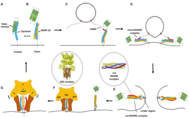
... 571. The endosomal system ... 57 2. SNARE-mediated membrane fusions ... 59
2.1 The SNARE complex structure and function ... 59 2.1.1 SNARE structure and classification ... 59 2.1.2 Main SNARE proteins ... 60 2.1.3 The SNARE cycle ... 61 2.2 SNARE specificity ... 64 2.3 SNARE cleavage by neurotoxins ... 67
3. Regulators of membrane trafficking: Rab proteins ... 70
3.1 Localization of Rab proteins ... 70 3.2 The Rab cycle ... 71
24
4. SNAREs at the synapse ... 73
4.1 SNARE proteins in synaptic vesicle exocytosis ... 73 4.2 Post-synaptic SNARE fusion machinery ... 79
C. Membrane trafficking in synaptic plasticity
... 821. Endosomal recycling and LTP ... 82
1.1 TfR in constitutive recycling... 82 1.2 Activity-dependent recycling ... 83
MATERIALS AND METHODS
1. Primary hippocampal Banker cultures ... 87 2. Organotypic hippocampal culture ... 88 3. Expression of exogenous proteins and shRNA ... 88 3.1 Plasmid constructs ... 88 3.2 Calcium phosphate transfection ... 89 3.3 Transduction with lentivirus ... 90 3.4 Single cell electroporation ... 91 4. Live cell imaging ... 92 4.1 Spinning disk confocal microscopy ... 92 4.2 Visualization of single exocytic events ... 94 a. SuperEcliptic pHLuorin (SEP) ... 94 b. Fusion events, imaging and analysis ... 94 4.3 Glycine treatment on live cells after photobleaching... 96 a. Chemical Long-term potentiation protocol ... 96 b. Fluorescence Recovery after Photobleaching ... 97 4.4 pH change for quantification of surface expression... 97 5. Immunocytochemistry and Transferrin recycling assay ... 98 6. Electron Microscopy ... 99 7. In- vitro electrophysiology ... 100 7.1 Whole-cell patch-clamp recordings ... 100 7.2 Long term potentiation induction ... 101 8. Statistical tests ... 101
25
RESULTS
VAMP4 controls constitutive recycling and sorting of post-synaptic receptors in neuronal dendrites ... 105
ABSTRACT ... 106 INTRODUCTION ... 107 RESULTS ... 109 VAMP4 is a marker of recycling endosome exocytosis in neuronal dendrites... 109 Downregulation of VAMP4 but not cleavage of VAMP2 reduces TfR exocytosis and recycling ... 111 VAMP4 exocytosis increases after chemical induction of LTP ... 112 VAMP4 KD does not impair the increase in RE exocytosis during cLTP induction . 113 VAMP4 KD accelerates AMPAR recycling and impairs its modulation during LTP induction ... 114 Effect of VAMP4 KD on synaptic transmission and plasticity ... 114 DISCUSSION ... 125 Involvement of VAMP4 in dendritic exocytosis ... 125 VAMP4 is necessary for endosomal sorting of AMPAR ... 126 A sequence of fusion events for the expression of LTP ... 128 REFERENCES ... 129
GENERAL DISCUSSION AND PERSPECTIVES
Further comments on the diversity of the endosomal system in dendrites ... 137 Implications for neuropathology ... 140 References ... 143
26
27
29
Neural signaling and plasticity
1. The neuronal cells
The basic unit of the nervous system is the nerve cell or the neuron. Neurons within the central and peripheral nervous system process information by generating sophisticated electrical and chemical signals across synapses. Signaling between interconnected neurons forms the circuitry which provides higher-level brain functions. The human brain with 100 billion neurons is the most cognitively able despite not being the largest among mammalian brains (Herculano-Houzel, 2009).
This introductory section is mainly from neuroscience books: The hippocampus book, Theoretical Neuroscience (Dayan, Abbott), Purves (3rd edition), Principles of neural science (4th edition), Molecular cell biology (7th edition).
1.1 Morphological properties of neurons
Neurons are highly specialized cells that generate electrical signals and transmit them to other cells via specialized morphological nerve fibers, the dendrites and the axons. Neurons connect and transmit information across junctions called synapses. The dendrites allow a neuron to receive inputs from multiple other neurons through synaptic connections. The structure of the dendrites or dendritic trees is very diverse (Figure1), likely reflecting diversity in the functional properties and the types of computations performed by different types of neurons (Sprutson, Hausser, & Stuart, 2013). Axons from a single neuron can traverse large brain fractions and carry the integrated neuronal output to other cells. In the mouse brain, cortical neurons typically send out an estimated 40 mm of axon which makes on average 180 synaptic connections with other neurons per mm of length. They have a total dendritic cable of approximately 10 mm and the dendritic tree receives 2 synaptic inputs per µm on average. The cell body or soma of a typical cortical neuron ranges in diameter from about 10 to 50 µm. It houses the nucleus and other structures that support the metabolic activity of the neuron.
1.2 Electrical properties of neurons
Neurons also have physiological specializations besides their morphological features. Most prominently, they harbor a wide variety of membrane-spanning ion channels that allow ions, mainly sodium (Na+), potassium (K+), calcium (Ca2+), and chloride (Cl-) to move across the
30
cell membrane. These ion channels open and close in response to voltage changes and other internal and external signals. The electric charges of these ions are important for many aspects of neuronal function, mainly the maintenance of the cell’s resting membrane potential and the generation of an action potential.
In quiescent cells, there are relatively more sodium and chloride ions outside the cell, whereas potassium and organic anions are typically found at higher concentrations within the cell than outside. This difference in concentrations provides a concentration gradient for ions to flow down their concentration gradients when their channels are open. As such, sodium and chloride ions will move into the cell, whereas potassium ion will flow out of the cell. However, of the three ions, the cell is most permeable to potassium, allowing it to have the greatest influence on the cell resting membrane potential. Thus, the resting membrane potential of neurons typically sits between -50 and -75 mv, a value that is closest to the equilibrium potential of potassium ions, and the cell is said to be polarized.
Ion pumps located in the cell membrane maintain concentration gradients that support this membrane potential difference. A change in voltage or concentration gradients across the membrane will allow the flow of ions into and out of a cell. Current in the form of positively charged ions flowing out of the cell (or negatively charged ions flowing into the cell) through open channels makes the membrane potential more negative, and the cell is hyperpolarized. In contrast, the current flowing into the cell changes the membrane potential to less negative or positive values leading to cell depolarization.
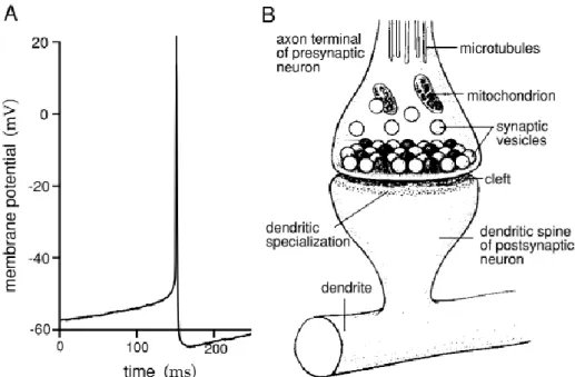
If a neuron is depolarized above a certain threshold, a positive feedback is initiated, and the neuron generates an action potential. An action potential is a 100 mV fluctuation in the electrical potential across the cell membrane lasting for about 1 ms (Figure 2A). A few milliseconds after the action potential, there is a hyperpolarization phase during which it may be impossible to initiate another spike, and the cell is said to be in the absolute refractory period.
Action potentials generated along axon processes can propagate rapidly over large distances. Axons terminate at synapses where the voltage transient of the action potential opens ion channels and calcium influx into the cell leading to neurotransmitter release (Figure 2B). The neurotransmitter binds to receptors at the post-synaptic membrane causing ion-conducting channels to open. Depending on the nature of the neurotransmitter release and the ion flow, the synapses can have an excitatory or an inhibitory effect on the post-synaptic neuron (discussed later in detail).
31
2. The Synapse
A Synapse is a term introduced by Charles Sherrington in 1897 and is derived from the Greek word Sinapsis meaning to “hold together”. It represents the precise location that transmits information from a pre- to a post-synaptic neuron allowing neuronal communication. Hence, the synapse consists of the pre-synaptic component; axonal bouton, the synaptic cleft of ~20 nm, and a post-synaptic component of a neighboring neuron (Figure 2B).
Large neurons are generally connected by thousands of boutons. The boutons may be opposed to dendrites of the receptor neuron (axodendritic synapses), to small projections of dendritic membrane or spines (axospinous synapses), to the perikaryon (axosomatic synapses), or the initial segment of the axon (axoaxonal synapses).
2.1 Synaptic Transmission
Synaptic transmission is the biological process by which a neuron communicates with a target cell across a synapse. There exist two main modalities of synaptic transmission: chemical and electrical, which coexist in most organisms and brain structures. At chemical synapses, a neurotransmitter is released from one neuron and detected by another, whereas in electrical synapses, adjacent cells are directly connected via gap junctions. The majority of the CNS synapses are chemical, while electrical synapses are much less common (Pereda, 2015).
2.1.1 The chemical synapse
The discovery of the chemical synapse was one of the most crucial in the history of neuroscience in the 20th century. It came from detailed studies on the functioning of the autonomic nervous system by T.R. Elliott, H. H. Dale, and O. Loewi (Tansey, 1991; Todman, 2008). The culmination of this work has led H. H. Dale together with O. Lowei to the Nobel prize in physiology or medicine in 1936 for the ‘discovery of chemical synaptic transmission’. The chemical synaptic transmission requires the release of neurotransmitter molecules from presynaptic axon terminals that are detected by the adjacent postsynaptic cell. The process is initiated when an action potential invades the terminal of a presynaptic neuron, which triggers the influx of calcium into the cell. Elevation of presynaptic calcium ion concentration, in turn, allows synaptic vesicles (SV) to fuse with the presynaptic plasma membrane and the release of neurotransmitters into the synaptic cleft. Following exocytosis, transmitters diffuse across the synaptic cleft and bind to their specific post-synaptic receptors (Figure3). This process plays
32
crucial functions in neuronal growth and development, synapse formation, and signal transduction.
One way of classifying synapses is whether the action of the neurotransmitter released tends to promote or inhibit the generation of an action potential in the postsynaptic cell. Therefore, neurotransmitters can either have excitatory or inhibitory effects on post-synaptic membrane. Excitatory postsynaptic potentials (EPSPs) are associated with a transmitter-induced increase in Na+ and K+ conductance of the synaptic membrane, resulting in net entry of positive charge carried by Na+ and membrane depolarization. Inhibitory postsynaptic potentials (IPSPs) are associated with a transmitter-activated influx of Cl− and membrane hyperpolarization. The majority of the excitatory synapses are found at dendrites, at the heads of spines, whereas the inhibitory synapses are found at the soma or the axon hillock, where excitation is generated and can be most effectively suppressed. Therefore, a single neuron receives a wealth of excitatory and inhibitory inputs through their synapses, which results in complex spatiotemporal signal integration involving current flow that ultimately converges in the axon until a fixed threshold for action potential firing is reached (Sprutson, Hausser, & Stuart, 2013).
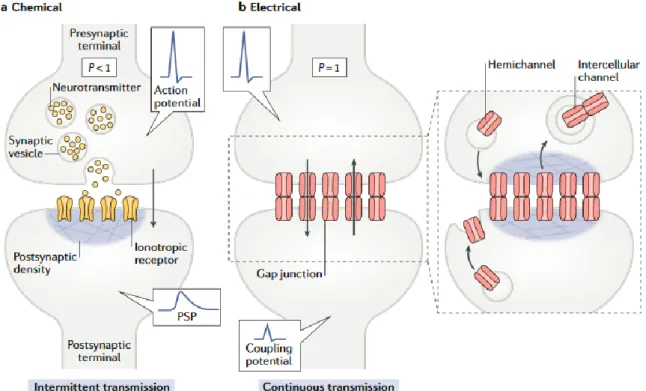
2.1.2 The electrical synapse
The electrical synaptic transmission is mediated by clusters of intracellular channels called gap junctions that directly communicate the interior of two adjacent cells, enabling the bidirectional passage of electrical currents and small molecules, for example, calcium, cyclic AMP and inositol-1,4,5-triphosphate). Gap junctions have a large internal diameter of ~1.2 nm and are formed by the docking of two hexameric connexin ‘hemichannels’ or ‘connexons’, one from each adjacent cell (figure3). Their bi-directionality enables them to coordinate the activity of a large group of interconnected neurons. Although they are a distinct minority, electrical synapses are found in all nervous systems, permitting direct, passive, and low resistance flow of electrical current from one neuron to another. Because gap junction communication occurs without the involvement of any intermediate messenger, they provide a fast mechanism for intercellular synaptic transmission (Curti and O’Brien, 2016). Electrical synapses are known to occur in the retina, inferior olive, and olfactory bulb (Pereda, 2014).
Gap junctions are highly dynamic structures, and their levels are maintained by a constant insertion and removal of channels providing a continuous and reliable conductance. By contrast, chemical synaptic communication is episodic given that it relies on the intermittent presence of an action potential at the presynaptic terminal which generates a transient increase in
33
intracellular calcium levels. Moreover, the neuro-transmitter release is probabilistic, and failures in trans-mission will occasionally occur despite the presence of an action potential in the presynaptic terminal (Alcami and Perada, 2019).
Electrical and chemical synapses can mutually co-regulate each other’s formation. Therefore, normal brain development and function relies on the interaction between these two forms of interneuronal communication (Jabeen and Thirumalai, 2018).
Figure 1. Neuronal diversity. (A) A cortical pyramidal cell: the primary excitatory neuron of
the cerebral cortex. (B) A Purkinje cell: present the most striking histological feature of the cerebellum and are the sole output of the cerebellar cortex. (C) A stellate cell of the cerebral cortex: they send inhibitory signals to the dendritic arbors of Purkinje cells. These figures are magnified about 150-fold. (Drawings from Cajal, 1911; figure from Dowling, 1992). Theoretical neuroscience book (Dayan, Abbott).
34
Figure 2. Action potentials and synapses. Intracellular recording of an action potential from
a cultural rat neo-cortical pyramidal cell. (B) Diagram of a chemical synapse. The synapse consists of the pre-synaptic component (bouton), the synaptic cleft, and the post-synaptic component (dendritic spine) of the next neuron. The axonal boutons contains mitochondria and small synaptic vesicles carrying neurotransmitter molecules that are released at the active zone. The presynaptic active zone is opposing the postsynaptic membrane containing a protein dense specialization called the postsynaptic density (PSD). Membrane fusion and exocytosis is triggered by a rise in intracellular calcium. Theoretical neuroscience book (Dayan, Abbott).
35
Figure 3. The general structures of a chemical and an electrical synapse. (A) The chemical
synapse requires transmitter release evoked by presynaptic action potentials, which activate calcium influx, and trigger synaptic vesicles exocytosis. Neurotransmitters released activate specific postsynaptic gated channels, eliciting a transient change in membrane permeability to cations or anions. Transmission at most chemical synapses is intermittent, as transmitter release is probabilistic (P <1) and depends on the presence of an action potential in the presynaptic terminal. (B) Electrical synapse transmission involves the transfer of electrical signals through gap junctions, that could be bidirectional. Continuous electrical communication is a highly regulated process that is maintained by a delicate balance between insertion and removal of gap junction channels Electrical transmission is continuous in nature (P= 1). Electrical currents underlying an action potential can directly spread to the postsynaptic cell, generating an electrical or a coupling potential. Figure from Alcami & Perada, 2019.
2.2 Neurotransmitter release
Neurotransmitters are endogenous molecules responsible for information transmission across chemical synapses. Over the years, several formal criteria have emerged that identify a substance as a neurotransmitter. The compound must be synthesized by the neuron; it must be released by the neuron in sufficient amounts to exhibit an effect on another neuron or effector organ; exogenous application in appropriate quantities must mimic the action of the endogenously released compound: and a mechanism must exist to remove the neurotransmitter from the site of action (Veca and Dreisbach, 1988). These criteria have led to the identification of more than 100 different neurotransmitter substances.
36
This large number of transmitters allows for tremendous diversity in chemical signaling between neurons. It is therefore useful to classify them into two major groups: (i) classic, such as amino acid derivatives, and (ii) neuropeptides. The most widely distributed classic transmitter substances in the nervous system are acetylcholine (ACh), glutamate, gamma-aminobutyric acid (GABA), dopamine, serotonin, norepinephrine, and glycine. The first substance identified as a neurotransmitter was ACh in 1914. The most abundant neurotransmitter in the CNS is glutamate, which is present in more than 80% of synapses and is the major excitatory neurotransmitter in the brain. In contrast, most inhibitory synapses use either GABA or glycine as neurotransmitters.
2.2.1 Different modes of neurotransmitter release
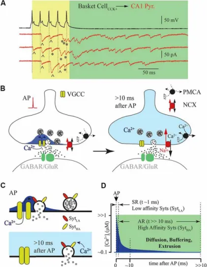
Most neuronal communication requires rapid information transfer within the CNS and relies upon the fast, synchronous release of neurotransmitters, which occurs within several milliseconds after an action potential invades a presynaptic bouton. However, neurotransmitter release could persist for tens or hundreds of msec after an action potential (asynchronous release), or in the absence of presynaptic depolarization stimulus (spontaneous release) (Rozov, Bolshakov, & Valiullina-Rakhmatullina, 2019; Kaeser and Regehr, 2014). The asynchronous release can influence network parameters including the efficacy of neurotransmission, synchronicity, and plasticity, whereas spontaneous release potentially affects synapse formation and connection strength (Chanaday et al., 2019).
Both synchronous and asynchronous releases are Ca2+ dependent, but the source of Ca2+ ions, and the Ca2+ sensors involved in both modes are different and have different binding kinetics. There is a general consensus that synchronous release is mainly triggered in the active zones by Ca2+ influx through presynaptic VGCC. Opening of these channels leads to a short-lasting and spatially restricted elevation of intraterminal Ca2+ at channel clusters known as nano- or microdomains. Furthermore, Ca2+ channels are closely associated with low-affinity vesicular synaptotagmines, usually, Syt1 and Syt2 at presynaptic release sites, which are suitable for triggering highly synchronized phasic release during the short-lived Ca2+ elevation within the microdomain (figure4) (Rozov, Bolshakov, & Valiullina-Rakhmatullina, 2019; Bukharaeva, 2015).
On the other hand, delayed asynchronous release requires a long-lasting elevation of free intraterminal Ca2+, but the source remains poorly identified. It has been proposed that VGCC may also provide a longer-lasting phase of Ca2+ influx that may contribute to asynchronous
37
release (figure4). In addition, it was hypothesized that synaptic activation may liberate sufficient ATP that activates presynaptic Ca2+ permeable P2X receptors. Intracellular Ca2+ is
suggested to bind to Syt7, which is a high-affinity calcium sensor mediating asynchronous release. However, the mechanism of its recruitment is not identified. Syt7 is also required for synaptic facilitation. It is found on the presynaptic plasma membrane and other internal membranes, but not on synaptic vesicles suggesting a non-canonical mechanism of vesicle exocytosis. (Rozov, Bolshakov, & Valiullina-Rakhmatullina, 2019; Kaeser and Regehr, 2014; Jackman et al., 2016).
2.2.2 Vesicular mechanism of neurotransmitter release
The foundations of presynaptic physiology were first established by the Nobel prize winner Bernard Katz and Ricardo Miledi during the 1950s and 1960s. Their pioneering work demonstrated the importance of presynaptic depolarization and Ca2+ influx for triggering the fast, synchronous transmitter release at nerve terminals. They have also shown that the synaptic transmitter is released in discrete packages called quanta. The discovery of the quantal release immediately raised the question of how such quanta are formed and discharged into the synaptic cleft. Later, Katz and colleagues revealed with electron microscopy the presence of synaptic vesicles in presynaptic terminals, which were then proposed to be loaded with the transmitter, and were thought to be the source of the quanta. These early findings are the basis for much of our current understanding of neurotransmitter release.
The ‘classical’ neurotransmitters are held in membrane-bound vesicles 40-50 nm in diameter near the synapse at the presynaptic cell. The exocytic release of neurotransmitters is triggered when electrical impulses in the form of an action potential invade the axon terminal. The transducers of electrical signals are voltage-gated Ca2+ channels (VGCC) localized at a region adjacent to the synaptic vesicles. The arrival of an action potential depolarizes the membrane and permits the influx of Ca2+ ions into the cytosol from the extracellular medium. This leads to a highly localized, transient increase in intracellular levels of Ca2+ ions in the region near the synaptic vesicles from <0.1µM, characteristic of a resting state, to 1-100 µM (Figure 4B). The increase in Ca2+ triggers the exocytosis of synaptic vesicles and thus the release of neurotransmitters. Ca2+ ions bind to a protein in the synaptic vesicle membrane called synaptotagmin (Syt), which is considered the key Ca2+ sensing protein that triggers vesicle fusion, initiating synaptic transmission (Lodish, Berk, & Zipursky, 2000; Sudhof, 2012). Syts do not act alone but require a cofactor called complexin, which is a small protein that binds to
38
SNARE complexes (soluble N-ethylmaleimide–sensitive factor attachment protein receptor), proteins that catalyze membrane fusion, triggering exocytosis (Sudhof, 2012). However, the mechanism by which Ca2+-Syt catalyzes the exocytic release of neurotransmitter remains largely unknown (Gundersen, 2019).
Figure 4. Synchronous vs asynchronous neurotransmitter release. (A) Example traces of
responses recorded from a presynaptic hippocampal cholecystokinin (CCK)+ basket cell shown in black and a postsynaptic CA1 pyramidal neuron shown in red (three traces recorded subsequently). Five action potentials triggered synchronized phasic IPSC (labeled with ^) and delayed responses (labeled with *). (B) Schematic drawing of a synapse after a single action potential. VGCC open transiently leading to an influx of calcium ions triggering phasic release (left panel). After closure of VGCC, Ca2+ concentration declines due to radial diffusion and binding to endogenous buffers (right panel). Two membrane transport proteins are responsible for maintaining pre-synaptic Ca2+ homeostasis: plasma membrane calcium-ATPase (PMCA) and the sodium/calcium exchanger (NCX). (C) Schematic drawing of vesicle fusion by Ca2+ micro/nano evoked by an action potential (upper panel). Phasic synchronous release arises from low affinity Syt (SytLA). The lower panel shows delayed vesicle fusion mediated by high affinity Syt (SytHA). (D) Schematic representation of Ca2+ time course at release site (blue) after an action potential. Dotted lines show time courses for synchronous and asynchronous release. Figure from Rozov, Bolshakov, & Valiullina-Rakhmatullina, 2019.
39
2.3 Glutamatergic Excitatory Synapses
Glutamatergic neurotransmission has been drawing substantial scientific interest owing to its implication in higher-order cognitive functions including learning and memory. Glutamate is a nonessential amino acid that was first speculated to have a metabolic function in the CNS. Then during the 1950s, it has been known that glutamate has an excitatory action in the mammalian brain and spinal cord (Meldrum, 2000). However, it was not until 1984 that it was acknowledged as fulfilling the criteria of a neurotransmitter and became widely recognized as the main excitatory transmitter within the vertebrate nervous system (Niciu, Kelmendi, & Sanacora, 2012). Glutamatergic excitatory synapses are now one of the best-understood synapses in the mammalian CNS (Siddoway, Hou, & Xia, 2011).
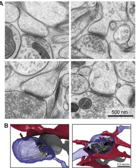
Glutamatergic synapses are excitatory relay points between presynaptic nerve terminals and postsynaptic spines. They are easily recognized via electron microscopy due to the appearance of the electron-dense region of the postsynaptic density (PSD) (Figure5). PSDs correspond to disks which are ~50 nm thick and 200-500 nm wide and may contain up to 100 types of proteins including membrane receptors, second messengers signaling molecules, anchoring and scaffolding proteins, and cytoskeletal components that provide structural and functional support to the synapse. The presynaptic terminal contains glutamatergic synaptic vesicles, which once released, targets its postsynaptic glutamate receptors (Siddoway, Hou, & Xia, 2011; Niciu, Kelmendi, & Sanacora, 2012).
2.3.1 Ionotropic glutamate receptors
Postsynaptic glutamate receptors can be divided into two broad categories: ionotropic and metabotropic receptors. The ionotropic receptor or iGluR family can be grouped into three subtypes that can all bind glutamate and are named based on their agonist selectivity: N-methyl-D-aspartate (NMDA), α-amino-3-hydroxy-5-methyl-4-isoxazole proprionic acid (AMPA), and kainate (KA).
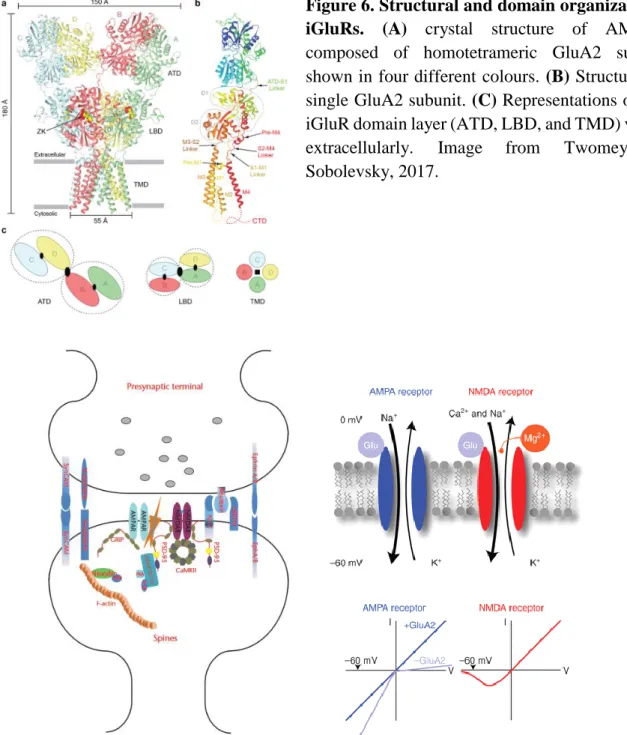
Ionotropic glutamate receptors are integral membrane proteins that assemble as tetramers of four intertwined subunits (>900 residues) that form an ion channel. Each subunit comprises an extracellular amino-terminal domain (ATD), a ligand-binding domain (LBD), a common pore-forming transmembrane domain (TMD), and an intracellular C-terminal domain (CTD) (Figure6). Assembly of subunits within the same functional class leads to the formation of a functional receptor. The AMPAR subunits (GluA1-GluA4) can form both homo- and
40
heteromers. Kainate receptors have five subunits (GluK1-GluK5), the subunits GluK1-GluK3 can also form both homo- and heteromers, but GluK4 and GluK5 are only functional when co-expressed with GluK1-GluK3. In addition, functional NMDA receptors require the assembly of two GluN1 subunits together with either two GluN2 subunits or a combination of GluN2 and GluN3 subunits. Different subunits are expressed in distinct brain regions and may serve different functions (Traynelis et al., 2010).
Figure 5. Architecture of glutamatergic synapses. (A) Two dimensional EM images of
synapses, showing presynaptic terminal containing vesicles, and postsynaptic electron-dense region of the PSD. (B) Reconstructed serial EM images, of axonal boutons (blue), dendritic spines (grey), and the astrocytic processes nearby (red). Image from Korogod et al., 2015.
The function of iGluRs is exhibited through ligand-gated, non-selective cation channels, which allow the passage of Na+, K+, and in some cases Ca2+. The neuronal excitatory postsynaptic
potential (EPSP) is mediated additively by AMPA and NMDA receptors (Figure 7), whereas KA receptors do not contribute significantly to synaptic transmission, except in specific synapses such as mossy fiber synapses in CA3 pyramidal cells (Siddoway, Hou, & Xia, 2011).
41
AMPA receptors have a lower glutamate affinity than NMDA receptors, but they have faster kinetics (millisecond timescale) and are responsible for the fast initial component of the EPSP (Meldrum, 2000). The rapid kinetics of AMPARs allows for fast depolarization of the postsynaptic membrane and high-fidelity basal synaptic transmission. In contrast, NMDA receptors have slower kinetics, use glycine as a co-agonist (which bind on the GluN1 subunits), and elicit relatively slow and long-lasting EPSPs. NMDAR is also considered a molecular coincidence detector that requires for activation both presynaptic release of glutamate and a sufficiently strong postsynaptic depolarization. The reason is that under basal conditions, magnesium ions block NMDAR pore, which can be removed upon adequate membrane depolarization allowing postsynaptic Ca2+ entry, activating downstream calcium-dependent signaling cascades. Therefore, NMDARs are not critical for basal transmission, but rather initiates changes in synaptic strength and plasticity as a result of their calcium permeability (Siddoway, Hou, & Xia, 2011). At some synapses, however, a minority of AMPARs (GluA2 subunit-lacking) are calcium-permeable and can trigger or contribute to various forms of synaptic plasticity (Greger, Watson, & Cull-Candy, 2017). Synaptic AMPA and NMDA receptors are clustered at the PSD, anchored by F-actin and other scaffolding and signaling proteins underneath (Figure 7) (Siddoway, Hou, & Xia, 2011).
2.3.2 Metabotropic glutamate receptors
Eight metabotropic glutamate receptors or mGluRs have been identified (mGlu1-8), and subdivided into three functional groups (Group I, II, or III) based on amino acid sequence homology, agonist binding, and activated downstream G protein signaling partners (Kim et al., 2008; Niciu, Kelmendi, & Sanacora, 2012). Group I mGluRs consist of mGlu1 and mGlu5, GroupII is mGlu2 and mGlu3, GroupIII is mGlu4,6,7. Both receptor families provide functional diversity and are widely expressed throughout the nervous system (Reiner and Levitz, 2018). mGluRs are present on both sides of the synapse but tend to be located perisynaptically and not within the synaptic zone (Sherman, 2014). They are members of the G-protein coupled receptors (GPCRs) superfamily which are constitutive dimers. Glutamate binding leads to activation of G protein signaling cascades that can modulate cell excitability and synaptic transmission. Group I mGluRs are primarily Gq-coupled, and elicit their downstream effects by
Ca2+ mobilization and activation of protein kinase C. Group II and III are Gi/o-coupled that are
42
cyclic adenosine monophosphate (cAMP), and inhibition of glutamatergic transmission (Niswender and Conn, 2010; Crupi et al., 2019).
Figure 6. Structural and domain organization of iGluRs. (A) crystal structure of AMPARs
composed of homotetrameric GluA2 subunits shown in four different colours. (B) Structure of a single GluA2 subunit. (C) Representations of each iGluR domain layer (ATD, LBD, and TMD) viewed
extracellularly. Image from Twomey and
Sobolevsky, 2017.
Figure 7. To the left, a schematic of the molecular organization of a glutamatergic excitatory
synapse showing AMPA and NMDA receptors localization at the PSD, and other anchoring and scaffolding proteins. Figure from Siddoway et al., 2011. To the right, major ionotropic glutamate receptors and their current voltage relationships (I-V) that is considered a biophysical signature for different receptors. AMPARs containing GluA2 show a linear I-V relationship, but are inward rectifying without GluA2. NMDARs have a more complex I-V curve because of the Mg2+ block at resting potentials. Figure from Luscher & Malenka, 2012.
43
3. Synaptic Plasticity
One of the most fascinating brain features is its capacity to adapt and modify neural synapses in response to ever-changing intrinsic and extrinsic stimuli. The idea that synapses could undergo dynamic changes in their activity was first proposed by the Canadian psychologist Donald Hebb, who postulated in 1949 that:
“When an axon of cell A is near enough to excite a cell B and repeatedly or persistently takes part in firing it, some growth process or metabolic change takes place in one or both cells such that A’s efficiency, as one of the cells firing B, is increased.” (Hebb, 1949).
These activity-dependent changes in synaptic transmission are the basis of synaptic plasticity (Langille & Brown, 2018). Synaptic transmission can be either enhanced or depressed, and the time span of such changes can be quite variable. Understanding the cellular and molecular mechanisms underlying synaptic plasticity is imperative given that it is the leading candidate for memory formation and storage (Citri & Malenka, 2008). And since the hippocampus is considered the brain’s memory hub, it has become one of the most extensively studied brain regions for synaptic plasticity to date.
3.1 The hippocampal formation and memory
The striking appearance of a distinct group of millions of neurons, buried deep within the medial temporal lobe of the human brain forming into an elegant, curved structure has captivated anatomists since the first dissections that took place at the Alexandrian school of medicine in classical Egypt. The structure resembles the coiled horns of a ram. Hence, the ancient scholars named the hippocampus cornu ammonis (CA), or the horns of Amun, an ancient Egyptian God, who is often represented as having a ram’s head. Later, the Bolognese anatomist Giulio Cesare Aranzi was the first to introduce the name ‘’Hippocampus’’ which comes from the greek hippokampos meaning sea horse (Figure 8) (The hippocampus book).
The emergence of microscopy has revealed the neatly organized cellular arrangement of the hippocampus that is condensed into single layers. Thus, the hippocampus was anatomically and functionally divided into four distinct subfields named CA1, CA2, CA3, and CA4, which
44
Figure 8. Dorsolateral view of the human hippocampus (left). Dissected and isolated human
hippocampus compared to the sea horse (Right). (The hippocampus book).
Figure 9.Basic anatomy of the hippocampus showing the EC-DG-CA3-CA1 circuitry. The
term hippocampal formation is a compound structure that refers to the DG, the hippocampus proper (cornu ammonis), and the subicular cortex. The entorhinal cortex sends projections from layers II, III, V, VI. Figure from Neves et al., 2008.
connect serially to form what is called a ‘trisynaptic loop’ (Figure 9). The major input to the hippocampus is provided by the entorhinal cortex (EC) which projects to the dentate gyrus (DG) granule cells via the perforant path (synapse1). The DG projects mossy fibers to the CA3 region (synapse2). CA3 pyramidal neurons project to the CA1 region via the Schaffer collateral
45
pathway (synapse3). Finally, CA1 pyramidal neurons project back to the entorhinal cortex, closing the loop. In addition, CA3 neurons provide feedback projections to the DG and make extensive recurrent connections onto other CA3 neurons. The EC also projects directly to the CA3 and CA1 regions (Knierim, 2015). Area CA2 is often excluded from circuit diagrams showing information flow through the hippocampus and has been seen as a transition zone between area CA1 and CA3. However, recent evidence indicates that area CA2 also receives direct excitatory inputs from both layers II and III of the EC (Chevaleyre and Siegelbaum 2010), and axons of CA2 pyramidal neurons project to both area CA1 and CA3 (Mercer et al. 2007). The hippocampus receives direct inputs from the olfactory bulb, and it was historically believed to function solely as an olfactory structure. The first link between the hippocampus and memory formation came from the observation of a case study of H.M. patient who had surgical removal of the medial temporal lobe including large parts of both hippocampi to cure epilepsy, which left him with profound global amnesia. Later, animal studies have confirmed the role of the hippocampus in various forms of memory including episodic memory; the memory of a particular single event, semantic memory; recall of general facts, spatial navigation, short-term memory. Given its crucial role in learning and memory, the hippocampus is indeed one of the earliest and most severely affected brain regions in Alzheimer’s disease (AD), the most common cause of dementia. Other roles include the regulation of emotional behavior, motor behavior and hypothalamic functions. (Anand & Dhikav, 2012; Bird & Burgess, 2008).
The memory function of the hippocampus has been correlated to the fact that activity-dependent synaptic plasticity is a prominent feature of hippocampal synapses. Therefore, the hypothesis that synaptic plasticity is the neural basis of information storage in the brain has remained to this day, an inference to the best explanation that has been accepted but yet difficult to prove in practice (Neves et al., 2008). Depending on the specific pattern of activation, synapses can either strengthen or weaken their connections, phenomena that are commonly known as long-term potentiation (LTP) or long-long-term depression (LTD) respectively. Synaptic plasticity has been studied and well-characterized extensively in the hippocampus due to its simple, laminar neuronal organization that enables the use of electrophysiological techniques to record synaptic events (Edelmann et al., 2017; Neves et al., 2008). However, it is now evident that synaptic plasticity is a property of many excitatory and inhibitory synapses across the brain (Castillo et al., 2011).
46
3.2 Long term potentiation
The first experimental evidence of LTP was performed by Bliss and Lomo in 1973(Bliss and Lomo, 1973), who demonstrated a long-lasting activity-dependent increase in synaptic efficacy, in a paper that is considered a breakthrough in the field of neuroscience. The broad definition of LTP is the long-lasting enhancement of synaptic strength in response to a brief high-frequency stimulation (Nicoll, 2017). Certainly, LTP exists in many mechanistically distinct forms, at different types of synapses, or even at the same synapse (Edelmann et al., 2017). Adding to the complexity, there are currently more than 100 proteins that have been claimed to be involved in LTP (Granger & Nicoll, 2014).
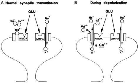
The classical form of LTP, or the Hebbian form of synaptic plasticity, is exhibited by the perforant path projection to granule cells of the dentate gyrus and by the Schaffer-collateral afferents to the CA1 pyramidal cells of the hippocampus. These synapses express a robust NMDAR-dependent LTP, which is blocked by D-AP5 (D (-)-2-amino-5-phosphonovaleric acid) or other NMDAR-antagonists. It is now generally accepted that LTP is induced by binding of glutamate to AMPA and NMDA receptors, depolarization of the postsynaptic membrane, Ca2+ influx through NMDARs, transient elevation of postsynaptic Ca2+ concentration, the release of Ca2+ from intracellular stores, and the subsequent activation of calcium/calmodulin-dependent protein kinase II (CAMKII) (Figure 10) (Nicoll, 2017). Activated CaMKII is necessary and sufficient for the induction of LTP (Lisman et al., 2012).
The exact mechanisms underlying LTP expression remain debated. A wide variety of induction protocols exist, each with potentially distinct expression mechanisms including high-frequency stimulation (HFS) or tetanus-induced LTP, theta-burst stimulation (TBS), pairing-induced LTP, spike-timing-dependent LTP, and chemically induced LTP (Bliss & Collingridge, 2013). Several key studies demonstrated a primarily postsynaptic locus of LTP expression, where the major contribution comes from increased current through postsynaptic AMPARs. Advances have been made in understanding the role of CAMKII in LTP expression. CAMKII diffuses to the synapse and interacts with the NMDA receptor (NR2B) forming a complex. CAMKII-NMDA receptor complex is believed to act as a molecular memory at the synapse, and is also a mechanism for LTP saturation. The use of Cyanogen bromide (CN) peptides that inhibit CAMKII binding to NMDARs can allow additional LTP induction by reversing saturated LTP (Sanhueza et al., 2011). In addition, activated CAMKII translocates to the PSD and enhances AMPAR-mediated transmission in two ways: phosphorylation of GluA1 (at serine 831),
47
increasing single-channel conductance, and phosphorylation of extrasynaptic stargazin (an AMPAR auxiliary protein), leading to AMPAR immobilization and trapping at the PSD (Lisman et al., 2012; Hayashi et al., 2000; Opazo et al., 2010).
In addition, strong evidence supports the idea that trafficking and exocytosis of new AMPARs lead to an increase in receptors number at the synapse and is involved in LTP expression. For instance, the use of neurotoxin which blocks exocytosis by cleaving some vesicular SNARE proteins blocks tetanus-induced LTP (Lledo et al., 1998) and HFS-induced LTP, but not short-term potentiation (STP) in the CA1 region of the hippocampus (Penn et al., 2017). Direct visualization of postsynaptic AMPARs exocytosis in dendritic shaft and spines during LTP is possible using pH-sensitive superecliptic pHluorin (SEP)-tagged AMPARs (Kopec et al., 2006, 2007a; Yudowski et al., 2007; Lin et al., 2009; Makino and Malinow, 2009; Petrini et al., 2009; Araki et al., 2010; Kennedy et al., 2010; Patterson et al., 2010; Cho et al., 2015; Tanaka and Hirano, 2012). Additionally, chemically induced LTP using glycine increases SEP-GluA1 exocytosis in dendrites and dendritic spines (Yudowski et al., 2007; Cho et al., 2015). Recent evidence from Choquet’s lab shows that surface cross-linking of exocytosed AMPARs blocks both HFS and TBS-induced LTP expression in the CA1 region of the hippocampus (Penn et al., 2017).
Surface AMPARs tune synaptic transmission via a constant exchange between synaptic and extrasynaptic sites (Heine et al., 2008). Accordingly, several studies accentuate the role of AMPAR lateral diffusion for the incorporation of the receptor at the synapse during LTP (Makino and Malinow, 2009; Penn et al., 2017). Given the fact that AMPAR exocytosis during LTP occurs at sites adjacent to the PSD, it would require these exocytosed receptors to be relocated via lateral mobility to synapses for synaptic potentiation. It is therefore proposed that pre-existing extrasynaptic AMPARs at the surface provide the reservoir for the initial phase of potentiation, whereas newly exocytosed AMPARs from the recycling endosomes are required for LTP maintenance (Figure 11) (Choquet, 2018).
48
Figure 10. Model for LTP induction in the hippocampal CA1 region. Under basal
conditions, glutamate could bind to both AMPA and NMDA receptos, but mainly AMPARs mediate basal synaptic transmission. Upon adequate depolarization of postsynaptic membrane, Mg2+ blockade is expelled, NMDA receptors are activated allowing the influx of cations, mainly calcium mediating downstream calcium-calmodulin signaling events. Figure from Nicoll, 2017
Figure 11.Trafficking of AMPA receptors during LTP. 1- AMPA receptors are trafficked
along microtubules to reach the target synapse. 2- Vesicles are exocytosed mainly at extrasynaptic sites in the dendritic shaft. 3- Receptors reach the synapse via lateral mobility. 4-They are stabilized at the PSD via interaction with scaffold proteins. 5- Receptors are endocytosed and can be recycled back to the plasma membrane. Figure from Choquet, 2018.
49
3.3 Long term depression
Long-term depression or LTD is the contrasting phenomenon of LTP, which is the persistent, use-dependent decrease in synaptic efficacy or strength. Two broad forms of LTD exist, heterosynaptic and homosynaptic. Heterosynaptic LTD occurs at inactive synapses, or in a non-conditioned input, whereas, homosynaptic LTD is usually induced in the non-conditioned input, thus is known to be input-specific (Figure 12). Homosynaptic LTD can be further divided into two main types: depotentiation, which is the depression of a potentiated response observed after LTP induction or de novo LTD which is observed from baseline conditions. These different forms of LTD have different molecular mechanisms and probably serve different functions. Generally, LTD is involved in some types of learning and memory, cognitive flexibility, acute stress-induced cognitive defects, drug addiction, and neurodegeneration (Collingridge et al., 2010).
The discovery of LTD came from Dunwiddie and Lynch in 1978, who first reported that LTD could be induced with low-frequency stimulation (LFS) (100 stimuli at 1Hz) (Dunwiddie and Lynch, 1978). However, interest in LTD began to accelerate later in 1992 when Dudek and Bear showed that prolonged trains of low-frequency stimulation (LFS) (900 stimuli at 1 Hz) induced reliable homosynaptic LTD in hippocampal slices. LTD induced with this protocol is long-lasting, input specific, and NMDAR-dependent (Dudek and Bear, 1992). Nevertheless, it was not clear at that time whether the induction of LTD in the hippocampal area CA1 was solely NMDAR-dependent or if the process can be triggered by mGluRs (Dudek and Bear, 1992; Bashir et al., 1993). In 1997, Oliet et al. confirmed the existence of both types of homosynaptic LTD and showed that it was possible to induce both types by changing the induction protocol. LTD can be induced by LFS (typically 900 stimuli at 1-3 Hz), by pairing baseline stimulation with depolarization (to -40 mV), appropriately timed back-propagating action potential (a form of spike-timing-dependent plasticity or STDP), or chemically induced using an NMDAR agonist, such as 3,5-dihydroxyphenylglycine (DHPG) or NMDA. Most synapses that undergo LTD are glutamatergic, and like LTP require NMDAR activation. The determinant of the synaptic modification polarity is widely assumed to be the kinetics, subunit composition, and the magnitude of activation of NMDARs. For example, the NR2A-containing NMDARs are important for LTP, whereas de novo LTD requires NR2B-containing receptors (Collingridge et al., 2004).