Dopamine–glutamate reciprocal modulation of release and
motor responses in the rat caudate–putamen and nucleus
accumbens of ‘‘intact’’ animals
Hélène N. Davida,b, Marc Ansseaua, Jacques H. Abrainib
aUnité de Psychologie Médicale, CHU Sart-Tilman, B 4000 Liège, Belgium
bUniversité de Caen - CNRS, UMR 6185, Centre CYCERON, Boulevard Henri Becquerel, 14074 Caen cedex,
France
KEYWORDS: Striatum; Nucleus accumbens; D1-like receptor; D2-like receptor; Glutamatergic ionotropic receptor; Glutamatergic metabotropic receptor; functional interactions; Locomotor activity; Neurochemistry
ABSTRACT
Functional interactions between dopaminergic neurotransmission and glutamatergic
neurotransmission are well known to play a crucial integrative role in the striatum, the major input structure of the basal ganglia now widely recognized to contribute to the control of motor activity and movements but also to the processing of cognitive and limbic functions. However, the nature of these interactions is still a matter of debate and controversy. This review (1) summarizes anatomical data on the distribution of dopaminergic and glutamatergic receptors in the striatum–accumbens complex, (2) focuses on the dopamine–glutamate interactions in the modulation of each other’s release in the striatum–accumbens complex, and (3) examines the dopamine–glutamate interactions in the entire striatum involved in the control of locomotor activity. The effects of dopaminergic and glutamatergic receptor selective agonists and
antagonists on dopamine and glutamate release as well on motor responses are analyzed in the entire striatum, by reviewing both in vitro and in vivo data. Regarding in vivo data, only findings from focal injections studies in the nucleus accumbens or the caudate–putamen of ‘‘intact’’ animals are reviewed. Altogether, the available data demonstrate that dopamine and glutamate do not uniformly interact to modulate each others’ release and postsynaptic modulation of striatal output neurons. Depending on the receptor subtypes involved, interactions between dopaminergic and glutamatergic transmission vary as a multiple and complex combination of tonic, phasic, facilitatory, and inhibitory properties.
Introduction
Dopamine–glutamate reciprocal modulations play a major integrative role in the striatum that is the major input structure of the basal ganglia now widely recognized to contribute not only to the control of motor activity and movements but also to the processing of cognitive and ‘‘limbic’’ (emotional and motivational) functions [17,96, 144]. Because of this, dopamine–glutamate interactions have been extensively studied, leading to a matter of debate and controversy for nearly three decades. Despite the fact that synaptic contacts between dopaminergic fibers, arising from the substantia nigra and the ventral tegmental area, and glutamatergic axon terminals, arising from the whole cerebral cortex, the thalamus, and limbic structures, are rare in the striatum [21], evidence has been accumulated that dopamine and glutamate, released from non-junctional varicosities and/or spilled over from the synaptic cleft, can interact directly through activation of extra-synaptic receptors (the so-called volume neurotransmission) or indirectly through striatal interneurons in a complex manner both at the presynaptic and postsynaptic levels [64].
Reciprocal modulation between dopamine and glutamate in the striatum is further complicated by the heterogeneity of receptors and subtypes of receptors activated by these neurotransmitters. Glutamate acts on two types of glutamatergic receptors: the ionotropic glutamatergic (iGlu) receptors that are ion channel-coupled receptors and comprise the N-methyl-d-aspartate (NMDA) receptors and the AMPA/kainate receptors (that include both the amino-3-hydroxy-5-methyl-4-isoxazoleproprionate (AMPA) receptors and the kainate receptors) [52]; and the metabotropic glutamatergic (mGlu) receptors that are G-proteins-coupled receptors and comprise three groups, namely the group I mGlu receptors (that includes the mGlu1 and mGlu5 receptors), the group II mGlu receptors (that includes the mGlu2 and mGlu3 receptors) and the group III mGlu receptors (that includes the mGlu4, mGlu6, mGlu7, and mGlu8 receptors) [160]. Dopamine acts on two classes of receptor subtypes that are coupled to G-proteins, namely the D1-like receptors (that include the D1 and D5 receptors) and the D2-like receptors (that include the D2, D3, D4 receptors) [186].
This review briefly summarizes anatomical data on the distribution of dopaminergic and glutamatergic receptors in the striatum complex (i.e., the caudate–putamen and the nucleus accumbens), and then focuses on particular and relevant aspects of dopamine–glutamate interactions, which are the reciprocal modulation of release and locomotor responses. The effects of dopaminergic and glutamatergic receptor selective ligands on dopamine and glutamate release as well as on locomotor responses are analyzed in the entire striatum, by reviewing both in vitro and in vivo data. Our purpose is not to cluster the dorsal and ventral striatum but more likely to highlight the differences and similarities between these two brain structures. Regarding in vivo data, only findings from focal injection studies in ‘‘intact’’ non-anesthetized animals are reviewed in order to focus on the intrinsic physiology of the striatum complex.
The striatum: caudate–putamen vs. nucleus accumbens
The striatum is the main input structure of the basal ganglia and is a key component of the motor system. It is divided into the dorsal striatum, which includes the caudate and the putamen, and the ventral striatum that is mainly composed of the nucleus accumbens. The caudate–putamen and the nucleus accumbens show differences in their input and output projections.
The caudate–putamen is mainly innervated by the primary motor cortex, the anterior premotor and cingulate areas, the substantia nigra pars compacta and the retrorubral nucleus. The caudate–putamen in turn projects to the globus pallidus, the substantia nigra pars reticulata and pars compacta and the retrorubral nucleus.
The ventral striatum receives inputs from numerous prefrontal areas, limbic structures, such as the hippocampus and the amygdala, the ventral tegmental area and the retrorubral nucleus; in turn, the ventral striatum sends projections to the ventral pallidum, the substantia nigra pars compacta and reticulata, the retrorubral nucleus, the ventral tegmental area, and the hypothalamus [88,96].
Therefore, while the dorsal striatum appears more allied with voluntary motor functions and is involved in the initiation, production and sequencing of motor behavior and in the development of addiction, the nucleus accumbens is more likely an interface between the limbic and the motor system and plays a major role in motivated and goaldirected behaviors as well as the development and expression of addiction [88,96,145]. However, there is no clear boundary between the caudate–putamen and nucleus accumbens.
The same neuronal cell types are present throughout the striatum. Striatal output neurons represent 95% of the striatal neurons and can be divided into two different subtypes: the striato-nigral GABAergic neurons and the striato-pallidal GABAergic neurons, which are characterized by containing different peptides and subtypes of dopamine receptors. The striato-pallidal GABAergic neurons contain the peptide enkephalin and express the D2-like receptor, whereas the striato-nigral GABAergic neurons contain the peptides dynorphin and substance P and express the D1-like receptor [5,66,114,238]. However, it is worthy to mention that these authors do not claim that striatal neurons that express one subtype of dopamine receptor cannot also express low levels of the other; but that the relative amount of D1-like and D2-like receptor subtypes varies dramatically according to the type of striatal output neurons that is considered. Thus, dopamine in fine differentially affects striato-nigral and striato-pallidal neurons as a consequence of the predominant expression of D1-like and D2-like receptors by these neurons. Nevertheless, in contrast with the dorsal striatum, where the vast majority of neurons that display D1-like receptors project to the substantia nigra, about 40 – 50% of neurons that express D1-like receptors in the nucleus accumbens project to the ventral pallidum [124,176]. But, like in the dorsal striatum, the vast majority of neurons that express D2-like receptors in the nucleus accumbens project to pallidum [124,176]. The second neuronal type found throughout the striatum is striatal interneurons, which represents 5% of the striatal neuronal population. Two subtypes of striatal interneurons can be found within the striatum: cholinergic interneurons and GABAergic interneurons [93].
Localization of dopaminergic and glutamatergic receptors
in the striatum complex (Fig. 1)
LOCALIZATION OF STRIATAL DOPAMINERGIC RECEPTORS
Localization studies have shown that D1-like (D1 and D5) receptors are located at the postsynaptic level, mostly on medium spiny output GABAergic neurons. D2-like (D2, D3, and D4) receptors are located both at the presynaptic level, on dopaminergic neurons where they act as autoreceptors and on non-dopaminergic afferent fibers to the striatum among which are the glutamatergic terminals, and at the postsynaptic level on GABAergic output neurons [14,77,139,187] (Fig. 1). In the caudate–putamen, all striato-nigral output neurons (100%) express D1-like receptors in the caudate–putamen while only 5% of the striato-pallidal GABAergic output neurons express them [66,238]; conversely, all striato-pallidal GABAergic output neurons express D2-like receptors, while only 5% of the striato-nigral GABAergic output neurons express them [66,83,114,115]. Although a similar anatomical distribution is found in the nucleus accumbens [114], approximately 40 – 50% of the striato-pallidal GABAergic neurons, mainly originating from the shell of the nucleus accumbens rather than from the core, express D1-like receptors [124,176]. Taken together, these data have provided the anatomical basis on the modulatory role of D1-like-and D2-like postsynaptic dopaminergic receptors on the activity of the striato-nigral GABAergic output neurons (that constitute the striato-nigral direct pathway) and of the striato-pallidal GABAergic output neurons (that is the first synaptic link of the striato-nigral indirect pathway), respectively [66,114,115,124,176].
At the receptor subtype level, D1, D2, and D5 receptors are expressed both in the caudate– putamen and in the nucleus accumbens. Although D3 receptors are poorly expressed in the caudate–putamen [20,193], they are largely distributed in the nucleus accumbens [20,50,51,112,113,115,117]. In contrast, only moderate and low levels of D4 receptors are found in the caudate–putamen and nucleus accumbens, respectively [173]. Moreover, localization of D2 and D5 receptors has been also demonstrated on cholinergic and GABAergic interneurons [4,7,14,100,116,125,162, 236,237]. Although there is some controversy about the dopaminergic receptor subtypes that are expressed on striatal glutamatergic terminals, either D2 receptors [36] or D4 receptors [13,205,208], there is, however, a general consensus as far today that the receptors involved in the control of glutamate release throughout the striatum belong to the D2-like, but not D1-D2-like, receptor family.
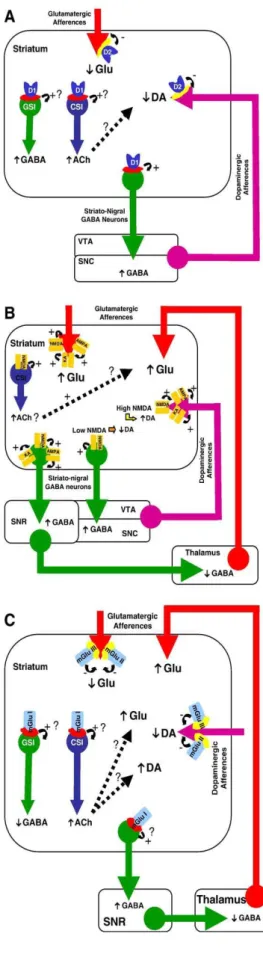
Fig. 1. Localization and relative abundance of D1-like and D2-like dopaminergic receptors; group I, group II and group III glutamatergic metabotropic receptors; and NMDA-, AMPA-and kainate glutamatergic ionotropic receptors in the caudate–putamen (CP) and nucleus accumbens (NAcc) on glutamatergic terminals arising from the cortex, the hippocampus and the amygdala (red arrow), on dopaminergic terminals arising from the retrorubral nucleus (RR), the substantia nigra pars compacta (SNC), and the ventral tegmental area (VTA; pink arrow), on GABAergic (green arrow) projection neurons and on GABAergic (green arrow; GSI) and cholinergic (blue arrow; CSI) striatal interneurons. AMPA: AMPA ionotropic glutamatergic receptors; D1: D1-like dopaminergic receptors; D2: D2-like dopaminergic receptors; EP: entopendocular nucleus; GPe: globus pallidus; KA: Kainate ionotropic glutamatergic receptors; mGlu I: group I metabotropic glutamatergic receptors; mGlu II: group II metabotropic glutamatergic receptors; mGlu III: group III metabotropic glutamatergic receptors; NMDA: NMDA ionotropic glutamatergic receptors; SNR: substantia nigra pars reticulata; STN: subthalamic nucleus; Thal: Thalamus.
LOCALIZATION OF STRIATAL IONOTROPIC GLUTAMATERGIC RECEPTORS
Localization studies of iGlu receptors suggested that NMDA-and AMPA/kainate iGlu receptors are expressed presynaptically in dopaminergic [62,205–207,239] and glutamatergic terminals [205–207], but also postsynaptically in striatal medium spiny projection neurons [15,69,205– 207] where they produce postsynaptic excitatory currents [146]. However, iGlu receptors are not located uniformly within the nucleus accumbens: while AMPA and kainate, as well as NMDA, receptors are expressed on the glutamatergic fibers arising from the cortex, only kainate and NMDA receptors, but not AMPA receptors, are expressed on the glutamatergic terminals arising from the hippocampus [205–207]. Moreover, a recent study has shown that AMPA receptors, but not NMDA receptors, are located on glutamatergic terminals arising from the cortex and the thalamus [63]. While NMDA and kainate receptors are also expressed on cholinergic and GABAergic striatal interneurons, AMPA receptors are expressed almost exclusively by GABAergic striatal interneurons [37,202].
LOCALIZATION OF STRIATAL METABOTROPIC GLUTAMATERGIC RECEPTORS
Over the last decade, histological studies have revealed that mGlu receptors are densely distributed within the striatum [3]. Although the vast majority (90%) of mGlu receptors is thought to be located postsynaptically since lesion of corticostriatal projections only results in a little effect on mGlu receptor binding quantity [230], studies utilizing lesion techniques or presynaptic markers have shown that mGlu2, mGlu3 (group II), mGlu4 and mGlu7 (group III) receptors, but not mGlu1 and mGlu5 (group I) receptors, are located at the presynaptic level on glutamatergic and/or dopaminergic terminals [24,25,105, 158,204,211].
In striatal output neurons, in situ hybridization and immunohistochemistry studies have demonstrated the presence of high levels of mGlu5 receptors [133,188,189,209] and low levels of mGlu1 receptors [105,209]. Interestingly, like D1-like and D2-like receptors, mGlu1 and mGlu5 receptors seem well segregated in striatal projection neurons: mGlu1 receptors are primarily present on striato-nigral output neurons whereas mGlu5 receptors are primarily expressed in striatopallidal projection neurons [98,210]. Similarly, mGlu3 (group II) and mGlu7 (group III) receptors show high levels of expression in striatal output neurons whereas mGlu2 (group II) and mGlu4 (group III) receptors only exhibit low levels of expression [24,25,105,133, 150,209]. Moreover, co-localization of mGlu1, mGlu3, mGlu5, and/or mGlu7 receptors has been shown on striato-nigral neurons or striatopallidal neurons [98, 210,211]. In addition to projection neurons and oncoming glutamatergic and dopaminergic neurons, it has been shown that at least mGlu1, mGlu2, and mGlu5 receptors are also expressed by cholinergic and GABAergic interneurons [12,105,163,164,203,209].
At the subcellular level, both group I and group III mGlu receptor subtypes, which are, respectively, concentrated in perisynaptic sites and pre- and postsynaptic sites [13,201, 204], share similar ultrastructural localization close to neurotransmitter release sites, whereas group II receptor subtypes have no close association with synapses [204]. In addition, unlike iGlu receptors, mGlu receptor subtypes, except for mGlu4 receptors, seem preferentially associated with fibers rather than soma [13,90,201].
Dopaminergic control of striatal dopamine and glutamate
release (Table 1)
MODULATION OF STRIATAL DOPAMINE RELEASE BY STRIATAL DOPAMINERGIC
RECEPTORS
DATA FROM IN VITRO AND EX VIVO STUDIES
Neurochemical studies on synaptosomes or striatal slices have repeatedly reported that activation of D2 receptors by the D2 receptor agonist quinpirole reduced the release of evoked dopamine [10,119,195,233] (Table 1). Ex vivo studies performed on slices of nucleus accumbens have evidenced that application of the D3 receptor agonist 7-OH-DPAT inhibited single pulse stimulated dopamine release in a concentration-dependent manner, and this inhibitory effect was partly blocked by the D2-like receptor antagonist haloperidol [155]. In addition, other data have revealed that activation of D1-like receptors can also decrease evoked dopamine release in the nucleus accumbens, while blockade of these receptors had no effect [10]. Finally, activation of both D1-like and D2-like receptors by the non-specific agonist apomorphine inhibited electrically stimulated dopamine release [155]. Taken together, these data supported the existence both of a direct inhibitory modulation of striatal dopamine release by D2-like autoreceptors and, given the lack of anatomical evidence for D1-like receptors on dopaminergic terminals [14,77,139], of an indirect inhibitory modulation of striatal dopamine release by D1-like receptors through feedback loops involving other neurotransmitters.
Table 1. Dopaminergic control of striatal dopamine and glutamate release
DATA FROM IN VIVO STUDIES (FIG. 2A)
Consistent with an autoreceptor role of D2-like receptors, in vivo studies using brain microdialysis in freely moving animals have revealed that focal administration of D2-like receptor agonists in the caudate–putamen reduced striatal dopamine release [79,213,222], while infusion of D2-like receptor antagonists increased it [79] (Fig. 2A). Additional studies performed in the caudate–putamen and the nucleus accumbens have revealed that application of the D3 receptor agonist 7-OH-DPAT decreased dopamine release [49,65]. However, the lack of selective D3 antagonist precluded any firm conclusion whether or not the decrease in striatal dopamine release was attributable to D3 receptors or other subtypes of D2-like receptors [49];
in that way, data from knock-out mice lacking the D3 receptor suggested that this receptor is not involved in dopamine autoreceptor function [104]. Activation of D1-like receptors by SKF 38393 or CY 208243 was found to decrease and to have no effect on striatal dopamine release [79,80]. But, given that the inhibitory effect of SKF 38393 on striatal dopamine release was not counteracted by co-infusion of a D1-like receptor antagonist, it seems possible that this effect might be nonspecific [79]. Conversely, blockade of D1-like receptors or of D2-like receptors in the caudate–putamen or the nucleus accumbens increased striatal dopamine release [79,80,168]. Administration of a D1-like receptor antagonist might stimulate dopamine release in the striatal complex by blocking D1-like receptors on GABAergic output neurons, thereby mediating negative feedback regulation to the substantia nigra pars compacta or ventral tegmental area, and/or by blocking D1-like receptor-mediated inhibition of interneurons. Such interneuron mechanisms may involve cholinergic interneurons, since it has been demonstrated that activation of the D1-like receptors excites cholinergic interneurons [7]; therefore, if one considers that dopaminergic terminals can be tonically inhibited by acetylcholine possibly through M2 or M4 receptors [57], blockade of D1-like receptors located on cholinergic interneurons would release this inhibition. Alternatively, it should be noted that activation of D2-like receptors inhibits cholinergic interneurons [162], although others did not find any effect on basal acetylcholine release in the nucleus accumbens following activation or blockade of D2-like receptors [99,169]. Given the anatomical distribution of dopaminergic receptors in the striatum complex, these results altogether are consistent with in vitro/ex vivo data of a direct inhibitory control of dopamine release by D2-like presynaptic autoreceptors and of an indirect control by D1-like receptors, likely acting through trans(poly)synaptic mechanisms [168].
MODULATION
OF
STRIATAL
GLUTAMATE
RELEASE
BY
STRIATAL
DOPAMINERGIC RECEPTORS
DATA FROM IN VITRO AND EX VIVO STUDIES
Few in vitro and ex vivo studies are available on the effects of dopamine and dopaminergic receptor agonists and antagonists on glutamate release in the striatum. However, they repeatedly show that both in striatal slices and synaptosomes dopaminergic receptor activation has an inhibitory action on glutamate release. Perfusion of dopamine at high concentration (100 µM) or of non-selective dopaminergic receptor agonists, such as apomorphine, inhibited KCl-evoked, but not spontaneous, glutamate release in striatal slices in a tetrodotoxin (TTX)-insensitive way [42,177]. Similar results were obtained both from slices and synaptosomes, and involvement of D2-like receptors was proposed on the basis of selectivity towards D2-like receptor antagonists [140]. Further experiments found that apomorphine and haloperidol, which are non-selective dopaminergic receptor agonist and antagonist, respectively, inhibited and enhanced KCl-evoked [3H]glutamate release from striatal slices [72], but other data did not report inhibitory modulation of KCl-stimulated [3H]glutamate release in slices during perfusion with selective D1-like or D2-like receptor agonists [194]. This lack of effect might be explained by a high level of receptor occupancy by endogenous dopamine as shown by Peris et al. [157] who confirmed the inhibitory effect of D2-like receptor activation in experiments monitoring [3H]d-aspartate (a tracer of glutamatergic transmission) efflux. These data obtained from striatal
slices were confirmed in a study using synaptosomal preparations showing that dopamine inhibited depolarization-evoked glutamate release via D2-like presynaptic receptors [135].
DATA FROM IN VIVO STUDIES (FIG. 2A)
Although a very few in vivo studies in the awake rat are available on the modulation of striatal glutamate release by striatal dopaminergic receptors, they confirm data from in vitro and ex vivo studies. Microdialysis studies employing D1-like and D2-like receptor agonists and antagonists showed that infusion of the D2-like receptor agonists quinpirole and LY 163502 in the striatum inhibited evoked glutamate release in a sulpiride (a D2-like receptor antagonist)-sensitive way [55,234], whereas injection of the D1 selective receptor agonist SKF 38393 showed no effect [234]. Subsequent studies using the push–pull technique have also demonstrated that the mixed D1–D2 receptor agonist apomorphine increased spontaneous glutamate efflux in the non-anesthetized rat [59,166]. Overall, consistent with a presynaptic localization of D2-like, but not of D1-like, dopaminergic receptors on glutamatergic terminals in the striatum, in vivo studies confirm what was found in in vitro studies that D2-like, but not D1-like, receptors are involved in an inhibitory control of evoked glutamate release, but further suggest that D2-like dopaminergic receptors may also modulate spontaneous glutamate release.
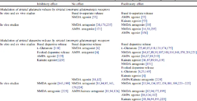
Fig. 2. (A) Illustration of the modulation of striatal dopamine- and glutamate release by striatal dopaminergic receptors. Taken together, data show a direct inhibitory control of dopamine release by D2-like autoreceptors and an indirect control by D1-like receptors, likely acting through trans(poly)synaptic mechanisms mediated via striatal interneurons and/or striato-nigral GABAergic output neurons. In addition, D2-like, but not D1-like, receptors are involved in an inhibitory control of glutamate release in the striatum. (B) Modulation of striatal dopamine and glutamate release by striatal ionotropic glutamatergic receptors. Ionotropic glutamatergic receptors might regulate glutamate release in the striatum by acting presynaptically although it is unclear how opening of ion-channel receptors may fit with a classical autoreceptor inhibitory mechanism; in fact, activation of ion-channel autoreceptors may favor release. In addition, trans(poly)synaptic striato-cortico-striatal mechanisms may be considered involving a positive feedback loop mediated through activation of postsynaptic NMDA receptors located on striato-nigral GABAergic projection neurons that may enhance glutamate release from corticostriatal and thalamostriatal afferents. Cholinergic interneurons might also play a key role in the positive regulation of striatal glutamate release. Alternatively, activation of NMDA receptors by physiological doses of NMDA inhibits striatal dopamine release and further leads to a concomitant increase of GABA release in the substantia nigra pars reticulata (SNR), suggesting the involvement of inhibitory feedback through the striato-nigro-thalamo-striatal loop. Such an inhibitory control of dopamine release in the striatum may overcome at higher NMDA receptor occupancy or high, possibly non-physiological, NMDA concentrations, leading to an increase in striatal dopamine release. Studies on AMPA and kainate receptors indicate that they exert a phasic facilitating effect on striatal dopamine release. (C) Modulation of striatal dopamine and glutamate release by striatal metabotropic glutamatergic (mGlu) receptors. Activation of group I mGlu receptors induces an increase in glutamate release. Given the lack of presynaptically located group I mGlu receptors within the striatum, it is likely that this facilitating effect results from trans(poly)-synaptic mechanisms that may involve striato-nigral GABAergic output neurons and/or striatal interneurons. Indeed, group I mGlu receptors have been demonstrated to stimulate cholinergic striatal interneurons (CSI): the release of acetylcholine may favor indirectly that of glutamate. GABAergic striatal interneurons (GSI) might also been involved since blockade of group I mGlu receptors stimulates GABA striatal release and activation of these receptors inhibits GABA-mediated IPSCs in corticostriatal slices: this may inhibit glutamate release in the striatum. In contrast, activation of group II or group III mGlu receptors inhibits glutamate release, results consistent with a presynaptic localization of these receptors on glutamatergic terminals within the striatum. Regarding the modulation of striatal dopamine release by metabotropic glutamate receptors, activation of group I mGlu receptors induces an increase in striatal dopamine release, which effect may result, as seen above for glutamate release, from trans(poly)synaptic mechanisms. Activation of group II or group III mGlu receptors decreases striatal dopamine, result consistent with a presynaptic localization of these receptors on dopaminergic fibers within the striatum. Ach: acetylcholine; AMPA: AMPA ionotropic glutamatergic receptors; D1: D1-like dopaminergic receptors; D2: D2-like dopaminergic receptors; DA: dopamine; Glu: glutamate; KA: Kainate ionotropic glutamatergic receptors; mGlu I: group I metabotropic glutamatergic receptors; mGlu II: group II metabotropic glutamatergic receptors; mGlu III: group III metabotropic glutamatergic receptors; NMDA: NMDA ionotropic glutamatergic receptors; SNC: substantia nigra pars compacta; SNR: substantia nigra pars reticulata; VTA: ventral tegmental area.
Glutamatergic control of striatal glutamate and dopamine
release
MODULATION OF STRIATAL GLUTAMATE AND ASPARTATE RELEASE BY
STRIATAL IGLU RECEPTORS (FIG. 2B)
Ex vivo studies on striatal slices have reported that application of agonists of AMPA/kainate receptors stimulated basal d-aspartate release, while activation of NMDA receptors had no effect [53] (Tables 2 and 3).
In vivo studies by means of microdialysis have reported consistently a facilitating effect of NMDA- and AMPA/kainate receptor agonists on glutamate and aspartate release [16,30,156,235], an effect that so far as NMDA receptors are concerned was blocked by pretreatment or co-administration of the non-competitive NMDA receptor antagonists MK-801 and phencyclidine [30,235]. In contrast, whether or not NMDA receptor antagonists may modulate glutamate release in the striatum is more controversial; indeed, while local infusion of MK-801 at 100 µM increased both aspartate and glutamate release in the striatum [30], MK-801 at the dose of 75 µM or phencyclidine at 1 mM did not [30,235]. However, since this facilitating effect has only been reported at high doses of MK-801, the observed effect might be unspecific and not be due to NMDA receptor blockade. Additionally, both the NMDA receptor antagonist CPP and the AMPA receptor antagonist LY 293558 attenuated the increase in glutamate release produced by the glutamate transporter inhibitor L-trans-PDC, without altering basal glutamate levels [171].
Although iGlu receptors might regulate glutamate release in the striatum by acting presynaptically, it is unclear how opening of ion-channel receptors may fit with a classical autoreceptor mechanism. However, one can presume that activation of iGlu receptors located at the presynaptic level would facilitate depolarization, which in turn would increase release of stored neurotransmitters in the vicinity of these activated receptors (consistent with what the bulk of the data show). This might be some kind of a ‘‘feed-forward’’ amplification function or a mechanism for amplifying certain signals when glutamatergic afferents are active [30,53,120,156]. Alternatively, since the issue of presynaptic iGlu receptors is controversial [15,69], the possibility has also been considered of a trans(poly)synaptic striato-cortico-striatal positive feedback loop, in which activating postsynaptic NMDA receptors would enhance action potential-dependent glutamate release from corticostriatal and thalamostriatal afferents through the substantia nigra and the thalamus [171,235]. Finally, cholinergic interneurons might also play a key role in the regulation of glutamate release. For instance, it has been demonstrated that activation and blockade of NMDA receptors in the dorsal striatum induced an increase and a decrease of striatal acetylcholine release, respectively [103]. Therefore, it is possible that such changes in striatal acetylcholine release may modulate in fine glutamate release.
Table 2. Control of striatal dopamine and glutamate release by ionotropic glutamatergic receptors
Table 3. Control of striatal dopamine and glutamate release by metabotropic glutamatergic receptors
MODULATION OF STRIATAL DOPAMINE RELEASE BY STRIATAL IGLU RECEPTORS
DATA FROM IN VITRO AND EX VIVO STUDIES
In vitro investigations have reported the facilitating action of NMDA, AMPA and kainate on spontaneous [3H]dopamine release in synaptosomes, which facilitating action was inhibited by competitive and non-competitive iGlu receptor antagonists [89,108,219]. In this model, NMDA and AMPA/kainate receptors work together to increase dopamine release, since AMPA/kainate receptor activation leading to membrane depolarization produces the removal of the voltage-dependent Mg2+ block and subsequently the NMDA channel opening [38,48].
Early observations from ex vivo studies on striatal slices have found that application of L-glutamate at millimolar concentrations increased spontaneous dopamine release in the striatum in a Mg2+-dependent way [22,40,85,174,175]; simultaneous application of a depolarizing stimulus, either KCl or electrical stimulation, potentiated the facilitating effect of L-glutamate on dopamine release and further lowered the threshold of L-glutamate effective concentrations from the millimolar to the micromolar range [6,23,178]. While in the absence of Mg2+, the effect of exogenously applied L-glutamate on spontaneous or evoked dopamine release was primarily mediated by NMDA receptors [6,149], in the presence of Mg2+ the effect of L-glutamate on electrically evoked dopamine release was mainly mediated by AMPA/ kainate receptors [6]. Similar findings were repeated afterwards with NMDA- and AMPA/kainate receptor agonists. Application of NMDA at micromolar concentrations (15–100 µM) or various iGlu receptor agonists was found to facilitate dopamine release [84,86,87,107,110,148,170,221]; in addition, submicromolar concentrations of NMDA or AMPA were further reported to potentiate dopamine release induced by high-frequency tetanic stimulation [149]. The NMDA effect was observed only in the absence of Mg2+. Evidence for a NMDA receptor involvement in the control of dopamine release has been also reported in slices from the nucleus accumbens [91,131]. Studies with tetrodotoxin confirmed localization studies from a functional perspective that NMDA and AMPA/kainate receptors are located both presynaptically and postsynaptically in the striatum [31,40,85,87,109,131, 147,192]. This excitatory effect of iGlu receptor agonists on dopamine release appears to be impulse-independent, since these compounds increase extracellular dopamine levels even after the cessation of impulse activity induced by application of tetrodotoxin [84,87,108,110]. Alternatively, although a few inhibitory [23,85] and excitatory effects [151] were observed, most studies performed ex vivo found that NMDA and AMPA receptor antagonists were ineffective at modulating spontaneous dopamine outflow in the striatum [6]. Altogether, these data rule out the existence of a glutamate-mediated facilitating tone on striatal dopamine release, and suggests that NMDA and AMPA/kainate receptors would exert a phasic facilitating control on striatal dopamine release.
However, in contrast with the facilitating effect of glutamate and iGlu receptor agonists on both spontaneous and evoked dopamine release in the striatum, there is a growing body of evidence that activation of iGlu receptors in the striatum may be inhibitory towards evoked dopamine release that is impulse-dependent (meaning that an impulse activity is necessary for dopamine release so that tetrodotoxin can counteract the effect on dopamine release) [84,229]. Data have shown in the presence of Mg2+ that application of the iGlu receptor agonist AMPA (100 µM) or kainate (10 AM– 1 mM) decreased evoked dopamine release in the striatum and that this effect was blocked by the non selective iGlu receptor antagonist kynurenate (100 µM), which by itself increased evoked dopamine release [229]. In the absence of Mg2+, a similar decrease in evoked dopamine release was observed following application of NMDA at 20, 50, and 100 µM, and this effect was blocked by the selective NMDA receptor antagonist AP5 [84,229]. Therefore, given these data and the considerable amount of evidence (see data above) that glutamate has excitatory effects on striatal dopamine release, it is likely that iGlu receptors may regulate both impulse dependent and impulse independent dopamine release in the striatum in a facilitatory and inhibitory fashion.
DATA FROM IN VIVO STUDIES (FIG. 2B)
Microdialysis investigations gave rise to similar controversial data. While high NMDA concentrations (0.3 –10 mM) were consistently reported to increase dopamine release within the striatum [33,94,95,134,152,159,181,199,223,224], lower concentrations (10– 100 µM) were reported to inhibit [143,199], to increase [226], or to leave unchanged [81,82] dopamine release in the caudate–putamen or the nucleus accumbens. The inhibitory effect on dopamine release of low NMDA concentrations was associated with a concomitant increase in GABA release in the substantia nigra pars reticulata [143], which suggests the involvement of a feedback inhibitory control through the striato-nigro-striatal loop. Such an inhibitory control of striatal dopamine release, which may occur through activation of intra- and/or extrastriatal inhibitory loops, may be predominant at low NMDA receptor occupancy and overcome at higher NMDA receptor occupancy or high NMDA concentration. Accordingly, inhibitory and stimulatory effects at low NMDA concentrations were observed when NMDA was perfused for short (i.e., 10 min) or longer (at least 30 min) time, respectively [143,199,226]. Regarding NMDA antagonists, most studies found that local perfusion of competitive or non-competitive NMDA receptor antagonists at micromolar concentrations failed to alter spontaneous dopamine release within the striatum ([81,94, 143,159,224], but [226]). Exceptions to these observations utilized the non-competitive NMDA receptor antagonist MK-801 at micromolar concentrations [82,199] or other NMDA receptor antagonists at high millimolar concentrations that may be non-selective [94,118,172] and reported that blocking NMDA receptors resulted in an increased dopamine release in the caudate–putamen and the nucleus accumbens. The agonist data and the low-dose antagonist data taken together suggest that NMDA receptors would exert a phasic excitatory control on striatal dopaminergic release (meaning that NMDA receptors are not always activated and when they are they stimulate dopamine release).
When dopamine release in the striatum is evoked by local infusion of the dopamine uptake blocker nomifensine [142] or electrical stimulation of the glutamatergic pathway that projects from the ventral subiculum to the nucleus accumbens [200], NMDA and AMPA/kainate receptor antagonists decrease striatal dopamine release. These data suggest that, depending on the level of striatal dopaminergic and/or glutamatergic activity, NMDA receptors may exert a tonic facilitatory control on dopamine release in the striatum.
Studies on AMPA and kainate receptors in the striatum consistently found that perfusion of AMPA or kainate in the micromolar range had a facilitatory effect on dopamine release in the striatum [81,87,94,126,182,191,223], while perfusion of AMPA/kainate receptor antagonists failed to alter spontaneous dopamine release [81,94,126]. This indicates that AMPA/kainate receptors would exert a phasic facilitating effect on dopamine release in the striatum.
MODULATION OF STRIATAL GLUTAMATE RELEASE BY STRIATAL MGLU
RECEPTORS
DATA FROM EX VIVO STUDIES
Despite the availability of Fgroup selective_ mGlu receptor ligands, very few data are available on the effects of mGlu receptors on striatal release of excitatory amino acids ex vivo. The effects of the group I/group II mGlu receptor agonist (1S,3R)-1-aminocyclopentane-1,3-dicarboxylic acid (ACPD), the group II mGlu receptor agonist (2S,3S,4S)-a-carboxycyclo-propylglycine (L-CCG-I), and the group III mGlu receptor agonist 2-amino-4-phosphono-butyrate (L-AP4) were studied in rat striatal slices on KCl-induced release of [3H]-glutamate and [3H]d-aspartate [121– 123]. Even at high concentrations up to 300 µM, L-AP4 had no effect, result that contrasts with anatomical data and further questions the functional role of group III mGlu receptors located presynaptically on glutamatergic terminals within the striatum. In contrast, L-CCG-I (EC50 = 0.5 µM) and ACPD (EC50 = 100 µM) reduced the KCl-induced increase in [3H]-glutamate, newly synthesized from [3H]-glutamine, and [3H]d-aspartate release. Given that group I mGlu receptors are not expressed in glutamatergic fibers afferent to the striatum, the effect induced by ACPD may be attributed in agreement with the action of L-CGI to group II mGlu receptor activation.
DATA FROM IN VIVO STUDIES (FIG. 2C)
Early investigations using in vivo microdialysis in freely moving rats found that mGlu receptor stimulation with the non-selective mGlu receptor agonist ACPD at relatively high concentrations increased basal glutamate release in the striatum [120]. Then, further experiments were conducted afterwards with selective mGlu receptor agonists and antagonists. In agreement with the data obtained from ex vivo studies, in vivo microdialysis investigations using group-selective mGlu receptors ligands have demonstrated a specific modulation of glutamate release in the striatum. Intrastriatal infusion of the group I mGlu receptor agonist DHPG or the mGlu5 receptor agonist (RS)-2-chloro-5-hydroxyphenylglycine (CHPG) induced an increase in glutamate release [161,198] that was blocked by either group I mGlu receptor antagonists or the mGlu5 receptor antagonist MPEP [161]. Given the lack of presynaptically located group I mGlu receptors within the striatum, it may be suggested that the facilitating effect of group I mGlu receptor agonists on striatal glutamate release resulted from trans(poly)synaptic mechanisms. Such mechanisms may involve striatal cholinergic or GABAergic interneurons since group I mGlu receptors are also expressed on these interneurons [12]. In addition, electrophysiological data showed that group I mGlu receptors stimulate striatal cholinergic interneurons [163], which may in fine stimulate glutamate release. GABAergic interneurons might also been involved since blockade of group I mGlu receptors stimulates striatal GABA release in vivo [11] and that activation of group I mGlu receptors inhibits GABA-mediated IPSCs in corticostriatal slices [11]. Therefore, GABAergic interneurons may tonically inhibit corticostriatal glutamatergic terminals and activating group I mGlu receptors may release this inhibition.
In contrast, intrastriatal perfusion of the group II mGlu receptor agonists APDC, DCG-IV, L-CCG-I produced a decrease in glutamate release [41,76,231], results consistent with a presynaptic location of group II mGlu receptors on glutamatergic terminals within the striatum. Conversely, intrastriatal infusion of group II receptor antagonists resulted in an increase in striatal
glutamate release [41,76,231] that suggests that group II mGlu receptors are tonically active at inhibiting glutamate release in the striatum. Group III mGluRs have also been demonstrated to control glutamate release in the nucleus accumbens. While activation of group III mGlu receptors decreased the extracellular level of glutamate, blockade of group III mGlu receptors increased it [232].
MODULATION OF STRIATAL DOPAMINE RELEASE BY STRIATAL MGLU
RECEPTORS
DATA FROM EX VIVO STUDIES
Weak evidence for an involvement of mGlu receptors in the control of striatal dopamine release ex vivo has been so far produced. The group I/group II mGlu receptor agonist ACPD has been demonstrated to stimulate spontaneous [3H]dopamine release in the presence of physiological concentration of Mg2+ (1.2 mM), which facilitating effect was blocked by the group I/group II mGlu receptor antagonist a-methyl-4-carboxyphenylglycine (MCPG) that showed no effect by itself when injected alone [6]. Given the lack of anatomical evidence of group I mGlu receptors on dopaminergic terminals that project to the striatum, this effect may be attributed to group II mGlu receptors located presynaptically on dopaminergic fibers afferent to the striatum that may exert a phasic facilitating control on striatal dopamine release. However, the meaning of these findings remains to be further investigated, since activation of mGlu receptors by ACPD did not seem to contribute to the facilitating effect of exogenously applied L-glutamate in the presence of Mg2+ on striatal dopamine release [6].
A recent study performed on corticostriatal slices demonstrated that activation of group I mGlu receptors, but not of group II or group III mGlu receptors, produced a dosedependent reduction of stimulated dopamine release; while blockade of group I mGlu receptors had no effect [240].
DATA FROM IN VIVO STUDIES (FIG. 2C)
Studies using in vivo microdialysis in freely moving rats revealed that mGlu receptor stimulation with the non-selective mGlu receptor agonist ACPD in the micromolar range increased dopamine release in the striatum [8,27,152,216]. In contrast to its facilitating effect on basal dopamine release and as reported above for glutamate, mGlu receptor stimulation was found to produce an inhibitory action on evoked striatal dopamine release, as revealed by the inhibitory action of ACPD on KCl-evoked dopamine release in the striatum [216]. Then, further experiments were conducted afterwards with selective mGlu receptor ligands. Administration of the group I mGlu receptor agonist DHPG induced an increase in striatal dopamine release [27]. Given the lack of presynaptically located group I mGlu receptors within the striatum, this facilitating effect on dopamine release may result, like glutamate release produced by group I mGlu receptor agonists, from trans(poly)synaptic mechanisms. An increase of striatal dopamine release has also been reported following application in the striatum of the mGluR5 agonist MPEP, but only at high dose (500 µM), which may reflect non-specific mechanism [67]. Otherwise, while infusion in the striatum of the group II mGlu receptor selective agonists led to a decrease in striatal dopamine ([70,76] but [27]), result consistent with a presynaptic location of group II mGlu receptors on dopaminergic fibers within the striatum, intrastriatal infusion of
group II receptor antagonists resulted in an increase in striatal dopamine [76]. This suggests that group II mGlu receptors would be tonically active at inhibiting dopamine release from nigrostriatal and mesostriatal terminals in the striatum. Other investigations revealed that group III mGlu receptors, like group II mGlu receptors, would also possess an inhibitory tonic action on striatal dopamine release [76,130], result consistent with both behavioral (see below) and anatomical data of a localization of group III mGlu receptors on dopaminergic terminals within the striatum area.
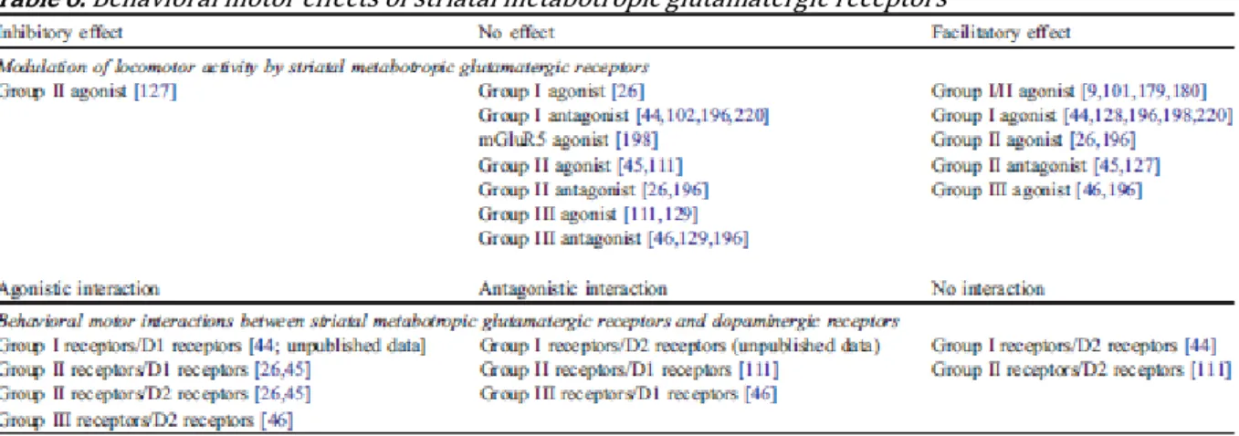
Behavioral motor intrinsic effects and interactions of
striatal dopaminergic and glutamatergic receptors
BEHAVIORAL MOTOR RESPONSES PRODUCED BY STRIATAL DOPAMINERGIC
RECEPTORS (TABLE 4; FIG. 3A)
The functional and behavioral effects of dopamine and dopamine receptor agonists and antagonists have been extensively investigated (Fig. 3A, Table 4). Injection of the D1-like receptor agonist SKF 38393 (0.3 – 10 µg, i.e., 0.9 – 30 µmol) in the nucleus accumbens induced a dosedependent increase in locomotor activity [39,44 – 47,56,68,137,138,197,227], which is assumed to reflect the predominant control of the direct pathway by D1-like receptors . Only rare studies show no effect on locomotion after injection of the D1 receptor agonist SKF 38393 in the nucleus accumbens [141,165] or of the D1 receptor agonist fenoldopam in the caudate– putamen [136]. Conversely, application of a specific D1-like receptor antagonist in the nucleus accumbens [137,153,165] or in the caudate–putamen [153] has been shown to decrease locomotor activity.
The physiology of D2-like receptors in the striatum appears more complex, probably due to the permissive, enabling, role of D1-like receptors on postsynaptic D2-like receptors activation [217,218,225], which mechanisms are still not elucidated. As a consequence, it is generally assumed that the effects observed following D2-like receptor agonist administration reflect dopaminergic processes placed under the control of D2-like autoreceptors. Consistent with a presynaptic localization of D2-like receptors on dopaminergic terminals arising from the substantia nigra and the ventral tegmental area, and in accord with the decrease in striatal dopamine release produced by D2-like receptor activation [79], administration in the nucleus accumbens of the D2-like receptor agonists LY 171555 (also named quinpirole; 1 – 15 µg, i.e., 4– 60 µmol), PD 128907 (0.5– 5 Ag, i.e., 1.75– 17.5 µmol) or of 7-OH-DPAT (2.5– 5 µg, i.e., 7.5 – 15 µmol) has been repeatedly found to decrease motor activity ([32,39,44 – 47,141,197] but [136]). However, depending on the dose used, others have found either a decrease or an increase in locomotor activity following administration of D2-like receptor agonists. Thus, infusion of LY171555 in the nucleus accumbens or in the caudate–putamen of awake rats has been reported to induce a biphasic effect with low doses (0.1 to 3 µg, i.e., 0.4 – 12 µmol) producing an increase in locomotor activity and high doses (5 to 40 µg, i.e., 20–160 µmol) a reduction of locomotor activity [56,60,68,215], although others have found opposite dose-dependent effects with the D2-like receptor agonist PD 128907 [167]. Finally, only rare studies found no significant effect on locomotor activity following activation of D2-like receptors [165]. These discrepancies may
depend on the level of basal locomotor activity and on D2-like autoreceptor occupancy [227]. Conversely, focal injection of D2-like receptor antagonists in the nucleus accumbens or the caudate–putamen resulted in a decrease in motor activity [74,153,165,214], which is assumed to reflect striatal postsynaptic mechanisms.
Whatever the compounds used, behavioral (but also electrophysiological) studies have repeatedly demonstrated that D1-like receptors and D2-like receptors exert synergistic effects. Co-infusion of D1-like and D2-like receptor agonists in the nucleus accumbens or the caudate putamen, a pharmacological condition thought to activate both striatal D1-like receptors and presynaptic and postsynaptic D2-like receptors [1,2,217,218,225], was consistently found to result in a dramatic increase in locomotor activity compared to the behavioral effect produced by administration of D1-like receptor agonists alone [32,39,43– 47,56,58,68,78,92,165].
BEHAVIORAL MOTOR EFFECTS OF STRIATAL IGLU RECEPTORS (TABLE 5B)
INTRINSIC BEHAVIORAL MOTOR RESPONSES PRODUCED BY IGLU RECEPTORS (FIG. 3B)
Behavioral motor effects of NMDA receptors
Studies on the locomotor effects of NMDA iGlu receptor agonists consistently found that the NMDA receptor agonist NMDA had a facilitating action on locomotor activity, when given in the nanomolar range (7– 70 nmol./side, i.e., 1 – 10 µg/ side) in the caudate–putamen ([181,212] but [183]) or the nucleus accumbens [18,29,47,54,71,228] (Table 5). Similar locomotor effects were found following injection in the nanomolar range (1– 50 nmol./side, i.e., 0.2 – 10 µg/side) of the NMDA receptor antagonists AP5 and AP7 in the nucleus accumbens [29,47,97,154] and the caudate– putamen [183, 212]. These effects of NMDA on locomotor activity can be attributed to NMDA receptor mechanisms. Indeed, administration in the caudate–putamen of AP7 at a subefficient dose of 0.5 nmol. (0.1 µg), which produced no effect by itself on locomotor activity, blocked the increase in locomotor activity induced by intrastriatal administration of NMDA [212]. Similar findings were found with the NMDA receptor antagonist AP5. Administration in the nucleus accumbens of AP5 at subefficient doses in the nanomolar range (1– 15 nmol, i.e., 0.2 –3 µg/side) led to a reduction of the NMDA-induced increase in locomotor activity [47,71], but showed no effect on the increase in locomotor activity produced by AMPA [47]. Understanding and explaining such results, i.e., that agonists and antagonists of NMDA receptors both induce an increase in locomotor activity, represents a major challenge that is still unresolved. However, several hypotheses that are not mutually exclusive may emerge regarding such an apparent paradox: subpopulations of functionally distinct NMDA receptors could be involved, possibly composed of different subunits, located on different pathways and/or activated in a different fashion (either tonically or phasically). But, clearly, much more studies combining neuropharmacological and immunohistochemical approaches are needed to understand thoroughly the modulation of striatal activity by NMDA receptors.
Behavioral motor effects of AMPA/kainate receptors
A similar locomotor activation was found following injection in the nanomolar range of the agonist AMPA (0.1–2.5 nmol/side, i.e., 0.02–0.5 µg/side) and antagonist CNQX (2–3 nmol./side, i.e., 0.6–1 µg/side) in the nucleus accumbens [19,29,47,54,75,190,228]. Other data in contrast
found no effect on locomotor activity following injection of CNQX in the nucleus accumbens, but confirmed that blockade of AMPA/kainate receptors by NBQX, which preferentially acts at AMPA receptors relative to kainate receptors, produced an increase in locomotor activity [39]. The locomotor-activating properties of AMPA were found to be selective. Administration in the nucleus accumbens of the AMPA/kainate receptor antagonist CNQX at 3 nmol./side (1 µg/side) decreased the locomotor activity produced by AMPA, but not that produced by NMDA [47]; similar findings were found with the AMPA/kainate receptor antagonist GAMS that preferentially acts at AMPA receptors relative to kainate receptors [190].
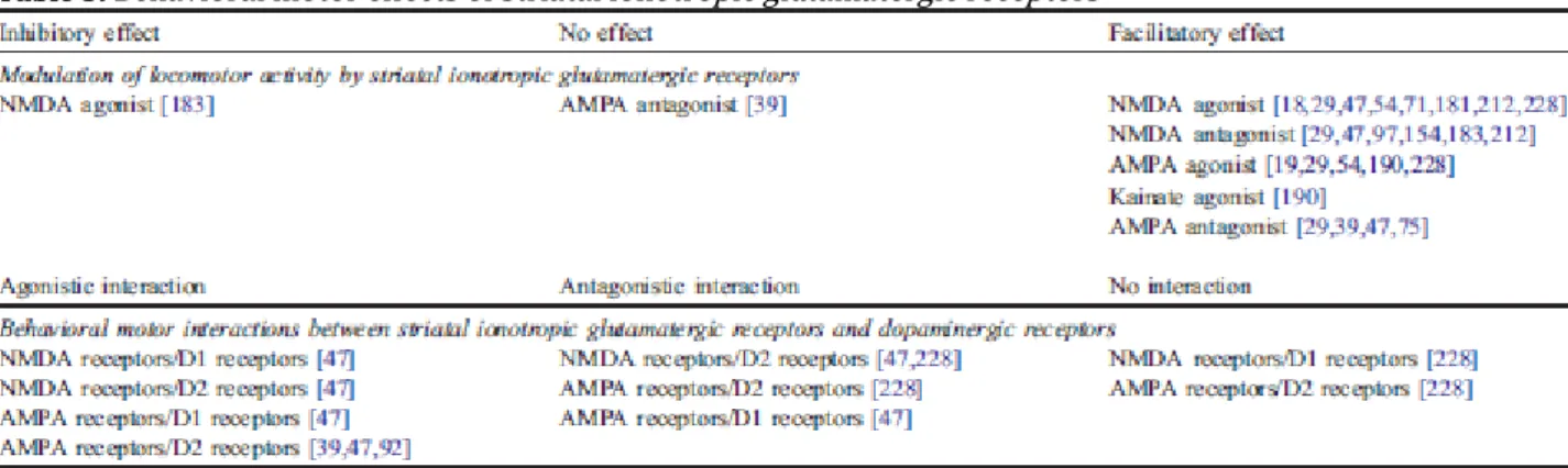
Fig. 3. (A) Illustration of the locomotor effects of striatal dopaminergic receptors. Activation of D1-like receptors increases locomotor activity, which is likely to be due to the predominant expression of D1-like receptors on the direct striato-nigral pathway. The physiology of D2-like receptors in the striatum appears more complex, probably due to the permissive, enabling, role of D1-like receptors on postsynaptic D2-like receptors activation, which mechanisms are still not elucidated. As a consequence, it is generally assumed that the effects observed after the administration of a D2-like receptor agonist alone reflect dopaminergic processes placed under the control of D2-like autoreceptors. Indeed, consistent with the decrease in striatal dopamine release produced by D2-like receptor activation, administration in the striatum of a D2-like receptor agonist is very generally found to produce a reduction of motor activity. However, it should be noted that other workers found an increase of locomotor activity following the injection in the striatum of a like receptor agonist. Infusion of non selective D1-like and D2-like receptor agonists or of a mixture of a selective D1-D2-like receptor agonist and of a selective D2-like receptor agonist, a pharmacological condition thought to activate both striatal D1-like receptors and presynaptic and postsynaptic D2-D1-like receptors, has been consistently found to exert synergistic effects and to produce a much greater increase in locomotor activity, as compared to the injection of D1-like receptor agonists. Blockade of D1-like and/or D2-like receptors reduces motor activity (not illustrated). (B) Locomotor effects of striatal ionotropic glutamatergic (iGlu) receptors. Investigations on the locomotor effects iGlu receptors consistently found that activation of these receptors in the striatum had a facilitating action on locomotor activity that is likely to be mediated in a phasic manner through the direct pathway. Emphasizing the complexity of the glutamatergic function in the striatum, it should be noted that facilitating locomotor effects are also found following blockade of iGlu receptors, which effects are likely to be mediated through the indirect pathway in a tonic fashion (not shown). (C) Locomotor effects of striatal metabotropic glutamatergic (mGlu) receptors. Activation of group I mGlu receptors increases locomotor activity that may result from a phasic facilitating action on the direct striato-nigral pathway. Consistent with both anatomical and neurochemical findings of a presynaptic localization of group II mGlu receptors on glutamatergic terminals in the striatum, blockade of group II mGlu receptors in the NAcc, but not in the CP, increases locomotor activity. Further emphasizing the complexity of the glutamatergic function in the striatum, activation of group II or group III mGlu receptors induces an increase in locomotor activity likely to be due to be mediated through the indirect pathway. AMPA: AMPA ionotropic glutamatergic receptors; D1: D1-like dopaminergic receptors; D2: D2-ike dopaminergic receptors; DA: dopamine; Glu: glutamate; GPe: globus pallidus; KA: Kainate ionotropic glutamatergic receptors; mGlu I: group I metabotropic glutamatergic receptors; mGlu II: group II metabotropic glutamatergic receptors; mGlu III: group III metabotropic glutamatergic receptors; NMDA: NMDA ionotropic glutamatergic receptors; SNR: substantia nigra pars reticulata; STN: subthalamic nucleus.
BEHAVIORAL MOTOR INTERACTIONS BETWEEN IGLU RECEPTORS AND DOPAMINERGIC
RECEPTORS (FIG. 4A)
Existing literature on behavioral motor interactions between dopaminergic receptors and iGlu receptors is not extensive as studied by local injections in the striatum complex of ‘‘intact dopamine’’ animals and gave rise, in some cases, to controversial findings (Fig. 4A).
Behavioral motor interactions between NMDA receptors and dopaminergic receptors.
Early data reported no behavioral interaction in the nucleus accumbens between NMDA receptor activation and D1-like receptor activation by the D1-like receptor agonist SKF 38393, but provided evidence of a blocking effect of the D2-like receptor agonist LY 171555 on the increase in locomotor activity produced by NMDA receptor activation [228]. In contrast, recent investigations utilizing 5-fold lower doses of NMDA (that led to no behavioral effect when given alone) and 4-fold lower doses of SKF 38393 and LY 171555 provided evidence of behavioral interactions between NMDA receptors and both D1-like receptors and D2-like receptors [47]. Given the dual effect of glutamate on striatal dopamine release, that is illustrated by the fact that low doses of NMDA inhibit dopamine release while high doses of NMDA increase it, the different doses of NMDA used between both studies may account for the discrepant data above. However, it should be emphasized that the potentiating and blocking effect of NMDA receptor activation, respectively, on the increase in locomotor activity produced by activation of D1-like receptors and co-activation of D1-like and D2-like receptors [47] is in good agreement with cellular, electrophysiological, and neurochemical investigations that have consistently shown, using in vivo, ex vivo or in vitro preparations from the caudate putamen, the nucleus accumbens, the cortex or the substantia nigra, a synergistic action between NMDA receptors and D1-like receptors [34,35, 61,73,132,184,185,241] and an antagonist action between NMDA receptors and D2-like receptors [106,132,241].
Altogether, these findings indicate that NMDA receptors interact both with D1-like receptors, located on striato-nigral medium spiny output neurons, and with D2-like presynaptic and postsynaptic receptors that are located, respectively, on dopaminergic and glutamatergic terminals and on striatopallidal spiny projection neurons in the striatum. In addition, these data further suggest that the glutamate neurotransmission mediated by NMDA receptors may interact with the dopaminergic neurotransmission mediated by D1-like receptors in a phasic excitatory fashion enabling a facilitatory control of striato-nigral neurons. They also suggest that the glutamatergic neurotransmission mediated by NMDA receptors may interact with the dopaminergic transmission mediated by D2-like postsynaptic receptors in a phasic fashion that would be devoted to an inhibitory control of these neurons.
Table 5. Behavioral motor effects of striatal ionotropic glutamatergic receptors
Behavioral motor interactions between AMPA/ kainate receptors and dopaminergic receptors. Early data reported no behavioral interaction in the nucleus accumbens between AMPA receptor activation and D1-like receptor activation by the D1-like receptor agonist SKF 38393, but provided evidence of a blocking effect of the D2-like receptor agonist LY 171555 on the increase in locomotor activity produced by AMPA receptor activation [228]. In contrast, recent investigations utilizing much lower doses of AMPA (25-fold less) and lower doses of SKF 38393 and LY 171555 (4-fold less) provided evidence of behavioral interactions between AMPA receptors and dopaminergic receptors. Infusion in the nucleus accumbens of AMPA or CNQX, an AMPA/kainate receptor antagonist, was found to reduce the increase in locomotor activity produced by the activation of D1-like receptors by SKF 38393 [47]. Administration of CNQX, but not of AMPA, further potentiated the decrease in basal locomotor activity induced by the D2-like receptor agonist LY 171555 [47] or the D2-like receptor agonist 7-OH-DPAT [39]. In addition, administration of AMPA and of CNQX, respectively, potentiated and reduced the locomotor-activating effects induced by co-infusion of SKF 38393 + LY 171555 [47,92].
Altogether these data indicate that AMPA/kainate receptors interact with D1-like and D2-like presynaptic and postsynaptic receptors. In addition, they further suggest that the glutamatergic neurotransmission mediated by AMPA/kainate receptors may interact with the dopaminergic transmission mediated by D1-like receptors in phasic manner that would be devoted to an inhibitory control of striato-nigral GABAergic output neurons. Glutamate–dopamine interactions mediated by AMPA/kainate and D2-like postsynaptic receptors, respectively, may involve phasic mechanisms of control allowing a facilitatory modulation of striato-pallidal neurons.
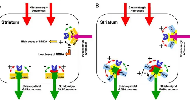
Fig. 4. Illustration of the interactions between dopaminergic receptors and both ionotropic glutamatergic (iGlu) receptors and metabotropic glutamatergic (mGlu) on locomotor activity. (A) NMDA receptors interact positively with D1-like receptors, leading to a facilitating action on locomotor activity mediated by D1-like receptor activation. In contrast, NMDA receptors interact negatively with D2-like postsynaptic receptors, leading to a reduction of the increase in locomotor activity induced by activation of these receptors. Consistent with neurochemical findings on glutamatergic control of striatal dopamine release, activation of NMDA receptors by low, physiological doses of NMDA potentiates the decrease in locomotor activity produced by activation of D2-like autoreceptors, whereas higher, possibly non-physiological doses of NMDA inhibits it. Constrating with the role of NMDA receptors, AMPA/kainate receptors interact negatively with like receptors, leading to a reduction of the locomotor activity mediated by D1-like receptor activation. It should be noted that blockade of AMPA/kainate receptors also inhibit the increase in locomotor activity induced D1-like receptor activation. Stimulation of AMPA/kainate receptors potentiate the locomotor activity produced by D2-like postsynaptic receptors activation but inhibit the decrease in locomotor activity produced by activation of D2-like autoreceptors; conversely, blockade of AMPA/kainate receptors potentiate the decrease in locomotor activity induced by D2-like autoreceptor activation (not shown). (B) Group I metabotropic glutamatergic (mGlu) receptors interact positively with D1-like receptors, leading to an increase of locomotor activity produced by D1-like receptor activation. In contrast, group I mGlu receptors interact negatively with D2-like postsynaptic receptors, leading to a reduction of the increase in locomotor activity induced by activation of these receptors. Some studies showed that activation of group II mGlu receptors potentiates the locomotor activity mediated by D1-like receptors, while others found that activation of group II mGlu receptors oppose the D1-like receptor locomotor response. In addition, group II mGlu receptors favor the locomotor response produced by D2-like receptors, leading to a potentiation of the increase of locomotor activity induced by D2-like postsynaptic receptor activation and of the decrease of locomotor activity produced by activation of D2-like autoreceptors activation. Group III mGlu interact negatively with D1-like receptors, leading to a decrease of the locomotor activity produced by activation of D1-like receptors, and positively with D2-like receptors, to leading to a potentiation of the increase of locomotor activity induced by D2-like postsynaptic receptor activation and of the decrease of locomotor activity produced by activation of D2-like autoreceptors activation. A/K: AMPA/kainate ionotropic glutamatergic receptors; D1: D1-like dopaminergic receptors; D2: D2-like dopaminergic receptors; mGlu I: group I metabotropic glutamatergic receptors; mGlu II: group II metabotropic glutamatergic receptors; mGlu III: group III metabotropic glutamatergic receptors; NMDA: NMDA ionotropic glutamatergic receptors.
BEHAVIORAL MOTOR EFFECTS OF STRIATAL MGLU RECEPTORS (TABLE 6)
INTRINSIC BEHAVIORAL MOTOR RESPONSES PRODUCED BY MGLU RECEPTORS (FIG.
3C)
Studies on the behavioral motor effects of mGlu receptors within the striatum started with behavioral investigations from Schoepp’s group [179,180] that showed that an acute unilateral injection of the non-selective group I/group II mGlu receptor agonist ACPD into the caudate– putamen produced rotation contralateral to the injection side (Table 6). Similar results showing behavioral activation following bilateral injection of ACPD into the caudate– putamen or the nucleus accumbens were obtained afterwards [9,101]. Then, further studies were conducted, using group selective mGlu receptor agonists and antagonists, on the role of mGlu receptors in the control of locomotor activity.
Behavioral motor effects of group I mGlu receptors.
Studies on the locomotor effects of the selective group I mGlu receptors consistently found that the selective group I mGlu receptor agonist DHPG had a facilitating action on locomotor activity, when infused locally in the caudate–putamen [128,220] or the nucleus accumbens ([44, 196,198] but [26]). This effect was blocked by co-treatment with group I mGlu receptor antagonists [41,196,220], but not group II or group III mGlu receptor antagonists [44]. All of the group I mGlu receptor antagonists used, either CPC-COEt at 10 nmol. (i.e., 2.5 µg) [196], PHCCC at 10 nmol (i.e., 3 µg) [220] or S-4-CPG at 10 nmol (i.e., 2 µg) [44], as well as AIDA at 9 nmol (i.e., 2 µg) [102], had no effect by themselves on locomotor activity when administered alone in the nucleus accumbens or the caudate–putamen; this agrees with anatomical data that showed that group I mGlu receptors are not located on glutamatergic terminals in the striatum, and further suggests that group I mGlu receptors-mediated glutamatergic transmission exerts a phasic facilitating control on striatal activity. In addition, data have shown that the effect of DHPG on locomotor activity was not altered by co-treatment with the mGlu5 receptor antagonist MPEP, indicating that DHPG may act preferentially at the mGlu1 receptor rather than at the mGlu5 receptor; in contrast with the motor-activating properties of DHPG, infusion in the nucleus accumbens of the mGlu5 selective agonist CHPG did not elicit an increase in locomotor activity [198].
It is not clear whether the locomotor-activating effects of DHPG injected directly in the caudate– putamen are independent of iGlu receptors. The NMDA receptor antagonist CPP did not alter the DHPG-induced behavioral activation [128]. However, AMPA receptor blockade has been reported to have no effect [128] or to inhibit [198] locomotor activity produced by local administration of DHPG.
Behavioral motor effects of group II mGlu receptors.
Investigations on the role and function of group II mGlu receptors gave rise to more controversial data. Depending on the ligands used and/or of the brain area explored, activation of group II mGlu receptors had no effect, or resulted in an increase or a decrease in locomotor activity. Infusion in the nucleus accumbens of the group II mGlu receptor agonists L-CCG-I [26] and DCG-IV [196] increased locomotor activity. The motor-activating properties of L-CCG-I and DCG-IV were, respectively, attenuated, but not blocked, by co-treatment with the group II/III
mGlu receptor antagonist MCCG at 50 nmol (i.e., 8.7 µg) [26] or the group II/III mGlu receptor antagonist MPPG at 10 nmol (i.e., 2.5 µg) [196], which had no effect by themselves on locomotor activity when administered alone. One potential limitation to this observation is the reported action of DCG-IV on NMDA receptors [28]. However, co-administration of the NMDA receptor antagonist CPP did not alter the increase in locomotor activity produced by DCG-IV [196], indicating that the locomotor-activating properties of DCG-IV are mediated through mGlu receptors. Alternatively, consistent with anatomical and neurochemical data of a presynaptic localization, as well as a postsynaptic localization, of group II mGlu receptors on glutamatergic terminals in the striatum, data have reported that infusion in the nucleus accumbens of the group II mGlu receptor antagonist LY 341495 induced a dose-dependent increase in motor activity, effect that was blocked by co-treatment with the group II mGlu receptor agonist APDC that had no behavioral effect by itself [45].
In contrast with its effect in the nucleus accumbens, administration in the caudate– putamen of L-CCG-I showed no effect [111] and DCG-IV led to a decrease in locomotor activity that was partly reversed by co-treatment with 10 nmol (i.e., 2.8 µg) MSOPPE, a group II mGlu receptor antagonist that produced a slight increase in locomotor activity when given alone [127].
Table 6. Behavioral motor effects of striatal metabotropic glutamatergic receptors
Behavioral motor effects of group III mGlu receptors.
Studies on the functional role of group III mGlu receptors found that infusion of the group III mGlu receptor agonist L-AP4 in the nucleus accumbens [46,196], but not in the caudate– putamen [111,129], led to an increase in locomotor activity that was blocked by cotreatment with the group III mGlu receptor antagonist MPPG [43]. Infusion of MPPG in the nucleus accumbens [46,196] or the caudate–putamen [129] was found to be ineffective by itself at changing locomotor activity.
BEHAVIORAL MOTOR INTERACTIONS BETWEEN MGLU RECEPTORS AND
DOPAMINERGIC RECEPTORS (FIG. 4B)
A few studies have investigated behavioral interactions between mGlu receptors and dopaminergic receptors within the striatum complex of Fintact_ animals by using locally applied drugs.