Science Arts & Métiers (SAM)
is an open access repository that collects the work of Arts et Métiers Institute of
Technology researchers and makes it freely available over the web where possible.
This is an author-deposited version published in:
https://sam.ensam.eu
Handle ID: .
http://hdl.handle.net/10985/19447
To cite this version :
Mario MEKHAEL, Chris LABAKI, Aren Joe BIZDIKIAN, Ziad BAKOUNY, Joeffroy OTAYEK, Fares
YARED, Abir MASSAAD, Wafa SKALLI, Ismat GHANEM, Ayman ASSI - How do skeletal and
postural parameters contribute to maintain balance during walking? Human Movement Science
-Vol. 72, p.1-10 - 2020
Any correspondence concerning this service should be sent to the repository
Administrator :
[email protected]
How do skeletal and postural parameters contribute to maintain
balance during walking?
Mario Mekhael
a, Chris Labaki
a, Aren Joe Bizdikian
a, Ziad Bakouny
a, Joe
ffroy Otayek
a,
Fares Yared
a, Abir Massaad
a, Wafa Skalli
b, Ismat Ghanem
a, Ayman Assi
a,b,⁎aFaculty of Medicine, University of Saint-Joseph in Beirut, Lebanon
bInstitut de Biomécanique Humaine Georges Charpak, Arts et Métiers ParisTech, Paris, France
A R T I C L E I N F O Keywords: Balance 3D gait analysis Spine Pelvis Hip Lower limbs A B S T R A C T
Introduction: Maintaining balance during gait allows subjects to minimize energy expenditure and avoid falls. Gait balance can be measured by assessing the relationship between the center of mass (COM) and center of pressure (COP) during gait. Demographics, skeletal and postural parameters are known to influence gait balance.
Purpose: What are the determinants of dynamic balance during gait in asymptomatic adults among skeletal and demographic parameters?
Methods: 115 adults underwent 3D gait analysis and full-body biplanar X-rays. Angles between the COM-COP line and the vertical were calculated in frontal and sagittal planes during gait: maxima, minima, and ROM were evaluated. Full-body 3D reconstructions were obtained; skeletal and postural parameters of the spine (lumbar lordosis, thoracic kyphosis, sagittal vertical axis SVA), pelvis (pelvic tilt and incidence, acetabular orientation in the 3 planes) and lower limbs (neck shaft angle femoral and tibial torsions) were calculated. A univariate followed by a mul-tivariate analysis were computed between the COM-COP parameters and skeletal and demo-graphic parameters.
Results: The univariate analysis showed that in the frontal plane, maximum (4.6°) of the COM-COP angle was significantly correlated with weight (r = 0.53), age (r = 0.28), height (r = 0.35), SVA (r = 0.23), T1T12 (r = 0.24) and pelvic width (r = 0.25).In the sagittal plane, maximum COM-COP (19.7 ± 2.8°) angle was significantly correlated to acetabular tilt (r = 0.25) and acetabular anteversion (r = 0.21). The multivariate analysis showed that, in the frontal plane, an increase in the maximum of the COM-COP angle was determined by a decreasing height (β = −0.28), an increasing weight (β = 0.48), being a male (β = −0.42), and an increasing posterior acetabular coverage (β = 0.22). In the sagittal plane, an increasing maximum COM-COP angle was determined by a decreasing height (β = −0.38) and an increasing SVA (β = 0.19).
Conclusion: Frontal imbalance appeared to be mainly correlated to demographic parameters. Sagittal imbalance was found to be correlated with weight, height, acetabular parameters and SVA. These results suggest that in addition to demographic parameters, acetabular parameters and SVA are important determinants of balance during gait.
⁎Corresponding author at: Laboratory of Biomechanics and Medical Imaging, Faculty of Medicine, University of Saint-Joseph, Campus of
Innovation and Sports, Damascus street, Beirut, Lebanon.
E-mail addresses:[email protected](W. Skalli),[email protected](A. Assi).
1. Introduction
Balance maintenance depends on multiple systems such as the vestibular, proprioceptive, visual and musculoskeletal systems (Maurer, Mergner, & Peterka, 2006). The concept of the conus of economy presents the variability in standing posture which allows the maintenance of static equilibrium while minimizing energy expenditure and fatigue on the muscles of the back and the lower limbs. An unstable standing posture can lead to subsequent pain and disability (JeanDubousset, 1994). In dynamic, balance consists in minimizing energy expenditure and avoiding fall during walking (Winter, 1995).
Several techniques for evaluating balance during gait have already been proposed. This has been evaluated either by determining local and orbital dynamic stability (Dingwell, 2006) by using tri-axial accelerometry (Auvinet et al., 2002) or by studying the center of mass/center of pressure COM-COP relationship (Lee & Chou, 2006). The center of mass (COM) is defined as the barycenter of the
subject, while the center of pressure (COP) is the application point of the ground reaction force captured by forceplates during gait. The angle between the COM-COP and the vertical can be evaluated in both frontal and sagittal planes, and its increase is correlated with increased instability. During the gait cycle, the sagittal COM-COP angle presents large variability in contrast with the frontal COM-COP, whose variations are limited (Lee & Chou, 2006). The variation of these two angles during gait has been shown to reflect
dynamic gait balance (Lee & Chou, 2006;Paul et al., 2014).
While demographic parameters (age, sex, weight and height) are known to affect gait (Gao et al., 2019;Hamacher et al., 2019), their relationship with gait stability is still unelucidated. Moreover, previous studies have determined that skeletal and postural alterations can affect gait balance. For instance, the correction of coronal and sagittal spinal malalignment in scoliotic patients decreased mediolateral excursion during gait (Paul et al., 2014). In another setting, patients with adult spinal deformity (ASD) are known to have alteration of their postural malalignment that affects not only their spine and pelvis but also the positioning of their head and lower limbs; a chain of compensatory mechanisms is established in order to maintain a balanced standing posture in the conus of economy (Diebo et al., 2019). Patients with ASD are known to have altered quality of life that affects their daily life activities such as walking. Several studies started to highlight gait abnormalities in patients with ASD (Assi et al., 2019;Haddas, Ju, Belanger, & Lieberman, 2018). However, it is still unknown how their skeletal and postural deformities are related to their gait balance. Any investigation on the relationship between skeletal or postural deformities and gait balance in the setting of ASD or other muscu-loskeletal pathologies would benefit from a baseline or normative relationship that is still unknown.
As afirst step, the aim of this study was to evaluate the normative relationship between gait balance and both demographic and skeletal and postural parameters in asymptomatic subjects.
2. Materials and methods
2.1. Study design
This is a cross-sectional IRB approved study (CEHDF285–2016) evaluating the different determinants of balance during gait in asymptomatic adult subjects recruited from our institution. All subjects signed an informed consent form. Subjects were excluded if they presented with prior history of self-reported back pain or orthopedic surgery. Exclusion criteria included also the presence of scoliosis (frontal Cobb > 10°), Scheuermann's kyphosis, leg length discrepancy or any radiologic determinant of adult spinal de-formity (Pellisé et al., 2014) detected on radiographs.
2.2. Data collection
Demographics (sex, age, weight and height) were collected. Subjects underwent three-dimensional gait analysis (3DGA) using a Vicon (Vicon Motion Systems, Oxford, UK) optoelectronic motion system (7 MX3 infrared cameras, 200 Hz). Marker placement was based on the modified Helen Hayes protocol and was applied as recommended in the Plug-in-Gait® model. Subjects were asked to walk along a 10-m walkway, at a self-selected speed, equipped with two calibrated forceplates (OR6-7-1000 AMTI®, MA, USA). Several trials were recorded and only those where the subjects placed a full foot on each of the forceplates were considered. The pipeline in Workstation® (Vicon Motion Systems, Oxford, UK) was used to process data: fill gap routine ± 10 frames and Woltring filter with a scale of 10. Consistent kinematic and kinetic curves were validated using Polygon® (Vicon Motion Systems, Oxford, UK) and inconsistent trials were eliminated. One representative trial was then considered.
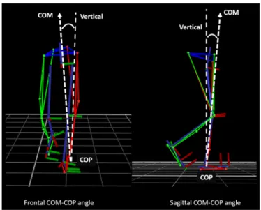
The center of mass (COM) was determined as the vector sum of the individual segment mass vectors, estimated from the kinematic centroid obtained from the Plug-In-Gait model. The center of pressure (COP) was determined as the application point of the ground reaction force obtained from the forceplates. The COP data were time-synchronized with motion data. During the double-stance phase, when the COP changed between the forceplates, a resultant COP was calculated for both feet (Jian, Winter, Ishac, & Gilchrist, 1993).The COM-COP angle with the vertical was calculated in both the frontal and sagittal planes during the gait cycle for each subject (Lee & Chou, 2006) (Fig. 1).
In the frontal plane, the maximum of the COM-COP angle was chosen as the highest value for the left or the right side, depending on which was higher. As for the sagittal plane, the maximum value was considered to be the highest positive value and therefore when the COP was in front of the COM. The minimum of the COM-COP was the lowest negative value, therefore the COP being behind the COM.
The maxima, minima and range of motion (ROM) of the COM-COP angle in the frontal and sagittal planes were considered as parameters of gait balance and were calculated over the whole gait cycle for each subject. An increase in any of these parameters
would indicate a decrease in balance.
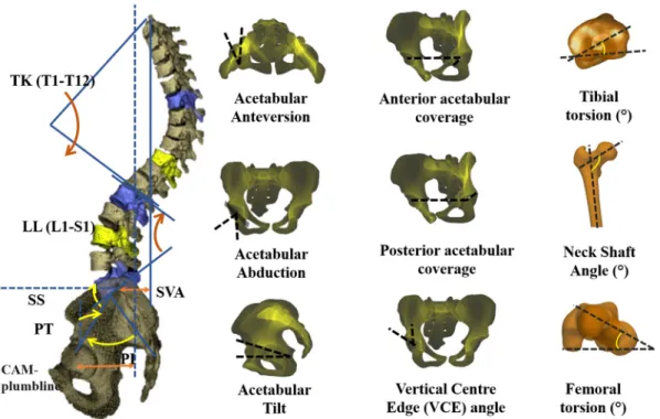
Following the gait analysis, subjects underwent a full body biplanar X-ray exam (EOS Imaging®, Paris, France). All subjects were placed in the consensual free-standing position with shouldersflexed at 45° and hands placed on the zygomatic processes, and with slightly shifted feet (Chaibi et al., 2012). This position was adopted to avoid the superimposition of subjects' arms over their spines, or the left and right femoral condyles, on lateral radiographs. Three-dimensional reconstructions of the spine and the lower limbs were obtained from the biplanar X-rays using SterEOS® (EOS imaging, Paris, France). Three-dimensional reconstruction of the pelvis was obtained using a specific software (Arts et Métiers ParisTech, Paris, France). Then, skeletal and postural radiological parameters of the spine, pelvis, lower limbs and global posture were calculated: sagittal vertical axis SVA (center of C7 vertebral body plumbline to the posterior corner of the sacral plate), CAM plumb line (center of auditory meatus plumbline to the middle of the hip axis), thoracic kyphosis (T1T12), lumbar lordosis (L1S1), pelvic incidence, pelvic tilt, sacral slope (Bendaya et al., 2015; J.Dubousset et al., 2008;
Dubousset, Charpak, Skalli, & Kalifa, 2007;Duval-Beaupere et al., 2002; J.Legaye, Duval-Beaupère, Hecquet, & Marty, 1998;Jean Legaye & Duval-Beaupère, 2005;Vialle et al., 2005), femoral neck length, neck shaft angle, femoral torsion, valgus/varus of the knee, femoral anteversion, tibial torsion, tibial mechanical angle, acetabular abduction, acetabular anteversion, acetabular tilt, vertical center edge angle (VCE) (Anda, Svenningsen, Dale, & Benum, 1986;Stem et al., 2006;Zilber, Lazennec, Gorin, & Saillant, 2004), anterior (AASA) and posterior (PASA) acetabular sector angles (Anda et al., 1986), percentage of acetabular coverage by the femoral head (Humbert, Carlioz, Baudoin, Skalli, & Mitton, 2008) and the sacro-acetabular angle (Fig. 2).
2.3. Statistical analysis
Descriptive statistics (mean and standard deviation) were calculated for the COM-COP angles in both the sagittal and frontal planes, as well as for the spino-pelvic, hip and lower limb parameters.
In order to evaluate the relationship between the gait balance (COM-COP) variables, and demographic and skeletal parameters, a univariate analysis was conducted using Pearson's correlation test. The level of significance was set at 0.05. P-values were adjusted using Benjamini-Hochberg procedure to account for multiple testing (n = 312 correlations).
Then, in order to evaluate the main determinants of gait balance from demographic and skeletal parameters while accounting for correlations between predictor variables, a multivariate analysis was conducted using a stepwise multiple linear regression (SMLR). Probability for entry was 0.05 / Probability for removal was 0.1. The dependent variables were the COM-COP angle parameters in the sagittal and frontal planes; the independent variables were demographics and skeletal parameters that showed significant correla-tions to COM-COP variables in the univariate analysis, as well as other non-significant parameters that were judged to be clinically important to be included in the model. In total, 5 models were run for the following COM-COP parameters: maximum frontal angle, ROM frontal angle, minimum sagittal angle, maximum sagittal angle and ROM sagittal angle. The level of significance of the model was adjusted using Bonferroni correction to account for multiple models (n = 5; thus, level of significance was 0.01 instead of 0.05). Moreover, the level of significance for each predictor variable was adjusted using the Benjamini-Hochberg procedure in order to account for the number of predictor variables included in the model (n = 18). The validity of multiple linear regression assumptions was checked. The adjusted R2of the models as well asβ and p-values of predictor variables were reported.
Statistics were undertaken using Xlstat® (version 2019.1.2, Addinsoft, Paris, France).
3. Results
In total, 115 asymptomatic subjects (age: 29 ± 7 years [18–59], weight: 70 ± 9 Kgs, height: 169 ± 10 cm, 57F: 58 M) were included in this study.
3.1. Descriptive statistics
Subjects had an average pelvic tilt of 12° with a pelvic incidence of 48°. Their L1S1 lumbar lordosis had a mean of 60° associated with a T1T12 thoracic kyphosis of 45°. Their C7 was positioned in the sagittal plane behind the sacrum (SVA = -12 cm) as well as their center of auditory meatus (CAM-HA = -25 cm). They had a pelvic width of 25 cm on average. The acetabulum was tilted with an average of 23°. The abduction had a mean of 55° and the anteversion was 17°. The acetabulum was covered posteriorly with a mean of 96° (PASA) and anteriorly 60° (AASA). The lateral coverage of the acetabulum (VCE) had a mean value of 30° (detailed values and other parameters are displayed inFig. 3).
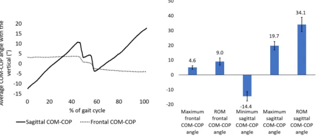
In the frontal plane, the maximum of the COM-COP angle reached 4.6 ± 1.3° and the ROM was 9.0 ± 2.4°. In the sagittal plane, the maximum of the COM-COP angle reached 19.7 ± 2.8°, the ROM was 34.1 ± 4.8° and the minimum was−14.4 ± 3.21° (Fig. 4).
3.2. Univariate analysis
As afirst step in assessing the relationship between demographics and skeletal parameters with gait balance, simple correlations were computed between the variables.
The COM-COP angles were significantly correlated to the following demographic and skeletal parameters (Fig. 5):
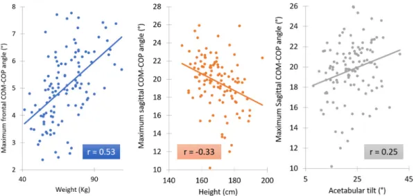
An increase of weight (r = 0.53,Fig. 6), age (r = 0.22 to 0.28), and height (r = 0.35 to 0.37) was correlated to an increase in the maximum and ROM of the frontal COM-COP angle. Regarding skeletal and postural parameters, an increase of a forward back (SVA: r = 0.2 to 0.23), thoracic kyphosis (T1T12: r = 0.22 to 0.24) and pelvic width (r = 0.24 to 0.25) was correlated to an increase in the maximum and ROM of the frontal COM-COP.
However, in the sagittal plane, an increase of height (r =−0.28 to −0.33,Fig. 6) was correlated to a decrease of maximum and ROM of the sagittal COM-COP. An increase of acetabular tilt (r = 0.25) and acetabular anteversion (r = 0.21) were correlated to an increase of the maximum sagittal COM-COP. In addition, a decrease in anterior acetabular coverage (r =−0.21) was correlated to an increase of the ROM sagittal COM-COP.
Fig. 3. Spino-pelvic, hip and global postural parameters calculated in 115 asymptomatic adults.
3.3. Multivariate analysis
Furthermore, in order to better investigate the main determinants of gait balance parameters among demographic and skeletal and postural parameters, multiple linear regressions were computed. The following parameters that were shown to be significant in the univariate analysis as well other parameters that were judged to be clinically significant were included in the multivariate models as dependent variables: age, weight, height, sex, SVA, CAM-HA, L1S1, T1T12, pelvic tilt, pelvic incidence, VCE, percentage of acetabular coverage over the femoral head, sacro-acetabular angle, acetabular tilt, acetabular abduction, acetabular anteversion, PASA, AASA.
Instability during gait was determined by the increase of the following parameters in each plane as shown in the multivariate analysis:
In the frontal plane, an increase in the maximum of the COM-COP angle was determined (p < .001) at 39% (adjusted R2) by an
increasing weight (β = 0.48, p < .001), being a male (β = 0.42, p < .001) and an increasing posterior acetabular coverage (β = 0.22, p = .006). An increasing ROM of the frontal COM-COP angle was determined (p < .001) at 35% by an increasing weight (β = 0.32, p = .002), being a male (β = 0.37, p = .001), and an increasing posterior acetabular coverage (β = 0.22, p = .007).
Fig. 4. Sagittal and frontal COM-COP angles calculated in 115 asymptomatic subjects.
In the sagittal plane, an increasing maximum of the COM-COP angle was determined (p < .001) at 13% by a decreasing height (β = −0.38, p < .001) and an increasing SVA (β = 0.19, p = .004). An increasing ROM of the sagittal COM-COP was solely determined (p = .01) at 7.3% by a decreasing height (β = −0.28, p = .002).
4. Discussion
In this study, the examination of skeletal parameters and gait of 115 asymptomatic adult subjects showed that gait balance in the frontal and sagittal planes was influenced by both anthropometric and skeletal parameters. Frontal imbalance appeared to be more pronounced with increasing weight, height, age, SVA, T1T12 and pelvic width. Sagittal imbalance was found to be associated with an increased acetabular tilt, acetabular anteversion and a decreased anterior acetabular coverage. In addition, sagittal COM-COP was shown to increase with decreasing height and weight.
The hip skeletal parameters of our population were found to be similar to those previously reported in studies concerning asymptomatic adults (Tannast, Hanke, Zheng, Steppacher, & Siebenrock, 2015). Similarly, spino-pelvic parameters of this study were closely comparable with the results shown in previous studies (Bakouny et al., 2018).
The COM-COP parameters of our population were also similar to the ones reported in previous studies (Lee & Chou, 2006). The female sex appeared as being an important determining factor (adjusted R2 > 0.33) of both the maximum (β: −0.426, M being the reference) and the ROM (β: −0.371) of the frontal COM-COP. It is known that the female pelvic anatomy is significantly different compared to males (Kersnic et al., 1996;Wang et al., 2004): the female pelvis being larger and wider than the male pelvis which is taller, narrower, and more compact. This anatomic property could influence balance during gait, thereby possibly explaining the increased frontal instability reported in women.
An increased weight appeared to be related to frontal imbalance. It was correlated to both the maximum (r = 0.53) and the ROM of the frontal COM-COP (r = 0.53). These results were reinforced by the multivariate analysis which showed that weight is a determinant of both frontal maximum and ROM COM-COP angles. Thesefindings are further illustrated by a comparison of two subjects with differing weights who present important variations in their maximum frontal COM-COP angles (Fig. 7).
On the other hand, pelvic width was also correlated to both the maximum (r = 0.25) and the ROM (r = 0.24) of the frontal COM-COP angle suggesting that a wider pelvis morphology may be associated with an increased imbalance in the frontal plane.
Furthermore, the sagittal vertical axis is a commonly used parameter to study global alignment. The subject is considered to have sagittal malalignment if the SVA drops in front of the femoral heads (Jackson & McManus, 1994). This parameter was used in the several classifications for adult spinal deformity subjects as an inclusion criteria for subjects having an SVA of over 4 or 5 cm (Pellisé et al., 2014;Schwab et al., 2012). Glassman showed that an increased forward bending of the trunk is associated with increased pain and decreased function (Glassman et al., 2005). The magnitude of these symptoms was also found to increase with augmented imbalance, i.e. when the SVA increases. An increased SVA is well known to affect a subject's quality of life and is therefore associated with altered HRQOL measures (Kim et al., 2017). In this study, the SVA was found to be positively correlated with the maximum (0.23) and the ROM (0.20) of the frontal COM-COP angles. SVA was also shown to be a determinant of maximum sagittal COM-COP angle (β = 0.19). Thus, an increase of SVA, that is usually seen in elderly people and especially in adults with spinal deformity (Diebo et al., 2019), is related to increasing imbalance during gait in both frontal and sagittal planes (Assi, Kawkabani, et al., 2019). Future studies should investigate about causes of gait imbalance in subjects with adult spinal deformity (ASD) and to verify if surgical correction of SVA can improve gait stability in these patients.
In addition to global posture, several acetabular parameters were found to affect gait, and especially in the sagittal plane. An increased acetabular tilt (r = 0.25) and acetabular anteversion (r = 0.21) were found to be correlated with increased maximum
sagittal COM-COP angle. Also, a decreased acetabular anterior coverage (r =−0.21) is correlated to an increased sagittal ROM of the COM-COP angle. Thesefindings are further illustrated by a comparison of two subjects with considerable variations in their acet-abular parameters who present with different maximum sagittal COM-COP angles (Fig. 8). Additionally, posterior acetabular cov-erage appeared to be an important determinant (adjusted R2 > 0.35) of both maximum (β = 0.22) and ROM (β = 0.22) of the
frontal COM-COP angle. These results show that when the acetabulum is more tilted in the sagittal plane, thus decreasing acetabular Fig. 7. Example of the maximum frontal COM-COP angle in two subjects with different weights.
anterior coverage and increasing posterior acetabular coverage, this could contribute to increase sagittal imbalance during gait. These modifications in acetabular orientation are seen in subjects with adult spinal deformity due to their increased pelvic tilt that contributes to an increase of their acetabular tilt associated with a decreased anterior coverage and an increase of the posterior coverage of the (Assi et al., 2019). Thus, these acetabular orientation parameters might be related to the gait imbalance in subjects with adult spinal deformity. Future studies should test this hypothesis in patients with ASD.
Moreover, these results suggest that the orientation of the proximal hip, which forms the anatomical hinge between the head and trunk segments, where the centre of mass is located, and the lower limbs, which are the principal actors in gait, is a very important factor that correlates with sagittal dynamic imbalance, and therefore instability during gait.
This is thefirst study to evaluate the normative relationship between demographic and skeletal parameters with gait stability. This study has some limitations; it did not take into account the muscular component when assessing the determinants of gait stability. Future studies should include the muscular factor, using EMG or musculoskeletal models, when evaluating stability during walking. As discussed earlier, future studies should also investigate gait balance in subjects with skeletal deformities in the spine and the hip such as in subjects with adult spinal deformity.
In conclusion, this study evaluated the demographic and skeletal determinants of gait balance in asymptomatic adult subjects. The results suggest that in addition to demographic parameters, acetabular orientation and SVA are important determinants of both frontal and sagittal balance during gait. Future studies should analyze the stability during gait in patients with spinal or hip de-formities, therefore allowing an understanding of the deformity and its repercussion on walking, leading to better patient-oriented treatment options.
Acknowledgment
This research was funded by the University of Saint-Joseph (grant FM183). The funding source did not intervene in study design; in the collection, analysis and interpretation of data; in the writing of the report; and in the decision to submit the article for publication.
Declarations of Competing Interest
None.
Author Statement
All the authors certify that they have seen and approved thefinal version of the manuscript being submitted. They warrant that the article is the authors' original work, hasn't received prior publication and isn't under consideration for publication elsewhere.
References
Anda, S., Svenningsen, S., Dale, L. G., & Benum, P. (1986). The acetabular sector angle of the adult hip determined by computed tomography. Acta Radiologica: Diagnosis, 27(4), 443–447.
Assi, A., Kawkabani, G., Saliby, R.-M., Mekhael, M., Lafage, V., Kharrat, K., ... Ghanem, I. (2019). How do spino-pelvic deformities and postural malalignment in adults affect gait patterns? Gait & Posture, 73(73), 33–34.https://doi.org/10.1016/j.gaitpost.2019.07.111.
Assi, A., Mekhael, M., Kawkabani, G., Saliby, R.-M., Karam, M., Mjaess, G., ... Skalli, W. (2019). P 128 - posterior coverage of the hip might be responsible of limiting pelvic retroversion in patients with adult spinal deformity. Gait & Posture, 73, 502–503.https://doi.org/10.1016/j.gaitpost.2019.07.295.
Auvinet, B., Berrut, G., Touzard, C., Moutel, L., Collet, N., Chaleil, D., & Barrey, E. (2002). Reference data for normal subjects obtained with an accelerometric device. 16, 124–134.
Bakouny, Z., Assi, A., Yared, F., Bizdikian, A. J., Otayek, J., Nacouzi, R., ... Kreichati, G. (2018). Normative spino-pelvic sagittal alignment of Lebanese asymptomatic adults: Comparisons with different ethnicities. Orthopaedics and Traumatology: Surgery and Researchhttps://doi.org/10.1016/j.otsr.2017.11.017.
Bendaya, S., Lazennec, J.-Y., Anglin, C., Allena, R., Sellam, N., Thoumie, P., & Skalli, W. (2015). Healthy vs. osteoarthritic hips: A comparison of hip, pelvis and femoral parameters and relationships using the EOS® system. Clinical biomechanics, 30(2), 195–204.https://doi.org/10.1016/j.clinbiomech.2014.11.010.
Chaibi, Y., Cresson, T., Aubert, B., Hausselle, J., Neyret, P., Hauger, O., ... Skalli, W. (2012). Fast 3D reconstruction of the lower limb using a parametric model and statistical inferences and clinical measurements calculation from biplanar X-rays. Computer Methods in Biomechanics and Biomedical Engineering, 15(5), 457–466.
https://doi.org/10.1080/10255842.2010.540758.
Diebo, B. G., Shah, N. V., Boachie-Adjei, O., Zhu, F., Rothenfluh, D. A., Paulino, C. B., ... Lafage, V. (2019). Adult spinal deformity. The Lancet, 394(10193), 160–172.
https://doi.org/10.1016/S0140-6736(19)31125-0.
Dingwell, J. B. (2006). Differences between local and orbital dynamic stability during human walking. Journal of Biomechanical Engineering, 129(4), 586.https://doi. org/10.1115/1.2746383.
Dubousset, J., Charpak, G., Skalli, W., de Guise, J., Kalifa, G., & Wicart, P. (2008). Skeletal and spinal imaging with EOS system. Archives de Pédiatrie, 15(5), 665–666.
https://doi.org/10.1016/S0929-693X(08)71868-2.
Dubousset, J, Charpak, G., Skalli, W., Kalifa, G., & Lazennec, J.-Y. (2007). [EOS stereo-radiography system: Whole-body simultaneous anteroposterior and lateral radiographs with very low radiation dose]. Revue de Chirurgie Orthopédique et Réparatrice de l'Appareil Moteur, 93(6 Suppl), 141–143.
Dubousset, J. (1994). Three-dimensional analysis of the scoliotic deformity. The Pediatric Spine. Retrieved fromhttp://scholar.google.com/scholar?hl=en&btnG= Search&q=intitle:Three-Dimensional+Analysis+of+the+Scoliotic+Deformity#0.
Duval-Beaupere, G., Marty, C., Barthel, F., Boiseaubert, B., Boulay, C., Commard, M. C., ... Touzeau, C. (2002). Sagittal profile of the spine prominent part of the pelvis. Studies in Health Technology and Informatics, 88, 47–64.
Gao, X., Wang, L., Shen, F., Ma, Y., Fan, Y., & Niu, H. (2019). Dynamic walking stability of elderly people with various BMIs. Gait & Posture, 68, 168–173.https://doi. org/10.1016/j.gaitpost.2018.11.027.
Glassman, S., Bridwell, K., Berven, S., Horton, W., & Schwab, F. (2005). The impact of positive sagittal balance in adult spinal deformity. The Spine Journal, 4(5), S113–S114.https://doi.org/10.1016/j.spinee.2004.05.231.
Journal, 0123456789.https://doi.org/10.1007/s00586-018-5569-1.
Hamacher, D., Liebl, D., Hödl, C., Heßler, V., Kniewasser, C. K., Thönnessen, T., & Zech, A. (2019). Gait stability and its influencing factors in older adults. Frontiers in Physiology, 9, 1955.https://doi.org/10.3389/fphys.2018.01955.
Humbert, L., Carlioz, H., Baudoin, A., Skalli, W., & Mitton, D. (2008). 3D evaluation of the acetabular coverage assessed by biplanar rays or single anteroposterior X-ray compared with CT-scan. Computer Methods in Biomechanics and Biomedical Engineering, 11(3), 257–262.https://doi.org/10.1080/10255840701760423. Jackson, R. P., & McManus, A. C. (1994). Radiographic analysis of sagittal plane alignment and balance in standing volunteers and patients with low Back pain
matched for age, sex, and size: A prospective controlled clinical study. Spine. 19(14), 1611–1618. Retrieved fromhttp://journals.lww.com/spinejournal/Fulltext/ 1994/07001/Radiographic_Analysis_of_Sagittal_Plane_Alignment.10.aspx.
Jian, Y., Winter, D., Ishac, M., & Gilchrist, L. (1993). Trajectory of the body COG and COP during initiation and termination of gait. Gait and Posture, 1(1), 9–22.
https://doi.org/10.1016/0966-6362(93)90038-3.
Kersnic, B., Iglic, A., Kralj-Iglic, V., Jaklic, A., Srakar, F., Pernus, F., & Antolic, V. (1996). Determination of the femoral and pelvic geometrical parameters that are important for the hip joint contact stress: Differences between female and male. European Journal of Physiology, 207–208.
Kim, Y. C., Lenke, L. G., Lee, S. J., Gum, J. L., Wilartratsami, S., & Blanke, K. M. (2017). The cranial sagittal vertical axis (CrSVA) is a better radiographic measure to predict clinical outcomes in adult spinal deformity surgery than the C7 SVA: A monocentric study. European Spine Journal, 26(8), 2167–2175.https://doi.org/10. 1007/s00586-016-4757-0.
Lee, H. J., & Chou, L. S. (2006). Detection of gait instability using the center of mass and center of pressure inclination angles. Archives of Physical Medicine and Rehabilitation, 87(4), 569–575.https://doi.org/10.1016/j.apmr.2005.11.033.
Legaye, J., Duval-Beaupère, G., Hecquet, J., & Marty, C. (1998). Pelvic incidence: A fundamental pelvic parameter for three-dimensional regulation of spinal sagittal curves. European Spine Journal, 7(2), 99–103.https://doi.org/10.1007/s005860050038.
Legaye, J., & Duval-Beaupère, G. (2005). Sagittal plane alignment of the spine and gravity a radiological and clinical evaluation. Acta Orthopaedica Belgica, 71(2), 213–220.
Maurer, C., Mergner, T., & Peterka, R. J. (2006). Multisensory control of human upright stance. Experimental Brain Research, 171(2), 231–250.https://doi.org/10. 1007/s00221-005-0256-y.
Paul, J. C., Patel, A., Bianco, K., Godwin, E., Naziri, Q., Maier, S., ... Errico, T. J. (2014). Gait stability improvement after fusion surgery for adolescent idiopathic scoliosis is influenced by corrective measures in coronal and sagittal planes. Gait & Posture, 40(4), 510–515.https://doi.org/10.1016/j.gaitpost.2014.06.006. Pellisé, F., Vila-Casademunt, A., Ferrer, M., Domingo-Sàbat, M., Bagó, J., Pérez-Grueso, F. J. S., ... Acaroglu, E. (2014). Impact on health related quality of life of adult
spinal deformity (ASD) compared with other chronic conditions. European Spine Journal, 24(1), 3–11.https://doi.org/10.1007/s00586-014-3542-1.
Schwab, F., Ungar, B., Blondel, B., Buchowski, J., Coe, J., Deinlein, D., ... Lafage, V. (2012). Scoliosis Research Society-Schwab adult spinal deformity classification: A validation study. Spine, 37(12), 1077–1082.https://doi.org/10.1097/BRS.0b013e31823e15e2.
Stem, E. S. E., O’Connor, M. I. M., Kransdorf, M. J. M., Crook, J., Stem, E. S., O’Connor, M. I., ... C. J. (2006). Computed tomography analysis of acetabular anteversion and abduction. Skeletal Radiology, 35(6), 385–389.https://doi.org/10.1007/s00256-006-0086-4.
Tannast, M., Hanke, M. S., Zheng, G., Steppacher, S. D., & Siebenrock, K. A. (2015). What are the radiographic reference values for acetabular under- and Overcoverage? Clinical Orthopaedics and Related Research.https://doi.org/10.1007/s11999-014-4038-3.
Vialle, R., Levassor, N., Rillardon, L., Templier, A., Skalli, W., & Guigui, P. (2005). Radiographic analysis of the sagittal alignment and balance of the spine. Methods..
https://doi.org/10.2106/JBJS.D.02043.
Wang, S. C., Brede, C., Lange, D., Poster, C. S., Lange, A. W., Kohoyda-Inglis, C., ... Garton, H. J. (2004). Gender differences in hip anatomy: Possible implications for injury tolerance in frontal collisions. Annual Proceedings. Association for the Advancement of Automotive Medicine, 48, 287–301. Retrieved fromhttp://www.ncbi. nlm.nih.gov/pubmed/15319131%0Ahttp://www.pubmedcentral.nih.gov/articlerender.fcgi?artid=PMC3217425.
Winter, D. A. (1995). Human blance and posture control during standing and walking. Gait & Posture, 3(4), 193–214.https://doi.org/10.1016/0966-6362(96) 82849-9.
Zilber, S., Lazennec, J. Y., Gorin, M., & Saillant, G. (2004). Variations of caudal, central, and cranial acetabular anteversion according to the tilt of the pelvis. Surgical and Radiologic Anatomy, 26(6), 462–465.https://doi.org/10.1007/s00276-004-0254-y.