UNIVERSITÉ DE SHERBROOKE Faculté de génie
Département de génie chimique et de génie biotechnologique
Développement et validation des conditions d’un
procédé en bioréacteur à perfusion pour la culture de
tissus pancréatiques
Development and validation of perfusion bioreactor
process conditions for the culture of pancreatic tissue
Thèse de doctorat Spécialité: génie chimique
Jamie SHARP
Jury: Professeur Patrick VERMETTE (Directeur) Professeur Denis GROLEAU (Rapporteur)
Professeure Marie-Josée BOUCHER Professeure Corrine HOESLI
i
RÉSUMÉ
La transplantation d’îlots pancréatiques offre un traitement potentielle pour le diabète de type 1 (T1DM). À ce jour, le succès mitigé de ce type de greffe est dû à plusieurs facteurs limitants comme le manque de revascularisation, la perte de la matrice extracellulaire (ECM) et le rejet par le système immunitaire du receveur. Dans les dernières années, l’utilisation de matrices tridimensionnelles (3D) et de bioréacteurs a amélioré le processus de transplantation et approfondi les connaissances sur le sujet. Le but de cette thèse est de mieux comprendre les effets des paramètres physiologiques (flux, concentration en oxygène dissous (D.O.) et pulsation) sur le tissu pancréatique dans un environnement 3D en utilisant un bioréacteur à perfusion.
Le premier chapitre présente une revue de la littérature détaillant le pancréas, les maladies qui lui sont associées ainsi que les techniques permettant son étude in vitro et in vivo. L’utilisation de matrices 3D en recherche sur le diabète est discutée en profondeur tout en mettant l’emphase sur l’incorporation de molécules de la ECM. La revue souligne comment des matrices 3D testées en combinaison avec différents bioréacteurs ont permis de mieux comprendre et améliorer la culture de cellules pancréatiques. Une brève conclusion met en lumière les applications futures des bioréacteurs dans la recherche sur le diabète.
La première étude de cette thèse traite de la culture de cellules de rat provenant d’insulinome (INS-1), encapsulées dans des matrices de fibrine en chambres de perfusion et cultivées dans un bioréacteur à perfusion. Un essai in situ de sécrétion d’insuline stimulée par le glucose fut développé pour comprendre les effets de la culture. Dans cette expérience, les effets bénéfiques des conditions contrôlées en bioréacteur à perfusion ont été démontrés et ont révélé une augmentation de l`indice de stimulation des cellules INS-1 avec le temps, une amélioration de la fonction GRIP, en plus d’une incidence moins élevée d’apoptose cellulaire en comparaison avec des témoins en culture statique, sans bioréacteur. Cette étude a été publiée dans la revue Biotechnology Progress.
ii
La deuxième étude décrit un design multifactoriel servant à l’identification des paramètres affectant des pancréas de rat dissociés mécaniquement, cultivés dans un bioréacteur à perfusion. Les effets uniques et combinés du flux, de la D.O. et de la pulsation ont été étudiés sur la culture de tissu pancréatique. Les conditions bénéfiques pour la culture en bioréacteur ont été identifiées. Le tissu pancréatique cultivé dans ces conditions bénéfiques a démontré une sécrétion d’insuline stimulée par le glucose, une plus grande activité métabolique, une coloration positive à l’insuline et au glucagon, des structures endothéliales multiples ainsi qu’un tissu plus intact en comparaison avec des cultures statiques cultivées en mode statique. Cette étude a été soumise à Biotechnology Progress.
Mots clés : bioréacteur à perfusion, diabète, sécrétion d’insuline, transplantation d’îlots,
iii
A
BSTRACT
Transplantation of pancreatic islets offers a potential cure for type 1 diabetes mellitus (T1DM). To date, the success of such a graft has been mired by a number of limiting factors including lack of revascularisation, loss of native extracellular matrix (ECM), and graft rejection by the recipient’s immune system. In recent years, new ways to understand and improve this process have been explored using three-dimensional (3D) matrices and bioreactors. This thesis aims to further understand the important effect(s) physiological parameters (flow, dissolved oxygen concentration (D.O.) and pulsation) have on pancreatic tissue in a 3D environment using a perfusion bioreactor with defined geometries.
The first chapter introduces a review of the literature detailing the native pancreas, its diseases, and how it is studied in vivo and in vitro. The use of 3D matrices in diabetes research is discussed with particular emphasis on the incorporation of ECM molecules. The review then highlights how 3D matrices have been used in combination with a host of different bioreactors to understand and improve pancreatic cell cultures. A brief conclusion about the future applications for the use of bioreactors in diabetes research is also discussed.
The first experimental work comprises the culture of rat insulinoma cells (INS-1) encapsulated in fibrin matrices in perfusion chambers and cultured under perfusion bioreactor conditions. An in situ glucose-stimulated insulin secretion assay was then developed to monitor the culture over time. With this work, the beneficial effects of perfusion bioreactor conditions were shown and revealed increasing functionality (glucose-stimulated insulin secretion) of INS-1 cells over time, and a lower incidence of apoptosis when compared to static control cultures. This study was published in Biotechnology Progress.
The second experimental work used a factorial design to identify process parameters affecting whole mechanically-disrupted rat pancreata in a perfusion bioreactor. Here, the singular and combinational effects of flow, dissolved oxygen concentration and pulsation were assessed on the outcome of pancreatic tissue. Beneficial bioreactor conditions were identified. Mechanically-disrupted rat pancreata cultured under these beneficial bioreactor conditions
iv
showed glucose-stimulated insulin secretion, higher metabolic activity, insulin- and glucagon-positive staining, extensive endothelial structures, and overall intact tissue when compared to static cultures. This study has been submitted to Biotechnology Progress.
Keywords: Perfusion bioreactor, diabetes, insulin secretion, islet transplantation, 3D matrices,
v
ACKNOWLEDGEMENTS
Firstly, I would like to acknowledge and thank my research director Prof. Patrick Vermette for the opportunity to work on this project in his lab. I would especially like to thank him for his guidance and support over the years.
I would like to thank the members of my jury (Prof. Marie-Josee Boucher, Prof. Corinne Hoesli, and Prof. Denis Groleau) for taking the time to read and evaluate this thesis. Their time and input is greatly appreciated.
I would also like to thank Prof. Edmond Rizcallah for his time and expertise in histology and his willingness to help.
I am most grateful to my current and previous group members, Rajesh Guruswamy, Tim Spitters, Parker Andersen, Evan Dubiel, Justin Dubois, Andriy Shkilnyy, Georges Sabra and Sylvain Vigier, at the Université de Sherbrooke, for sharing the literature, scientific discussions, and invaluable assistance. A very special Thank You to Carina Kuehn and Jocelyne Ayotte for all of their friendship and support over the years.
Words cannot express how grateful I am to my partner, Sam, and my family for all the love and endless support. Thank you for doing what you do. I am eternally grateful.
vi
TABLE OF CONTENTS
RÉSUMÉ i ABSTRACT iii ACKNOWLEDGEMENTS v LIST OF FIGURES xiLIST OF TABLES xiv
LIST OF ABBREVIATIONS xv
Chapter 1 INTRODUCTION 1
Chapter 2 Introduction of Literature 4
2.1 The Pancreas 5
2.2 Exocine-endocrine Axis 6
2.3 Diabetes Mellitus 7
2.4 Current Treatments for T1DM 8
2.4.1 Exogenous Insulin 8
2.4.2 Whole Pancreas Transplantation 9
2.4.3 Pancreatic Islet Transplantation 9
2.5 Diabetes Research Platforms 10
vii
2.5.2 Stem Cells and their Application for T1DM 11
2.5.3 3D Static Systems 12 2.6 Extracellular Matrix Molecules in the Native Pancreas 13 2.7 Biomaterials - 3D Hydrogel Matrices 14
2.7.1 Fibrin 14
2.7.2 Collagen 15
2.7.3 Alginate 16
2.8 Three-dimensional Matrices Limitations 18 2.9 Bioreactors 20
2.10 Bioreactors in Diabetes Research 20 2.10.1 Spinner Flask 20
2.10.2 Rotating Wall Vessel Bioreactors 21 2.10.3 Perfusion Bioreactors 22
2.11 Bioreactor Limitations 25
2.11.1 Sterilisation 25
2.11.2 Quantification and Imaging of 3D Cultures in Bioreactor Systems ..27
2.12 Conclusion 28
Chapter 3 An in-situ Glucose-Stimulated Insulin Secretion Assay Under Perfusion Bioreactor Conditions 29
Foreword 30 Resume 31
viii
Abstract 32
3.1 Introduction 33
3.2 Materials and Methods 34 3.2.1 Reagents, Cells and Antibodies 34
3.2.2 Bioreactor Design, Cell Culture Chamber and Sterilisation 35
3.2.3 Cell Culture and Fibrin Gel Preparation 35
3.2.4 Dynamic Perfusion Bioreactor Cultures 36 3.2.5 Three-dimensional Static Control Preparation 36 3.2.6 Two-dimensional Static Control Cultures 36
3.2.7 Glucose-stimulated Insulin Secretion (GSIS) 37 3.2.8 Immunofluorescent Staining and TUNEL Apoptosis Assay 40 3.2.9 Data Analysis 40
3.3 Results and Discussion 41 3.3.1 Assessment of INS-1 Cell Insulin Expression 41
3.3.2 Glucose-stimulated Insulin Secretion (GSIS) 42
3.3.3 Apoptosis Assessment by TUNEL Assay 47
3.3.4 Assessment of Proliferation by Immunofluorescence 50
3.4 Conclusion 52
3.5 Acknowledgements 52
Chapter 4 A Factorial Design to Identify Process Parameters Affecting Whole Mechanically-Disrupted Rat Pancreata in a Perfusion Bioreactor 53
ix
Foreword 54
Resume 55
Abstract 56
4.1 Introduction 57
4.2 Materials and Methods 59 4.2.1 Reagents, Cells and Antibodies 59
4.2.2 Bioreactor Design, Cell Culture Perfusion Chambers and Sterilis... 60
4.2.3 Whole Primary Rat Pancreatic Tissue 60 4.2.4 Pancreatic Tissue Medium 61
4.2.5 Insulin Adsorption in the Perfusion Bioreactor 61
4.2.6 Enzyme-linked Immunosorbent Assay (ELISA) 61
4.2.7 Factorial Design 61
4.2.8 Selected Bioreactor Conditions 66
4.2.9 Statistical Analysis 68
4.3 Results and Discussion 68 4.3.1 Screening of Bioreactor Conditions Effects by Factorial Design 68 4.3.2 Investigation of Selected Bioreactor Conditions 79
4.4 Conclusion 86
4.5 Acknowledgements 87
Chapter 5 CONCLUSIONS 88
x
APPENDIX A 110
APPENDIX B 114
xi
LIST OF FIGURES
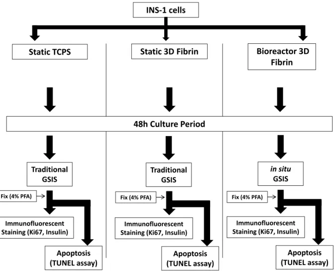
Figure 1: A schematic representation of the experimental design. ... 377 Figure 2: Percentage distribution of insulin secreted by fibrin-embedded INS-1 cells into the
medium and that remaining within the fibrin gel after 24h of culture. ... 39
Figure 3: Immunofluorescence staining for insulin (green). INS-1 cells incubated for 48h in
fibrin in the perfusion bioreactor ... 41
Figure 4: Total insulin secreted from INS-1 cells cultured either (A) on TCPS or (B) in fibrin
in a cell culture incubator for 48h. ... 43
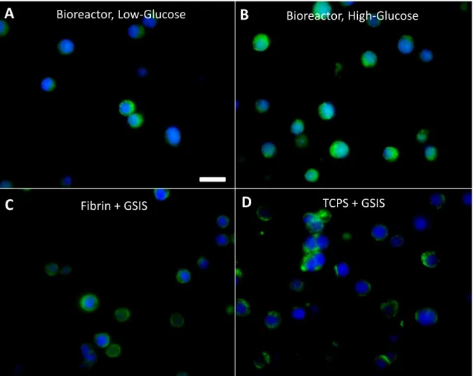
Figure 5: (A) Insulin secretion from INS-1 cells embedded in fibrin in the perfusion chamber
under perfusion bioreactor conditions after 48h of culture. The cells were then exposed to either low-glucose (2.8mM), shown as ‘low’, or high-glucose (28mM), shown as ‘high’ for 24h.. ... 45
Figure 6: (A) Percentage of apoptotic INS-1 cells cultured in fibrin under perfusion bioreactor
conditions for 48h and then exposed to either low-glucose (2.8mM) or high-glucose conditions (28mM) for 24h. Additionally, percentage of apoptotic INS-1 cells cultured in fibrin or on TCPS in a cell culture incubator for 48h and then exposed to a traditional GSIS. ... 49
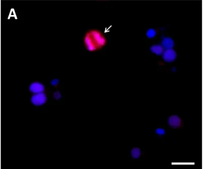
Figure 7: (A) Immunofluorescence staining for Ki67 proliferation protein of INS-1 cells
embedded in fibrin and cultured under perfusion bioreactor conditions for 48h and then exposed to high-glucose (28mM) for 24h. (B) Assessment of INS-1 cell proliferation under perfusion bioreactor conditions after 48h of culture and then exposed to either high-glucose (28mM) or low-glucose (2.8mM) for 24h. ... 51
Figure 8: Insulin secretion per volume (mL) of mechanically-disrupted rat pancreas
suspension, as measured in circulating supplemented medium (5.6 mM glucose) over 72h of culture under the respective bioreactor process conditions. ... 70
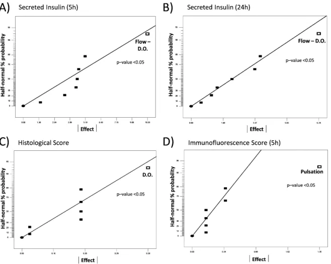
Figure 9: Half-normal plots of the effects of flow, D.O. and pulsation bioreactor conditions
and their interactions on: A) Secreted insulin at 5h; B) Secreted insulin at 24h; C) Histological score after 72h of culture; D) Immunofluorescence score after 72h of culture. The p-values of the significant effects are shown on each graph, where p<0.05 is considered statistically significant. ... 72
Figure 10: Results from whole mechanically-disrupted rat pancreas suspension in a tissue
xii
maintained in a traditional cell culture incubator. A) Insulin secretion per tissue volume (mL) over time. Representative images of B) Haematoxylin and eosin staining as well as C) Immunofluorescent insulin staining (green) and DAPI staining (blue) after 72h of culture. In panel A, the three profiles (square, triangle, and diamond shapes) refer to 3 independent experiments. In panels B and C, the histological (Histo Score) and immunofluorescence (IF Score) scores are indicated at the bottom right of the image. ... 74
Figure 11: Representative haematoxylin and eosin (H&E) staining of whole
mechanically-disrupted rat pancreata cultured in supplemented medium (5.6 mM glucose) after 72h of culture under the respective bioreactor process conditions. ... 76
Figure 12: Representative immunofluorescent insulin staining (green) and DAPI staining
(blue) on mechanically-disrupted rat pancreata cultured in supplemented medium (5.6 mM glucose) after 72h under the respective bioreactor process conditions ... 78
Figure 13: Insulin secretion per volume (mL) of whole mechanically-disrupted rat pancreas
suspension cultured in high flow, low D.O. and pulsation bioreactor conditions, as measured in circulating supplemented medium (5.6 mM glucose) over 24h (A, C, and E) and their respective insulin response to high glucose concentration (20 mM) for 6h, and IBMX (50 μM) made in a high glucose concentration solution (20 mM) for 6h (B, D, and F). Three independent experiments were performed. Arrows indicate the time points of injection of high glucose and IBMX. ... 81
Figure 14: A) Insulin secretion per volume (mL) of whole mechanically-disrupted rat
pancreata cultured in supplemented medium (5.6 mM glucose) in TCPS flasks maintained in a traditional cell culture incubator over 24h and B) the subsequent respective insulin secretion in response to high glucose concentration (20 mM) for 6h followed by another 6h in high glucose concentration (20 mM) + IBMX (50 μM). C) Delta of insulin concentration from whole mechanically-disrupted rat pancreata cultured under bioreactor conditions upon stimulation with high glucose concentration (20 mM) between 24-30h (total amount of insulin at 24h subtracted from total amount of insulin at 30h), and high glucose concentration (20 mM) + IBMX (50 μM) between 30-36h (total amount of insulin at 30h subtracted from total amount of insulin at 36h). D) Metabolic (MTT) assessment of pancreatic tissue at time 0, as well as after culture and stimulation studies were performed on bioreacted and non-bioreacted cultures, as measured by optical density.. ... 83
xiii
Figure 15: Representative staining of whole mechanically-disrupted rat pancreata cultured in
supplemented medium (5.6 mM glucose) after 24h of culture and stimulation with high glucose, as well as high glucose + IBMX under bioreactor conditions (high flow, low D.O. and pulsation) for A) Haematoxylin and eosin (H&E). C) Insulin and glucagon (counterstained with DAPI). E) α-SMCA and BS-lectin. Non-bioreacted static control cultures in TCPS flasks stained with B) haematoxylin and eosin (H&E). D) Insulin and glucagon (counterstained with DAPI). F) α-SMCA and BS-lectin (counterstained with DAPI). ... 84
xiv
LIST OF TABLES
Table 1: Upper and lower thresholds of the bioreactor operating conditions used in the
factorial design experiments. ... 62
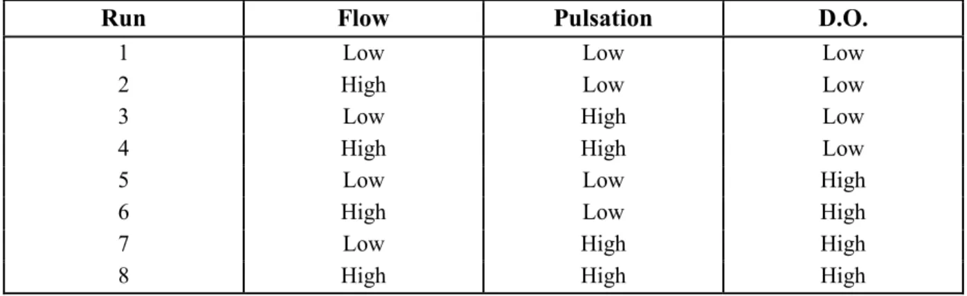
Table 2: Experimental bioreactor operating conditions studied in the factorial design
experiments (8 runs in total). ... 62
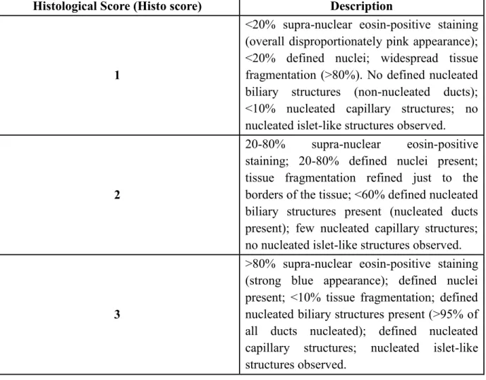
Table 3: Histological scoring criteria used for assessing haematoxylin and eosin (H&E)
staining. ... 65
xv
LIST OF ABBREVIATIONS
2D two dimensional
3D three dimensional
ANOVA analysis of variance
BM basement membrane
BSA bovine serum albumin
CaCl2 calcium chloride
CMD carboxymethyl-dextran
CSTR continuously-stirred tank reactor
CO2 carbon dioxide
DAB 3,3'-diaminobenzidine
DAPI 4’, 6-diamidino-2-phenylindole, dihydrochloride DLS duct-like structures
DMEM Dulbecco's modified Eagle medium
DNA deoxyribonucleic acid
ECM extracellular matrix
ELISA enzyme-linked immunosorbent assay
FBS fetal bovine serum
FDA Food and Drug Administration
FN fibronectin
xvi
GSIS glucose-stimulated insulin secretion
HG high glucose
HI high glucose + IBMX
HBSS Hank’s balanced salt solution
IBMIR instant blood-mediated inflammatory reaction IBMX 3-isobutyl-1-methylxanthine
IEQ islet equivalent
IFN interferon
IL interleukin
ILA islet-like aggregate INS-1 rat insulinoma cells
KRBH Krebs-Ringer buffer with HEPES
LG low glucose
LPS lipopolysaccharides
MIN-6 mouse insulinoma cells MMP matrix metalloproteinase
MTT 3-(4,5-dimethylthiazol-2-yl)-2,5-diphenyltetrazolium bromide
P probability
PANC-1 pancreatic carcinoma cell line PBS phosphate buffered saline
xvii
Pdx1 pancreatic and duodenal homeobox 1 RPMI Roswell Park Memorial Institute medium
RGD Arg-Gly-Asp
S.E.M. standard error of the mean RWV rotating wall vessel reactor
RT room temperature
α-SMCA α-smooth muscle cell actin T1DM type 1 diabetes mellitus T2DM type 2 diabetes mellitus TCPS tissue culture polystyrene TNF tumor necrosis factor
τp time constant
1
Chapter 1
2
The World Health Organisation (WHO) recently reported that there are over 422 million people worldwide suffering from diabetes (WHO, 2016). In Canada, in 2016, the Canadian Diabetes Association (CDA) listed a record 3.5 million Canadians as suffering with diabetes with an associated healthcare cost of $3.4 billion (CDA, 2016). Between 5 and 7 % of these diabetic patients suffer from T1DM (Shapiro et al., 2017). One treatment option offering a possible long-term treatment for T1DM is pancreatic islet transplantation. Here, pancreatic islets are mechanically and enzymatically cleaved from a donor pancreas and transplanted into the hepatic portal vein of the recipient (Shapiro et al., 2000). However, due to the shortage of donor organs this procedure has primarily only been offered to brittle T1DM patients with hypoglycemic unawareness. In addition, patients who do undergo this procedure generally enjoy insulin-independence for less than 5 years and, thus, require exogenous insulin supplements thereafter (Gruessner, 2011; Shapiro et al., 2017).
Some of the limiting factors in the success of the graft include: 1. the need for multiple donor organs for a sufficient islet graft, 2. immediate and long-term immune rejection, 3. islet damage due to the aggressive nature of isolation (loss of extracellular matrix and mechanical damage), and 4. poor revasculatisation post-transplantation (Tatum et al., 2017).
Improving the outcome of islet transplantation has been a particular focus in the diabetes research community and there has been a drive to understand the mechanisms that underlie graft failure and graft success. Recent tissue engineering approaches to model the mechanisms of successful and unsuccessful grafts have focused on culturing encapsulated pancreatic islets and insulin-producing β-cells in tridimensional (3D) scaffolds made of biomaterials, using different bioreactors. Here, cells are exposed to microenvironments closer to their native ones and so are enticed to behave more natively.
This thesis aims to explore the effects important physiological parameters (flow, dissolved oxygen concentration and pulsation) have on pancreatic tissue outcome. By exploring and understanding these effects, this knowledge could be applied to improve pancreatic islet graft outcome by highlighting beneficial pre-culture conditions pre-implantation.
3
In Chapter 2, a review of the literature is introduced detailing the native pancreas, its diseases, and how it is studied in vivo and in vitro. The use of 3D matrices in diabetes research is discussed at length with important consideration given to the incorporation of ECM molecules. The review then highlights how 3D matrices have been used in combination with a host of different bioreactors to culture pancreatic tissues and insulin-producing cells with the global goal to improve the success of transplanted pancreatic islet grafts.
In Chapter 3, entitled “An in-situ glucose-stimulated insulin secretion assay under perfusion bioreactor conditions”, INS-1 cells (a rat insulinoma cell line) were encapsulated in fibrin and cultured in a perfusion bioreactor. Briefly, it was found that these cells showed glucose-stimulated insulin secretion, increasing functionality (glucose-glucose-stimulated insulin secretion) and lower levels of apoptosis when compared to non-bioreacted 3D and 2D static control cultures.
In Chapter 4, entitled “A factorial design to identify process parameters affecting whole mechanically-disrupted rat pancreata in a perfusion bioreactor”; a factorial design was used to identify process parameters affecting whole mechanically-disrupted rat pancreata in a perfusion bioreactor. The singular and combination effects of flow, dissolved oxygen concentration and pulsation were assessed on the outcome of pancreatic tissue. Beneficial bioreactor conditions which supported pancreatic tissue attributes were identified. Mechanically-disrupted rat pancreata cultured under these beneficial bioreactor conditions showed glucose-stimulated insulin secretion, higher metabolic activity, insulin- and glucagon-positive staining, extensive endothelial structures, and overall intact tissue when compared to static cultures.
In Chapter 5, entitled “Conclusion”, the outcome of this thesis is discussed and future application and directions are suggested.
4
Chapter 2
5
2.1 The pancreas
The pancreas is located behind the stomach and connected to the liver, the spleen, and the small intestine. The main functions of the pancreas are to produce exocrine enzymes to aid digestion, and endocrine hormones to regulate blood glucose levels. The exocrine pancreas represents approximately 98% of the pancreatic mass and is comprised of acinar cells responsible for synthesis, storage and secretion of digestive enzymes (e.g., pancreatic lipase and amylase, phospholipase, nucleases) into the duodenum (Jouvet and Estall, 2017). The endocrine pancreas represents approximately 2% of the total mass of the pancreas and is made up of islets of Langerhans (Hogan and Hull, 2017). These are highly vascularised structures that contain 5 distinct cell types; α-, β-, δ-, ε-, and γ (PP) cells. Although the islets of Langerhans account for 1-2% of the total mass of the pancreas yet receive 15% of the blood flow. This is important in rapidly sensing glucose changes (Jansson et al., 2016).
The main cell population in pancreatic islets are β-cells (50%), which secrete amylin, C-peptide and, more importantly, insulin. The other main cell type is the α-cell (40%) which is responsible for the secretion of glucagon. The small proportion of δ-cells (<10%) and ε-cells (<1%) secrete somatostatin and ghrelin, respectively. In addition to the aforementioned cell types, PP cells (<5%) are also present and are responsible for producing pancreatic polypeptides which acts locally within the pancreas to regulate endocrine function and to regulate gastrointestinal secretion (Kojima et al., 2007). Much emphasis has been placed upon the hormones insulin and glucagon due to their essential role in the regulation of glucose homeostasis. Insulin is produced and secreted by the β-cells in response to glucose although insulin is also produced and secreted in response to other non-glucose stimulants and neural peptides but to a far lesser degree (Komatsu et al., 2013). Insulin is then released by the β-cells into the blood stream and binds to its receptors found on most tissues including the liver, muscle and fat cells, to facilitate glucose uptake and storage (Olerud et al., 2011).
6
2.2 Exocrine-endocrine axis
The endocrine and exocrine components of the pancreas share an anatomical and physiological relationship. Pancreatic islets are scattered and embedded throughout the exocrine tissue, void of any special capsule or membrane. Given this, it has become increasingly evident that these two structures may not be so independent of each other. Although the relationship between these two counterparts is still under investigation, the parts which are known may give some insight into the special relationship they share.
In a healthy human adult pancreas, acinar tissue and pancreatic islets are fed separately from the pancreatic capillary plexuses which are formed from the major arteries surrounding the pancreas. However, the outward flow of blood from the pancreatic islets drains directly into the acinar capillary network, aptly named the ‘insulo-acinar axis’ (Evert et al., 2005; Weir and Bonner-Weir, 1990). The acinar tissue receives high concentrations of pancreatic islet hormones suggesting a possible effect of pancreatic islets on the exocrine pancreas. To date, the nature of this effect remains unclear.
In addition, acinar cells secrete enzymes as zymogen granules into the ducts of the pancreas. These secretions are highly viscous and acidic in nature. To counter this, ductal epithelial cells secrete alkaline fluid with low viscosity. Furthermore, lithostanthin S, an enzyme which keeps the calcium of the pancreatic juice in a soluble state, is present in this secretion. In combination, these processes inhibit stone formation within the duct (Chakraborty and Chowdhury, 2015).
In the diseased endocrine pancreas, such is the case in type 1 diabetes mellitus (T1DM), the effect of chronic hypoinsulinemia causes major functional, morphological, and histological changes in the exocrine tissue. These widespread changes include acinar atrophy, fibrosis, fatty degeneration, and changes to the ductal system (Ewald et al., 2009; Frier et al., 1976; Lankisch et al., 1982; Williams et al., 2012). The exact mechanisms responsible for these changes are still under investigation but may be a result of multiple factors including inflammation and lack of insulinotropic effects due to the loss of pancreatic β-cells (Campbell-Thompson et al., 2015).
7
In exocrine pancreatic disorders (for example chronic pancreatitis), increased pancreatic tissue pressure and hypoxia have been observed causing alterations to the microcirculation of the pancreas. This causes surrounding stellate cells to proliferate and secrete collagen. The net result of this is fibrosis of the pancreas which results in a progressive loss of endocrine function (Watanabe et al., 2004). In addition, chronically high blood glucose levels have also been shown to have a stimulatory effect on stellate cells in the pancreas. This effect may increase and amplify the progression of fibrosis in the pancreas (Nomiyama et al., 2007).
It is clear that although this endocrine-exocrine relationship is still not fully understood, the research so far highlights the important interplay the endocrine and exocrine may have on normal pancreatic function.
2.3 Diabetes mellitus
Diabetes mellitus (DM) is a broad term used to characterise a group of metabolic diseases. To date, it remains a treatable but incurable disease. Diabetes may be subdivided into two main types; type 1 (T1DM) and type 2 (T2DM).
In T1DM, pancreatic β-cells are destroyed by the autoimmune system which usually leads to absolute insulin deficiency. This autoimmunity is most likely a combination of genetic susceptibility and a host of environmental triggers such as diet, viruses, stress, and exogenous toxins (Kuehn et al. 2014b). In a recent study, it was revealed that a host of proinflammatory cytokines including interleukin-6 (IL-6), IL17, transforming growth factor-β (TGF-β), C-reactive protein (CRP), and tumour necrosis factor-α (TNF-α) strongly correlated with disease progression (Qiao et al., 2017). However, the exact mechanisms that are responsible for causing this inflammation remain unknown.
Although T1DM makes up just 5-7% of the cohort all diabetes diagnoses, it remains the most common endocrine and metabolic condition occurring in childhood (Lysy et al., 2016).
In T2DM, there is a progressive loss of β-cell insulin secretion as a result of chronically high blood glucose levels leading to insulin resistance and later insulin deficiency (Katsiki et al., 2017). This type of diabetes is generally considered to be a result of chronic obesity, low or
8
reduced activity, and poor diet/malnutrition. However, a review of the literature identified more than 36 genetic susceptibility factors associated with disease progression (Herder and Roden, 2011).
To put this pathology into clinical perspective, the World Health Organisation (WHO) stated that there are over 422 million people worldwide suffering from diabetes (WHO, 2016). In Canada in 2016, the Canadian Diabetes Association (CDA) listed a record 3.5 million Canadians as suffering with diabetes with an associated healthcare cost of $3.4 billion (CDA, 2016). This is a 76% increase from 2006.
Together, these reports cement the notion that diabetes is of epidemic proportion and new ways to diagnose, treat, and understand the pathophysiology of the disease is of great clinical and socioeconomic importance.
2.4 Current treatments for T1DM
2.4.1 Exogenous insulin
As previously mentioned, T1DM is a treatable but incurable disease. The most common treatment deployed by healthcare professionals to patients is exogenous insulin therapy. Here, patients monitor their blood glucose levels multiple times daily using a finger-prick test and accordingly titrate exogenous insulin, subcutaneously. In general, this method serves the insulin-dependent diabetic population with a viable option for transiently correcting blood glucose levels. However, this treatment method relies on strict life-time adherence by the patient. In addition, insulin is a growth factor and is administered in a non-physiological manner. Long-term use of exogenous insulin has been associated with higher incidences of cancer, cardiovascular disease, blindness, and peripheral limb amputation (Currie et al., 2009; Currie et al., 2013).
9
2.4.2 Whole pancreas transplantation
Allogenic pancreas transplantation has been considered a treatment option offering a long-term treatment option for brittle T1DM patients. This is considered major surgery and thus carries all of the associated risks. The patient is also required to adhere to a strict life-long immunosuppressive medication regime. However, due to the sparse organ donor availability and the biocompatability of available donors, just (c.a.) 25,000 transplantations have been performed since 1966(Gruessner, 2011).
2.4.3 Pancreatic islet transplantation
Islet transplantation is much less surgically invasive than whole pancreas transplantation and also offers a possible cure for T1DM.
Briefly, the pancreas is surgically excised from the donor. The pancreas is then cannulated, treated with collagenase, and mechanically agitated (Ricordi chamber). The resulting islets are then purified and transplanted into the portal vein of a recipient. Since the advent of the Edmonton protocol, islet isolation and islet procurement have improved significantly resulting in a higher viable islet yield and improved clinical outcome. To date, 1500 grafts have been performed since its advent in 2000 (Shapiro et al., 2000; Shapiro et al., 2017). However, due to the shortage of donor organs, this procedure has primarily only been offered to brittle T1DM patients with hypoglycemic unawareness. In addition, patients who do undergo this procedure generally enjoy insulin-independence temporarily and, thus, require exogenous insulin supplements thereafter. In addition, the patient is required to take life-long immunosuppressive drugs (Gruessner, 2011).
Some of the limiting factors in the success of the graft include: 1. the need for multiple donor organs for a sufficient islet graft, 2. immediate and long-term immune rejection, 3. islet damage due to the aggressive nature of isolation (loss of extracellular matrix and mechanical damage), 4. poor revasculatisation post-transplantation (Tatum et al., 2017).
10
Many studies have attempted to understand and improve the clinical outcomes of this procedure from the lab bench and in vivo studies alike. Islet transplantation still represents a viable and realistic longer-term treatment option for T1DM.
2.5 Diabetes research platforms
2.5.1 Pancreatic islets and β-like cell lines
Human pancreatic islets isolated from cadaveric donors are the primary cell source approved for clinical use, but are limited due to donor availability. Instead, non-human islet sources have been investigated as an alternative. To date, researchers have favoured porcine islets as an alternative due to their unlimited supply, insulin homology (porcine and human insulin vary by just one amino acid) (Dufrane and Gianello 2012), and have been shown to have lower sensitivity to destruction by recurrent T1DM autoimmunity than human islets (Koulmanda et al., 2003). In addition, porcine islets which have been genetically-modified to reduce expression of pro-inflammatory antigens, thus reducing the host immunogenic response, have also been explored (Cowan et al., 2016). Furthermore, xenotransplantation studies where adult porcine islets were encapsulated in alginate and subcutaneously grafted into non-human primates have showed promising clinical outcomes including survival of the graft and correction of diabetes without the need for immunosuppression for up to six months post-transplantation (Dufrane et al., 2010; Marigliano et al., 2011).
Although cell lines have less clinical relevance than islets, they serve as an invaluable tool for research purposes due to their ease of handling and abundant availability. β-like cell lines come from a host of different species but tend to take their origins from cancer cells (Amer et al., 2014). However, their use in developing strategies for tissue engineering approaches is supported by their characteristic glucose-stimulated insulin release. Some commonly used cell lines used in diabetes research include INS-1 cells (a rat insulinoma cell line) (Asfari et al., 1992; Sharp and Vermette, 2017), MIN-6 cells (a transgenic mouse insulinoma cell line)
11
(Edén et al., 2015; Ishihara et al., 1993), and β-TC cells (a mouse insulinoma cell line) (Efrat et al., 1988; Kim et al., 2015).
2.5.2 Stem cells and their application for T1DM
Stem cells are undifferentiated cells that can divide and expand, or can differentiate into specialised cells such as insulin-producing cells. Insulin-producing cells derived from human mesenchymal stem cells (MSCs), embryonic stem cells (hESCs), and induced pluripotent stem cells (iPSCs) are a promising treatment for T1DM (van de Vyver, 2017). They continue to receive much attention in the diabetes research community because, in principle, large numbers of functional cells could be produced and transplanted, therefore, offering a possible cure for T1DM. To date, numerous in vivo studies have been performed on small animal models. Multiple groups have reported that they can cure or prevent diabetes in mice by transplanting insulin-producing cells derived from stem cells in cell numbers ranging from 1.25 to 7 x 10⁶ (D’Amour et al., 2006; Espes et al., 2017; Kroon et al., 2008; Pagliuca et al.,
2014; Rezania et al., 2014). More recently, islet-sized ‘neo-islets’ were produced by aggregating pancreatic islet cells (β- and α-cells) with MSCs and transplanted these into the peritoneal space of mice. These mice achieved long-term glyceamic control without the need for immunosuppression (Westenfelder et al., 2017). In another recent study, hESC-derived pancreatic endoderm cells were transplanted into diabetic mice in a prevascularised subcutaneous site. These cells were able to correct long-term hyperglyceamia by secreting insulin in glucose-regulated manner (Pepper et al., 2017).
One promising outlook for the treatment of T1DM in humans has come from ViaCyte; a company specialising in the induction/derivation of PSCs into pancreatic β-cells. Here, PSCs are induced/differentiated into progenitor pancreatic β-cells. These cells are then encapsulated in a flat immune-evading capsule and then transplanted subcutaneously into the patient. The progenitor β-cells then mature into insulin-producing β-cells in vivo. This approach has successfully corrected hyperglyceamia in rat and mice models (Lee et al., 2009; Pagliuca et al., 2014). These cells combined with this technology have now entered Phase I-II clinical trials in humans (Trounson and DeWitt, 2016).
12
Although the current perspective for use of stems cells in the treatment of T1DM cannot be dismissed, there are some special considerations to be noted. In addition, murine studies have reported the formation of teratomas when hESCs were transplanted (Kelly et al., 2011; Kroon et al., 2008).
2.5.3 3D static systems
To date, the majority of cell-based assays use traditional two-dimensional (2D) monolayer cells cultured on flat and rigid surfaces made primarily of glass or tissue culture polystyrene (TCPS). This method has proven to be a valuable tool for cell-based studies, however, its limitations have increasingly become recognised. Since almost all cells in the in vivo environment are surrounded by other cells and by ECM in a three-dimensional (3D), 2D cell cultures do not adequately take into account the natural 3D microenvironment of cells.
In 2D systems, cells are plated as monolayers and receive a heterogenous supply of nutrients and oxygen from the medium reservoir. Cellular waste products are also secreted and remain in the medium reservoir. Dying cells detach and are washed away during medium change. The remaining cells are normally flat and have a restricted cell morphology to planar geometries that inhibit the formation of more complex tissue architecture, unlike their native environment. Additionally, concentration gradients often form which can effect cellular behaviour (Edmondson et al., 2014; Tibbitt and Anseth, 2012).
It has repeatedly been demonstrated that cells that are exposed to a 3D environment with and without the addition of ECM cues show large differences when compared to those in 2D systems. These studies have reported increased viability, increased proliferation, and more dynamic morphology alike to their native environment (Hanjaya-Putra et al., 2011; Lewis et al., 2015; Mori et al., 2009; Zhu et al., 2017).
Given this, there has been a surge in 3D matrix design for the intention of cell culture applications.
13
2.6 Extracellular matrix molecules in the native pancreas
Multiple researchers have reported the merits of using extracellular matrix molecules in the design of 3D matrices for research purposes.
The ECM is made of a basement membrane (BM) and of an interstitial ECM. The BM is organ-specific meaning its structure, composition, and function varies. It separates the epithelial and endothelial tissue from the connective tissue and is generally made up of laminin, microfibrils, fibronectin, laminin, and different collagens. Generally, these are glycoproteins and fibrous proteins that provide the 3D structural support, and cell adhesion, and they have been shown to activate intracellular signalling pathways (Daoud et al., 2011; Kuehn et al., 2014b; Stendahl et al., 2009).
The interstitial ECM is highly varied and is generally composed of structural proteins and of polysaccharide chains of glycosaminoglycans (GAGs) which interact with proteins to form proteoglycans (Palombo et al., 2014).
To date, the exact composition and arrangement of the pancreatic ECM is still under investigation and little is known about ECM homology across species. However, some studies have identified important components present including laminins and collagens, and heparan sulfate proteoglycan (HSPG) families providing binding and interaction sites with integrins (Cross et al., 2017; Wang and Rosenberg, 1999a).
Most recently, our group has reported some advances in the identification of native ECM molecules within the pancreata of mice and pigs. Briefly, our group used scanning electron microscopy, proteomics, and immunohistochemistry to identify the presence and location of some important ECM molecules from decellularised matrices. In mice, they showed that the α4 and β2 chains of laminin were localised inside the islet but were absent around the islet when the islets were surrounded by an ECM made of collagen IV and V. In addition, collagen type I, III, and VI were identified in pig pancreatic ECM. Isoforms of collagen type IV and type V were also present, however, in a much lower abundance (Vigier et al., 2017).
14
As mentioned previously, in addition to offering biochemical cues to mediate cell behaviour, the ECM is also, at least in part, responsible for maintaining the 3D structure of the islet. These findings are reinforced when considering the poor clinical outcome of islets transplanted in T1DM patients. During islet isolation, much of the ECM is lost due to the isolation technique and some researchers have attributed this to be one of the main factors for the poor clinical outcome of transplanted islets (Davis et al., 2012; Nagata et al., 2001).
In light of this, researchers have used 3D scaffolds made of biomaterials, and more specifically, hydrogels, in an attempt to replicate the native 3D microenvironment of the cell with their native ECM molecules.
2.7 Biomaterials - 3D hydrogel matrices
2.7.1 Fibrin
Fibrin is a soluble, fibrous, transient ECM, FDA-approved protein formed at the site of vascular injury by the polymerisation of fibrinogen by the protease thrombin (Laurens et al., 2006; Mosesson, 2005). More importantly, fibrin contains the Arg-Gly-Asp (RGD) amino acid sequence which is a ligand for 24 known integrin receptors (Arnaout et al., 2005; Riopel et al., 2015). In an in vitro setting, fibrinogen has been purified from a host of species and crosslinked with thrombin to form hydrogels which enable encapsulation of both pancreatic islets and pancreatic β-cell lines. In the context of pancreatic islet transplantation, fibrin has shown potential for improving islet viability, function, and preservation of native islet morphology (Andrades et al., 2007; Beattie et al., 2002a; Kidszun et al., 2006; Kim et al., 2012; Lim et al., 2009; Maillard et al., 2011). We have previously shown that encapsulation of young porcine islets in fibrin matrices had a significantly positive protective effect against apoptotic stimuli (hydrogen peroxide) (Kuehn et al., 2014a). More recently, we showed that INS-1 cells (a rat insulinoma β cell line) encapsulated in fibrin matrices exhibited improved functionality and lower levels of apoptosis when compared to INS-1 cells plated on TCPS (Sharp and Vermette, 2017). In another study, INS-1 cells cultured within and on fibrin using 2D and 3D culture techniques showed improved glucose-stimulated insulin secretion, formed islet-like clusters, and showed increased αvβ3 protein expression; a major ligand for RGD
15
(Riopel et al., 2013). In another study, porcine islets were embedded in fibrin matrices and cultured for 7 days in a traditional cell culture incubator. Here, the islets dissociated in fibrin over time (Dubiel et al., 2014).
Given this, it has been proposed that purifying fibrinogen from a human islet graft recipient’s blood could both improve the quality of the islet graft and reduce the intensity of the immediate immune response post-transplantation in humans.
2.7.2 Collagen
Hydrogels which incorporate different collagens are gaining widespread popularity as scaffolds for tissue engineering due to the abundance of collagen in native extracellular matrix (ECM). Briefly, collagen comprises 25% (by dry weight) of total structural protein in vivo (Antoine et al., 2014; Drury and Mooney, 2003). The aforementioned collagens I, V, and VI are the most abundant isoforms within the islet-exocrine interface of the developed human pancreas (Di Lullo et al., 2002). Collagen IV appears less abundantly in the mature pancreas but is strongly expressed in foetal pancreatic tissue (Cirulli et al., 2000).
It has previously been shown that rat islets plated on TCPS plates coated with collagen I or IV, remained 60% and 89% viable, respectively. In contrast, over 90% of rat islets left in suspension underwent apoptosis after 48h (Pinkse et al., 2006).
In another study, murine islets embedded in 3D collagen matrices significantly improved graft survival post-transplantation. Here, chronic hyperglyceamia was corrected and less islets were required to correct the hyperglyceamia compared to non-collagen support controls (Jalili et al., 2011). Further, transplanted collagen IV-adsorbed 3D scaffolds containing mouse islets significantly improved glycemic control faster than scaffolds coated with either fibronectin, laminin, or serum (Salvay et al., 2008).
More recently, human islets have been microencapsulated in collagen IV-reinforced alginate matrices and showed improved glucose-stimulated insulin secretion and improved basal insulin secretion when compared to islets encapsulated in alginate matrices alone. Interestingly, this effect was concentration-dependent, where the lowest concentration of
16
collagen IV used showed the largest effect. This may mimic the native in vivo conditions of the mature human pancreas where collagen IV is in low abundance (Llacua et al., 2016).
2.7.3 Alginate
Alginate is an unbranched, anionic polysaccharide made from repeating units of α-L-guluronate (G) and β-D-mannuronate (M) and is found in the cell walls of brown algae. Historically, it has been a major candidate for use as a biomaterial as it is noted for exhibiting a low immunogenic profile, has a non-toxic effect, and can undergo sol-gel transition under physiological conditions (Amer et al., 2014; Strand et al., 2003; Strand et al., 2017).
Alginate beads/matrices can be easily manipulated by the researcher and are formed by dripping an alginate solution containing G-, and M-, and GM-monomers/polymers of various lengths into a calcium dihydrate solution. The properties of the alginate beads are determined by the length and conformation of the resulting polymers, thus, each bead may contain a different ratio of G-, M-, and GM-monomers/polymers (Martinsen et al., 1989).
As cells do not have receptors that recognise alginate, it may be considered an inert compound, thus, some researchers have chosen to supplement alginate with other ECM molecules to entice cellular behaviour (Andersen et al., 2015). Given this, one of the main research goals for encapsulation of islets in alginate has been to circumvent the problems associated with the immediate and long-term immune responses by the host upon islet transplantation by creating an alginate ‘shield’. The use of alginate encapsulation may evade the need for the lifelong immunosuppression regime required post-transplantation (both of these severely impact the clinical outcome of the graft) (Strand et al., 2001).
To date, there are few recent studies detailing the application of alginate-encapsulated islets in the treatment of humans with T1DM. One study followed up 4 patients who had alginate-encapsulated islets grafted into their portal vein, 3 years post-transplantation. Here, all 4 patients showed reduced immune responses to the graft when compared to non-encapsulated islet grafts. In addition, all patients showed a basal and stimulatory response from glucose (serum C-peptide). However, none of the patients ever achieved insulin independence and
17
only one patient was able to transiently reduce exogenous insulin intake (Basta et al., 2011). This finding confirmed the results of a previous study. Here, rat islets were encapsulated in purified alginate. These islets were then transplanted into the microcapsules in the peritoneal cavity of diabetic rats for up to 12 months. Glyceamic control was achieved within 5 days post-transplantation but hyperglyceamia reoccurred within 6-20 weeks post-transplantation. Interestingly, purified alginate-encapsulated islets showed a higher biocompatibility and, thus, lower immune responses compared to non-purified alginate-encapsulated islet grafts and free non-encapsulated islet grafts (De Vos et al., 1997).
In another study, human islets were encapsulated in alginate (500 µm diameter) and transplanted into the abdominal cavity of 10 non-immunosuppressed T1DM human patients. Here, all participants showed improved glyceamic control however none of the participants were ever able to achieve insulin-independence (Calafiore et al., 2006). In another large mammal study, pig islets were macroencapsulated in alginate and transplanted these macrocapsules subcutaneously in T1DM-induced primates. Here, diabetes was corrected (insulin-independent) for up to 6 months. Interestingly, a strong humoral response was observed post-transplantation in these primates however the physical properties of the alginate capsules prevented diffusion of IgG into the graft for up to 20 weeks (Dufrane et al., 2010). This suggests that the composition of the alginate, in this case 3% wt/vol was used, should be an important consideration when encapsulating islets for transplantation with a goal of evading the immune system.
In 2007, a brief communication was published describing a T1DM patient who received a transplantation of alginate-encapsulated porcine islets. The patient was then followed up for 18 years. Although the patient never achieved insulin-independence, a marked decrease in his exogenous insulin supplement was needed to achieve euglygeamia. The graft remained functional for 18 years. This study highlights both the possible use of alginate as an encapsulation matrix and the use of porcine islets as a surrogate to treat T1DM (Elliott et al., 2007).
18
In another study,alginate-encapsulated human pancreatic cells were grafted into the peritoneal cavity of totally immunocompromised diabetic mice. The grafts exhibited rapid secretory responses to glucose and glucagon after 5 weeks. Free grafts (non-alginate-encapsulated pancreatic cells) showed reduced function, reduced viability, and a higher incidence of necrosis over the same period of time (Jacobs-Tulleneers-Thevissen et al., 2013).
These findings may still suggest an important role for alginate to evade the destructive nature of the immune response on the graft. These findings also suggest that alginate in combination with another ECM molecule may yield a better clinical outcome (insulin independence).
Together, these studies highlight the positive clinical effects alginate encapsulation may potentially have on islet graft outcome. However, it is noteworthy to mention that ion-exchange (with sodium) and chelating ions such as phosphates make alginate beads susceptible to destabilisation by depletion of calcium ions under physiological conditions thus creating a major barrier for longer-term culture periods (Morch et al., 2007). Using Ba2+ or
Sr2+ as chelating ions have been shown to produce stronger, yet still biocompatible alginate
gels (Morch et al., 2006). An alternative mechanism to achieve alginate gelation is to covalently cross-link alginate using photo cross-linking. Briefly, alginate can be mixed with methacrylate and cross-linking can be achieved using an argon ion laser. This method has been used to seal corneal perforations indicating that this method may be a suitable candidate for other tissue engineering applications. The rate of degradation is slower compared to ion-exchange preparations of alginate (Smeds et al., 2001).
2.8 Three-dimensional matrices limitations
As reported, the beneficial effects of using 3D hydrogels give cells a 3D microenvironment and so encourage cells to behave more natively. However, static 3D cell and tissue culture systems also have a host of limitations. For example, there is no mixing or circulation of the culture medium which causes heterogeneous distributions of nutrients and metabolites causing concentration gradients (Tibbitt and Anseth, 2009). In addition, the dimensions of the matrix/scaffold should be an important consideration. In the native cell environment,
cell-19
capillary distances range from 20 to 200 µm (Volkmer et al., 2008). The capillaries assist the cells in nutrient and oxygen acquisition and in the removal of cellular waste products. Due to poor oxygen diffusivity in aqueous solutions, oxygen diffusion in in vitro systems is limited to a distance of 100 –200 µm (Carrier et al., 2002; Lovett et al., 2009). Recently, it has been demonstrated that cells which reside at the base of the matrix (furthest from the oxygen source) undergo hypoxic changes and ultimately cell death (DelNero et al., 2015).
Accurate quantification of these systems also remains a challenge. For example, researchers have favoured quantifying the secreted protein of interest from the medium reservoir which lies on top of the 3D matrix construct (Carriel et al., 2012; Sodian et al., 2002a). This method may negate the accumulated protein that remains within the matrix. Our group has previously demonstrated that disproportionate amounts of proteins remain within the matrices (Kuehn et al., 2013; Sharp and Vermette, 2017). Efforts to quantify the remaining protein in the matrix by dismantling/degrading the matrix using digestive enzymes have been unsuccessful. It was found that digestive enzymes also affect cells within the matrix, which can cause cell damage and even cell death, thus, preventing an accurate quantification of the culture (Sabra and Vermette, 2011).
Imaging 3D cultures in situ also remains a challenge. Normal bright-field and fluorescent microscopy is hindered by the dimensions of the matrix/construct. Instead, more advanced imaging techniques are required including confocal microscopy, magnetic resonance imaging (MRI), positron emission tomography (PET), and computed tomography (CT).
In combination, these limitations have grave implications for higher cell seeding densities, size of construct, and are limiting factors in the scale-up of in vitro 3D cell culture systems.
Given these challenges, researchers have refocused their efforts to create more dynamic 3D systems which improve oxygen and nutrient delivery, removal of cellular waste products, and homogeneity of cultures. The use of bioreactors in combination with cell-containing 3D matrices may circumvent these challenges.
20
2.9 Bioreactors
In the realms of research, the word bioreactor is a broad term used to describe a system in which an extra-cellular device is added to a cell or tissue culture system that can closely assist, monitor, and tightly control environmental and operating conditions including flow, temperature, pressure, pH, pulsation etc. (Martin et al., 2004). Biorectors are used to circumvent the aforementioned limitations of static 3D culture systems and help in furthering our understanding of how cells behave in vivo by more closely replicating the native in vivo environment more than the capabilities of static 3D culture systems.
In addition to their widespread use in an industrial setting, bioreactors have also been widely used in tissue engineering areas such as bone engineering, cardiac regeneration, and tumour modelling. To date, the use of bioreactors in the field of diabetes research remains an emerging field with some important advances having being made using a host of different bioreactors.
2.10 Bioreactors in diabetes research
2.10.1 Spinner flask
A spinner flask reactor is a variation of a constantly stirred-tank reactor (CSTR) (Bijonowski et al., 2013). To counter the problems associated with low seeding efficiency and other limitations present in static culture systems, spinner flask bioreactors offer convective flow and produce hydrodynamic forces that attempt to increase mass transport in a given culture (Gaspar et al., 2012). They come in a host of simple designs but are generally composed of a cylindrical container complete with an inlet/outlet port for introduction or removal of cells and medium. They also contain a basal stirring element to allow constant mixing of culture medium and oxygen, thus, reducing the boundary layer formation at the cell-medium interface (Yeatts and Fisher, 2011). Cell cultures can be suspended without a matrix or in a scaffold/matrix which is normally suspended in a fixed position and anchored to the top of the cylindrical container (Martin and Vermette, 2005a).
21
Spinner bioreactors have been used extensively in bone tissue applications but little is known on the effects these bioreactors have on pancreatic tissue. In one of the few studies on such a system for pancreatic applications, Chawla et al. (2006) inoculated spinner bioreactors with collagenase-dissociated pancreatic porcine neonatal tissue. The inoculated spinner bioreactors were then placed into a traditional cell culture incubator and cultured for 9 days. The effects of serum-free medium and pH were also investigated. The pH was manually adjusted. The results showed that dissociated neonatal pancreatic porcine tissue reformed into aggregates, showed massive cellular proliferation (7.5 times increase), and exhibited glucose-responsive behaviour at pH 7.3 (Chawla et al., 2006). Aforementioned, these systems are simple in design and so lack the ability for automation (automatic control of pH, temperature, and dissolved oxygen concentration) creating a barrier for the researcher using more complex tissue types.
2.10.2 Rotating wall vessel bioreactors
The rotating wall vessel (RWV) was first developed by NASA to create a microgravity environment (Wong, 1997). Principally, these reactors are composed of two cylinders (one encased in the other) where the inner cylinder is static and has a porous wall used for gas exchange (Varley et al., 2017). The outer cylinder is the rotating vessel for the medium containing cells/tissue. The cells/tissues normally require a supporting matrix such as a collagen or alginate hydrogel. These cell/tissue constructs are then added to the medium in the outer cylinder and are suspended in a simulated zero gravity state achieved by rotating the vessel (Begley and Kleis, 2002). RWVs exhibit low shear stress and turbulence on the cell/tissue culture. In addition, the researcher is able to control the gas exchange in the system. In combination, these are major advantages when comparing RWV to CSTR systems (Martin and Vermette, 2005a).
In some of the first studies to use this technology, it was observed that cells aggregated and tissue-like spheroids formed highlighting its potential for a host of tissue engineering applications, especially its use in the study of diabetes (pancreatic islets are essentially aggregates/spheroids) (Granet et al. 1998; Manley & Lelkes 2006).
22
In a recent study, Samuelson and Gerber (2013) cultured murine pancreatic β-cells (β-TC-6 cells) on collagen beads in a RWV for 2 weeks. RWV cultures showed the formation of islet-like clusters, robust proliferation, enhanced transcriptional signalling, improved translation of pancreatic genes, and markedly higher glucose stimulated insulin secretion compared with two-dimensional static culture (Samuelson and Gerber, 2013).
In another study, Tanaka et al. (2013) cultured MIN6 cells in a RWV to produce 250 µm spheroids. These spheroids showed increased upregulation of several β-cell genes (including insulin 1, insulin 2, and glucokinase genes) compared to non-spheroid 2D MIN6 cells cultured on TCPS. The spheroids also showed increased insulin secretion upon glucose stimulation. In addition, the researchers then transplanted these spheroids into the portal vein of streptozotocin-induced diabetic mice for 30 days. They reported that the spheroids corrected hyperglyceamia in a series of glucose challenges compared to non-spheroid MIN6 cell transplants.
In combination, these studies support previous documented observations on the use of RWV in the culture of pancreatic cells (Rutzky et al., 2002; Stepkowski et al., 2006; Wong, 1997).
The positive applications for the use of RWVs in spheroid production have been described, however, due to the low shear stress and turbulence exhibited by this system, culturing more complex tissues, where shear has been shown to have a negative effect on tissue outcome, may hinder their use.
2.10.3 Perfusion bioreactors
The importance of pancreatic perfusion has been well documented in the literature in animal models. Briefly, it has been repeatedly demonstrated that there is decreased pancreatic perfusion in T1DM-induced animals of different species when compared to their healthy counterparts (Jansson and Hellerstrom, 1983; Lifson et al., 1980; Nyman et al., 2010). More recently however, positive electron tomography (PET) was used to highlight the disparity in pancreatic perfusion in T1DM humans. Here, it was shown total pancreatic perfusion was on average 23% lower in individuals with T1DM under fasting conditions compared to sex-, age-, and BMI-matched healthy control individuals. Upon glucose-stimulation, there was a 48%
23
higher pancreatic perfusion in healthy individuals compared to the T1DM cohort (Carlbom et al., 2016). In combination, these studies highlight the important role perfusion may play in pancreas homeostasis.
Unlike RWV or CSTR systems, perfusion bioreactors can provide more uniform mixing of the medium, thus, allowing for better environmental control and physical stimulation of cells in larger cell or tissue constructs. In addition, they use flow to perfuse medium through the construct (Wang et al., 2015).
In general, the design of a perfusion reactor is fairly constant, consisting of a peristaltic pump, a culture medium vessel, tubing to complete the circuit, and a cell-, or, tissue-containing perfusion chamber (Gaspar et al., 2012; Tapia et al., 2016). There are many different variations of perfusion chambers, thus, this is normally the main differentiating factor amongst perfusion bioreactors. However, more recently additions such as gas exchange units and pressure transducers have been added.. Given this, perfusion bioreactor systems are sometimes referred to as ‘dynamic’ systems as they can allow the researcher to test a range of physiological parameters in unison on a given cell or tissue culture; a major advantage over other bioreactor systems. Some of these parameters are flow rate, dissolved oxygen concentration, pulsation, temperature, pH, and pressure.
In one of the few studies using a perfusion bioreactor with human islets, human islets were successfully dedifferentiated into proliferative duct-like structures (DLS). Here, they encapsulated human pancreatic islets in collagen-fibronectin matrices and further encased these in a 3D-printed poly(DL-lactide-co-glycolide) scaffold. They then incorporated these scaffolds into a perfusion bioreactor with dielectric spectroscopy capabilities. The scaffolds were then perfused with medium. Dielectric spectroscopy measurements were taken over the culture period (9 days) and the spectra were successfully assigned to the different stages of dedifferentiation into DLS (Daoud et al., 2015). This study highlights the benefits of using a perfusion bioreactor to monitor a culture over time with little or no invasive techniques. To my knowledge, this is the only published study on the use of a perfusion bioreactor for culturing human pancreatic islets.
24
In my own study, INS-1 cells (rat insulinoma β-cells) were encapsulated in fibrin matrices inside a hollow-fibre perfusion chamber and cultured using a proportionally-, and integrally- controlled (PI) perfusion bioreactor. Here, set points for temperature, dissolved oxygen, and pH were selected and stringently controlled using the PI system. In addition, cells were also exposed to flow using a peristaltic pump. After a 48h culture period, the cells were then glucose challenged and compared to non-bioreactored cultures prepared in the same way. Bioreacted cultures showed improved glucose-stimulated insulin secretion and a lower incidence of apoptosis compared to non-bioreacted cultures 3D static cultures (Sharp and Vermette, 2017).
Aforementioned, the use of perfusion bioreactor cultures for diabetes applications is currently under-represented in the literature. However, the effect(s) of flow have also been investigated in other bioreactor systems. For example, Sankar et al. (2011) used a microfluidic device to demonstrate that direct flow on free pancreatic mouse islets dampened insulin-secreting β-cell function at the periphery of the islet (those cells directly exposed to shear) compared to cells closer to the core.
Another study has reported the effect(s) of pulsatile flow on the pancreas. Lesser et al. (2004) showed that a higher viable and functional yield of human islets for transplantation was obtained when the surgically-removed pancreata, from heart-beating donors, were exposed to pulsatile flow compared to those that were not. However, the exact mechanism that governs this finding has yet to be identified.
In conclusion, perfusion bioreactors offer diabetes research a currently under-explored and exciting approach for the culture of pancreatic tissue.