Skeletal deformities induced by the intraperitoneal administration of
deoxynivalenol (vomitoxin) in mice
C. Debouck1, E. Haubruge2, P. Bollaerts2, D. van Bignoot2, Y. Brostaux2, A. Werry1, M. Rooze1,3 1 Department of Anatomy and Embryology, University of Brussels, Brussels, Belgium
2 Unit of General and Applied Zoology, Faculty of Agronomic Science, Gembloux, Belgium
3 Department of Anatomy and Embryology, University of Brussels. Route de Lennik, 808 (CP619), 1070-Brussels, Belgium
Abstract
The contamination of drinking water by organic acids, selenium deficiency and the ingestion of fungal
mycotoxins are the three main aetiological factors in the development of Kashin-Beck disease. An avian tibial chondrodysplasia induced by mycotoxins has been reported. Deoxynivalenol (DON) is one of many mycotoxins produced by the most common contaminating species of fungi. The pattern of skeletal malformations induced by its administration intraperitoneally to pregnant mice is reported. Costo-vertebral segmentation abnormalities were the main deformities observed. The chondrodysplasia previously described was not seen.
Resumé
La contamination de l'eau potable par des acides organiques, une déficience en sélénium et une intoxication par
des mycotoxines sont les trois principaux facteurs etiopathogéniques proposes dans la maladie de Kashin-Beck. Une chondrodysplasie tibiale a été décrite chez l'Oiseau. Elle était provoquée par des mycotoxines. Le désoxynivalenol (DON) est une mycotoxine communément produite par divers taxons de champignons qui contaminent les céréales et la nourriture. L'administration intrapéritonéale de DON à la souris gestante a provoque des malformations squelettiques diverses. Les anomalies de segmentation costo-vertébrales sont les plus fréquentes dans le cadre du schéma choisi d'administration de la drogue. Nous n'avons pas pu reproduire dans cette étude préliminaire un syndrome de chondrodysplasie tibiale analogue a celui décrit chez l'Oiseau.
Introduction
Kashin-Beck disease (KBD) is a human endemic osteo-articular condition whose aetiology remains unclear. Various authors have reported the characteristic clinical features. The most precise description was made by Mathieu et al. [8]. The clinical aspects suggest a lesion of the growth plate [1]. The appearance of the bony and articular lesions during childhood and the association with shortening of the limbs or some form of dwarfism may suggest that the lesion affects the early development of the bone rudiments during prenatal development. Various aetiological factors have been suggested, including the contamination of drinking water by organic acids, selenium deficiency and fungal mycotoxins [9].
Fulvic acids in an in vitro chondrocyte culture induce cellular lesions and lipid peroxydation [10] as well as a disturbed procollagen biosynthesis [14]. The same family of organic acids may also interfere with selenium absorption from the intestine. The areas where KBD is endemic coincides with the areas where there is selenium deficiency. With regard to fungal mycotoxins, the presence of fungi such as Fusarium oxysporum and
Alter-naria alternata contribute to an increase in the content of semiquinolone radicals in cereals. A statistically
significant link has been established between cereal contamination by some species of fungi and the incidence of KBD [3]. Some histopathological observations made on patients suffering from KBD are similar to those seen in experimental mycotoxin administration. Tibial chondrodysplasia may be induced in an avian model by trichothecenes [12]. There is little data on the effects of trichothecenes on mammalian development. In this preliminary study we describe skeletal defects induced by the administration to pregnant mice of deoxynivalenol (DON), a mycotoxin belonging to the trichothecene group. DON (vomitoxin) is a mycotoxin produced by the Fusarium species detected worldwide in cereals. The detected levels of toxin ranged from trace amounts to 40 mg/kg, with an average of less than 5 mg/kg. The degree of contamination of food varies with environmental factors such as temperature and humidity, and thus there are significant seasonal variations. Tricothecene mycotoxins are resistant to high temperatures, thus the toxin may withstand baking processes. Humans may be exposed through cereals and animal products. DON is a clinically potent immunosuppressor and an inhibitor of protein synthesis. It causes gastro-intestinal symptoms (feed refusal, weight reduction, vomiting and malabsorption) and increases susceptibility to infection.
The teratogenic potential of DON is currently poorly documented. Khera et al. [6] have described a high level of resorption, and severe abnormalities in the rare surviving foetuses after doses of 10-15 µg/g of DON were given to pregnant mice. Bergsjo et al. [2] have shown that DON may induce delayed ossification in the chick.
The present study was therefore undertaken to establish the skeletal teratogenic potential of DON given intraperitoneally to pregnant mice, and thus to develop an experimental model to allow testing of the prenatal effects of the putative causal effects involved in KBD.
Materials and methods
Animals. Three-month-old nulliparous female NMRI mice weighing about 30 g were obtained from Iffa Credo
(Brussels, Belgium). After 1-week acclimatization, they were mated with mature males; the morning that a vaginal plug was observed was designated as day 0 of gestation. Fertilized females were randomly assigned to the control or treated groups. Mice were Yfed with commercial pellets (Pavan service, Carfil quality, Oud-Turnhout, Belgium) and tap water throughout the experimental period.
Injections. DON was purchased from Sigma-Aldrich (Bornem, Belgium) in a 98% pure desiccated form. Toxin
was dissolved in a 1:2 mixture of propylene glycol/saline and injected intraperitoneally on gestation days 7 and 9 at levels of 3, 4, 5 and 10 mg/kg in the first experiment and daily on gestation days 7 to 10 at levels 1.5, 2.5 and 3 mg/kg in the second experiment.
Experimental design. Dams were sacrificed on day 18 of gestation. The total numbers of implants, resorptions,
dead and live foetuses were recorded. Resorption was considered as early if foetal structures were resorbed, and late if some recognizable foetal tissue remained. Live foetuses were examined for external malformation, weighed and then submitted to double skeletal staining with Alcian blue and Alizarin red according to the technique described by Watson [13]. Foetuses were then stored in 80% glycerol for microscopic skeletal examination.
Embryonic weights from control and treated groups were compared using the Kruskol-Wallis test based on mean ranks. Statements of significance are based on P<0.05.
Results
Maternal effects
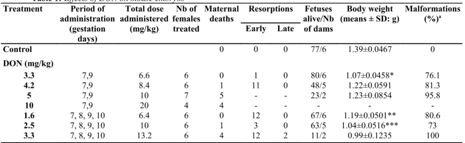
The effects of intraperitoneal administration of DON to pregnant mice on maternal death, percentage of resorptions, percentage of live foetuses and foetal body weights are summarized in Table 1. As would be expected, higher maternal mortalities were associated with higher doses of toxin. However, the same total dose of DON had a lower impact on maternal mortality if given for 4 rather than 2 days.
Effects on embryos
Resorptions
The percentage of implants resorbed per group was higher in treated animals than controls and was dose-dependent. In contrast to the maternal effects, the total dose of DON given at 2 days had the same effect on early resorption rates.
Skeletal abnormalities
All foetuses in the control group showed normal skeletal development. The major appendicular and axial skeletal defects are summarized in Table 2. The most frequent deformities were in the axial skeleton; the appendicular skeleton being spared. This has to be related to the choice of gestational day exposure. The skull is affected (mainly by exencephaly; Fig. 1) when the dose level of DON rises to 75 or 100 µg/30 g during 4 days. Neural arch defects or fusion occur frequently with the higher dose and short exposure. Compared with the 4-day administration experiment, it is seen that only the 100 µg/30 g dose has the same effect. This suggests that there is a threshold dose required to induce abnormalities of the neural arch. However, this observation has no statistical value.
Table 1: Effects of DON on mouse embryos Treatment Period of administration (gestation days) Total dose administered (mg/kg) Nb of females treated Maternal
deaths Resorptions alive/Nb Fetuses of dams Body weight (means ± SD: g) Malformations(%)a Early Late Control 0 0 0 77/6 1.39±0.0467 0 DON (mg/kg) 3.3 7,9 6.6 6 0 1 0 80/6 1.07±0.0458* 76.1 4.2 7,9 8.4 6 1 11 0 48/5 1.22±0.0591 81.3 5 7,9 10 7 5 - - 23/2 1.23±0.0854 95.8 10 7,9 20 4 4 - - - - -1.6 7, 8, 9, 10 6.4 6 0 12 0 67/6 1.19±0.0501** 80.6 2.5 7, 8, 9, 10 10 6 1 3 0 63/5 1.04±0.0516*** 73 3.3 7, 8, 9, 10 13.2 6 4 12 2 11/2 0.99±0.1235 100
a Percentage of fetuses showing minimum of one malformation *P<0.05, **P<0.01, ***P<0.001
Table 2: Number (percentages) of skeletal malformations following DON treatment
DON - period of administration
Gestation days 7, 9 7, 8, 9, 10
Dose level (mg/kg) 3.3 (n=80) 4.2 (n=48) 5 (n=23) 1.6(n=67) 2.5 (n=63) 3.3 (n=11)
Axial skeleton
Skull - 3 (6.3) - 2(3) 10(15.9) 2(18.2)
Neural arch defect 15(18.7) 10 (20.8) 7 (30.4) 9(13.4) - 1(9.1)
Fused neural arch 23 (28.7) 19(39.6) 12 (52.2) 7(10.4) 7(11.1) 4 (36.4)
Atlas fused with occipital 8(10) 4(8.3) 1 (4.3) - - 1(9.1)
Vertebral body divided 16 (20) 14 (29.2) 6(26.1) 13(19.4) 3 (4.8) 1(9.1)
Vertebral body scrambled 34 (42.5) 9(18.8) 13(56.5) 20(30) 13 (20.6)
-Fused bodies 7(8.7) 4(8.3) 2 (8.7) 1(1.5) 1(1.6) 8 (72.7) Hemivertebra 16 (20) 10 (20.8) 6(26.1) 23 (34.3) 6(9.5) 3 (27.3) Cervical rib 17(21.2) 22 (45.8) 17(73.9) 10(14.9) 23 (36.5) 4 (36.4) Fused ribs 8(10) 17(35.4) 5(21.7) 16(23.9) 21 (33.3) 4 (36.4) Branched ribs 3(3.7) 1 (2.1) 1 (4.3) 3 (4.5) 2 (3.2) -Asymmetric sterno-costal junction 6(7.5) 7(14.6) - 6(9) 11 (17.5) 2(18.2)
One sternebra missing Tail 2(2.5) 1 (2.1) 2 (8.7) 19(28.4) 26(41.3) 5 (45.5)
Limbs
Forelimb - 2 (4.2) - - -
-Hindlimb - - -
Fig. 2: Vertebral body fusion associated with a hemivertebra (upper arrow) and a further hemivertebra (lower
arrow). Note the asymmetric last ribs and an isolated cartilaginous nodule between two vertebral bodies
Fig. 3: Complex rib fusions (arrows) with a distal duplication. Such deformities are associated with various
vertebral deformities such as those illustrated in Fig. 2
The vertebral bodies show various deformities. Destruction or division of the vertebral body is common with any dose given in the 2-day experiment, and in the 4-day experiment with a dose level of 50 µg/30 g. Hemivertebrae (Fig. 2) appear with a frequency of around 25% throughout the experiment (except with the 75 µg/30 g dose given during 4 days). A high rate of vertebral fusion (Fig. 3) was seen with the higher dose given for 4 days. There is also fusion of the ribs or distal duplication ("branched" ribs). The percentage of such abnormalities is poorly related to dose. Cervical ribs are seen in both experiments. In the 2-day experiment they are dose-dependent, with the highest frequency seen with the highest dose. In the 4-day experiment the incidence is lower.
Discussion
The aetiology of KBD remains unknown. Selenium deficiency, high organic matter content in drinking water and contamination of food by mycotoxins have been suggested as causal agents. These environmental factors may act together to increase the oxidative stress on cartilage. Different species of fungi produce a wide range of mycotoxins. Among them, DON has been implicated in various health problems in animals [11]. It may not be the only mycotoxin involved in the aetiology of KBD. However, although it is one of the most widely encountered mycotoxins, its teratogenic potential has been little studied.
In this preliminary report, we have studied the teratogenic effects of DON when administered intraperitoneally to pregnant mice. This experimental procedure does not reflect the effects of chronic ingestion of the mycotoxin. DON may be absorbed by the digestive or respiratory systems. The intraperitoneal injection bypasses the
gastro-intestinal tract and the metabolites of the mycotoxin which reach the embryo may not be the same as those which follow physiological absorption. Intraperitoneal injections may not be the most suitable method of inducing chronic overdose of toxin. However, we chose this experimental procedure as a first attempt to investigate the teratogenic potential of DON, hoping to reproduce patterns of malformations similar to those seen in KBD and in avian tibial chondrodysplasia [12]. Further experimental studies are required to determine the period of greatest sensitivity to DON. We were not able to explore a wide range of doses and administration protocols. The choice of experimental procedure was intended to expose the embryos to the mycotoxin during the early organogenetic period. Few significant results were obtained concerning the effects of a single dose and these were therefore not reported.
Injecting DON on days 7 and 9 covers the period of organogenesis of the early axial skeleton and also the beginning of limb bud development. The 4-day injection experiment allows the mycotoxin to affect the beginning of limb morphogenesis. The teratogenic effect of DON on limb development was limited. One of the reasons may be that the drug was given too early and that the period of exposure was too limited, which allowed some repair of the induced damage. However, it may be that the dose administered was too small to provoke a skeletal defect or that the limb bud is less sensitive to the drug. This seems unlikely, however, bearing in mind the well-described sensitivity of the limbs to teratogenic agents. It seems that this procedure does not allow the mycotoxin to interact during the short periods when specific teratogenicity takes place. Various costo-vertebral segmentation anomalies induced by DON were revealed. The fused vertebral bodies, hemivertebrae and rib deformities which were seen even with low doses of DON are similar to those in some human sporadic or familial malformation syndromes such as spondylocostal dysplasia, Jarcho-Levin syndrome, spondylothoracic dysplasia and costovertebral dysplasia. The frequency at which these deformities were seen may allow this model to be used when studying the aetiology of such abnormalities.
The development of cervical ribs shows an important abnormality in which a cervical vertebra acquires some characteristics of a thoracic segment. Similar abnormalities have been observed using the retinoic acid. This drug allowed a "reprogramming" of the vertebral development by modifying the expression of genes of the Hox family [4, 5]. Similar abnormalities have been observed in mice "knocked out" for some Hox genes [7]. Further histological studies on the early effects of DON on vertebral blastemes are required to explain these defects. Although we were unable to reproduce chondrodysplasia in the skeletal rudiments of the limb, these preliminary results are significant. The murine model allows us to explore the effects of DON and other mycotoxins. The effects of multiple toxin exposure must be considered because a single toxin is only rarely present in contaminated food. It has previously been shown that exposure to a mixture of toxins has a more serious, synergistic effect than exposure to a single toxin.
References
1. Allander E (1994) Kashin-Beck disease. An analysis of research and public health activities based on a bibliography 1849-1992. Scand J Rheumatol Suppl 99:1-36
2. Bergsjo B, Herstad O, Nafstad I (1993) Effects of feeding de-oxynivalenol-contaminated oats on reproduction performance in White Leghorn hens. Br Poult Sci 34:147-159
3. Chasseur C, Suetens C, Nolard N, Begaux F, Haubruge E (1997) Fungal contamination in barley and Kashin-Beck disease in Tibet [letter]. Lancet 350:1074
4. Kessel M (1992) Respecification of vertebral identities by retinoic acid. Development 115:487-501
5. Kessel M, Grass P (1991) Homeotic transformations of murine vertebrae and concomitant alteration of Hox codes induced by retinoic acid. Cell 67:89-104
6. Khera KS, Arnold DL, Whalen C, Angers G, Scott PM (1984) Vomitoxin (4-eoxynivalenol): effects on reproduction of mice and rats. Toxicol Appl Pharmacol 74:345-356
7. Le Mouellic H, Lallemand Y Brulet P (1992) Homeosis in the mouse induced by a null mutation in the Hox-3.1 gene. Cell 69:251-264
8. Mathieu F, Begaux F, Lan ZY, Suetens C, Hinsenkamp M (1997) Clinical manifestations of Kashin-Beck disease in Nyemo Valley, Tibet. Int Orthop 21:151-156
9. Peng A, Yang C, Rui H, Li H (1992) Study on the pathogenic factors of Kashin-Beck disease. J Toxicol Environ Health 35: 79-90
10. Peng A, Wang WH, Wang CX, Wang ZJ, Rui HF, Wang WZ et al. (1999) The role of humic substances in drinking water in Kashin-Beck disease in China. Environ Health Perspect 107: 293-296
11. Rotter BA, Oh YN (1996) Mycotoxin fumonisin B1 stimulates nitric oxide production in a murine macrophage cell line. Nat Toxins 4:291-294
12. Walser MM, Morris VC, Levander OA (1988) Effect of dietary selenium on the development of Fusarium-induced tibial dyschondroplasia in broiler chickens. Avian Dis 32:84-88
13. Watson AG (1977) In toto Alcian blue staining of the cartilaginous skeleton in mammalian embryos. Anat Rec 187: 743
14. Yang CL, Bodo M, Notbohm H, Peng A, Muller PK (1991) Fulvic acid disturbs processing of procollagen II in articular cartilage of embryonic chicken and may also cause Kashin-Beck disease. Eur J Biochem 202:1141-1146