1
Université de Bordeaux
U.F.R. DES SCIENCES MÉDICALES
Année 2019 Thèse N°3033 Thèse pour l’obtention du
DIPLÔME D’ÉTAT DE DOCTEUR EN MÉDECINE Spécialité Anesthésie-Réanimation
Présentée et soutenue publiquement le 19 avril 2019
Par Rabia SIBAÏ
Née le 10 mai 1989 à Enghien-Les-Bains (95)
Ventilation non invasive prophylactique puis curative : étude de la
mécanique ventilatoire après résection pulmonaire en chirurgie thoracique
Directeur de thèse Monsieur Le Docteur Benjamin REPUSSEAU
Membres du Jury
Monsieur le Professeur Alexandre OUATTARA Président du jury Monsieur le Docteur Guillaume CARTEAUX Rapporteur Monsieur le Professeur Frédéric VARGAS Juge Monsieur le Docteur Hadrien ROZÉ Juge
2
Remerciements
A mon directeur de thèse, Mr le Docteur Benjamin REPUSSEAU, PH en Anesthésie- Réanimation Thoracique, GH Sud, CHU de Bordeaux,
Je te remercie de m’avoir proposé ce sujet de thèse, de ton aide précieuse tout au long de ce travail qui m’a permis de le mener à bien. Merci pour ta sérénité, ta gentillesse et ton soutien. Je tiens à exprimer toute mon amitié et ma reconnaissance.
A mon président du jury Mr le Professeur Alexandre OUATTARA, PU-PH, Chef de Service d’Anesthésie-Réanimation GH Sud, CHU de Bordeaux,
Vous m’avez fait l’honneur d’accepter la présidence de ce jury de thèse. Je vous remercie pour votre disponibilité, pour votre engagement dans notre formation tout au long de cet internat. Soyez assuré de mon profond respect et de ma sincère reconnaissance, ce sera avec plaisir que j’intégrerai prochainement votre service.
A mon rapporteur de thèse Mr le Docteur Guillaume CARTEAUX, MCU-PH en Réanimation Médicale à l’AP-HP, CHU Henri Mondor,
Je suis honorée que vous ayez accepté de rapporter ce travail. Je vous remercie d’avoir pris de votre temps pour lire cette thèse et pour la réponse rapide que vous avez apporté. Votre expertise dans ce domaine rend votre jugement précieux. Soyez assuré de ma sincère reconnaissance et de mon profond respect.
3
A Mr le Docteur Hadrien ROZE, membre du jury, PH en Anesthésie-Réanimation Thoracique, GH Sud, CHU de Bordeaux,
Merci de m’avoir conseillée durant ce travail, d’avoir significativement participé à la réalisation des statistiques et d’avoir accepté de juger cette thèse. Tes travaux et tes connaissances m’ont permis d’approfondir les miennes dans le domaine de la ventilation en chirurgie thoracique. Ton expertise dans ce domaine rend ton jugement précieux. Je tiens à exprimer tout mon respect et ma reconnaissance.
A Mr le Professeur Frédéric VARGAS, membre du jury, PU-PH en Réanimation Médicale au CHU de Bordeaux,
Vous m’avez fait l’honneur d’accepter de juger ce travail. Je vous remercie pour votre disponibilité, soyez assurés de ma profonde estime et de notre respect. Votre expertise dans ce domaine rend votre jugement précieux.
4
Merci, merci et merci,
À mes amis d’externat, qui m’ont appris à parler français, À mes amis d’internat, qui m’ont appris à picoler,
À mes amis proches, pour tout l’amour et l’amitié que je vous dois. Je vous ai à l’œil, de la rue de Kater et Dandicolle jusqu’aux 4 coins de la France. Au quatuor de promo, j’espère que je verrai un jour vos enfants se marier et vos sourires édentés,
À toutes les équipes médicales, paramédicales et de cointernes qui m’ont appris à pousser du blanc, mettre des tuyaux à n’importe quelle heure, avec rigueur et bonne humeur, surtout l’équipe actuelle de choc et la future du cœur. La poésie ça ne coûte pas cher.
Un big up à Périgueux, dont les membres du bloc et de l’internat ont durablement investi une partie de mon cœur,
À la Vaginale qui m’a sortie de l’autisme profond en me mettant une trompette dans les mains, à Los Teoporos (et les Boobbbbs) qui ont poursuivi la psychothérapie et tenté de relever mon niveau de groove (sans franc succès), à mes parrains princiers,
Aux amis en dehors de médecine, parce que ça fait tellement du bien,
A toutes ces personnes de mon entourage qui cultivent la sérénité et la bienveillance, qui sont au cœur de notre métier et la clé du bonheur,
À mes parents, qui ont réussi à faire de moi quelqu’un qui ressemble à quelque chose, pourtant ce n’était pas gagné. Moi qui trouve toujours le moyen de me cogner aux coins de portes, vous m’avez toujours remis dans le droit chemin. Vous pouvez dorénavant vous reposer sur moi, j’espère être à la hauteur.
À ma sœur, qui me supporte en croyant l’inverse, tu es parfaite et je t’aime comme tu es. À San Antonio et YF, qui ont irrémédiablement pimenté mon esprit et ma psychopathologie, Aux francs-maçons, parce qu’il y en a partout,
Et à toi, qui cherche ton nom depuis le début, je te frustrerai une fois de plus avec un clin d’œil. Merci pour tout, absolument tout.
5
Table des matières
ABRÉVIATIONS ... 7
INTRODUCTION ... 9
1- Particularités de la chirurgie pulmonaire ... 9
2- Complications postopératoires ... 10
3- Ventilation non invasive en chirurgie thoracique ... 11
4- Particularités de la mécanique respiratoire en ventilation spontanée ... 13
5- Controverses de la VNI et rationnel de l’étude ... 17
6- Objectif de l’étude ... 18
ARTICLE ORIGINAL ... 19
I. Abstract ... 20
II. Introduction ... 24
III. Materials and Methods ... 27
III. Results ... 30
IV. Discussion ... 32
V. Conclusion ... 35
VI. References ... 36
VII. Tables and Figures ... 39
DISCUSSION ... 47
1- Contrôle des volumes courants en VNI ... 47
2- Pressions motrices et compliance ... 48
3- Avantages et limites de l’étude ... 49
4- Perspectives ... 50
CONCLUSION ... 53
6
Tables et Figures
Table 1: Baseline characteristics of patients who required or not intubation ... 39
Table 2: Ventilation and blood gas parameters in preventive and curative NIV ... 40
Table3: Ventilation and blood gas data in curative NIV for intubated and not intubated ... 41
Table 4: Ventilation and blood gas parameters before and after intubation ... 42
Table 5: Parameters of survivors and non survivors ... 42
Figure 1 : Flow Chart ... 43
Figure 2: Parameters for preventive NIV, curative NIV and controlled ventilation ... 44
Figure 3: NIV parameters of intubated and not intubated patients ... 45
Figure 4: VT PBW and VT anat in curative NIV ... 46
7
ABRÉVIATIONS
APRV : Air Pressure Release Ventilation ASA : American Society of Anaesthesiologists
BPCO : Broncho Pneumopathie Chronique Obstructive COPD : Chronic Obstructive Pulmonary Disease CRF : Capacité Résiduelle Fonctionnelle (RFC) Crs : Compliance du Système Respiratoire ∆P : Driving Pressure ou Pression Motrice
∆PL : Lung Driving Pressure ou Pression motrice transpulmonaire FR : Fréquence Respiratoire (RR)
HTP : Hypertension Artérielle Pulmonaire IGS2 : Indice de Gravité Simplifié
IRA : Insuffisance Respiratoire Aiguë OAP : Œdème Aigu du Poumon PAV : Proportional Assist Ventilation
PAVM : Pneumopathie Acquise Sous Ventilation Mécanique Paw: Air Way Pressure
PBW/PP: Predicted Body Weight (Poids Prédit) PEEP: Positive End Expiratory Pressure
Pmus : Pression générée par les Muscles Inspiratoires Ppl : Pression Pleurale
Pplat : Pression de Plateau
8
PS: Pressure Support ou Aide Inspiratoire
P-SILI: Patient Self Inflicted Lung Injury PTP : Pressure-Time Product
RFE : Recommandations Formalisées d’Experts Rrs : Résistance du Système Respiratoire
SAOS : Syndrome d’Apnée Obstructive du Sommeil (OSAS) SDRA : Syndrome de Détresse Respiratoire Aiguë
SOFA: Sequential Organ Failure Assessment VD : Espace Mort (Dead Volume)
Vm : Ventilation Minute
VEMS : Volume Expiratoire Maximal par Seconde (FEV1) VILI : Ventilation Induced Lung Injury
VNI : Ventilation Non Invasive (NIV) VS : Ventilation Spontanée (SV)
VSAI : Ventilation Spontanée en Aide Inspiratoire VT : Volume Courant (Tidal Volume)
VT PP ou VT PBW : VT rapporté au Poids Prédit
9
INTRODUCTION
1- Particularités de la chirurgie pulmonaire
La chirurgie pulmonaire est le traitement de référence des cancers non à petite cellule localisés et sa fréquence est en constante augmentation. Elle est responsable d’une réduction du volume pulmonaire, qui peut être estimée à l’aide de mesures scannographiques préopératoires (1), ou à l’aide de la formule de Juhl et Frost : le volume pulmonaire à soustraire après résection : A Gauche : Pneumonectomie : -45% Lobectomie Supérieure : -20% Lobectomie Inférieure : -25% A Droite : Pneumonectomie : -55% Lobectomie Supérieure : -15% Lobectomie Moyenne : -10%; Lobectomie Inférieure : -25%
Ce calcul participe à la prédiction de la fonction respiratoire postopératoire et peut même venir contre-indiquer la chirurgie.
Même si les abaques de calcul du volume courant (VT) rapporté au poids prédit (PP) (6 à 8mL.kg-1) sont couramment utilisés chez les patients ayant eu une résection pulmonaire (2), il est important de prendre en compte cette modification anatomique et physiopathologique dans notre raisonnement notamment lorsqu’il s’agit de complications respiratoires postopératoires.
La question de la ventilation en péri-opératoire de chirurgie pulmonaire se pose à différents stades : En peropératoire, en ventilation uni-pulmonaire, plusieurs études ont tenté de trouver la ventilation uni-pulmonaire optimale : Katz & al. avaient préconisé dans les années 80 une ventilation à VT PP = 10 mL.kg-1 et 0 de pression télé-expiratoire (PEEP) afin de minimiser le shunt intra-pulmonaire, responsable d’hypoxémie peropératoire (3). Plus tard, les études se sont penchées sur le risque de surdistension du poumon dépendant (ventilé), et sur les conséquences du shunt intra-pulmonaire, lié au poumon non dépendant (non ventilé) collabé et aux atélectasies présentes sur le poumon dépendant. Ainsi devant un risque majoré de
10
complications postopératoires sous ventilation conventionnelle à hauts volumes courants (4), on préconise une ventilation protectrice, avec des VT PP à 5-6 mL.kg-1 et une PEEP élevée, tout en respectant des limites de pression de plateau (5,6). Il est donc logique de poursuivre cette protection pulmonaire en postopératoire en cas de ventilation non invasive, en cas de réintubation suite à une complication postopératoire ou en cas de sevrage respiratoire.
2- Complications postopératoires a. Etiologie :
La chirurgie et la période péri-opératoire sont sources de complications postopératoires indépendantes liées à (7,8) :
a) L’inflammation locale ;
b) L’atélectasie liée à la compression mécanique du poumon par le chirurgien et à l’altération du surfactant, associée à l’altération du rapport ventilation/perfusion ;
c) Aux dysfonctions diaphragmatiques liées à la lésion peropératoire du nerf phrénique ou à une atteinte fonctionnelle ;
d) L’amputation de la fonction respiratoire liée à la résection, et l’altération de la mécanique ventilatoire liée à la thoracotomie et à la pleurotomie (responsables d’une modification de l’élastance pulmonaire et pleurale) ;
e) La ventilation uni-pulmonaire, responsable d’une part d’un shunt intra-pulmonaire (allant jusqu’à 50%), créant une hypoxémie peropératoire plus ou moins contrôlée par la vasoconstriction hypoxique, et d’autre part d’une altération du drainage lymphatique peropératoire et le syndrome d’ischémie reperfusion du poumon collabé, participant à l’inflammation locorégionale. On note également une surdistension du poumon ventilé ; f) L’inhibition de la toux et de l’inspiration profonde en postopératoire par deux mécanismes :
la lésion des nerfs intercostaux et la douleur, qui favorisent l’encombrement bronchique et l’atélectasie ;
g) La douleur est également responsable d’une augmentation du tonus musculaire abdominal et d’une diminution de l’ampliation thoracique. Elle est source de consommation importante de morphiniques en postopératoire et de ses conséquences (hypoventilation, hypovigilance, nausées/vomissements, risque d’inhalation). Mais l’avènement de la thoracoscopie d’une part et la démocratisation de l’anesthésie péridurale (avec le bénéfice sur l’analgésie mais
11
aussi sur la réhabilitation précoce postopératoire et sur la mortalité (9)), du bloc para-vertébral et d’autres techniques d’analgésie locorégionale d’autre part, ont permis de contrôler la douleur postopératoire et de limiter la consommation morphinique ;
h) L’anesthésie générale, par le risque d’inhalation gastrique per-anesthésique, la diminution du tonus ventilatoire, les atélectasies générées par la ventilation uni-pulmonaire ;
i) Au terrain du patient : cancer, dénutrition, âge avancé, tabagisme actif, pathologie pulmonaire sous-jacente (bronchopneumopathie chronique oblitérante (BPCO), hypertension pulmonaire (HTP), Emphysème…), obésité (et syndrome restrictif), chimiothérapie néo-adjuvante, tabagisme actif.
b. Complications et morbimortalité (10–14)
La mortalité postopératoire à 1 mois est évaluée entre 0,79% et 4% pour les lobectomies, entre 4 et 12% pour les pneumonectomies. L’incidence des complications postopératoires est de 17 à 40% selon les études. L’incidence des syndromes de détresse respiratoire aiguë (SDRA) postopératoires est de 2 à 6%, avec une mortalité allant jusqu’à 65%. Parmi ces complications, on retrouve 17% de pneumopathies, 12% d’hémorragies postopératoires, 5% de fistules broncho-pleurales, 4,5% d’empyèmes intra-thoraciques. La réintubation après résection pulmonaire est grevée d’une mortalité pouvant aller jusqu’à 80%, probablement du fait de la gravité du tableau respiratoire sous-jacent. D’où l’intérêt d’une prise en charge précoce de ces complications.
3- Ventilation non invasive en chirurgie thoracique a. Définition
Outil de plus en plus répandu, il regroupe l’ensemble des méthodes d’assistance ventilatoire en pression positive prenant en charge tout ou une partie du travail respiratoire, à l’aide d’une interface non invasive, afin d’assurer une ventilation alvéolaire satisfaisante. Ses principales indications intrahospitalières sont l’œdème aigu du poumon (OAP) cardiogénique, la décompensation de BPCO, l’insuffisance respiratoire aiguë (IRA) hypercapnique, la prévention du sevrage respiratoire difficile en post-extubation en réanimation chez les patients à risque, la prévention et le traitement des atélectasies en postopératoire chez les patients et/ou les chirurgies à risque (15).
12
b. Modalités de ventilation en VNI
La particularité de ce mode ventilatoire, par rapport à la ventilation invasive, est de détecter et de compenser les fuites par l’interface, afin d’adapter le trigger inspiratoire et le temps inspiratoire. Les paramètres ventilatoires sont globalement réglables de manière similaire à la ventilation invasive en aide inspiratoire (VSAI). Un schéma continue ou intermittent peut être défini, avec une titration des paramètres statiques et dynamiques en fonction de l’efficacité et du confort du patient, afin d’optimiser l’adhésion et l’efficacité thérapeutique.
c. Concernant la VNI prophylactique
L’intérêt de la VNI prophylactique en postopératoire d’une chirurgie thoracique serait de prévenir ou de traiter les atélectasies par un recrutement par la pression télé-expiratoire positive (PEEP), permettant d’améliorer la capacité résiduelle fonctionnelle (CRF), de réduire le travail musculaire respiratoire et d’améliorer les échanges gazeux, dans le but d’accélérer la récupération et de diminuer la durée d’hospitalisation. Plusieurs études prospectives et méta– analyses ont étudié l’intérêt de la VNI prophylactique en postopératoire d’une chirurgie pulmonaire (16–23) et autres chirurgies majeures (abdominale, cardiaque et aortique) (24–27). Les résultats de ces études sont mitigés : certaines retrouvent globalement une amélioration de la gazométrie, de la spirométrie, une diminution du taux de réintubation, une diminution des pneumopathies, de la réadmission en réanimation et de la mortalité (11,16,24). D’autres (17– 20,22,23) ne retrouvent pas de bénéfice à la VNI prophylactique, notamment pas de réduction de la mortalité, du taux de réintubation ni des insuffisances respiratoires aiguës postopératoires. Cependant, la méta-analyse de Torres(17) regroupe 6 études randomisées assez hétérogènes, avec des populations de patients de niveaux de risque différents (notamment des proportions de BPCO et de cardiopathies hétérogènes), et une VNI prophylactique conduite de manière hétérogène. Les conditions de réalisation de l’étude de Lorut (18) font que le groupe VNI était désavantagé et l’effet de la VNI sous-estimé. De façon similaire, l’étude de Hernandez (19) - qui ne retrouve pas d’avantage à une VNI prophylactique - possédait des effectifs bas et des durées de séances limitées. Dans l’étude de Perrin (16), les patients avaient une acidose respiratoire postopératoire, rendant le caractère prophylactique de la VNI discutable. Cette divergence pourrait également dépendre de la fonction respiratoire (évaluée par le volume expiratoire maximal en 1 seconde ou VEMS) préopératoire du patient et de la présence d’une BPCO préopératoire (16). La VNI prophylactique a en effet fait ses preuves chez les patients atteints de BPCO en post-extubation (28) avec une diminution significative de l’insuffisance
13
respiratoire aiguë, de la mortalité et de la durée de séjour en réanimation. Dans ce contexte, les recommandations formalisées d’experts (RFE) de 2019 sur la VNI en postopératoire de chirurgie thoracique vont probablement préciser la place de la VNI prophylactique.
d. Concernant la VNI curative :
L’intérêt de la VNI curative s’inscrit dans la prise en charge d’une détresse respiratoire aiguë, en dehors des critères d’intubation. Son succès est évalué à 85% (29) avec une amélioration de la morbimortalité et une éviction de l’intubation, ces chiffres étant plus marqués chez les BPCO (30). Un essai clinique randomisé multicentrique (11) retrouve une efficacité de la VNI dans les IRA postopératoires avec une réduction du recours à l’intubation et de la mortalité. On note des durées de VNI de 14h+/- 2,8h sur 2 à 4 jours, ce qui est plus élevé que la majorité des autres études et qui pourrait expliquer ces résultats positifs. D’autre part, même si la VNI curative avait un important taux de réussite dans l’étude descriptive de Lefebvre (29), avec un bénéfice marqué surtout dans le groupe des patients hypercapniques, cette étude retrouve une mortalité importante (46%) en cas d’échec de VNI, probablement à cause du retard à l’intubation. Les résultats d’une autre étude observationnelle vont également dans ce sens (31), rejoignant ainsi la controverse de l’indication de la VNI dans l’IRA hypoxémique de novo.
4- Particularités de la mécanique respiratoire en ventilation spontanée a. Rappels physiologiques
La ventilation spontanée présente des particularités quant à ses régimes de pression, avec des conséquences bénéfiques et potentiellement délétères, qui font l’objet d’une multitude d’études cliniques et expérimentales. Ce chapitre traitera de la physiopathologie de la ventilation spontanée en pression positive, sans différencier le caractère invasif ou non invasif.
La ventilation est générée grâce à une pression d’insufflation, déclenchée par le patient. La pression d’insufflation dépend de la force générée par le respirateur, des résistances des tuyaux mais également des propriétés élastiques et résistives de l’arbre bronchique, du parenchyme pulmonaire et de la paroi thoracique. Cette pression est partagée entre le ventilateur (Paw) et les efforts du patient (Pmus). L’équation du mouvement fait intervenir : Paw + Pmus = PEEP + [VT/Crs] + [Rrs × Débit], la résistance et la compliance étant des constantes et les pressions, les volumes et les débits étant des variables. De cette équation, nous
14
pouvons déduire qu’à débit nul (en occlusion télé-inspiratoire), Paw + Pmus = Pplat = VT/Crs + PEEP, ce qui nous permet théoriquement de calculer une pression motrice ∆P=VT/Crs. La pression motrice est un paramètre important communément utilisé en ventilation mécanique invasive passive. Il permet de normaliser les volumes courants à la compliance pulmonaire (∆P=VT/Crs) et est associé à la mortalité des patients en SDRA (32). Un seuil de 14 cmH2O pourrait être utilisé dans la ventilation des patients en SDRA, pour optimiser le rapport recrutement/surdistension pulmonaire (33) et il présente une bonne corrélation au risque de survenue du Ventilation Induced Lung Injury (VILI). Plus récemment, certaines études proposent son utilisation dans certaines conditions en ventilation spontanée (34–36). D’autres paramètres permettent de quantifier le travail respiratoire en ventilation spontanée, tels que la mesure (électrique ou échographique) du travail diaphragmatique, les mesures du produit Pression-Temps (PTP) et autres mesures à partir de la pression œsophagienne et gastrique (37), mais ne font pas partie des pratiques courantes.
Un autre paramètre important dans la ventilation spontanée est la pression transpulmonaire PL (38). Celle-ci reflète la force de distension appliquée sur le poumon par le ventilateur. En ventilation spontanée, elle reflète le gradient de pression durant l’effort inspiratoire permettant de passer de la position de repos (CRF) à la pression négative nécessaire à l’inflation du poumon d’un certain volume courant. Elle se calcule à partir de la différence entre la pression inspiratoire à l’entrée des voies aériennes (Paw) et la pression pleurale (Ppl) (estimée à partir de la manométrie œsophagienne en occlusion) (PL = Paw - Ppl). Cependant, cette pression présente une distribution variable, liée aux propriétés élastiques du poumon et du diaphragme, au gradient vertical de la pression pleurale et à l’altération de l’élastance pulmonaire par une potentielle pathologie. Elle varie également selon le mode ventilatoire (en pression positive ou négative) puisque le comportement du poumon, du diaphragme et de la plèvre diffèrent (39). Pour mieux caractériser les pressions transpulmonaires en ventilation spontanée, il est donc plus judicieux de parler de pression motrice transpulmonaire ou de la variation de la PL entre l’inspiration et l’expiration (40).
b. La surdistension en ventilation spontanée :
Avant de définir les lésions de surdistension en ventilation spontanée, nous allons rapidement définir le VILI (41) :
Le « Ventilation Induced Lung Injury » est une entité englobant l’ensemble des lésions pulmonaires secondaires à la ventilation mécanique. Plusieurs mécanismes sont impliqués : le
15
barotraumatisme (lung stress), le volotraumatisme (lung strain), la toxicité de l’O2, l’altération du surfactant, l’inflammation et le remaniement de l’épithélium broncho-alvéolaire, le retentissement hémodynamique, etc. D’un point de vue mécanique, des pressions transpulmonaires excessives génèrent une surdistension régionale du parenchyme pulmonaire, et ce pour des raisons anatomiques : les compliances régionales varient dans le parenchyme (meilleure compliance à l’apex et dans les zones proximales qu’aux bases et dans les zones distales, contraction diaphragmatique inhomogène se répercutant sur les pressions pleurales avoisinantes) mais également physiopathologiques (meilleure compliance dans les zones saines que dans les zones malades). D’un point de vue histologique, de microlésions dans l'espace interstitiel, libérant des molécules de bas poids moléculaire responsables d’une cascade pro-inflammatoire (biotraumatisme), augmentent la perméabilité de la barrière alvéolo-capillaire, responsable d’un œdème interstitiel et alvéolaire (42). Dans le SDRA, le poumon malade se comporte comme un babylung avec un parenchyme inhomogène et des zones ventilables (zones non dépendantes) réduites. En chirurgie thoracique, la physiopathologie est différente mais le poumon est néanmoins mécaniquement réduit, avec des zones inflammatoires inhomogènes. Du fait de cette distribution hétérogène, certaines alvéoles resteront collabées au cours d’un cycle respiratoire tandis que d’autres subiront une surdistension.
Les VILI ont longtemps été décrits comme des lésions spécifiques du ventilateur. Une autre entité s’est progressivement définie, appelée P-SILI pour Patient Self-Inflicted Lung
Injury, comme des lésions induites par la ventilation spontanée, de novo ou se surajoutant aux
lésions pulmonaires préexistantes.
Lorsque les rapports ventilation/perfusion sont altérés, la commande ventilatoire augmente la ventilation spontanée afin de compenser au maximum l’espace mort et de maintenir le pH du LCR au niveau du tronc cérébral dans les limites de la normale. Ces augmentations de commande respiratoire et de ventilation spontanée peuvent être excessives en termes d’étirements sur un parenchyme pulmonaire déjà lésé. Des études expérimentales ont démontré l’apparition de lésions pulmonaires spécifiques liées à l’hyperventilation sur un poumon sain en respiration spontanée (43) ou sur un poumon lésé ventilé (44) avec une altération de la compliance, de l’hématose et du parenchyme pulmonaire, et ce indépendamment de la ventilation mécanique. Les principales causes d’hyperventilation en ventilation spontanée peuvent être liés : aux réglages du ventilateur (aide inspiratoire (AI) trop élevée), à l’interaction ventilateur-patient (asynchronies, double-déclenchements et désadaptation patient-machine) (45) ou au patient lui-même, par la stimulation maximale de ses commandes ventilatoires
16
d’origine extra-respiratoire (fièvre, sepsis, acidose métabolique, état de choc, délirium…) ou d’origine respiratoire (hypoxémie, hypercapnie).
Les mécanismes en jeu (46) :
- L’augmentation des pressions transpulmonaires : Alors que les pressions d’insufflation (Paw) sont moins élevées en ventilation spontanée qu’en ventilation contrôlée, cela ne se traduit pas sur la pression appliquée sur le poumon (PL). En ventilation spontanée, à la pression positive appliquée par le respirateur s’ajoute la valeur absolue d’une pression pleurale négative importante liée aux efforts du patient. Une augmentation du travail respiratoire peut être responsable de pressions pleurales négatives importantes et par conséquent de pressions transpulmonaires positives excessives devenant délétères (PL > 25cmH2O), même avec de faibles niveaux d’aide inspiratoire.
- La fréquence respiratoire élevée pourrait être responsable d’hyperinflation et de surdistension pulmonaire par augmentation de la ventilation minute et diminution du temps expiratoire. Elle fait partie, avec les pressions transpulmonaires et le volume courant, d’une entité plus globale qui est la puissance mécanique (énergie par unité de temps) appliquée sur le poumon et intervient donc dans le phénomène de VILI (47).
- A l’expiration, la mise en jeu d’une contraction active des muscles expiratoires augmente la pression pleurale durant cette phase, au-delà de la pression intra-alvéolaire, responsable d’un collapsus télé-expiratoire des alvéoles et d’un dérecrutement cyclique (atelectrauma).
- Le phénomène de Pendelluft : Une négativation importante des pressions pleurales sur un poumon lésé peut être responsable, sans augmentation des volumes courants ni de la pression de plateau, d’un déplacement de l’air insufflé des zones non dépendantes (zones bien aérées, au comportement fluide) vers les zones dépendantes (zones atélectasiées, au comportement solide) et causer une majoration de la pression transpulmonaire et une surdistension alvéolaire régionale (48).
De ces phénomènes résulte une augmentation des pressions négatives interstitielles et alvéolaires, et une augmentation du gradient de pression transmurale au niveau des capillaires, favorisant l’œdème lésionnel interstitiel et alvéolaire (49).
L’occurrence des VILI en ventilation spontanée est mal connue. La surdistension régionale est difficile à mesurer en pratique clinique et donc difficile à détecter et à traiter. La
17
pression alvéolaire globale peut être estimée grâce à la pression transpulmonaire, reflet de la compliance pulmonaire. On peut évaluer le stress du poumon par sa force de distension ou ∆PL
(= PL inspiratoire – PL expiratoire) dont l’origine serait les muscles respiratoires (en respiration spontanée), le ventilateur (en ventilation contrôlée) ou les 2 (en ventilation spontanée). Une étude de Bellani & al. a démontré que, à volume et débit égaux, sur des propriétés pulmonaires comparables (mêmes compliance et résistance), la variation de la pression transpulmonaire (∆PL) et les pressions motrices sont similaires, que la ventilation soit contrôlée ou spontanée (35).
Le mécanisme et le caractère délétère restent néanmoins mitigés, et sont sujets à débat (50–52). Certains auteurs ont stratifié le VILI en VS en fonction de la sévérité des lésions (44,51): Sur un poumon modérément lésé, la ventilation spontanée serait bénéfique par le recrutement et l’optimisation du travail respiratoire, mais elle serait au contraire plus délétère sur un poumon gravement lésé par majoration de l’inflammation et de la consommation d’oxygène, avec la présence de pressions transpulmonaires très élevées pouvant passer inaperçues (53).
5- Controverses de la VNI et rationnel de l’étude
Même si la VNI (prophylactique et curative) a sa place en postopératoire de chirurgie pulmonaire, son efficacité et son innocuité dans l’IRA hypoxémique sont débattues.
Plusieurs problématiques se posent chez nos patients :
- Les études sur les VILI en VS concernent essentiellement la VS chez les patients intubés ou en respiration spontanée. Peu d’études concernent les patients ventilés en VNI.
- La majorité des études sur la surdistension sont faites chez des patients non chirurgicaux et de fait sans résection pulmonaire.
- Les hauts volumes courants expirés en VNI semblent également être impliqués et apparaissent dans certaines études comme facteurs prédictifs d’échec de la technique et de recours à l’intubation dans les populations de patients médicaux. En chirurgie thoracique, la question des volumes courants expirés cibles se pose d’autant plus que le parenchyme pulmonaire est réduit et mécaniquement lésé.
18
6- Objectif de l’étude
L’objectif principal est de comparer les volumes courants sous ventilation des patients ayant une détresse respiratoire aiguë postopératoire en 3 temps : lors de la prophylaxie avant la détresse, à l’acmé de l’hypoxémie, puis une heure après l’intubation chez ceux qui l’ont été.
19
20
Prophylactic and Curative Non Invasive Ventilation: A Study of Ventilatory Mechanics after Lung Resection in Thoracic Surgery
Authors: RABIA SIBAÏ*, BENJAMIN REPUSSEAU M.D1, HADRIEN ROZÉ M.D., Ph.D.2, ALEXANDRE OUATTARA, M.D., Ph. D.3
Addresses & Affiliations:
1. Anaesthesiology and Critical Care Department, Haut Lévêque Hospital, CHU Bordeaux, 33600 PESSAC
2. Anaesthesiology and Critical Care Department, Haut Lévêque Hospital, CHU Bordeaux, 33600 PESSAC
3. Anaesthesiology and Critical Care Department, Haut Lévêque Hospital, CHU Bordeaux, 33600 PESSAC Emails: rabia.sibai@chu-bordeaux.fr benjamin.repusseau@chu-bordeaux.fr hadrien.roze@chu-bordeaux.fr alexandre.ouattara@chu-bordeaux.fr (*) Corresponding author
Rabia Sibaï, Anaesthesiology and critical care department, Haut Lévêque Hospital, CHU Bordeaux, Avenue Magellan, 33600 PESSAC
21
ABREVIATIONS
APRV : Air Pressure Release Ventilation ARDS: Acute Respiratory Distress Syndrome ARF : Acute Respiratory Failure
ASA : American Society of Anaesthesiologists COPD : Chronic Obstructive Pulmonary Disease Crs : Compliance of the Respiratory System ∆P : Driving Pressure
∆PL : Lung Driving Pressure
FEV1 : Forced Expiratory Volume at 1 second IGS2 : Indice de Gravité Simplifié
NIV : Non Invasive Ventilation
OSAS : Obstructive Sleep Apnea Syndrome PAV : Proportional Assist Ventilation Paw: Air Way Pressure
PBW: Predicted Body Weight
PEEP: Positive End Expiratory Pressure Pmus : Respiratory Muscles Pressure Ppl : Pleural Pressure
Pplat : Plateau Pressure
PPNV : Postoperative Prophylactic Non-invasive Ventilation PS: Pressure Support
22
PTP : Pressure-Time Product
RFC : Residual Functional Capacity RR : Respiratory Rate
Rrs : Resistance of the Respiratory System SOFA: Sequential Organ Failure Assessment SV: Spontaneous Ventilation
VD : Dead Volume
VILI : Ventilation Induced Lung Injury Vm : Minute Ventilation
VT : Tidal Volume
VT PBW : Tidal Volume to Predicted Body Weight
23
I. Abstract
Introduction: Lung resection surgery is a source of inflammation, atelectasis and postoperative
complications, which can be prevented or treated by noninvasive ventilation (NIV). However, spontaneous ventilation at high tidal volumes above 8mL.kg-1 of predicted body weight (V
T
PBW) can lead to overdistension and high transpulmonary pressure, by decreasing the respiratory
system’s compliance (Crs). The aim of this study was to compare the VT PBW of patients
receiving NIV for acute respiratory failure (ARF) following lung resection at 3 stages: during prophylaxis, at the acme of hypoxemia, and then one hour after intubation (if it occurred).
Methods: This retrospective cohort study included 21 patients between July 2016 and
December 2017. Postoperative VT were estimated with Juhl & Frost formula and called VT anat.
Results: Both VT PBW and VT anat were higher in curative NIV compared to prophylaxis (VT PBW
=8.0 [6.5-10] versus 11.2 [9.9-12.7] mL.kg-1 and VT anat= 10.5 [8.5-12.2] versus 14.6
[12.1-16.3] mL.kg-1 (p<0.05). Inspiratory pressure levels were similar in both types of NIV (5 [5-8] and 5 [4-6] cmH2O). Patients needing intubation showed, during curative NIV a higher
respiratory rate compared to not intubated patients (RR= 33 [26-36] versus 26 [24-30], p= 0.05) but a lower PaO2/FiO2 ratio (105 [96-112] versus 131 [112-217] p= 0.02). In the intubated group
Crs= 13 [12-17] ml.cmH2O-1, predicting high driving pressures.
Discussion: Tidal volumes during curative NIV were all excessive despite low levels of
inspiratory pressure. Low Crs predicted high driving pressure under curative NIV. Alveolar overdistension in spontaneous ventilation after lung resection is therefore frequent under curative NIV.
Conclusion: The only way to control compensatory hyperventilation observed in ARF after
lung resection, is probably early sedation and intubation, in order to guarantee protective ventilation.
24
II. Introduction
Pulmonary resection is responsible for lung volume reduction, which can be estimated by using preoperative CT scan measurements (1) or the Juhl and Frost formula1. Pulmonary surgery causes various pathophysiological phenomena (local inflammation, diaphragmatic dysfunction, pulmonary and pleural elastance modification, postoperative pain, atelectasis ...) in patients presenting multiple comorbidities (age, cancer, undernutrition, chronic obstructive pulmonary disease (COPD), obesity, obstructive sleep apnoea syndrome (OSAS), neoadjuvant chemotherapy etc.) (2,3). Postoperative complications are estimated between 17 and 40% and overall mortality at 1 month varies between 0.79% and 4% (4–6).
Postoperative prophylactic non-invasive ventilation (PPNV) is used to prevent or treat atelectasis by positive end-expiratory pressure (PEEP) recruitment, thereby improving functional residual capacity (FRC), reducing respiratory work and improving gas exchange, with the aim of accelerating postoperative recovery and reducing the length of inhospital stay. Several prospective studies and meta-analysis have investigated the efficiency of PPNV after lung surgery. Results are mixed: some find improvements in blood gas results, spirometry, less intubation, pneumonitis, readmission to intensive care unit (ICU) and mortality (5,7,8). Others
(9–12) do not find any profit. In this context, future French recommendations in 2019 should specify the role of PPNV in thoracic surgery.
Non-invasive ventilation (NIV) is also used to treat acute respiratory failure (ARF), apart from intubation criterias. Its success is estimated at 85% (13), reducing morbidity and intubation rate (5,14). However, several studies carried out on curative NIV in de novo ARF showed that, owing to an increased respiratory drive, patients have very high tidal volumes (VT> 9.5mL.kg-1) of predicted body weight (PBW) despite targets set between 6 and 8mL.kg-1
PBW (15,16). High VT seems to be a predictor of intubation and could be a source of pulmonary
overdistension injuries or "Ventilator Induced Lung Injury" (VILI). In order to understand these overdistension lesions, it is important to understand ventilatory mechanics during spontaneous ventilation. Inspiratory pressure is shared between the ventilator (airway pressure, Paw) and patient's efforts (muscular pressure, Pmus). Respiratory efforts increase the negative pleural
1Juhl & Frost formula: left lung: a superior lobectomy takes off 20% of the parenchyma, an inferior lobectomy takes off 25% of the parenchyma and 45% for pneumonectomy. Right lung: a superior lobectomy takes off 15% of the parenchyma, the middle lobe resection takes off 10%, and an inferior lobectomy takes off 25% and 55% for right pneumonectomy.
25
pressure and, consequently, the transpulmonary pressure (PL). The latter reflects the pressure gradient needed to move from rest position (FRC) to the negative pressure, in order to inflate the lung of a certain tidal volume (17). Driving pressure is also an important parameter that reflects lung compliance. It seems to be associated with mortality in acute respiratory distress syndrome (ARDS) and is well correlated to VILI (18). More recently, Bellani & al. showed its applicability under certain conditions in spontaneous ventilation (SV) (19,20).
Some clinical situations may be responsible for an increase in respiratory drive, (hypoxemia, hypercapnia, fever, anxiety, metabolic acidosis, and poor patient - ventilator synchronization (21). When respiratory drive under ventilation increases, respiratory work increments as well, and thus transpulmonary pressure, becoming deleterious (PL> 25cmH2O). Transpulmonary pressure is heterogeneously distributed in the lung (better compliance at the apex and in the proximal areas than at the bases and in the distal areas, better compliance in healthy areas compared to sick areas), generating local injurious forces. In addition, Occult
Pendelluft phenomenon2 (22) and polypnea (causing hyperinflation by increasing minute ventilation and decreasing expiratory time (23)) lead to overdistension, creating barotrauma, volotrauma, inflammatory reactions (biotrauma) as well as an increase in the capillary transmural pressure, favouring interstitial and alveolar oedema (24). All of these events are called P-SILI for Patient Self Inflicted Lung Injury, and are comparable to VILI.
The occurrence of P-SILI is poorly known. Regional overdistension is difficult to measure in clinical practice and therefore difficult to detect and treat. Global alveolar pressure can be estimated by transpulmonary pressure. Lung stress can be assessed by its distension force or transpulmonary swings (ΔPL = PLinsp - PLexp). Mechanisms and harmfulness, however, remain uncertain, and are subject to debate (25–27).
Considering our cohort, even though NIV is possibly beneficial in the postoperative period, its efficacy and safety in hypoxemic ARF are uncertain. Several issues arise for our patients: 1) Studies on P-SILI are related to invasive spontaneous pressure support and few studies concern NIV. 2) Most studies are done on non-surgical patients, free from lung resection. 3) If the high tidal volumes in NIV appear to be harmful for ARF population, the
2OccultPendelluft: Yoshida et al. showed that, without increasing tidal volume nor plateau pressure, spontaneous ventilation with a high respiratory drive could induce major negative pleural pressure, causing an internal redistribution of the inspired tidal volume and generating local injurious forces.
26
question of target VT arises especially as the lung parenchyma is reduced and mechanically
injured.
The main objective of this study is to assess the expired tidal volume under NIV based
on PBW (VT PBW), and on lung volume estimated by the Juhl and Frost formula after pulmonary
resection (anatomical VT or VT anat). All in two clinical situations, during prophylactic NIV and
during curative NIV, in order to compare the VT according to the clinical evolution of the
27
III. Materials and Methods
1- Study design
This was an observational, single-centre, retrospective study, realized in the thoracic surgery ICU of 12 beds at Magellan Hospital, University Hospital Centre of Bordeaux.
2- Population
a. Inclusion criteria
We analysed the records of all the patients who stayed in thoracic ICU after undergoing pulmonary resection, over a period of 18 months (July 2016 - December 2017), and who received prophylactic and curative NIV for postoperative acute respiratory distress. Acute respiratory distress was defined by the association of clinical signs (polypnea, clinical symptoms of respiratory distress, desaturation) and blood gas impairment (PaO2/FiO2< 300).
The emergence of radiographic infiltrates was collected but not taken into account, being often difficult to analyse following thoracic surgery (presence of chest drains, resected lobes, pleural suffusion, and atelectasis).
b. Exclusion criteria Missing data;
Urgent surgery;
Less than 48 hours hospitalization; NIV without acute respiratory distress.
Given the retrospective type of study, informed consent was waived for analysis at the time of data collection. Moreover, according to the French law, the analysis on retrospective data and not on human subjects, did not need ethics approval.
3- Study outcomes
The primary endpoint was to assess tidal volume relative to the predicted body weight (VT PBW) in preventive then in curative NIV. The secondary endpoints were:
28
Ventilatory parameters measured in preventive, curative NIV and then in controlled ventilation;
Rate of intubation; Mortality.
4- Data collection
We collected the parameters of preventive and curative NIV in the 21 patients included. For prophylactic NIV, patients received one-hour sessions, 3 to 4 times a day. In case of a curative treatment, patients received sessions of 1 to 2 hours every 4 hours (ie 6 to 12 hours per day). Patients with OSAS also received nocturnal ventilation with the same parameters. Levels of positive pressure support were theoretically adjusted for a goal of VT PBW= 6-8mL.kg-1.
Clinical, biological and imaging data were collected from DxCare® computerized medical
record and Metavision® ICU software (iMdSoft®, USA); Demographic data (age, weight, sex,
predicted body weight, body mass index), comorbidities (ASA (American society of anaesthesiologists) score, active or former smoking, COPD, OSAS, ischemic heart disease), respiratory functional explorations data, surgery related data (site, technique, regional analgesia) and hospitalization data (IGS2 scores (Simplified severity index), SOFA score (Sequential organ failure assessment), total days spent in ICU, death during hospitalization) were collected for each patient.
The following parameters were also collected during a NIV session (prophylactic or curative):
Respiratory parameters: FiO2, pressure support (PS), PEEP, VT PBW (mL.kg-1), minute
ventilation (L/min), VT anat (mL.kg-1), interface leaks (%);
Clinical variables (respiratory rate, heart rate, SpO2, mean arterial pressure) averaged over one hour of NIV;
Usage of Norepinephrine;
Blood gas results: PaO2, PaCO2, pH and PaO2/FiO2 at the end of a NIV session.
Tidal volumes were measured by the respirator and recorded in Metavision® software. The first expiration per minute, every minute, was recorded. We took the first tidal volume recorded every 5 minutes and averaged them over one-hour session (i.e. an average of 12 measurements). We chose the best quality NIV session (minimal leak, well conducted session over an hour, homogeneous respiratory rate and volumes over an hour, presence of a blood gas
29
sample at the end of the NIV session). This was feasible because of the small number of patients and the completeness of the Metavision® software. The parameters of a PPNV session were
collected on the first postoperative day, the curative NIV session was collected either on the day of intubation or the day when the respiratory distress was the greatest (for patients who were not intubated). In controlled ventilation, ventilatory and blood gas parameters were collected one hour after intubation, sedation and neuromuscular blockade.
The driving pressure is not measurable in NIV, while it is easily calculated during controlled ventilation. Driving pressure (ΔP) was therefore calculated for intubated patients ΔP = Pplat - PEEP, as well as the respiratory system’s compliance (Crs = ΔV / ΔP). Assuming that Crs did not change between the curative NIV period and after intubation, we extrapolated the results to calculate the theoretical driving pressure under NIV.
5- Statistical analysis
Qualitative variables were expressed as numbers and percentages. Continuous variables were expressed as numbers and medians (1st -3rd quartiles). Given the small size of the cohort, non-parametric tests were used. Groups were compared using the Chi-square or Fischer Exact tests for qualitative variables, and the Wilcoxon test for paired continuous variables and the Mann-Whitney test for independent continuous variables. A p value of less than 0.05 was considered statistically significant. Analysis were performed with Prism® software (version 5.0).
30
III. Results
1- Population
During the study period, 90 patients received postoperative NIV after lung resection surgery. Sixty-nine patients were excluded for different reasons and 21 patients were included in the study, as detailed in the flow chart (Figure 1). Patients’ characteristics are shown in Table 1. Twelve patients (57%) had COPD, 11 of whom had mild or moderate stages (GOLD 1 and 2), 17 (81%) had FEV1> 70%. Moreover; 81% presented pneumonitis, of whom 65% had bacteriological documentation. There were also 2 cases of atelectasis (9.5%), 1 case of bronchopulmonary fistula (4.8%) and 1 case of heamothorax (4.8%). Seventeen patients (81%) had thoracotomy. When video-thoracoscopy was performed, or when epidural analgesia failed or was contraindicated (8 patients, 38%), the surgeon implanted a paravertebral catheter. 8 patients (38.1%) developed ARF requiring intubation, of whom 7 (33.3%) had septic shock and 4 patients (19%) died. Intubated and not intubated patients had similar demographic characteristics (Table 1). COPD was less frequent in the intubated group and, when present, it was less severe. We also noted that there was less epidural analgesia and longer ICU stay in the intubated group.
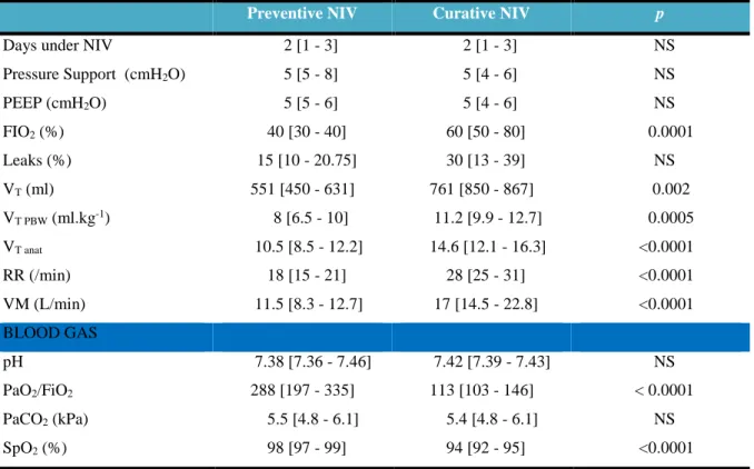
2- Ventilation
The adjusted ventilatory parameters were minimally modified between the "preventive" and the "curative" period (Table 2). Median PS and PEEP levels were around 5 cmH2O. In curative NIV, respiratory rate was significantly higher (RR= 18 [15-21] in
preventive NIV than in curative NIV RR= 28 [25-31], p <0.0001), with no difference of leaking (p = 0.17). VT PBW and VT anat were significantly increased in curative NIV compared to those in
preventive NIV (VT PBW = 11.2 ml.kg-1 versus 8 ml.kg-1 p = 0.0005 and VT anat = 14.6 ml.kg-1
versus 10.5 ml.kg-1 p = 0.0001) (Figure 2). Similarly, minute ventilation was significantly higher in the curative NIV group (11.5 L/min [8.3-12.7] versus 17 L/min [14.5, 22.8], p <0.0001).
Patients receiving curative NIV were significantly more hypoxic (SpO2 = 94%
[92-95] versus 98% [97-99], p <0.0001) and more hypoxemic (PaO2/FiO2 = 113 [103-146] versus
31
groups (5.4 kPa [4.8-6.1] versus 5.5 kPa [4.8-6.1] p> 0.05). Similarly, there was no significant difference in the pH.
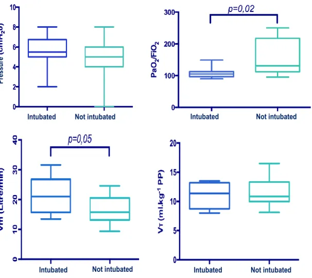
Table 3 compared ventilatory parameters in patients who received curative NIV successfully, to those who required intubation afterwards. Both groups had the same levels of PS and PEEP, and had the same percentage of air leaking. VT PBW and VT anat were not
significantly different (p = 0.8). Patients spent significantly shorter time under NIV in the
intubated group, and were also more hypoxemic (PaO2/FiO2 = 105 [97-112] versus 131
[112-217], p = 0.026), with similar PaCO2 levels (5.2 kPa [4.8-6.3] versus 5.5 kPa [4.8-6.6] p> 0.05).
Respiratory rate was also higher in the intubated group (33.5 L/min [26.2-36]) versus non-intubated group 26 L/min [24.2-30] p = 0.05) as well as minute ventilation (21 L/min [15.7- 26.8] versus 15.8 L/min [13.2-20.5] p = 0.05) (Figure 3).
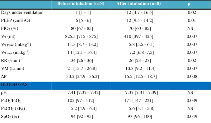
Table 4 compared intubated patients before and after intubation. After intubation, VT
PBW administered to patients were reduced by 48.6% compared to VT PBW under curative NIV
(11.3 ml.kg-1 [8.7-13.2] versus 5.8 ml.kg-1 [5.5-6.1] p = 0.007). PEEP was significantly increased (6 cmH2O [5; 6] versus 12 cmH2O [9-14] p = 0.01). Respiratory rate was also
significantly reduced (RR= 34 [26-36] versus 26 [24-27] p = 0.02) as well as the minute ventilation, with a significant improvement in oxygenation (PaO2/FiO2 = 105 [97-113] versus
171 [147, 221] p = 0.039) and oxymetry (SpO2= 94% [92-95] versus 98% [97-100] p = 0.049),
with no effect on PaCO2 and without significant modification of FiO2. VT anat were significantly
higher than VT PBW in curative NIV (14.6 ml.kg-1 [12.1-16.3] versus 11.2 ml.kg-1 [9.9-12.7]
respectively, p = 0.001) (Figure 4). Driving pressure was significantly lower in controlled ventilation than in spontaneous ventilation, ΔP = 30 cmH2O [25-36] against 16.5 cmH2O
[12.5-18.7] p = 0.008 (Figure 5). Compliance seemed lower (12.2 mL/cmH2O [10.7-14] versus 16.6
mL/cmH2O [13-28.1]) and driving pressure seemed higher (33.1 cmH2O [28.5-9.3] versus 26.6
32
IV. Discussion
Our study demonstrated the effects of respiratory complications on patients’ ventilation, under postoperative NIV. There was a significant increase in VT PBW and VT anat as
well as in respiratory rate without any PS or PEEP modification. The respiratory drive was responsible for an increase in the minute ventilation because of a deterioration of the ventilation / perfusion ratio, which led to acute respiratory failure. PaO2/FiO2 ratio decreased without an
impairment of pH nor PaCO2. The increase of VT was therefore related to significant
compensatory hyperventilation.
Carteaux & al. found high VT under curative NIV in de novo ARF (16), predicting
NIV failure when VT exceeded 9.5 ml.kg-1 over 4 hours. In our study all VT under NIV were
above 8 ml.kg-1 and there was no difference in VT between the intubated and the not intubated
groups.
Similarly, several studies have observed high VT in NIV during hypoxemic ARF
(15,28). Compared to these studies our ventilatory parameters were lower (median PS between 7 and 9 cmH2O in Carteaux and Frat studies) and thus not incriminated in the increase of the
tidal volumes. We have been able, thanks to the automated collection of ventilatory parameters and the small number of patients, to control the quality of the NIV sessions and the interface leaks, which is rarely found in the literature. We have also found that air leaking is not significantly increased between the preventive and curative NIV sessions, nor between the intubated or not intubated groups, which could have skewed our results.
In addition, we noticed a few peculiarities: Even in preventive NIV, patients had high VT. Several elements can be involved: 1) Respiratory drive does not adapt to the new pulmonary
volume in the immediate postoperative period. 2) Hyperventilation would be predictive of degradation, as suggested Carteaux & al. 3) A resected lung cannot be treated like a healthy lung. Regional inflammation, altered elastance, deformed chest wall, and general and regional anaesthesia (29) can significantly alter its function. Thus, non-protective ventilation in this population may have a deleterious effect by potentiating the underlying lesions (30). 4) With a reduced gas exchange surface and an unchanged anatomical dead space (VD), in order to maintain an effective minute ventilation (Vm = VT × RR), and an efficient VD/VT ratio for
clearing CO2, it is necessary to increase the tidal volume and / or the RR.
Moreover, we found a significant increase in RR, Vm, and hypoxemia under curative NIV in the intubated group, compared to the non-intubated group. These results are consistent
33
with the Frat & al. study (15), revealing that VT PBW> 9mL.kg-1 over an hour and PaO2/FiO2
<200 is a predictor of intubation in the NIV group. VT PBW> 9mL.kg-1 was also an independent
risk factor for mortality in the NIV group.
Driving pressure is a strong parameter whose strict control could reduce mortality during controlled protective ventilation of ARDS (18,31). Its benefit in spontaneous ventilation
is poorly known but seems to be present (20). We calculated the driving pressure under NIV
through the equation Crs = VT/ΔP, assuming that: 1) the pulmonary compliance is sparsely
modified between the curative NIV period and immediately after intubation, and 2) the
pulmonary compliance is not modified immediately after general anaesthesia (32). However,
this approximation is probably significant because the Crs varies according to the aerated zones of the parenchyma, which are modified under controlled ventilation and with PEEP changes
(33,34).
Interestingly, prior to intubation, clinicians tolerated high levels of VT, driving
pressures, and high RR. Once intubated and sedated, patients were immediately provided with protective ventilation with low VT (median VT PBW 5.8 mL.kg-1), with a marked reduction in
driving pressure and a significantly higher PEEP. We also found a rapid improvement in oxygenation.
The main risk of this hyperventilation is P-SILI lesions, and therefore several questions arise as to how to avoid them: Should these patients be sedated earlier in order to reduce respiratory drive and compensatory hyperventilation? If yes, at what time? The VT PBW is not
discriminating and the RR is around 30/min, therefore it would be very difficult to find predictive threshold values. Moreover, the success rate of curative NIV in our study is relatively high (61.9%). High flow oxygenation could be alternatively used because of the absence of inspiratory pressure support (even if it was weak in our study), but the risk is firstly that the VT
PBW remains high without being monitored, and secondly that the PEEP generated by high flow
cannula is insufficient in a situation of pulmonary collapse. For these cases of respiratory distress with underlying lung lesions the solution would not be to stop non-invasive ventilation but rather stop spontaneous ventilation.
To our knowledge, tidal volumes have never been specifically assessed in postoperative ventilation after pulmonary resection. The originality of this study arises from the peculiarity of this type of surgery, which alters the charts used to evaluate the optimal volumes and pressures to apply for patients during ventilation. It also alters the mechanical and pathophysiological properties of lungs and the chest wall (35). However, this study had several
34
limitations. Its small size has limited the statistical analysis. A centre effect bias was present in this monocentric study, in a structure that practices a lot of NIV, which limited the extrapolation of our results. The retrospective nature was a source of selection bias since at least 11 patients were excluded for missing data. Several measurement biases could be raised: The analysis of expired tidal volume was at the centre of this study, however it depended on several parameters that could not be collected (inspiratory slope, expiratory cycling). Thus, we probably collected an insufficient number of current volumes and leaks compared to the totality of the sessions (up to 12 hours a day for several days).
A debate is launched on the consequences of hyperinflation generated in spontaneous ventilation on an injured lung, in critical care patients but also in experimental cases (25–27,36). P-SILI and its consequences raises the prospect of prophylactic treatment (intubation and neuromuscular blockade). However, one crucial point remains at the centre of this study: we cannot be sure that high tidal volumes in thoracic surgery are responsible for P-SILI lesions. This has never been proven in the literature. Our study aimed to describe our practices, and to note excessive volumes under NIV despite an already limited inspiratory support. In order to optimize the ventilatory strategy to prevent these potential P-SILI lesions, it is important to find tools for assessing overdistension in daily practice. ΔPL, via oesophageal manometry, and driving pressure can be considered, however their use in current practice remains difficult to implement and the literature concerning them in NIV is very limited.
Finally, when overdistension is suspected or confirmed, the simplest treatment remains sedation and controlled protective ventilation (37). However, in our patients under NIV, this would involve intubation, with the potential complications that would ensue. Target volume ventilation models have been proposed to optimize PSV ventilation, such as APRV (Air Pressure Release Ventilation) (38), but are not applicable to NIV. We can also imagine, after suppressing favouring factors of elevated respiratory drive, an artificial decrease of the latter by the use of sedatives (39), or even low doses of neuromuscular blockade (40). These drugs may effectively decrease respiratory drive, but at the cost of increased patient-ventilator asynchrony, disturbance of consciousness, hypercapnic acidosis and haemodynamic impairment, in patients whose airways are not protected. These practices do not seem advisable.
35
V. Conclusion
Our study shows that in postoperative NIV after thoracic surgery, high tidal volumes appear during respiratory distress without changing the ventilator parameters. The increase in respiratory drive allows the maintenance of pH and PaCO2 levels, due to hyperventilation, with
an increase in respiratory rate and minute ventilation. Several clinical and experimental settings suggest that spontaneous ventilation at such high volumes can lead to overdistension lesions, called P-SILI. The monitoring tools are missing to diagnose it. Nevertheless, clinicians must precociously draw their attention to this phenomenon in order to adapt the therapy and not to harm the patient.
36
VI. References
1. Fourdrain A, De Dominicis F, Lafitte S & al. Quantitative computed tomography to predict postoperative FEV1 after lung cancer surgery. J Thorac Dis. 2017;9(8):2413‑8.
2. Bouvier G, Kouatchet A, Jaffré S, & al. La ventilation non invasive en postopératoire. 3. Bignami E, Saglietti F, Di Lullo A. Mechanical ventilation management during
cardiothoracic surgery: an open challenge. Ann Transl Med 2018 /PMC6212361
4. Tomizawa K, Usami N, Fukumoto K, & al. Risk assessment of perioperative mortality after pulmonary resection in patients with primary lung cancer: the 30- or 90-day mortality. Gen Thorac Cardiovasc Surg. 2014;62(5):308‑13.
5. Auriant I, Jallot A, Hervé P, & al. Noninvasive Ventilation Reduces Mortality in Acute Respiratory Failure following Lung Resection. Am J Respir Crit Care Med. 2001;164(7):1231‑5.
6. Kometani T, Okamoto T, Yoshida S & al. Acute respiratory distress syndrome after pulmonary resection. Gen Thorac Cardiovasc Surg. 2013;61(9):504‑12.
7. Perrin C, Jullien V, Vénissac N & al. Prophylactic use of noninvasive ventilation in patients undergoing lung resectional surgery. Respir Med. 2007;101(7):1572‑8.
8. Chiumello D, Chevallard G, Gregoretti C. Non-invasive ventilation in postoperative patients: a systematic review. Intensive Care Medicine. 2011;37(6):918‑29.
9. Torres MF, Porfirio GJ, Carvalho AP & al. Non-invasive positive pressure ventilation for prevention of complications after pulmonary resection in lung cancer patients. Cochrane Database of Systematic Reviews 2015 doi/10.1002/14651858.CD010355.pub2/
10. Lorut C, Lefebvre A, Planquette B, & al. Erratum to: Early postoperative prophylactic noninvasive ventilation after major lung resection in COPD patients: a randomized controlled trial. Intensive Care Med. 2014;40(3):469.
11. Guerra Hernández E, Rodríguez Pérez A, Freixinet Gilard J, & al. Prophylactic use of non-invasive mechanical ventilation in lung resection. Eur Rev Med Pharmacol Sci. 2018;22(1):190‑8.
12. Liao G, Chen R, He J. Prophylactic use of noninvasive positive pressure ventilation in post-thoracic surgery patients: A prospective randomized control study. J Thorac Dis. 2010;2(4):205‑9.
13. Lefebvre A, Lorut C, Alifano M, & al. Noninvasive ventilation for acute respiratory failure after lung resection: an observational study. Intensive Care Med. 2009;35(4):663‑70. 14. Parquin F, Cuquemelle E. La ventilation non invasive en chirurgie pulmonaire.
Réanimation. 2015;24(1):37‑45.
15. Frat J-P, Ragot S, Coudroy R, & al. Predictors of Intubation in Patients With Acute Hypoxemic Respiratory Failure Treated With a Noninvasive Oxygenation Strategy. Crit Care Med. 2017;
37
16. Carteaux G, Millán-Guilarte T, De Prost N, & al. Failure of Noninvasive Ventilation for De Novo Acute Hypoxemic Respiratory Failure: Role of Tidal Volume. Crit Care Med. 2016;44(2):282‑90.
17. Silva PL, Rocco PRM. The basics of respiratory mechanics: ventilator-derived parameters. Annals of Translational Medicine 2018 /19973
18. Amato MBP, Meade MO, Slutsky AS & al. Driving Pressure and Survival in the Acute Respiratory Distress Syndrome NEJM 2015 doi/10.1056/NEJMsa1410639
19. Bellani G, Grasselli G, Teggia-Droghi M & al. Do spontaneous and mechanical breathing have similar effects on average transpulmonary and alveolar pressure? A clinical crossover study. Crit Care 2016 /PMC4862136
20. Bellani G, Grassi A, Sosio S, Foti G. Plateau and driving pressure in the presence of spontaneous breathing. Intensive Care Medicine. 2019;45(1):97‑8.
21. Beitler JR, Sands SA, Loring SH & al. Quantifying unintended exposure to high tidal volumes from breath stacking dyssynchrony in ARDS: the BREATHE criteria. Intensive Care Med. 2016;42(9):1427‑36.
22. Yoshida T, Torsani V, Gomes S & al. Spontaneous Effort Causes Occult Pendelluft during Mechanical Ventilation. Am J Respir Crit Care Med. 2013;188(12):1420‑7.
23. Cressoni M, Gotti M, Chiurazzi C & al. Mechanical Power and Development of Ventilator-induced Lung Injury. Anesthesiology. 2016;124(5):1100‑8.
24. West JB. Invited review: pulmonary capillary stress failure. J Appl Physiol. 2000;89(6):2483-2489.
25. Gattinoni L. Ventilation-induced lung injury exists in spontaneously breathing patients with acute respiratory failure: We are not sure. Intensive Care Med. 2017;43(2):256‑8.
26. Brochard L, Slutsky AS, Pesenti A. Reply: “A Word of Caution Regarding Patient Self-inflicted Lung Injury and Prophylactic Intubation” and “Hyperventilation (Not Ventilator)-induced Lung Injury”. Am J Respir Crit Care Med. 2017;196(7):937‑8.
27. Brochard L. Ventilation-induced lung injury exists in spontaneously breathing patients with acute respiratory failure: Yes. Intensive Care Med. 2017;43(2):250‑2.
28. Thille AW, Contou D, Fragnoli C & al. Non-invasive ventilation for acute hypoxemic respiratory failure: intubation rate and risk factors. Crit Care. 2013;17(6):R269.
29. Rozé H, Germain A, Perrier V& al. Effect of flumazenil on diaphragm electrical activation during weaning from mechanical ventilation after acute respiratory distress syndrome. Br J Anaesth. 2015;114(2):269‑75.
30. Hernandez LA, Coker PJ, May S, & al. Mechanical ventilation increases microvascular permeability in oleic acid-injured lungs. J Appl Physiol. 1990;69(6):2057‑61.
38
31. Guérin C, Papazian L, Reignier J, & al. Effect of driving pressure on mortality in ARDS patients during lung protective mechanical ventilation in two randomized controlled trials. Crit Care 2016;20(1):384.
32. Unoki T, Mizutani T, Toyooka H. Changes in respiratory physiological dead space and compliance during non-abdominal, upper abdominal and lower abdominal surgery under general anaesthesia: European Journal of Anaesthesiology. 2004;21(4):302‑8.
33. Carvalho ARS, Jandre FC, Pino AV & al. Effects of descending positive end-expiratory pressure on lung mechanics and aeration in healthy anaesthetized piglets. Critical Care. 2006;10(4):R122.
34. Grieco DL, Russo A, Romanò B, Anzellotti GM, Ciocchetti P, Torrini F, et al. Lung volumes, respiratory mechanics and dynamic strain during general anaesthesia. Br J Anaesth. 2018;121(5):1156‑65.
35. Miserocchi G, Beretta E, Rivolta I. Respiratory mechanics and fluid dynamics after lung resection surgery. Thorac Surg Clin. 2010;20(3):345‑57.
36. Mauri T, Cambiaghi B, Spinelli E, Langer T, Grasselli G. Spontaneous breathing: a double-edged sword to handle with care. Ann Transl Med 2017. /PMC5537122/
37. Brochard L, Slutsky A, Pesenti A. Mechanical Ventilation to Minimize Progression of Lung Injury in Acute Respiratory Failure. Am J Respir Crit Care Med. 2017;195(4):438‑42. 38. Perinel-Ragey S, Baboi L, Guérin C. Variability of Tidal Volume in Patient-Triggered
Mechanical Ventilation in ARDS. Respir Care. 2017;62(11):1437‑46.
39. Costa R, Navalesi P, Cammarota G, Longhini F, Spinazzola G, Cipriani F, et al. Remifentanil effects on respiratory drive and timing during pressure support ventilation and neurally adjusted ventilatory assist. Respir Physiol Neurobiol. 2017;244:10‑6.
40. Doorduin J, Nollet JL, Roesthuis LH, van Hees HWH, Brochard LJ, Sinderby CA, et al. Partial Neuromuscular Blockade during Partial Ventilatory Support in Sedated Patients with High Tidal Volumes. Am J Respir Crit Care Med. 2017;195(8):1033‑42.