Université de Montréal
Regulation of lipid metabolism in adipocytes and
hepatocytes by hexarelin through scavenger receptor
CD36
par
Amélie Rodrigue-Way
Département de Biochimie Faculté de Médecine
Thèse présentée à la Faculté des études supérieures en vue de l’obtention du grade de Philosophiae Doctor (Ph.D.)
en Biochimie
Avril 2011
Faculté des études supérieures
Cette thèse intitulée :
Regulation of lipid metabolism in adipocytes and hepatocytes by hexarelin through scavenger receptor CD36
présentée par : Amélie Rodrigue-Way
a été évaluée par un jury composé des personnes suivantes :
Luis Rokeach, président-rapporteur André Tremblay, directeur de recherche
Grant Mitchell, membre du jury Frédéric Picard, examinateur externe Pierre Haddad, représentant du doyen de la FES
Résumé
Les sécrétines de l’hormone de croissance (GHRPs) sont de petits peptides synthétiques capables de stimuler la sécrétion de l’hormone de croissance à partir de l’hypophyse via leur liaison au récepteur de la ghréline GHS-R1a. Le GHRP hexaréline a été utilisé afin d’étudier la distribution tissulaire de GHS-R1a et son effet GH-indépendant. Ainsi, par cette approche, il a été déterminé que l’hexaréline était capable de se lier à un deuxième récepteur identifié comme étant le récepteur scavenger CD36. Ce récepteur possède une multitude de ligands dont les particules oxLDL et les acides gras à longue chaîne. CD36 est généralement reconnu pour son rôle dans l’athérogénèse et sa contribution à la formation de cellules spumeuses suite à l’internalisation des oxLDL dans les macrophages/monocytes. Auparavant, nous avions démontré que le traitement des macrophages avec l’hexaréline menait à l’activation de PPARƔ via sa liaison à GHS-R1a, mais aussi à CD36. De plus, une cascade d’activation impliquant LXRα et les transporteurs ABC provoquait également une augmentation de l’efflux du cholestérol. Une stimulation de la voie du transport inverse du cholestérol vers les particules HDL entraînait donc une diminution de l’engorgement des macrophages de lipides et la formation de cellules spumeuses. Puisque CD36 est exprimé dans de multiples tissus et qu’il est également responsable du captage des acides gras à longue chaîne, nous avons voulu étudier l’impact de l’hexaréline uniquement à travers sa liaison à CD36. Dans le but d’approfondir nos connaissances sur la régulation du métabolisme des lipides par CD36, nous avons choisi des types cellulaires jouant un rôle important dans l’homéostasie lipidique n’exprimant pas GHS-R1a, soient les adipocytes et les hépatocytes.
L’ensemble de mes travaux démontre qu’en réponse à son interaction avec l’hexaréline, CD36 a le potentiel de réduire le contenu lipidique des adipocytes et des hépatocytes. Dans les cellules adipeuses, l'hexaréline augmente l’expression de plusieurs gènes impliqués dans la mobilisation et l’oxydation des acides gras, et induit également l’expression des marqueurs thermogéniques PGC-1α et UCP-1. De même, hexaréline
augmente l’expression des gènes impliqués dans la biogenèse mitochondriale, un effet accompagné de changements morphologiques des mitochondries; des caractéristiques observées dans les types cellulaires ayant une grande capacité oxydative. Ces résultats démontrent que les adipocytes blancs traités avec hexaréline ont la capacité de se transformer en un phénotype similaire aux adipocytes bruns ayant l’habileté de brûler les acides gras plutôt que de les emmagasiner. Cet effet est également observé dans les tissus adipeux de souris et est dépendant de la présence de CD36. Dans les hépatocytes, nous avons démontré le potentiel de CD36 à moduler le métabolisme du cholestérol. En réponse au traitement des cellules avec hexaréline, une phosphorylation rapide de LKB1 et de l’AMPK est suivie d’une phosphorylation inhibitrice de l’HMG-CoA réductase (HMGR), l’enzyme clé dans la synthèse du cholestérol. De plus, la liaison d'hexaréline à CD36 provoque le recrutement d’insig-2 à HMGR, l’étape d’engagement dans sa dégradation. La dégradation de HMGR par hexaréline semble être dépendante de l’activité de PPARƔ et de l’AMPK. Dans le but d’élucider le mécanisme d’activation par hexaréline, nous avons démontré d’une part que sa liaison à CD36 provoque une déphosphorylation de Erk soulevant ainsi l’inhibition que celui-ci exerce sur PPARƔ et d’autre part, un recrutement de l’AMPK à PGC-1α expliquant ainsi une partie du mécanisme d’activation de PPARƔ par hexaréline.
Les résultats générés dans cette thèse ont permis d’élucider de nouveaux mécanismes d’action de CD36 et d'approfondir nos connaissances de son influence dans la régulation du métabolisme des lipides.
Mots-clés : Adipocytes, hépatocytes, CD36, hexaréline, PPARƔ, PGC1α, biogenèse
mitochondriale, UCP-1, oxydation des acides gras, LKB1, AMPK, HMGR, insig-2, Erk, OSBPs.
Abstract
Growth hormone releasing peptides (GHRPs) are small synthetic peptides aimed at stimulating GH release from the pituitary through their binding to ghrelin receptor known as growth hormone secretagogue receptor 1a (GHS-R1a). Using the GHRP, hexarelin to study tissue distribution of GHS-R1a and its GH-independent effect, it was observed that hexarelin was capable of binding to a second receptor identified as scavenger receptor CD36. While having multiple ligands, CD36 is mainly known for binding and internalizing oxLDL and long chain fatty acids. CD36 is thought to play a detrimental role in macrophage derived foam cell formation and development of atherosclerosis. Previously, we have shown that in macrophages, expressing both GHS-R1a and CD36, hexarelin promoted an activation of PPARƔ via GHS-R1a but also through its binding to CD36. This activation led to the induction of the LXRα-ABC transporters pathway and an increase in cholesterol efflux, reducing lipid-laden macrophage content. This positive effect on macrophages was reproduced in apolipoprotein E-null mice on a high fat diet treated with hexarelin. A significant reduction in the size of atherosclerotic lesions was observed while similar increases in the expression of PPARƔ, LXRα and ABC transporters occurred in isolated peritoneal macrophages. CD36 also plays a role in fatty acid uptake, and to further investigate the impact of the interaction of hexarelin with CD36, we aimed at evaluating the role of CD36 in regulating lipid metabolism in cells devoid of GHS-R1a such as adipocytes and hepatocytes.
In the present thesis, we demonstrated through its interaction with hexarelin, the ability of CD36 to decrease intracellular lipid content in both adipocytes and hepatocytes. In adipocytes, hexarelin was able to increase the expression of several genes involved in fatty acid mobilization, fatty acid oxidation but also to induce the expression of the thermogenic markers, PGC-1α and UCP-1. In addition, hexarelin increased the expression of genes involved in mitochondrial biogenesis which was accompanied by mitochondrial morphological changes in agreement with what is usually seen in highly oxidative cells. In
support of these findings, we also observed an increase in the activity of cytochrome c oxidase (a component of the respiratory chain) which could reflect an increase in oxidative phosphorylation. The results generated with cultured white adipocytes suggest the ability of hexarelin to promote changes toward a brown fat-like phenotype which also occurred in vivo and was dependent on the presence of CD36. In hepatocytes, CD36 was capable of regulating cholesterol metabolism by rapidly phosphorylating LKB1 and AMPK which subsequently resulted in the inactivating phosphorylation of HMG-CoA reductase, the rate-limiting enzyme in cholesterol synthesis. Hexarelin via CD36 also induced the recruitment of insig-2 to HMGR, the committed step in HMGR degradation while lifting the exerted inhibitory effect of Erk on nuclear receptor PPARƔ activity, and promoting the recruitment of AMPK to PPARƔ coactivator PGC-1α, suggesting an enhanced transcriptional potential of PPARƔ.
The results generated during my graduate studies represent unique and novel mechanisms by which CD36 is capable of regulating lipid metabolism.
Keywords : Adipocytes, hepatocytes, CD36, hexarelin, PPARγ, PGC1α, mitochondrial biogenesis, UCP-1, fatty acid oxidation, LKB1, AMPK, HMGR, insig-2, Erk, OSBPs.
Table of contents
Résumé...iii Abstract………...v List of Tables...xi List of Figures...xii Abbreviation list...xiv Acknowledgements...xxi CHAPTER 1: Introduction ... 11 Growth hormone secretagogues ... 1
1.1 Design of growth hormone releasing peptides ... 1
1.2 Peptidomimetic growth hormone secretagogues ... 5
1.3 Growth hormone secretagogues today ... 6
2 Growth hormone secretagogue receptor and its natural ligand ... 7
2.1 Identification of an alternative pathway for GH release ... 7
2.2 Identification of the receptor for GHS ... 8
2.3 Distribution of GHS-R1a ... 9
2.4 The natural ligand of GHS-R1a: ghrelin ... 11
2.4.1 Discovery of ghrelin: classical example of reverse pharmacology ... 11
2.5 Role of ghrelin and its binding to GHS-R1a ... 13
2.6 Therapeutic interest of ghrelin and GOAT ... 14
3 The growth hormone releasing peptide, hexarelin ... 15
3.1 A novel GHRP ... 15
3.2 The dual action of hexarelin ... 15
4 Scavenger receptors ... 16
4.1 The role of scavenger receptor in the formation of atherosclerotic lesions ... 17
5 Scavenger receptor, CD36 ... 21
5.1 Identification of CD36 and its various designations ... 21
5.2 Tissue distribution ... 22
5.3 The role of the multi-ligand receptor CD36 in different biological processes .... 23
5.3.1 Thrombospondine-1 (TSP-1) and CD36 ... 23
5.3.2 Modified LDL particles, atherosclerosis and inflammation ... 25
5.3.3 Phagocytosis of apoptotic cells ... 25
5.3.4 Bacterial infection ... 26
5.3.5 Parasitic infection ... 26
5.3.6 Long chain fatty acids ... 26
5.4 CD36 gene ... 27
5.4.1 CD36 alternative forms and mutations ... 28
5.5 CD36 structure and post-translational modifications ... 31
5.6 Binding sites on CD36 ... 33
6 Regulation of CD36 ... 35
6.1 Regulation of CD36 gene expression ... 35
6.1.1 In atherosclerosis ... 35
6.1.2 Adipose tissue and adipocytes ... 40
6.1.3 Liver and hepatocytes ... 41
6.2 CD36 localization ... 43
6.2.1 In lipid rafts and caveolae ... 43
6.2.2 Mitochondrial CD36 ... 46
6.3 Ligand-dependent signaling pathways and CD36 movement ... 46
6.3.1 TSP-1/platelets ... 48
6.3.2 LDLox/macrophages ... 48
6.3.3 Fatty acids ... 49
6.4 Mouse and human CD36 deficiency ... 54
7.1 The origin of adipocytes ... 56
7.2 Differentiation of adipocytes... 57
7.3 Adipokines and the adipose tissue ... 58
7.4 Fatty acid transport into adipocytes ... 60
7.5 Storage of fatty acids in lipid droplets ... 61
7.6 Lipolysis ... 63
7.7 White to brown transdifferentiation of adipocytes... 65
7.8 Mitochondria and fatty acid oxidation ... 66
7.8.1 Mitochondrial Biogenesis ... 67
7.8.2 FAO and oxidative phosphorylation in white adipocytes ... 69
7.8.3 Respiratory uncoupling and thermogenesis in brown adipocytes ... 73
8 PPAR gamma ... 75
8.1 PPARγ isoforms, structure and heterodimerizaton ... 76
8.2 Regulation of PPARγ activity ... 77
8.2.1 Natural and synthetic ligands ... 77
8.2.2 Corepressors ... 78
8.2.3 Coactivators ... 79
8.3 Post-translational modifications of PPARγ ... 81
8.4 Role of PPARγ in adipocytes ... 82
8.5 Role of PPARγ in hepatocytes ... 83
9 Regulation of cholesterol synthesis ... 84
9.1 AMP-activated protein kinase (AMPK) ... 84
9.1.1 AMPK in the liver ... 87
9.2 HMG-CoA reductase ... 88
9.2.1 Phosphorylation of HMGR ... 90
9.2.2 Gene expression regulation of HMGR ... 90
9.3 Regulation of cholesterol synthesis by sterols ... 91
9.3.2 Sterol-regulated SCAP-SREBP pathway ... 92
9.3.3 Degradation of HMGR ... 94
10 Effect of hexarelin through its interaction with CD36 ... 95
11 Hypothesis and project objectives ... 98
CHAPTER 2: Results ... 100
1 Paper 1 ... 101
2 Paper 2 ... 134
CHAPTER 3: General discussion, perspectives and conclusions ... 181
1 Discussion ... 181
1.1 Elucidation of the impact of CD36 on lipid metabolism ... 181
1.1.1 CD36 in Adipocytes ... 181
1.1.2 CD36 in hepatocytes ... 191
2 Perspectives ... 203
2.1 Studies to complement our results ... 204
2.2 Signaling cascade(s) involving the binding of hexarelin to CD36 ... 205
2.3 Other pathways affected by CD36/hexarelin interaction ... 206
2.4 Lipid rafts and CD36 ... 207
2.5 The impact of hexarelin on lipid metabolism mediated by other nuclear factors 207 3 Conclusions ... 208
List of tables
Table 1. Evolution of growth hormone releasing peptides and their unique related properties ... 5 Table 2. Tissue- or cell-specific distribution of GHS-R1a and CD36 ... 11 Table 3. Exons, introns and mutations in the human CD36 gene ... 31 Table 4. Adipokines and their role in inflammation, energy metabolism or insulin
List of figures
Figure 1. The hypothalamic-pituitary axis. (A) Functional anatomy of the hypothalamus and pituitary gland, (B) Regulation of the secretion of growth
hormone by the hypothalamus ... 2
Figure 2. Growth hormone-releasing hormone and growth hormone secretagogue pathways involved in GH release from the pituitary ... 8
Figure 3. Classification of scavenger receptors and their proposed structural features ... 21
Figure 4. Schematic representation of CD36 gene and protein ... 28
Figure 5. Schematic representation of CD3 protein, ligand-binding sites and post-translational modifications ... 32
Figure 6. CD36 ligand-specific downstream events ... 48
Figure 7. Adipocytes: Differences between white and brown adipocytes ... 56
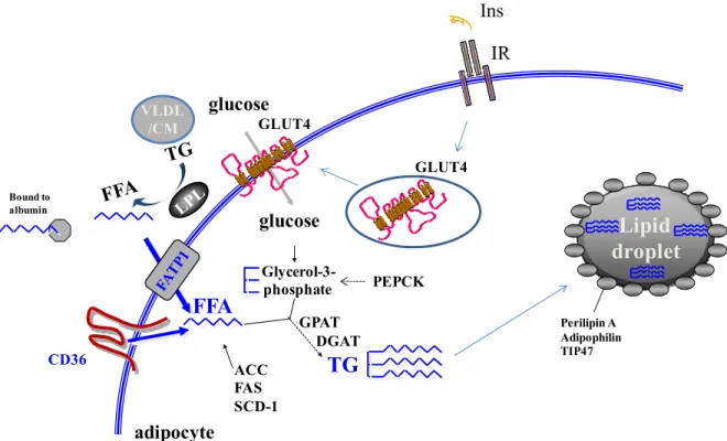
Figure 8. Adipocyte differentiation (A) Determination of adipocyte fate from mesenchymal precursors, (B) factors regulating white adipocyte differentation 57 Figure 9. Fatty acid uptake, triglyceride synthesis and lipid droplets ... 61
Figure 10. Regulation of lipolysis in adipocytes ... 64
Figure 11. Mitochondrial Biogenesis, protein import and translation ... 68
Figure 12. Steps in β-oxidation of fatty acids, oxidative phosphorylation coupled with ATP production ... 69
Figure 13. Initial Steps Fatty Acid Oxidation ... 70
Figure 14. β-oxidation reaction of fatty acids ... 71
Figure 15. Oxidative phosphorylation ... 73
Figure 16. Adaptive thermogenesis and uncoupling protein-1 in brown adipocytes ... 75
Figure 17. PPARγ isoforms, structure, and heterodimerization with RXR ... 77
Figure 18. Regulation of PGC-1α activity ... 80
Figure 20. Cholesterol synthesis pathway and structure of its rate-controlling enzyme, HMG-CoA reductase ... 89 Figure 21. Mechanistic schema showing how sterols and insigs regulate post-translational proteolysis and activation of SREBPs ... 93 Figure 22. Mechanistic schema showing how sterols and insigs regulate HMGR
degradation ... 95 Figure 23. Hexarelin-mediated activation of the PPARγ-LXRα-ABC metabolic
pathway in macrophages (Demers et al., 2008) ... 98 Figure 24. The effect of hexarelin’s interaction with CD36 on lipid metabolism in
Abbreviation list
9-,13-HODE 9- and 13-hydroxyoctadecadienoic acid 15-HETE 15-hydroxyeicosatetraenoic acid [Ca2+]i intracellular calcium concentration ABCA1 ATP-binding cassette transporter AI AC adenylate cyclase
ACC acetyl-CoA carboxylase acLDL acetylated low density liprotein AF-1, -2 activation function 1 and 2
AICAR 5-amino-4-imidazolecarboxamide ribonucleoside AMPK 5' adenosine monophosphate-activated protein kinase ANGPTL2 angiopoietin-like protein 2
aP2 adipocyte protein 2 ApoB100 apolipoprotein B-100 ApoE apolipoprotein E
ATGL adipose triglyceride lipase ATP adenosine triphosphate BAT brown adipose tissue bHLH basic helix-loop-helix
CaMKK Ca2+/calmodulin-dependent protein kinase kinase cAMP adenosine 3′,5′-cyclic monophosphate
CAT carnitine acylcarnitine transferase
CBP/p300 cAMP response element binding protein (CREB) binding protein CCL2 Chemokine (C-C motif) ligand 2
CD36 Cluster of Differentiation 36 CDCA chenodeoxycholic acid
cDNA complementary deoxyribonucleic acid CE Cholesterol ester
C/EBP CCAAT/enhancer-binding protein CEOOH cholesterol ester hydroperoxides CHD coronary heart disease
ChIP-Seq chromatin immunoprecipitation sequencing cLDL carbamylated low density lipoprotein CLESH CD36 LIMP-II Emp sequence homology CNS central nervous system
CPT-I carnitine palmitoyltransferase I CsA cyclosporine A
CSF colony stimulating factor
CXCL16 Chemokine (C-X-C motif) ligand 16 DAG diacylglycerol
DBD DNA binding domain
DEXA dual-energy x-ray absorptiometry DGAT diacylglycerol acyltransferase EMSA electrophoretic mobility shift assay ER endoplasmic reticulum
Erk extracellular signal-regulated protein kinases 1 and 2 FA fatty acids
FABPpm plasma membrane fatty acid binding protein FACS fatty acyl-CoA synthetase
FAK focal adhesion kinase FAO fatty acid oxidation FAS fatty acid synthase FAT fatty acid translocase FATP fatty acid transporter protein FFA free fatty acid
G-3-P glycerol-3-phosphate
GH growth hormone
GHRH growth hormone releasing hormone GHRP growth hormone releasing peptide GHS-R1a GHS receptor 1a
GLUT-4 glucose transporter-4 GOAT ghrelin O-Acyltransferase
GPAT glycerol-3-phosphate acyltransferase GPCR G protein-coupled receptor
GPI-anchored glycophosphatidylinositol GPIV glycoprotein IV
GS glycogen synthase HDL high density lipoprotein HFD high fat diet
HMGR HMG-CoA reductase
HSL hormone-sensitive lipase HNE 4-hydroxynonenal
IBMX 3-isobutyl-1-methylxanthine IFN interferon
IGF-1 insulin-like growth factor 1 IL Interleukin
Insig-1,-2 insulin-inducible genes-1 and -2 proteins IP3 inositol (1,4,5)-trisphosphate
JNK c-Jun N-terminal kinase LBD ligand binding domain LCFA long chain fatty acid
LDLR low density lipoprotein receptor LKB1 liver kinase B1
LOX-1 lectin-like oxLDL receptor-1 LPL lipoprotein lipase
LPS lipopolysaccharide LTA Lipoteichoic acid
MAPK mitogen-activated protein kinase M-CPT-1 muscle carnitine palmitoyltransferase I MEFs mouse embryonic fibroblasts
MEKK2 mitogen-activated protein (MAP) kinase kinase kinase 2 M-FABP muscle fatty acid binding protein
MGL monoacylglycerol lipase
mmLDL minimally modified low density lipoprotein mRNA messenger ribonucleic acid
MRP mitochondrial ribosomal protein mTOR mammalian target of rapamycin MW molecular weight
NAFLD non-alcoholic fatty liver disease NADH nicotinamide adenine dinucleotide
NAMPT nicotinamide phosphoribosyltransferase NASH non-alcoholic steatohepatitis
NCoR Nuclear receptor corepressor protein NPY neuropeptide Y
Nrf2 Nuclear factor E2-related factor 2 oxLDL oxidized low density lipoprotein oxPCCD36 oxidized phosphatidylcholine
PEPCK cytosolic phosphoenolpyruvate carboxykinase
PfEMP1 Plasmodium falciparum erythrocyte membrane protein 1
PFK2 6-phosphofructokinase-2 PGC-1α PPARγ coactivator-1 alpha
PGC-1β PPARγ coactivator-1 beta PHA Phytohemagglutinin PI3K phosphoinositide 3-kinase PKA, PKC protein kinase A and C
PL phospholipid
PLAP placental alkaline phosphatase PMA Phorbol 12-myristate 13-acetate
PPAR peroxisome proliferator-activated receptor
PPRE peroxisome proliferator-activated receptor response element PRDM16 PRD1-BF1-RIZ1 homologous domain containing 16
PUFA polyunsaturated fatty acid RIP140 receptor interacting protein 140 RUNX3 Runt-related 3
RXR retinoid X receptor
SCAP SREBP cleavage-activating protein SCD-1 stearoyl-CoA desaturase-1
SFRP5 secreted frizzled-related protein 5 siRNA small interfering RNA
shRNA small hairpin RNA SMC smooth muscle cells
SMRT silencing mediator of retinoid and thyroid hormone receptors SR scavenger receptor
SR-A scavenger receptor type A SR-BI scavenger receptor type B SRC steroid receptor co-activator
SREBP sterol-regulatory element binding protein
SREC scavenger receptor expressed by endothelial cells proteins SRIF somatotropin release-inhibiting factor
SSD sterol-sensing domain TAK1 TGFβ-activated kinase-1 TCA cycle tricarboxylic acid cycle TG triglycerides
TGF tumor growth factor
TIM translocase of the inner membrane TIP47 tail-interacting protein 47
TLR Toll-like receptors
TM transmembrane
TNF-α tumor necrosis factor alpha
TOM translocase of the outer membrane TR4 testicular orphan nuclear receptor 4
TR4RE testicular orphan nuclear receptor 4 response element TRAIL TNF-related apoptosis-inducing ligand
TRAP220 thyroid receptor associated protein 220 TSC-2 tumor suppressor tuberous sclerosis 2 TSP-1 thrombospondin 1
TZD thiazolidinedione UCP uncoupling protein UTR untranslated region
VLDL very low density lipoprotein VSMC vascular smooth muscle cells WAT white adipose tissue
Acknowledgements
The desire to return to graduate school stemmed from a few years of working in biotech and pharmaceutical companies. And while I loved that experience, the wish to return to basic research prompted me to reconsider my future. At a given point, I grabbed that opportunity and decided to finally stop asking that nagging question: Should I? So I did and I certainly don`t regret it. It seemed like such a long journey, long enough that my life no longer resembles what it was when I first started. People have entered my life while others have left forever but for most, they have remained as supportive and essential to me, especially throughout this period. I`d like to think that I`ve experienced all those wonderful interactions as best as I possibly could.
A special thanks to my research advisor, André Tremblay who has given me the opportunity to fulfill my dream. You have provided an environment and a training that permitted us to improve our skills, share our knowledge and excel in what we do. I am especially grateful to you for providing enough guidance without impeding my independence and for always being there when needed. I have learned so much from your ability to always put an interesting spin to papers and projects. Although it might seem trivial, thank you for building an incredible team of people for otherwise this experience would not have been the same.
Aux filles du labo pour lesquelles j’ai une affection sans pareil! To Mélanie Sanchez, my companion throughout my chocolate rampage and other delicious goodies: Thanks for all the laughs, for sharing the joy of simply being silly, not to mention your endless help and guidance. You are one of the few people I know who can be absolutely hilarious without overshadowing their professionalism. This journey would have not been the same without the girls. A special thanks to Nathalie Picard, Karine Sauvé, Mélanie Sanchez and Annie Demers with whom I have worked along the longest. Thanks to Véronique Caron, Stéphanie Bilodeau, Méryl Hassan, Julie Martin and Julie Lepage for all our discussions (personal and professional) and for all the help that you have provided. Because of the embedded willingness to help, this amazing quality that you all possess
almost goes unnoticed. Your team-spirit should be an example to all. I would be ever so lucky and honored to work by your side again one day. Kudos!
A special thanks to Hugues Bernard and Steve Tirrell for their much appreciated time and help.
On a more personal note, I am grateful to my wonderful love, Eric, for being my rock, my pillar of strength. Thanks for your patience, and for encouraging and supporting me throughout my graduate studies. And in particular, thanks for picking up the slack during the last few months. Your love and support are so important to me. To my greatest accomplishment of all: Sarah. My little angel with the propensity to lighten up the world around her; whom I love more than life itself. You have transformed me more than I could have ever imagined and all for the best. Thanks for reminding me to take a deep breath and enjoy the simple things especially when it seems like there`s no time left. You are too young to realize why I had so little time in the past few months but I will do my best to make it up to you. To Pop, your great knowledge, and among other things your continuous fascination and enthusiasm for science have always encouraged me to follow my own passions and dreams. To Maggie and Liam who have become incredible individuals. Thanks to my family for their continuous encouragements and support.
In memory of Cathy Way (2004), Claude René (2008), Cheryl Irving (2009) and Patrick Errada (2010) for their courage that never faltered in the face of cancer. We wish you were still here with us. To my friend Patrick who has always shown such passion and love for life, I miss you.
To my dearest friends: Isabelle Michaud, Sylvie Bernier, Johanne Laporte, Mary Donoghue, Sue Acton, Barb Sullivan and Colleen Mullen for their support, guidance and input, and above all for lending an ear.
CHAPTER 1: Introduction
1 Growth hormone secretagogues
Growth hormone secretagogues are a family of synthetic peptides (also called growth hormone releasing peptides) and peptidomimetic agonists designed to increase the secretion of growth hormone in GH-deficient patients.
1.1 Design of growth hormone releasing peptides
Growth hormone releasing peptides (GHRPs) research stemmed from studies on synthetic analogs of opioid peptides during the 1970s. Opioids such as enkephalins, endorphins, dynorphins, and endomorphins are produced by the body in response to pain while naturally occurring opiates such as morphine and codeine are extracted from opium poppy. Enkephalins are pentapeptides that regulate nociception or pain perception via peripheral nerves in order to control or lessen the pain signal sent to the central nervous system (CNS). There are 2 forms of enkephalin which are products of the same proenkephalin gene: leu-enkephalin Gly-Phe-Leu) and met-enkephalin (Tyr-Gly-Gly-Phe-Met) (Udenfriend & Kilpatrick, 1983). In 1975, enkephalins were identified as the endogenous ligands of the morphine receptor, opioid receptor μ widely distributed throughout the central and peripheral nervous system (Hughes et al., 1975). With enkephalins as the prototype, opioid analogs were synthesized in order to develop more potent and less addicting analgesic compounds. Soon after, Cyril Y Bowers, Frank Momany and colleagues noticed that certain opioids stimulated growth hormone (GH) secretion (Bowers et al., 1977).
Also called somatotropin, GH is 191-aa hormone secreted by the pituitary gland (hypophysis), more precisely by the anterior part of the gland (Figure 1A). The pituitary gland is controlled by the hypothalamus, connected by the pituitary stalk (or infundibular stem). The hypothalamus directly controls the endocrine system by secreting factors that stimulate or inhibit the secretion of hormones released from the pituitary. The latter
contains 2 sections: the anterior pituitary and the posterior pituitary. The anterior pituitary is responsible for the secretion of various hormones including GH. The secretion of GH is regulated by the hypothalamic-pituitary axis (Figure 1B). Somatocrinin or growth hormone-releasing hormone (GHRH) is produced by the hypothalamus (Figure 1A) and secreted towards the pituitary (Guillemin et al., 1982). GHRH binds to somatotropes and induces secretion of GH into the blood circulation which will then bind to its receptor present in peripheral tissues. In response to GH, the liver secretes insulin-like growth factor 1 (IGF-1) causing a negative feedback on the production of GH. The resulting increase in somatotropin release-inhibiting factor (SRIF) production competes with GHRH for the same receptor on somatotropic cells and reduces secretion of GH. The interplay between GHRH and SRIF results in the observed pulsatile release of GH (Brazeau et al., 1973).
A B Hypothalamus Pituitary gland or hypophysis Pituitary stalk Anterior pituitary Somatotropic cells GH Neurosecretory neurons Posterior pituitary SRIF GHRH Pituitary -+ Hypothalamus GHRH SRIF GH Liver IGF-1 IGF-1 Target Tissue Growth
Figure 1. The hypothalamic-pituitary axis. (A) Functional anatomy of the hypothalamus and pituitary gland, (B) Regulation of the secretion of growth hormone by the hypothalamus
Regardless of the cause, in newborns and young children, a GH deficiency results in hypoglycemia and growth retardation. Although rare, a GH deficiency in adults (due to traumatic brain injury, cancer, radiotherapy) can result in prolonged fatigue, a loss in bone
density and a loss of muscle mass. Prior to the advent of recombinant DNA technology in 1985, the treatment of choice for GH deficiency was the rather expensive injections of purified human GH. Although due to the seriousness of contracting the degenerative neurological disorder, Creutzfeldt-Jakob disease (CJD) from contaminated human GH samples, the use of hGH was soon banned from Europe and North America (Huillard et al., 1999). This hormone represents a 191-aa protein with a molecular mass of 215 kDa and a low oral absorption (Nargund et al., 1998). In addition, daily injections masked the physiological regulatory feedback and therefore resulted in the absence of pulsatile release of GH. In certain cases, a deficiency in GH was not primarily due to an insufficient synthesis/secretion of GH but rather a poor signaling from the hypothalamus. Taking those two facts into account, an alternative approach was considered in which the biologically active portion of the 44-aa GHRH peptide would be used to stimulate GH secretion from the pituitary. This 29-aa synthetic bioactive truncated GHRH had the advantage of maintaining the pulsatile release of GH, and therefore several analogues of GHRH were tested in humans as an alternative to GH replacement therapy (Grossman et al., 1984). Following the arrival of recombinant GH on the market, studies performed on GHRH and its analogues could not demonstrate the advantage of using GHRH in terms of efficacy and bioavailability in comparison to recombinant GH (Campbell et al., 1995).
Following the previously mentioned observation in 1977 of Bowers, Momany and colleagues that enkephalins stimulated GH secretion, this team redirected their research from opioids and undertook the tedious task of synthesizing more potent analogues as a therapeutic approach to treating GH deficiency and doing so by using met-enkephalin as their prototype (Table 1). The peptide sequence was modified to produce analogues capable of eliciting a stronger GH secretion on isolated rat pituitary glands without affecting opioid receptors (Bowers et al., 1980;Momany et al., 1981). The analogue of met5-enkephalin, Tyr-DTrp-Gly-Phe-Met-NH2, illustrated in Table 1, was the first to elicit a stronger in vitro
GH secretion; however, when tested in rat, no in vivo activity was found. The D conformation of the substitute aromatic amino acid, tryptophan brings a stabilizing element
to this peptide making it more resistant to proteases; while the amine group (NH2) in C-terminal renders the peptide biologically active (Kreil, 1997). Indeed, it is estimated that half of peptide hormones have an amine group at their C-terminus and is required for optimal biological activity (Kim & Seong, 2001). Theoretical conformational studies by Momany helped in the design of more energetically favorable and potent GHRPs (Momany
et al., 1981;Momany & Bowers, 1996). While some were a thousand times more active
than their predecessor, they still did not possess any in vivo activity. The main feature of all GHRPs synthesized that needed to be preserved in the following screenings was the presence, position and stereochemistry of the Trp residu at position 2.Using a trial and error approach, the first in vivo bioactive GHRP called GHRP-6 (His-D-Trp-Ala-Trp-D-Phe-Lys-NH2) was finally synthesized (Momany et al., 1984). The three dimensional structure of GHRP-6 indicated that the N-terminal end of histidine and the C-terminal end of lysine were in close proximity, adopting a folded conformation. The hydrophobic indole ring of each tryptophan (D-Trp2 and Trp4) was directed toward one another while the phenyl unit of D-Phe5 interacted with the amino unit of Lys6. These particular modifications seemed to give GHRP-6 its bioactive feature. Unfortunately, GHRP-6 and others that followed had a very poor oral absorption and a short half-life. For example, oral GHRP-6 had a bioactivity of 0.3% to that of injected GHRP-6 with a half-life of 20 minutes (Bowers et al., 1992). This lack of bioavailability provided the opportunity for other research teams to search for nonpeptidyl compounds imitating GHRPs but with the added feature of being orally active.
Table 1. Evolution of growth hormone releasing peptides and their unique related properties Met-enkephalin: Tyr-Gly-Gly-Phe-Met Analogue of met5-enkephalin: Tyr-D-Trp-Gly-Phe-Met-NH2 Structure Name:
Peptide sequence Modification(s) Applied Reported effect(s)Function or
N/A Opioidligandμ receptor
NH2in C-terminal confers to this peptide an increase
in bioavailability
D-Trp, conformation renders peptide resistant
to proteases
GHRP-6:
His-D-Trp-Ala-Trp-D-Phe-Lys-NH2
GH secretion from isolated pituitary in
vitro but not in vivo
First peptide capable of stimulating in vitro
AND in vivo GH secretion
Methyl group provides an increased stability to
this peptide. First peptide that can be
taken orally. Several modifications were
made to generate hexapeptide with a second
aromatic aa with a D conformation
Methyl added in position 2 in aromatic group of D-Trp
Hexarelin:
His-D2MeTrp-Ala-Trp-D-Phe-Lys-NH2
1.2 Peptidomimetic growth hormone secretagogues
Roy Smith and colleagues from Merck assumed the task of designing such compounds. To do so, they took into consideration all of the particular features that made GHRP-6 a bioactive peptide such as the amine group, the aromatic amino acids in position 2, 4 and 5 as well as the unnatural D-Trp and combined them with benzodiazepine, known to imitate small peptides. They synthesized an array of compounds capable of secreting GH called benzolactames (Smith et al., 1993). The bioavailability of benzolactames, although superior to GHRPs, turned out to be relatively low (Leung et al., 1996). Focusing on the concept of “privileged structures” by Ben E. Evans and colleagues that stated that certain molecular units had the capacity of interacting with various receptors, the same team
screened different compounds within the company’s various internal projects to finally focus on agonists containing a spiropiperidine group (Evans et al., 1988;Jacks et al., 1996). More precisely, spiroindoline sulphonamide (also called MK-0677) was found to elicit GH release in rat pituitary cell culture assay with greater potency. MK-0677 was identified as a specific GHS with an elevated bioactivity, a high bioavailability of more than 60% and a half-life of 5 to 6 hours in dogs (Jacks et al., 1996). Merck selected MK-0677 for safety assessment studies which then entered clinical trials (Nargund et al., 1998).
1.3 Growth hormone secretagogues today
Despite tremendous research and effort at designing efficacious GHS that will at the same time maintain the pulsatile release of GH and have an elevated oral bioavailability, injections of recombinant GH continues to be the treatment of choice for GH-deficient patients, mainly due to its low-cost production. Furthermore, in a high percentage of cases, a decrease in GH is the result of an improper activity of the pituitary gland; therefore, injection of GH is the logical approach to treat GH-deficiency. However, throughout the years, new applications were considered for GHS. For example, they have been used as a diagnostic tool to detect GH deficiency. In Japan, the second generation of GHRP-6, GHRP-2 is used as a kit to detect GH deficiency in adults (Arita et al., 2008). In addition, MK-0677 is being considered for its role in counteracting the reduced basal metabolic rate resulting from aging, from calorie-restricted diets and from wasting syndrome (cachexia) seen in patients suffering from chronic diseases (Nass et al., 2008;Smith et al., 2007;Murphy et al., 1998). However, due to its effect on GH secretion and its potential in improving athletic performance, GHS are amongst the list of banned substances published by the World Anti-Doping Agency (WADA). GHS have also found their way to the black market for their use in bodybuilding. Recently, GHRP-2 was detected in over-the-counter nutritional supplemental tablets (Thomas et al., 2010).
2 Growth hormone secretagogue receptor and its
natural ligand
2.1 Identification of an alternative pathway for GH release
As illustrated in Figure 1B, it was first presumed that the pulsatile GH secretion was regulated by only two hormones: GHRH and SRIF. Following the discovery of GHRPs, studies pertaining to their mechanism of action have permitted to identify a second activation pathway for the release of GH. Based on their preliminary results, Bowers and colleagues suggested early on that GHRPs acted on a different pathway than that of GHRH. They observed that combining GHRP-6 with GHRH had an additive effect on GH secretion in rats (Bowers et al., 1984;Sartor et al., 1985). In addition, Roy Smith and colleagues at Merck reported that in isolated rat somatotropic cells, repeated treatments with GHRP-6 resulted in the desensitization of cells to GHRP-6 without affecting its response to GHRH; and inversely, treatment of cells with a GHRH antagonist had no effect on the response of cells to GHRP-6 (Cheng et al., 1989). Similarly to opioid receptors, GHRH receptor (GHRHR) belongs to the family of G protein-coupled receptors (GPCRs) (Figure 2). One main feature of the binding of GHRH to its receptor is the increase in intracellular cyclic AMP (cAMP) via stimulation of adenylate cyclase (AC) by Gsα subunit (Mayo, 1992). Cyclic AMP activates protein kinase A (PKA) which in turn activates a range of factors responsible for the expression of GH and its subsequent processing and secretion (Cohen et
al., 1999). In support of an alternative pathway for GH release, several studies have used
GHS such as GHRP-6 and MK-0677 to reveal the following findings:
- GHS had no effect on intracellular cAMP levels (Cheng et al., 1989)
- Phorbol 12-myristate 13-acetate (PMA), an activator of protein kinase C (PKC) was capable of imitating the additive effect of GHS when combined with GHRH (Cheng et al., 1991)
- GHS causes a rapid increase in intracellular free calcium, [Ca2+]i (Herrington & Hille, 1994;Bresson-Bepoldin & Dufy-Barbe, 1994)
- The secretion of GH by GHS involved the inositol (1,4,5)-trisphosphate/ diacylglycerol (IP3/DAG) pathway (Adams et al., 1995;Mau et al., 1995)
Somatotropic cell
GHRH GHS
Adapted from Kojima et al., TRENDS in
Endocrinology & Metabolism 2001, 12(3):
118-126. AMPc [Ca2+]i Natural ligand GH GH Gαs AC ATP AMPc PKA PKC IP3 Gα11
Figure 2. Growth hormone-releasing hormone and growth hormone secretagogue pathways involved in GH release from the pituitary
2.2 Identification of the receptor for GHS
In 1996, under the supervision of Lex Van der Ploeg and Roy Smith, the research team at Merck were the first to clone the receptor for GHS (Howard et al., 1996). Using the expression-cloning strategy in which size-fractionated poly A+ RNA from pig pituitaries were microinjected into Xenopus oocytes along with cRNA of selected G protein and aequorin. Using aequorin, a bioluminescent probe known to bind intracellular calcium, they measured levels of [Ca2+]i released following treatment with MK-0677. Exhaustive stepwise fractionation of positive pools finally resulted in the identification of an orphan
receptor. The gene for GHS receptor, called GHS-R1a codes for a 366-aa GPCR (41 kDa) belonging to GPCR, class A (rhodopsin-like receptors) with the typical conserved 7 transmembrane (7-TM) α-helices and classic sequence for G protein interactions. A second isoform (GHS-R1b) was also cloned that represented a truncated 289-aa protein missing TM 6 and 7, which did not respond to GHS and was a product of pre-mRNA splicing. Formation of GPCRs homo- and hetero-oligomers is thought to play an important role in ligand binding and cell signaling (Maggio et al., 2005). It was recently shown that GHS-R1b acts as a dominant-negative form of GHS-R1a to lessen its constitutive activity by forming heterodimers and when its expression exceeds that of GHS-R1a, trafficking of GHS-R1a to the cell surface is attenuated (Leung et al., 2007).
2.3 Distribution of GHS-R1a
The tissue distribution of GHS-R1a is more widespread than first anticipated suggesting a role beyond that of stimulator of GH secretion from the pituitary. Table 2 depicts the detection GHS-R1a in various tissues or cell types. Listed also is the expression of scavenger receptor CD36 which will be discussed in section 5.2. The expression of GHS-R1a is elevated in several sections of the brain including the hypothalamus and pituitary gland (Guan et al., 1997). The hypothalamic nuclei play a major role in the regulation of food intake and energy homeostasis (Gao & Horvath, 2008;Horvath, 2005). In addition, GHS-R1a is also expressed in endocrine tissues such as pancreas, adrenal glands, thyroid, ovaries and testicles (Guan et al., 1997;Gnanapavan et al., 2002;Gaytan et al., 2005;Tena-Sempere et al., 2002). In relation to the cardiovascular system, GHS-R1a is present in the aorta, the left atrium and ventricle, and more precisely in cardiomyocytes as well as in smooth muscle cells and microvascular endothelial cells (Nagaya et al., 2001;Kleinz et al., 2006;Li et al., 2007a). Looking at the digestive system, GHS-R1a was detected in the intestine and stomach (Dass et al., 2003;Wu et al., 2004;Shuto et al., 2001). GHS-R1a is also found in spleen and in leukocytes such as monocytes/macrophages, lymphocytes and neutrophiles (Gnanapavan et al., 2002;Demers et al., 2004;Hattori et al.,
2001). Its presence in adipose tissue has been suggested (Choi et al., 2003;Davies et al., 2009) while in other studies it has not been detected (Gnanapavan et al., 2002;Muccioli et
al., 2004). To support its absence in adipose tissue, GHS-R1a was undetectable in cultured
mouse adipocytes, 3T3-L1 (Zhang et al., 2004;Rodrigue-Way et al., 2007). To explain this discrepancy, macrophages can be present in adipose tissue and their infiltration can contribute to inflammation in obese subjects (Weisberg et al., 2003). It is therefore possible that detection of GHS-R1a in adipose tissue in certain studies is due to contaminating macrophages known to express GHS-R1a. GHS-R1a is also absent in liver, primary hepatocytes and human hepatocellular carcinoma cell line, HepG2 (Smith et al., 2007;Thielemans et al., 2007;Gauna et al., 2005). GHS-R1a neither was detectable in skeletal muscle nor in mouse differentiated myoblastic cell line, C2C12 (Ueberberg et al., 2009;Filigheddu et al., 2007). It was also untraceable in colon, kidney and prostate (Ueberberg et al., 2009).
It is noteworthy to mention that GHS-R1a is not expressed in tissues involved in lipid metabolism such as adipose tissue (adipocytes), liver and skeletal muscle.
Table 2. Tissue- or cell-specific distribution of GHS-R1a and CD36
√ -mRNA or protein detected
X -Undetected ? - Unknown
Tissue or cell type GHS-R1a CD36 Reference(s)
Hypothalamus √ √ Guan et al. , 1997; Le Foll et al., 2009
Pituitary gland √ √ Guan et al. , 1997; Ong et al., 1998b
Heart or cardiomyocyte √ √ Kleinz et al. , 2006; Van Nieuwenhoven et al., 1995
Vascular smooth muscle cell √ √ Kleinz et al., 2006; de Oliveira et al., 2008
Microvascular endothelial cell √ √ Li et al ., 2007a; Swerlick et al., 1992
Spleen √ √ Gnanapavan et al. , 2002; M emon et al., 1998
Pancreas √ √ Guan et al., 1997; Noushmehr et al., 2005a
Stomach √ √ Shuto et al. , 2001; Chen et al., 2001
Intestine √ √ Dass et al. , 2003;Wu et al. , 2004; Chen et al., 2001
Adrenal gland √ √ Gnanapavan et al. , 2002; Zhang et al., 2003
Macrophage √ √ Demers et al. , 2004; Endemann et al., 1993
Lymphocyte √ √ Hattori et al. , 2001; Won et al., 2008
Neutrophile √ √ Hattori et al. , 2001; Suchard et al., 1992
Ovary √ √ Gaytan et al. , 2005; Zhang et al., 2003
Testicule √ √ Tena-Sempere et al. , 2002; Gillot et al., 2005
Adipose tissue and 3T3-L1 adipocyte X √ Gnanapavan et al. , 2002; Zhang et al ., 2004; Harmon & Abumrad, 1993
Liver, primary hepatocyte and HepG2 X √ Smith et al. , 2007; Gauna et al. , 2005; Thielemans et al ., 2007; M emon et al., 1998;M alerod et al., 2002 N9 microglia cell X √ Bulgarelli et al., 2009
Kidney X √ Ueberberg et al. , 2009; Susztak et al., 2004
Colon X √ Ueberberg et al. , 2009; Chen et al., 2001
Skeletal muscle or C2C12 myoblast X √ Ueberberg et al. , 2009; Filigheddu et al. , 2007; Van Nieuwenhoven et al., 1995
Prostate X √ Ueberberg et al. , 2009; Vallbo & Damber, 2005
Platelet/megakaryocyte ? √ Clemetson et al ., 1977
Breast ? √ Clezardin et al. , 1993
Pneumocytes ? √ Guthmann et al. , 1999
Airway epithelium ? √ Atsuta et al. , 1997
Dendritic cell ? √ Juhlin, 1989
Retinal pigment epithelium ? √ Ryeom et al. , 1996
Gustatory cell ? √ Fukuwatari et al. , 1997 Thyroid √ X Patey et al. , 1999
2.4 The natural ligand of GHS-R1a: ghrelin
2.4.1 Discovery of ghrelin: classical example of reverse pharmacology
The endogeneous ligand of GHS-R1a was isolated in 1999 by Kenji Kangawa and his team in Japan (Kojima et al., 1999). The discovery of ghrelin became a classical example of reverse pharmacology (Libert et al., 1991). Their approach consisted of treating
stable GHS-R1a-transfected CHO cells with tissue extracts from brain, lung, heart, kidney, stomach and gut; and to monitor for changes in [Ca2+]i. Following a positive response from
stomach extracts, they proceeded to go through intensive chromatography purification steps, including reversed-phase HPLC. From the final purified sample, they characterized and sequenced the potential GHS-R1a ligand by mass spectrometry. Finally, they identified a 28-aa protein with a n-octanoylated (C7H15CO) serine residue at position 3. This unique
post-transcriptional modification was essential for its GH-releasing activity. The authors names this peptide “ghrelin” after the word root “ghre” in Proto-Indo-European languages meaning “grow” and superposed with “rel” for “release” for its GH-releasing ability (Kojima, 2008). It took a little less than a decade for Joseph Goldstein and Andrew Brown and colleagues, to discover the enzyme responsible for the acylation of ghrelin with the fatty acid, octanoate (Yang et al., 2008). Ghrelin O-Acyltransferase (GOAT) is the only member of its family capable of attaching a small fatty acid to a peptide. Due to its unique role, GOAT has become an interesting target in the treatment against obesity and diabetes (Chen et al., 2009). The majority of ghrelin in the gastrointestinal tracts is produced by a distinct endocrine cell type called X/A-like cells in the oxyntic gland (Date et al., 2000). However, ghrelin is also expressed to a smaller degree throughout the digestive system from the stomach to the colon and also in the pancreas, adipose tissue, heart, kidney, lung, adrenal gland, thyroid, pituitary and hypothalamus (Kojima et al., 1999;Ueberberg et al., 2009). In addition to its role in secreting GH, ghrelin is an appetite-stimulating peptide hormone and the only gastrointestinal peptide with orexigenic powers (Asakawa et al., 2001;Woods, 2004).
2.4.1.1 Deacyl-ghrelin
The deacylated ghrelin does not bind to GHS-R1a and was considered as a non-functional peptide until recently (Chen et al., 2009). Since deacyl-ghrelin was the major form secreted in the circulation, it seemed improbable that it had no physiological relevance. Indeed, the ratio deacyl-ghrelin:ghrelin was found to be between 2.5:1 and 9:1 (Tsubone et al., 2005;Broglio et al., 2004;Yoshimoto et al., 2002). The ratio
deacyl-ghrelin:ghrelin turned out to have important physiological consequences on energy balance since it was discovered that deacyl-ghrelin had opposite effects to that of ghrelin on food intake, for example (Asakawa et al., 2005). In obese patients, circulating deacyl-ghrelin levels decreased greatly while ghrelin levels increased; and in obese diabetic patients, this change in ratio was further accentuated (Rodriguez et al., 2009). Despite its inability to bind GHS-R1a, deacyl-ghrelin is thought to bind a yet unknown receptor since some studies have shown binding sites on cardiomyocytes and C2C12 skeletal muscle cells (Filigheddu et al., 2007;Lear et al., 2010).
2.5 Role of ghrelin and its binding to GHS-R1a
Given the broad distribution of GHS-R1a and ghrelin, it wasn’t surprising to discover that their influence on energy homeostasis went far beyond that of simply controlling GH secretion. As stated previously, a major role was given to ghrelin as an appetite-stimulating hormone (Asakawa et al., 2001). In addition, central and peripheral injections of ghrelin in mice provoked a decrease in energy expenditure, an increase in respiratory quotient (RQ= CO2 eliminated /O2 consumed) and a decreased in oxygen
consumption (Tschop et al., 2000;Asakawa et al., 2005). Using neuropeptide Y (NPY)-deficient mice, injections of ghrelin were shown to increase body weight and adiposity independently from its orexigenic effect due to the absence of NPY, the regulator of food intake (Tschop et al., 2000). Ghrelin decreased adipocyte thermogenesis suggesting a GHS-R1a-independent effect (Tsubone et al., 2005). Adipogenesis is a carefully controlled event during which the timing in the expression of specific genes is important in the induction of the differentiation program and proper functioning of adipocytes. Adipogenesis is further discussed in Section 7.2. Both chronic intracerebroventricular infusion of ghrelin in rats and treatment of human visceral adipocytes resulted in the increased expression of 2 genes involved in adipocyte differentiation, nuclear receptor peroxisome proliferator-activated receptor gamma (PPARγ) and sterol-regulatory element binding protein-1 (SREBP-1) as well as several genes involved in adipocytic function such as lipoprotein lipase (LPL),
acetyl-CoA carboxylase (ACC), fatty acid synthase (FAS) and stearoyl-CoA desaturase-1 (SCD-1) (Rodriguez et al., 2009;Theander-Carrillo et al., 2006). Ghrelin is shown to activate 5' adenosine monophosphate-activated protein kinase (AMPK) in tissues expressing GHS-R1a such as the hypothalamus and the heart while inhibiting AMPK in GHS-R1a-negative tissues such as the liver and adipose tissue (Kola et al., 2005). However, ghrelin had no effect on AMPK in skeletal muscle. AMPK is a key regulator of energy homeostasis further presented in Section 9.1. The GH-independent cardioprotective effect of ghrelin might be due in part to the activation of AMPK (Frascarelli et al., 2003). In general, the orexigenic effect of ghrelin ties macronutrient composition with regulation of energy balance by the CNS but it also has direct GHS-R1a-dependent and -independent effects of peripheral tissues.
2.6 Therapeutic interest of ghrelin and GOAT
Based on its role in energy balance, ghrelin has become an interesting therapeutic target for certain pathophysiological conditions. In cancer patients as well as in patients with severe chronic wasting diseases such as chronic obstructive pulmonary disease and renal failure, infusion of ghrelin resulted in a marked improvement in appetite, food intake and nutrient absorption (Neary et al., 2004;Nagaya et al., 2005;Ashby et al., 2009). Along with the identification of GOAT in 2008 and the importance in the ratio deacyl-ghrelin:ghrelin, much attention is given to GOAT and its role in controlling ghrelin’s action (Romero et al., 2010). However, the use of an agonist or antagonist of GOAT still remains to be determined since the physiological effects of ghrelin are multiple, sometimes opposite and difficult to interpret based on the various sites of action of ghrelin independent and dependent of GHS-R1a.
3 The growth hormone releasing peptide, hexarelin
3.1 A novel GHRP
Hexarelin made its appearance in 1994 when Vittorio Locatelli and colleagues in Italy synthesized a derivative of GHRP-6 in which they substituted D-Trp for D-2-methyl-Trp (see Table 1) making this peptide chemically more stable than GHRP-6 and above all the first orally active GHRP (Deghenghi et al., 1994;Ghigo et al., 1994). Initially called EP 23905, hexarelin was synthesized during the era when intensive search for a highly orally active GHS was conducted. Despite the superior bioavailability of peptidomimetic GHS, studies using GHRPs, including hexarelin, were pursued in humans and various animal models in the hopes of understanding its biological effect but also to mainly justify its use as a therapeutic or a diagnostic tool (Micic et al., 1999). Hexarelin studies were quickly undertaken in humans to verify its efficacy on GH secretion. Hexarelin was well tolerated in humans without any reported side-effects and elicited a substantial elevation in plasma GH concentrations in a dose-dependent manner (Imbimbo et al., 1994). Even though hexarelin was orally active, it still did not mirror the efficacy of the orally active GHS, MK-0677. Because of the highly vascularized nasal cavity, intranasal administration was proposed for hexarelin as a therapeutic tool for GH deficiency instead of IV injections (Laron et al., 1994;Laron et al., 1995;Pontiroli, 1998). Today, hexarelin is used mainly in research but it also has become a popular choice as a performance enhancement drug.
3.2 The dual action of hexarelin
Prior to the detection of GHS-R1a in the myocardium, GH-independent studies suggested that hexarelin possessed cardioprotective properties distinct from that of GH (Berti et al., 1998;Rossoni et al., 1998). It was suggested that perhaps in addition to GHS-R1a, GHRPs were capable of binding to a sub-type of GHRP receptor. In 1998, the group of Magnus Nilsson performed binding assays on membrane extracts from human, bovine
and porcine anterior pituitaries to identify hexarelin’s receptor (Ong et al., 1998b). They developed a photoreactive analogue of hexarelin containing a photoactivatable amino acid, p-benzoyl-L- phenylalanine and iodine-125 labeled ([125I]iodoTyr-Bpa-Ala-Hexarelin) (Dorman & Prestwich, 1994). They identified a 57 kDa photolabeled protein in all samples analyzed that was distinct from the 41-kDa GHS-R1a protein. Another protein capable of interacting with hexarelin and with a molecular weight of 84 kDa was detected in heart (Ong et al., 1998a). However, when the heart photolabeled protein was treated with N-glycosidase F, a decrease in its MW to ~57 kDa was observed suggesting that this receptor contained oligosaccharide chains and was therefore heavily N-glycosylated in the heart but not in the pituitary gland (Bodart et al., 1999). In addition, the binding was specific to hexarelin since MK-0677 and EP51389 (another potent GHRP) were unable to compete with the photoactivatable hexarelin for this unidentified receptor. In support of these findings, another study showed that hexarelin had a different binding pattern than that of MK-0677 or ghrelin (Papotti et al., 2000). Binding of hexarelin was detected in decreasing order in heart, adrenal gland, gonad, artery, lung, liver, skeletal muscle, kidney, pituitary, thyroid, adipose tissue, vein, uterus, skin and lymph node. In 2002, the second receptor for hexarelin was identified as the scavenger receptor, CD36 (Bodart et al., 2002).
4 Scavenger receptors
The role of certain cell types as scavengers was first suggested by Joseph Goldstein, Andrew Brown and associates (Goldstein et al., 1979). They observed that in presence of acetylated 125I-labeled low density lipoprotein (125I-acLDL) cultured macrophages would internalize these particles at a much higher rate than native LDL particles. This uptake resulted in the accumulation of intracellular cholesterol and a transformation of cells into foam cell-like phenotype similar to what is observed in atherosclerotic plaques. These results implied the presence of receptors capable of binding modified LDL particles and were therefore referred to as scavenger receptors.
4.1 The role of scavenger receptor in the formation of
atherosclerotic lesions
It was later determined that the formation of oxidized LDL (oxLDL) particles rather than acLDL was a likely occurrence since oxLDL particles were present in atherosclerotic plaques (Palinski et al., 1989;Yla-Herttuala et al., 1989).The first step in the development of atherosclerotic plaques is the appearance of fatty streaks consisting primarily of foam cells loaded with lipids and T lymphocytes within the vessel’s subendothelial space or intima (Daugherty & Roselaar, 1995). Oxidation of LDL particles is thought to occur not in the circulation but rather in the intima following their infiltration in specific locations of a vessel (Chow et al., 1998;Rangaswamy et al., 1997). The oxidation of imprisoned LDL particles is a long and complex process that is still considered a key step in the development of atherosclerosis (Stocker & Keaney, Jr., 2005;Steinberg, 2002). The core of a LDL particle contains cholesterol esters (CE) and triglycerides (TG) while the surface is covered with a single layer of phospholipids (PL), including phosphatidylcholine, molecules of non-esterified cholesterol and a single molecule of apolipoprotein B-100 (apoB100) that specifically interacts with the LDL receptor on neighboring cells (Steinberg, 2002).While cholesterol is less susceptible to oxidation, an important variety of oxidized PL was detected within lesions and especially in oxLDL (Berliner et al., 2001).When phospholipids are minimally modified (mmLDL), they become negatively charged, have an anti-apoptotic effect on scavenger cells, and stimulate secretion of chemokines and cytokines from neighboring endothelial cells lining the blood vessels (Berliner et al., 1995;Boullier et al., 2006). Consequently, monocytes are recruited from the circulation toward the site of inflammation. Subsequently, within the intima, the infiltrated monocytes differentiate into macrophages. The constant recruitment of inflammatory cells results in increased cytokine secretion and continuous oxidation of mmLDL. Phospholipids are increasingly oxidized on LDL while apoB100 undergoes modifications and unfolds (Hamilton et al., 2008). Oxidized LDL is no longer recognized by LDLR but becomes a ligand for scavenger
receptors. Macrophages, endothelial cells and even vascular smooth muscle cells (VSMC) take up oxLDL via their scavenger receptors. This process allows especially macrophages to clear the intima from the harmful presence of oxLDL. In addition, the oxidized lipids taken up by the macrophage serve as ligands to nuclear receptor, PPARγ and induce the expression of genes involved in the reverse cholesterol transport such as ATP-binding cassette transporter AI (ABCA1) which shuttles internalized cholesterol into nascent HDL particles for clearance by the liver (Chawla et al., 2001). However, when macrophages become overloaded and overwhelmed by oxLDL, an imbalance occurs between the uptake and the clearance of lipids and cells become consequently lipid-laden macrophages, or foam cells (Faggiotto et al., 1984). In atherosclerosis, progression in the formation of foam cells, but also in the increase in inflammation, in cellular necrosis, and thinning of the fibrotic plaque eventually lead to plaque rupture and thrombosis (Lusis, 2000). Therefore, scavenger receptors are thought to play a detrimental role in the pro-atherogenic effect of modified LDL particles.
4.2 Scavenger receptor classes
Scavenger receptors (SR) have been identified and grouped based on their capacity to bind modified lipoprotein particles and their contribution to the development of atherosclerosis. They have been categorized into different classes based mainly on their structural features and functional domains (Krieger, 1997;Horiuchi et al., 2003). Figure 3 provides an illustration of the different classes of SR. Members of class A have a single TM region, a trimeric formation (α-helices) and possess an affinity for acLDL and oxLDL. This class includes SR-A type 1 and type 2 (SR-A1 and SR-A2) which are encoded by the same gene and alternatively spliced (Kodama et al., 1990;Rohrer et al., 1990). Members of class A, mainly expressed in macrophages, play a major role in the development of atherosclerosis (Sakaguchi et al., 1998;Suzuki et al., 1997). A marked decrease in lesion size was observed in SR-A knockout mice on either an apoE- or a LDLR-deficient background (atherosclerotic models). The main feature of members of Class B, such as
CD36 and SR-BI, consists of two TM domains separated by N- and C-terminal cytosolic extremities, forming a large loop-like extracellular structure. CD36 will be discussed further in the following section. SR-BI binds to oxLDL, acLDL, native LDL, apoptotic cells and also lipopolysaccharide (LPS) found on bacteria. It is however mainly known for its ability to bind HDL particles and its central role in reverse cholesterol transport (Acton
et al., 1996). SR-BI is expressed on macrophage, endothelial cell as well as in liver, adrenal
gland, placenta and gonad (Landschulz et al., 1996). SR-BI is also highly glycosylated and comparisons between SR-BI and CD36 show several conserved extracellular cysteine residues; however their TM and cytosolic domains share little resemblance (Krieger, 1999). Class C initially thought to be expressed in mammals is only found in drosophila (dSR-CI) and was characterized based on its ability to bind acLDL (Pearson et al., 1995). Class D features CD68 which is mostly present in the macrophage endosome (Ramprasad et al., 1996). When the cell is activated, CD68 is translocated to the cell surface for binding to oxLDL, acLDL and native LDL. CD68 is heavily O-glycosylated accounting for two thirds of its molecular weight (Holness & Simmons, 1993). Class E is represented by lectin-like oxLDL receptor-1 (LOX-1) expressed mainly on endothelial cells but also on macrophages, smooth muscle cells (SMC) and platelets (Sawamura et al., 1997;Apostolov et al., 2009). LOX-1 contains a type C lectin-like domain that recognizes specific carbohydrate structures (Drickamer, 1988). Special attention is given to LOX-1 since in addition to binding to oxLDL and acLDL, it also binds to carbamylated LDL (cLDL) particles, recently associated with oxidative stress and inflammation (Apostolov et al., 2009). Class F is defined by the scavenger receptor expressed by endothelial cells proteins (SREC-I and – II) which have repeats of EGF-like cysteine-rich motifs (Ishii et al., 2002;Adachi et al., 1997). While their expression pattern is similar (endothelial cells, macrophages and SMC), they differ in their ligand recognition. SREC-I recognizes oxLDL, acLDL and bacterial surface proteins. SREC-II does not bind native or modified LDL particles, technically not qualifying as a scavenger receptor. However, due to its EGF-like domain, SREC-II participates in cell aggregation by interacting with SREC-I in absence of modified LDL
particles (Ishii et al., 2002). Class G protein, SR-PSOX/CXCL16 binds oxLDL and phosphatidylserine, and contains a chemokine domain and mucin-like domain (Shimaoka et
al., 2000;Matloubian et al., 2000). Phosphatidylserine, usually in the inner-leaflet of the
membrane, becomes exposed on cell surface of apoptotic cells (Fadok et al., 1992). Proteolysis of the chemokine portion of this receptor results in the release of the soluble CXCL16 and acts to attract CXCR6-positive lymphocytes (van der Voort et al., 2010). PSOX/CXCL16 is expressed on macrophages, and also on dendritic, endothelial and smooth muscle cells (Sheikine & Sirsjo, 2008). All these classes, with the exception of class C, have been present in atherosclerotic lesions and are involved in foam cell formation. Scavenger receptors on phagocytic cells act primarily to detect abnormal specific motifs and are considered multi-ligand receptors. Each class possesses distinct properties; however, their ligand-recognition ability often overlaps and complicates our understanding of their role and downstream effects. It is clear however that in a healthy individual, the role of scavenger receptors is to clear the body of infection, of apoptotic cells and of modified lipoprotein that might be potentially harmful. For the purpose of this thesis, the focus will remain on CD36.
C C C C C C N N N N N N Cystein-rich Collagen α-Helix SR-AI SR-AII CLASS A CLASS B dSR-C CD36 C N N C C N SR-BI C N SR-D CD68 N-glycosylated O-glycosylated SR-G SR-PSOX/ CXCL16 SR-F SREC-I SR-E LOX-1 C N Lectine -like type C C N C N EGF-like Chemokine
Adapted from van Berkel et al., Current Opinion in Lipidology 2005, 16:525–535
Figure 3. Classification of scavenger receptors and their proposed structural features
5 Scavenger receptor, CD36
5.1 Identification of CD36 and its various designations
Blood platelets play a crucial role in hemostasis and coagulation. When activated following an injury, platelets adhere to the solid surface of a vessel to form aggregates with other platelets and prevent hemorrhage (Cooper et al., 1976). Little less than forty years ago, studies on polypeptides and glycoproteins located on the surface of platelets were undertaken to determine the physiological role and biochemical nature of this anucleated cell. Three types of glycoprotein (I, II and III) were identified as major platelet surface proteins involved in adherence and aggregation (Phillips, 1972;Nachman & Ferris, 1972). Glycoprotein IV (GPIV) also present on platelets was identified as a protein with a MW of ~88 kDa (Clemetson et al., 1977). During the 1980’s, more than a hundred ninety