Stillbirth following severe symmetric fetal growth
restriction due to reactivation of Epstein–Barr virus
infection in pregnancy
jog_1662 1..6Xuan-Hong Tomai
Department of Obstetrics and Gynecology, University of Medicine and Pharmacy, Ho Chi Minh City, Vietnam
Abstract
Epstein–Barr virus (EBV) infection in pregnancy and consequent fetal outcomes are rarely reported. The majority of cases described strongly support the possibility of transmission of this virus in utero and during delivery, resulting in stillbirth and/or congenital defects. We present a case of EBV reactivation in pregnancy that caused a severe symmetrical fetal growth restriction (FGR) and ultimately spontaneous fetal death. A 36-year-old woman, whose infection status was undetermined, was diagnosed with severe FGR at 24 weeks’ gestation. The fetal karyotype was normal. EBV DNA was detected in the amniotic fluid and maternal immunoglobulin G antibodies were positive. At 30 weeks’ gestation, the fetus died spontaneously. Placental examination found evidence of deciduitis and villitis. Reactivation of EBV infection appears to be related to FGR and warrants further research to determine the optimal management strategy in pregnancy.
Key words: Epstein–Barr virus infection, fetal growth retardation, reactivation of Epstein-Barr virus infection.
Introduction
Epstein–Barr virus (EBV) is a herpes virus that infects more than 90% of asymptomatic people.1 If the
indi-vidual is infected during adolescence and adulthood, EBV can cause a benign lymphoproliferative disease known as infectious mononucleosis.2 In pregnancy,
similarly to cytomegalovirus (CMV), varicella zoster virus and human herpes virus (HHV) types 6, 7 and 8, EBV shares the ability to establish latency, but can become reactive at a later time.3Structural fetal
abnor-malities can result from intrauterine infection and transmission of the infection during pregnancy, and transmission by blood at the time of delivery may result in neonatal disease.3
However, EBV-related pregnancy outcome data is conflicting regarding adverse outcomes: according to Eskild et al., while pregnancy is a state of altered
immu-nity, it is not associated with increased EBV reactiva-tion, and thus there is no association between EBV antibody status and fetal death.4Haeri et al. reported on
EBV reactivation as a biological marker of stress in pregnancy, since there is mounting evidence that stress promotes latent EBV reactivation in non-pregnant adult populations (with a rate of 20–30%).5 In addition,
although cases of birth defects, prematurity and low birth weight have been observed, they could not be definitively linked to EBV infection.2Case reports and
studies of EBV in amniotic fluid suggest that in utero infection does indeed occur.2,6 A study by Meyohas
et al. determined that a significant reactivation of EBV
infection in the first part of pregnancy was associated with earlier delivery and lower birth weight in still-born and live-still-born children.2
This case report of the presence of EBV in amniotic fluid, symmetric fetal growth restriction (FGR) and Received: December 21 2010.
Accepted: April 7 2011.
Reprint request to: Dr Xuan-Hong Tomai, Department of Obstetrics and Gynecology, Faculty of Medicine, University of Medicine and Pharmacy, 217 Hong Bang Street, District 5, Ho Chi Minh City, Vietnam. Email: tomaixuanhong@yahoo.com;
fetal demise, gives a further example of the conse-quences of EBV in pregnancy. In the light of the pub-lished research described above, this report indicates the need for further investigation into the conse-quences of maternal EBV infection during pregnancy.
Materials and Methods
A quantitative real-time polymerase chain reaction (PCR) (Bio-Rad Laboratories, Philadelphia, PA, USA) was applied for amniotic fluid. A multicolor real-time PCR has been used with the following fluorophores containing DNA probes (TaqMan probe; IDT, San Jose, CA, USA):
• Fluorophore containing Fam for detecting EBV, toxo-plasmosis, parvovirus B19, adenovirus and tre-ponema DNA
• Fluorophore containing Hex for detecting CMV DNA
• Fluorophore containing Cy5 for detecting HHV DNA.
We quantified the number of copies of DNA virus based on three standard levels: S1, 10 000 copies/mL; S2, 1000 copies/mL; and S3, 100 copies/mL.
Case Report
A 36-year-old woman, gravida 2, para 1, attended routine follow up for her pregnancy at 24 weeks’ ges-tation, at which time the fetus was diagnosed with FGR. The obstetric history of this pregnant woman was unremarkable with no abortion, fetal and/or child
malformation. Her first daughter had been delivered in 2005 by cesarean section at term. There was no evi-dence of infection in the first trimester, confirmed by laboratory tests, which were negative for toxoplasmo-sis, CMV, rubella, hepatitis B virus, human immunode-ficiency virus (HIV) and syphilis.
Fetal development was normal until the 24thweek of
gestation: the fetal nuchal translucency thickness was 1.1 mm (crown rump length = 50 mm) and the possi-bilities of trisomy 21 and 18 were calculated using a combined test as 1/398 and 1/20 176, respectively. At the 24thweek of gestation, routine morphological
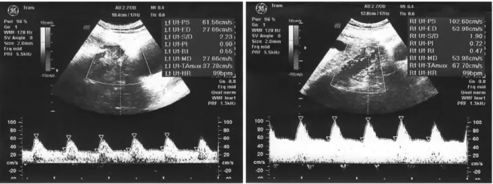
scan-ning showed the presence of hyperechoic bowel (grade 2) and symmetrical FGR. Doppler velocimetry of the uterine artery was normal, there was no notch sign (resistance index [RI] = 0.55 in left uterine artery and RI = 0.47 in the right uterine artery) (Fig. 1), and an absence of end-diastolic frequencies was diagnosed in the umbilical artery (Fig. 2). Doppler velocimetry of the middle cerebral artery and ductus venous were normal (Figs 3,4).
The patient and her husband gave their consent for amniocentesis to diagnose fetal chromosomal aberra-tions. The fetal karyotype was confirmed to be normal (Fig. 5). Toxoplasmosis, parvovirus, adenovirus, tre-ponema, CMV, HHV 1 and 2, and EBV were routinely screened by using antibody measurements in the maternal blood and real-time PCR in the amniotic fluid. EBV was incidentally detected in the amniotic fluid, with 26 800 copies/mL. The patient was then further tested for EBV, and it was demonstrated to be present in her peripheral blood: immunoglobulin (Ig)G antibodies to the EBV capsid antigen were
positive (174 IU/mL), and IgM antibodies were negative.
After informed consent, the patient and her husband preferred to pursue the pregnancy for as long as possible. Fetal growth was reviewed every two weeks by measurement of biparietal diameter (BPD), head circumference (HC), femur length (FL) and abdominal circumference (AC). All fetal biomet-ric parameters were consistently found to be lower than the third percentile line (Fig. 6). Doppler veloci-metry of the umbilical artery, the middle cerebral artery, and the ductus venous were performed routinely every two weeks. There was a consistent
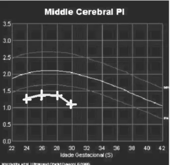
absence of end-diastolic frequencies in the umbilical artery, the pulsatility index (PI) of middle cerebral artery was found to be below the third percentile (Fig. 7) and the ductus venous continued to show a normal a-wave.
At the 30thweek of gestation, the fetus demised
spon-taneously in utero, and after an induced labor, the patient delivered a female infant transvaginally. The stillborn baby weighed 900 grams and the placenta was small, weighing 200 grams. Histopathological exami-nation of placental tissues demonstrated deciduitis and villitis with a significant mononucleocyte infiltration, trophoblastic necrosis and capillary endothelial damage. The couple refused an autopsy for religious
Figure 2 Absent end-diastolic frequencies in the
umbili-cal artery. This sign was consistent from the 24thto the
30thweeks of pregnancy.
Figure 3 Normal Doppler velocimetry of the middle cerebral artery at 24 weeks’ gestation (no notch sign, normal pulsatility index [PI] and resistance index [RI]).
Figure 4 Normal Doppler velocimetry of the ductus venous at 24 weeks’ gestation (normal a-wave, normal pulsatility index [PI] and resistance index [RI]).
reasons, further histopathological and immunohis-tochemical testing is not currently possible in our resource-limited setting.
Discussion
This case report demonstrates symmetrical FGR and early intrauterine death as apparent consequences of reactivation of EBV infection in pregnancy. There is very little known about maternal EBV infection and pregnancy outcomes, since most published material are case reports: some reported cases suggest a rela-tionship between EBV and congenital heart disease,7,8,9
and FGR and multiple congenital anomalies;10,11others
support the concept that entry of a viral genome into the amniotic fluid occurred in normal pregnancies without any perinatal impacts.12 With evidence of a
poor fetal outcome and proven EBV infection in preg-nancy, we think that EBV infection during pregnancy may indeed produce an effect on perinatal and neonatal outcomes.
Eskild et al. consider that adverse pregnancy out-comes are not associated with EBV reactivation.4
Despite this conclusion, evidence has accumulated in the last few years highlighting the complexity of the interaction between EBV and the human host.13 The
Figure 6 Development of fetal biometry (biparietal diameter, head circumference, femur length and abdominal
actual damage to the developing embryo or fetus from maternal herpes simplex virus and EBV appears to be very small.14,15But both herpes simplex virus and EBV
seem to be able to cross the placenta and cause, as described by several investigators, placental infection manifested by deciduitis and villitis. These placental changes potentially cause fetal damage: Brown and Stenchever reported on an infant exposed to EBV from conception to delivery: the infant was delivered at 42 weeks’ gestation weighing 1720 g, showing sym-metrical growth retardation and died several minutes after delivery; post-mortem examination revealed mul-tiple anomalies of the craniofacial complex, kidneys, lungs, heart and brain.7 This case demonstrates that
EBV may cross the placenta causing placental and fetal infection.10,15
In our case report, reactivation of EBV infection during pregnancy was suggested because of a presence of a high titer of IgG antibodies in maternal peripheral blood (17 times higher than our laboratory cut-off [10 IU/mL]) and real-time PCR evidence of EBV DNA in the amniotic fluid. In addition, the IgM antibody titer was negative, so there was no evidence of a primary infection. We propose that the impact of EBV was to disrupt placental development, which then caused a symmetrical FGR and an absence of end-diastolic frequencies in the umbilical artery, and finally fetal demise. We are limited in not being able to give
more certain evidence of EBV reactivation through his-topathology and immunohistochemistry testing of the placenta; however, this case is consistent with the study of Eskild et al., who demonstrated that a reacti-vation of EBV infection is associated with lower birth weight in stillborn and live-born children.4
There is no published evidence in the literature that EBV has an effect on the developing embryo or fetus.5,15,16Similarly, in this case, we found that the fetal
karyotype was normal (46, XX) and no fetal structural abnormality was detected on ultrasonography. We also conclude that EBV reactivation during pregnancy does not represent a major teratogenic risk to the fetus as Miller et al. affirmed.12
However, there is a need for further studies to verify the actual impact of EBV exposure on the fetus itself during pregnancy. In the majority of the cases describ-ing the influence of maternal EBV infection on preg-nancy outcome, there seems to be placental, cardiac and visual involvement.7,8,10,11,15,16Avgil et al. found two
cases of congenital heart anomalies (ventricular septal defect and persistent foramen ovale) in women who had a recurrent EBV infection during the first trimester of pregnancy.16They ruled out the possible association
of congenital heart disease and an exposure to EBV from conception to delivery.16 Recently, Werler et al.
demonstrated that EBV reactivation resulting from multiple challenges to the immune response, such as pregnancy, age and toxic exposure might be related to a risk of gastroschisis.17Therefore, they propose further
research into gastroschisis caused by EBV and other herpes viruses. The role of EBV in pregnancy, accord-ing to Haeri et al., should be examined.5
Although reports of pregnant women with EBV are rare,15 EBV infection in pregnancy does seem to
have a close relationship with stillbirth and congenital defects.10 Our results support the studies that
dem-onstrate an association between EBV reactivation and placental damage during pregnancy.10,11,13,16We do
not recommend adding EBV testing in the universal TORCH (toxoplasmosis, rubella, cytomegalovirus, herpes simplex and HIV) screening for infection during pregnancy, because the seroprevalence of this virus is very rare in this population. Our message in this case report is that the obstetricians should be aware of the need for testing EBV infection during specific pregnancies when trying to elucidate the possible cause of early fetal growth restriction and/or intrauter-ine death. We acknowledge that these findings need confirmation through future studies for prognosis of pregnancy with EBV infection.
Figure 7 Abnormal pulsatility index (PI) curve of the middle cerebral artery.
Acknowledgments
The author is grateful to Dr Sarah Hoskins for her assistance in editing this manuscript.
References
1. Fauci AS, Braunwald E, Kasper DL et al. Harrison’s Principles
of Internal Medicine, 17th Edition, Manual of Medicine. New
York: Mc Graw-Hill, 2009; 574–575.
2. Meyohas MC, Marechal V, Desire N, Bouillie J, Frottier J, Nicolas JC. Study of mother-to-child Epstein-Barr virus trans-mission by means of nested PCRs. J Virol 1996; 70: 6816–6819. 3. Haun L, Kwan N, Hollier LM. Viral infections in pregnancy.
Minerva Ginecol 2007; 59: 159–174.
4. Eskild A, Bruu AL, Stray-Pedersen B, Jenum P. Epstein-Barr virus infection during pregnancy and the risk of adverse pregnancy outcome. BJOG 2005; 112: 1620–1624.
5. Haeri S, Baker AM, Boggess KA. Prevalence of Epstein-Barr virus reactivation in pregnancy. Am J Perinatol 2010; 27: 715– 719.
6. Baschat A, Baschat AA, Towbin J, Bowels NE, Harman CP, Weiner CR. Prevalence of viral DNA in amniotic fluid of low-risk pregnancies in the second trimester. J Matern Fetal
Neonatal Med 1993; 13: 381–384.
7. Brown Z, Stenchever M. Infectious mononucleosis and congenital anomalies. Am J Obstet Gynecol 1978; 131: 108–109.
8. Fleisher G, Bolognese R. Persistent Epstein-Barr virus infec-tion and pregnancy. J Infect Dis 1983; 147: 982–986.
9. Fleisher G, Bologonese R. Epstein-Barr virus infections in pregnancy: A prospective study. J Pediatr 1984; 104: 374–379. 10. Icart J, Didier J, Dalens M, Chabanon G, Boucays A. Prospec-tive study of Epstein Barr virus (EBV) infection during preg-nancy. Biomedicine 1981; 34: 160–163.
11. Goldberg GN, Fulginiti VA, Ray CG, Ferry P, Jones JF, Cross H. In utero Epstein-Barr virus (infection mononucleosis) infection. JAMA 1981; 246: 1579–1581.
12. Miller JL, Harman C, Weiner C, Baschat AA. Perinatal out-comes after second trimester detection of amniotic fluid viral genome in asymptomatic patients. J Perinat Med 2009; 37: 140–143.
13. Levitsky V, Masucci MG. Manipulation of immune response by Epstein-Barr virus. Virus Res 2002; 88: 71–86.
14. Ornoy A, Dudai M, Sadovsky E. Placental and fetal pathology in infectious mononucleosis. Diagn Gynecol Obstet 1982; 4: 11–16.
15. Avgil M, Ornoy A. Herpes simplex virus and Epstein-Barr virus infection in pregnancy: Consequences of neonatal or intrauterine infection. Reprod Toxicol 2006; 21: 436–445. 16. Avgil M, Diav-Citrin O, Shechtman S, Arnon J, Wajnberg R,
Ornoy A. Epstein-Barr virus infection in pregnancy—a pro-spective controlled study. Reprod Toxicol 2008; 25: 468–471. 17. Werler MM. Hypothesis: Could Epstein-Barr virus play a role
in the development of gastroschisis? Birth Defects Res A Clin
![Figure 3 Normal Doppler velocimetry of the middle cerebral artery at 24 weeks’ gestation (no notch sign, normal pulsatility index [PI] and resistance index [RI]).](https://thumb-eu.123doks.com/thumbv2/123doknet/6093761.154291/3.892.457.799.116.373/figure-normal-doppler-velocimetry-cerebral-gestation-pulsatility-resistance.webp)