The MIT Faculty has made this article openly available. Please share how this access benefits you. Your story matters.
Citation Bellott, Daniel W et al. “Cost-Effective High-Throughput
Single-Haplotype Iterative Mapping and Sequencing for Complex Genomic Structures.” Nature Protocols 13, 4 (March 2018): 787–809 © 2018 Macmillan Publishers Limited, part of Springer Nature
As Published http://dx.doi.org/10.1038/nprot.2018.019
Publisher Nature Publishing Group
Version Original manuscript
Citable link http://hdl.handle.net/1721.1/118374
Terms of Use Article is made available in accordance with the publisher's
policy and may be subject to US copyright law. Please refer to the publisher's site for terms of use.
Cost-‐effective, high-‐throughput, single-‐haplotype iterative mapping and sequencing for complex genomic structures
Daniel W. Bellott1&4, Ting-‐Jan Cho1&4, Jennifer F. Hughes1, Helen Skaletsky1&2, David C. Page1-‐3
1. Whitehead Institute, Cambridge, Massachusetts, USA
2. Howard Hughes Medical Institute, Whitehead Institute, Cambridge, Massachusetts, USA
3. Department of Biology, Massachusetts Institute of Technology, Cambridge, Massachusetts, USA
4. These authors contributed equally to this work.
Correspondence should be addressed to D. W. B. ([email protected])
Abstract
Reference sequence of structurally complex regions can only be obtained through highly accurate clone-‐based approaches. We and others have successfully employed Single-‐Haplotype Iterative Mapping and Sequencing (SHIMS 1.0) to assemble
structurally complex regions across the sex chromosomes of several vertebrate species and in targeted improvements to the reference sequences of human
autosomes. However, SHIMS 1.0 was expensive and time consuming, requiring the resources that only a genome center could command. Here we introduce SHIMS 2.0, an improved SHIMS protocol to allow even a small laboratory to generate high-‐ quality reference sequence from complex genomic regions. Using a streamlined and parallelized library preparation protocol, and taking advantage of high-‐throughput, inexpensive, short-‐read sequencing technologies, a small group can sequence and assemble hundreds of clones in a week. Relative to SHIMS 1.0, SHIMS 2.0 reduces the cost and time required by two orders of magnitude, while preserving high sequencing accuracy. Introduction
Ampliconic sequences, euchromatic repeats that display greater than 99% identity over more than 10 kilobases, are the most structurally complex regions in the genome and are notoriously difficult to assemble. These complex repetitive structures mediate deletions, duplications, and inversions associated with human disease1,2, but the absence of accurate reference sequence of these regions has impeded comprehensive studies of genomic structural variation as well as the mechanisms that govern the rearrangements associated with ampliconic sequences. Furthermore, experiments based on aligning short reads to existing reference sequences – such as genome and exome resequencing, RNA-‐seq, and ChIP-‐seq – are necessarily limited by the quality and completeness of the reference sequence. Reanalysis of short-‐read datasets in the light of improved reference sequences can immediately provide rich annotation of structurally complex regions for studying their role in human variation in health and disease.
Only extremely long and accurate reads can discriminate between amplicon copies, and generate a correct reference sequence from structurally complex regions. The human genome was assembled from the sequences of BAC (Bacterial Artificial Chromosome) clones. Each BAC clone was shotgun sequenced in Sanger reads and painstakingly, and largely manually, assembled into a synthetic long read of ~150kb with error rates as low as one in 1,000,000 nucleotides3. However, this process was both slow and expensive, and subsequent generations of sequencing technology have prioritized driving down sequencing costs at the expense of read length and accuracy. Whole-‐genome shotgun strategies based on Sanger reads forfeited the ability to assemble ampliconic sequences, and assemblies of shorter Illumina and SOLiD reads struggle to traverse smaller and more numerous genome typical interspersed repeats4. Single-‐molecule sequencing technologies like PacBio
or Nanopore sequencing offer longer read lengths that can span most genome typical repeats, but they lack the accuracy to assemble ampliconic sequences5. We developed our Single-‐Haplotype Iterative Mapping and Sequencing (SHIMS) approach, which is the only sequencing technology capable of assembling ampliconic regions, in the context of the human genome project. Utilizing BAC clones derived from a single haplotype allowed us to discriminate between paralogous amplicon copies that are more similar than alleles, and accurately
assemble the intricate repetitive structures of the human Y chromosome6. Since that time, our approach has been instrumental in producing the reference sequences of sex chromosomes from several vertebrate species7-‐13. Here we describe how we have advanced this technique to combine the advantages of a hierarchical, clone-‐ based strategy with new high-‐throughput sequencing technologies (Fig. 1). SHIMS 2.0 reduces both the time and cost by two orders of magnitude, while maintaining read length and accuracy.
Development
The human reference sequence was assembled from a patchwork of BAC clones derived from the genomes of 16 diploid individuals14. While this strategy was suitable for single-‐copy regions of the genome, it proved inadequate to accurately assemble structurally complex repetitive sequences15. Ampliconic sequences can differ from each other by less than one base pair in 10,000, an order of magnitude less than the average difference between alleles16. In the assembly of ampliconic regions, only these rare differences, what we call sequence family variants (SFVs), distinguish truly overlapping BACs from those that belong to paralogous ampliconic sequences. When a mix of haplotypes is used, the noise of frequent differences between alleles overwhelms the signal of the rare differences between paralogous amplicon copies. In the multi-‐haplotype assembly of the human genome, amplicons were misassembled or mistakenly skipped as redundant11,15,17-‐19.
We developed SHIMS 1.0 to cope with the ampliconic sequences of the human Y chromosome20. We deliberately restricted ourselves to BAC clones from one man’s Y chromosome, avoiding polymorphisms that would confound our ability to assemble and map sequences within ampliconic regions. Because only complete and accurate BAC sequences can reveal the SFVs that distinguish amplicon copies, mapping and sequencing became coupled and iterative processes. Sequencing an initial tiling path of BACs, selected by low-‐resolution fingerprint or STS mapping, reveals SFVs that allow us to refine our map and select BACs for subsequent rounds of sequencing. SHIMS requires sequencing clones with substantial (>10kb) overlaps to detect SFVs that guide us to accurately distinguish and assemble near-‐identical amplicons. This painstaking approach produced a complete and accurate
representation of the repeat architecture of a single Y chromosome6, and as a result, we were able to predict and characterize rearrangements mediated by that
architecture throughout the human population21-‐29. These recurrent Y-‐chromosome rearrangements are the most common genetic cause of spermatogenic failure in men, and have been shown to play predominant roles in sex reversal, Turner syndrome, and testicular germ cell tumors. None of these insights would have been
possible without a complete and accurate reference sequence of the amplicons of the human Y chromosome.
SHIMS 1.0 proved successful in generating reference sequence across several vertebrate sex chromosomes, and it remains the only technique capable of accessing structurally complex ampliconic regions. However, we relied on the Sanger
sequencing pipelines and the expertise of dedicated genome finishers at genome centers to assemble each BAC clone. This process was expensive and time
consuming; each BAC clone cost about $9000 to sequence and assemble, and each iteration of mapping and sequencing took around six months. We therefore sought to adopt new technologies to reduce costs, increase speed and efficiency, and bring SHIMS within the reach of a single lab (Fig. 1). SHIMS 2.0 takes advantage of the low cost and high consensus accuracy of Illumina reads to sequence indexed pools of hundreds of BAC clones. We streamlined and parallelized library production to bring sequencing costs down to $50 per BAC clone and shorten mapping and sequencing iterations to a single week. In contrast to earlier BAC pooling
strategies30-‐33, we tag each clone with a unique barcode to assemble the reads from each clone separately. This allows us to pool clones from the same amplicon without endangering the integrity of the assembly of the entire pool. It is rare to encounter closely related interspersed repeats within a single BAC clone, and therefore long interspersed repeats typically do not frustrate the assembly of individual BAC clones from Illumina reads as they do in whole genome shotgun approaches. When we encounter internally repetitive clones, we use long reads from single molecule sequencing technologies to scaffold the short read assemblies, eliminating much of the need for manual finishing. Together, these optimizations allow a small
independent laboratory to do the work that once required a fully staffed genome center.
Overview of SHIMS 2.0
The most critical resource for a SHIMS project is a large-‐insert clone library derived from a single haplotype. For sex chromosomes, where ampliconic sequences are abundant, this is a trivial requirement because any library constructed from an individual of the heterogametic sex (males for X and Y; females for Z and W) contains a single haplotype for each sex chromosome (at half the coverage of the autosomes). For many model organisms, a library constructed from an inbred strain will provide a single haplotype of the autosomes. For diploid organisms where inbreeding is not possible, special sources of single-‐haplotype DNA are required. A single-‐haplotype BAC library was constructed for the human genome, using DNA from a hydatidiform mole, an abnormal conceptus that arises when an enucleated egg is fertilized by a single X-‐bearing sperm17-‐19. For some plant species, haploid cell lines or haploid plants can be used as a source of DNA for library
construction. Ideally the BAC library should have greater than 10x coverage of the chromosome of interest; coverage lower than 5x will inevitably result in a
fragmented assembly due to gaps in library coverage. When there is prior
knowledge about the size of ampliconic repeat units, it is useful to choose a library with an average insert size smaller than the repeat unit of the amplicon – if multiple
amplicon copies are present within a single insert, the clone assembly will collapse. For smaller ampliconic repeat units, fosmids can substitute for BACs.
After choosing or constructing a single-‐haplotype BAC library, the next step is to select an initial tiling path for sequencing and iterative refinement. A variety of mapping methods can be used to identify clones of interest from a BAC library, including fingerprint maps, end sequences, screening high-‐density filters by
hybridization, or screening high-‐dimensional pools of BACs for STS content by PCR. Typically, the cost to confirm each positive clone by another round of end-‐
sequencing or PCR will exceed the cost to obtain draft sequence using the SHIMS 2.0 protocol, making sequencing the most efficient way to confirm the identity of clones. SFVs that distinguish between amplicon copies can be identified using draft sequences from the initial tiling path of clones. We scrutinize the differences in the apparent overlaps between clones using a graphical editor, such as Consed34 or Gap535. We typically limit ourselves to single nucleotide differences supported by high quality bases in the majority of reads. Variants in short tandem repeats are not reliable; these are not always accurately assembled, and differences between clones often represent mutations that occur during propagation of the BACs in E. coli rather than true differences between paralogous amplicons. After using newly identified SFVs as markers to refine the sequence map and resolve all paralogous amplicons, we order and orient the resulting sequence contigs by a complementary method, such as RH mapping or metaphase FISH. Whenever possible, we estimate the size of the remaining gaps, either by RH mapping or extended chromatin FISH.
Early attempts to adapt next-‐generation sequencing technologies for BAC assembly pooled many clones in a single sequencing run. While this approach was faster and more cost-‐effective than traditional Sanger sequencing, it had the major shortcoming that assemblies either collapsed at genome-‐typical repeats shared among clones, or worse, contained chimeric contigs from two or more clones in the pool. Therefore we adopted the practice of adding unique “barcodes” or “indexes” to each clone during library preparation, and only pooling material from different clones after these indexes were added. This allows us to automatically assign each read to a single clone, making the assembly of each clone less prone to artifacts. We typically use a set of TruSeq-‐compatible adapters with 384 unique 8nt indexes (Supplementary Table 1), and pool 192 clones in a single MiSeq run, but in principle, this procedure can be extended to use larger numbers of indexes, or even dual indexes, to sequence larger pools of clones on higher-‐throughput sequencing machines.
A major challenge in pooling hundreds of samples is to ensure an adequate representation of each sample. When each sample in the pool has widely varying concentration, some samples will have wasteful coverage that is greater than what is required for an adequate assembly, while others will have too few reads to generate any assembly, necessitating another round of library preparation to join the next pool. We use a short course of library amplification (20 cycles of PCR), using limiting primers. This is sufficient to normalize each library within 2-‐3 fold of the median concentration. Although PCR amplification can introduce errors and biases into Illumina libraries, we find that our consensus sequence accuracy does
not suffer, and it saves a large amount of tedious and exacting labor in measuring each library and diluting them to the same concentration.
Simple Sequence Repeats (SSRs) represent the chief obstacle to contiguous BAC assemblies with short Illumina reads. Reads that cover SSRs are subject to stutter noise from replication slippage in library preparation (producing reads with inaccurate SSR array length) and cluster generation (reducing quality scores in the SSR)36. As a result, most assemblers fail to assemble SSRs, leaving gaps flanked by SSR sequence. We use a combination of long (300bp) reads and large (1000-‐1200 bp) fragment sizes to scaffold over the vast majority (>99.8%) of SSRs. One drawback to fragment sizes greater than about 600bp is that they must be size-‐ selected by gel purification to eliminate any smaller fragments. This gel-‐purification would be onerous and expensive if each clone were purified separately, but our indexing and normalization procedures allow us to perform a single size selection on a pool of libraries from hundreds of clones.
Advantages of SHIMS 2.0
SHIMS produces de novo sequence assemblies of greater accuracy and contiguity than any other technique, and is the only technique that has successfully produced accurate reference sequences of ampliconic regions. These advantages are rooted in the clone-‐based nature of SHIMS. Each clone assembly represents the highly
accurate sequence of a single long molecule – with error rates as low as one in 1,000,000 nucleotides. Any observed SFV can be verified by resequencing a clone of the same molecule, increasing the confidence and resolution of the SFV map.
In contrast to whole genome shotgun sequencing using short-‐read, or even Sanger technologies, clone-‐based approaches produce a much more contiguous and accurate assembly. While genome-‐typical interspersed repeats like SINEs, LINEs, or ERVs are the primary obstacle to WGS assembly, they only rarely confound the assembly of individual clones. Furthermore, a hierarchical, clone-‐based approach guarantees that all sequence contigs are unambiguously mapped within a single clone in the assembly, and that clone is, in turn, mapped by long, perfect overlaps with neighboring clones.
Continuous long-‐read technologies offer improvements in contiguity relative to short-‐read shotgun sequencing, but their accuracy is far too low to resolve
ampliconic sequences37,38. Error correction with short reads can improve accuracy in single-‐copy regions37,39,40, but this process tends to obscure SFVs by correcting long reads to the consensus of paralogous amplicons, resulting in collapsed assemblies. For example, the recent assembly of the Gorilla Y chromosome with a mixture of Illumina and PacBio reads was able to identify ampliconic sequences and estimate their copy number, but was unable to resolve their structural
organization41.
Synthetic long read technologies produce more accurate reads than continuous long read technologies, but are neither accurate nor long enough to assemble ampliconic sequences. Synthetic long reads have an error rate of ~1 in 10,000 nucleotides42,43, or two orders of magnitude higher than clone based
opportunity to resequence the same molecule to resolve discrepancies between reads. Synthetic long reads are also 1-‐2 orders of magnitude shorter than BAC clones42,43, limiting their power to resolve long ampliconic sequences that can differ by less than 1 in 10,000 nucleotides6,10,12.
Optical mapping techniques provide long range scaffolding information that can help increase the contiguity of genome assemblies by generating restriction maps of DNA fragments 0.1-‐1 Mb in size that can be compared against in-‐silico restriction maps of WGS contigs44. In general, these restriction maps do not have sufficient resolution to uncover the single nucleotide differences that constitute reliable SFVs, and do not sample molecules long enough to resolve many ampliconic sequences. Even combined with PacBio and Illumina reads, optical mapping was unable to resolve the ampliconic sequences on the human Y chromosome45.
Limitations of SHIMS 2.0
The SHIMS approach provides access to longer and higher identity ampliconic sequences than any other sequencing technique, but the clone-‐based nature of this approach imposes several limitations. First, the maximum size of BAC inserts limits SHIMS to resolving duplications with less than 99.999% identity. Second, SHIMS is limited to sequences that can be cloned into E. coli. Third, SHIMS is frustrated by repeated sequences shorter than a single clone.
The average BAC clone size limits the power to resolve paralogous amplicons to those that differ by more than one nucleotide in 100,000, so that each clone can be mapped by one or more SFVs. Clones with longer inserts, such as YACs, could potentially capture SFVs that distinguish paralogous amplicons at lower rates of divergence, but, in practice, YACs are subject to high rates of chimerism46, deletion, and rearrangement, making them far too unreliable for sequencing ampliconic regions. This limitation will remain until long-‐read technologies can surpass BAC sequencing in both read length and accuracy, or a reliable cloning technology emerges that exceeds the insert size of BACs.
SHIMS is also limited to sequences that can be cloned. Sequences that are toxic to E. coli are underrepresented in BAC and fosmid libraries. An exhaustive search through the library will sometimes turn up deleted clones, where the cloning process has selected for clones with rearrangements that eliminate the toxic
sequences. Gaps of this nature can be resolved by directed efforts that avoid cloning in E. coli. For example, orthologous ampliconic sequences on the human and
chimpanzee Y chromosomes contained an unclonable sequence that led to deleted clones in both human and chimpanzee BAC libraries – this ~30 kb unclonable region was eventually sequenced from a long-‐range PCR product.
Arrays of repeated sequences shorter than the clone insert size present special problems for clone-‐based sequencing. The centromere (171 bp repeat in 6 kb secondary unit), long-‐arm heterochromatin (degenerate pentamer repeat in 3.5 kb secondary unit), and TSPY gene array (20.4 kb unit) were not resolved on the human Y chromosome. Arrays with short repeat units may cause library gaps, as restriction sites will either appear too frequently, or not at all, so that no fragments covering the array are successfully cloned in the library. In this case, libraries
produced by random shearing, like Fosmid libraries, may produce better coverage than those generated by restriction digest. The presence of many highly identical repeats within a single clone will cause the sequence assembly to collapse multiple repeats into a single short contig. Whenever possible, it is best to choose clone libraries with an insert size that matches the expected repeat unit size. However, sequencing with fosmids will increase costs relative to BACs, as it requires many more clones to cover the same amount of sequence. In some cases, continuous long read technologies applied to individual BAC clones can resolve internal repeats, albeit at higher cost and with lower per-‐base accuracy.
Applications of SHIMS 2.0
SHIMS has been repeatedly applied to resolve ampliconic sequences across vertebrate genomes, particularly the sex chromosomes, where ampliconic sequences are most abundant and elaborate. SHIMS was used to resolve the ampliconic sequences of the human, chimpanzee, and mouse Y chromosomes, the human X chromosome, and the chicken Z and W chromosomes6-‐8,10-‐12,20. SHIMS has also been applied to the human immunoglobulin gene cluster19 and other
structurally complex regions on human autosomes17,18 using a single haplotype library derived from a hydatidiform mole.
The SHIMS approach is applicable to any genomic sequence where amplicons and other structurally complex regions complicate WGS assembly. A library of large insert clones from a haploid or inbred diploid source is required to resolve
ampliconic sequences, but a library derived from an outbred diploid source could be used to generate a phased diploid genome assembly covering non-‐ampliconic
regions.
Existing instruments (Illumina HiSeq series) already generate sufficient numbers of reads to sequence a tiling path of BAC clones across the human genome in a single run costing around $14,00047; the costs of sample preparation and library generation therefore dominate cost considerations. With our current SHIMS 2.0 approach, assembling the entire human genome would cost around $2,000,000, or three orders of magnitude less than the cost to generate the original BAC-‐based reference sequence. The cost could be reduced further with future extensions of the indexing and pooling strategy we describe here to reduce reagent costs and labor required for sample preparation and library generation. This could potentially make it cost-‐effective to apply SHIMS 2.0 across whole genomes, even in the absence of the extensive resources, like BAC fingerprint maps and end sequences, available to common model organisms.
Experimental Design
The primary time and cost savings of SHIMS 2.0 over traditional BAC sequencing come from the ability to process many clones in parallel and sequence them in a single pool. While each step can be performed by hand with a multichannel pipette, all operations, especially size selection with SPRI beads, will be more accurate with a liquid handling robot. This need not be expensive – adequate used instruments can
be purchased for less than $5000, and many core facilities will offer access to a liquid handling robot.
We made several optimizations to adapt standard DNA extraction and library preparation techniques for our purposes. To support growth of E. coli carrying single-‐copy BACs, clones are grown in 2x LB. SPRI beads are added to the isopropanol precipitation step to recover more DNA compared to a standard
alkaline lysis preparation. In a crude preparation of low-‐copy plasmids like BACs or fosmids, there will be 10-‐30% E. coli genomic DNA contamination; sequencing costs are low enough that it is not cost-‐effective to take special measures to reduce this contamination fraction any further.
In our experience, the smallest fragments present in the library determine the average sequenced fragment size. We find that a Covaris focused ultrasonicator is invaluable for generating DNA fragments with a reproducibly tight size distribution as input for the library generation protocol. We have optimized our shearing
conditions for a Covaris LE220 focused ultrasonicator; other makes and models may require slightly different conditions to achieve 1 kb fragments. The Covaris 96 microTUBE plates are costly but necessary for consistent shearing across wells. After library generation is complete, a gel-‐based size selection assures the tightest size distribution around 1 kb. Because each sample is individually barcoded, it is possible to pool all samples in a single well before size selection, drastically reducing the labor involved.
We use a custom set of 384 8-‐mer indexes for barcodes; Illumina offers sets of 96 and 384 barcodes through a dual-‐indexing scheme. More elaborate dual-‐index schemes48 could potentially allow for larger pools on higher throughput Illumina machines. We selected the MiSeq because of its combination of long reads, short run times, and low cost, and believe it offers the right combination of features for a single lab to perform SHIMS 2.0 on targeted genomic regions. It is certainly possible to scale up to higher-‐throughput instruments for genome-‐scale sequencing projects, but this would only reduce sequencing reagent costs by a modest amount -‐-‐ the bulk of costs are in the library preparation.
A SHIMS 2.0 project requires significant bioinformatics expertise to proceed from raw reads to finished, annotated sequence. The state-‐of-‐the-‐art software advances rapidly, so that any specific software recommendations are likely to become outdated very quickly. Demultiplexed fastq-‐format files should first be trimmed to remove Illumina adapter sequences and low quality bases, and then screened for contamination from the host genome and vector sequences. Filtered sequences are then used for assembly, scaffolding, and gap closure. We use
cutadapt49 to trim adapters and low quality sequences, bowtie250 to screen out vector and E. coli genomic DNA contamination, SPAdes51 for assembly, BESST52 for scaffolding, and Gap2Seq53to fill gaps. Some clone assemblies will require manual finishing; we rely on Consed34 for visualizing discrepant bases, separating collapsed duplications, and merging overlapping contigs.
Several controls ensure the accuracy and quality of a SHIMS 2.0 assembly. A cell line derived from the same individual or strain as the BAC or fosmid library permits FISH experiments and long-‐range PCR. An independent FISH, radiation hybrid, or optical map can be used to confirm the order and orientation of contigs in
the clone map, as well as estimate the size of any remaining gaps. Finally, the error rate of the assembly can be calculated from the number of discrepancies observed in the long (>10kb) redundant overlaps between adjacent clones; for BACs, it is
possible to achieve error rates as low as one in 1,000,000 nucleotides. There are important quality control checkpoints in both the library preparation and bioinformatic analysis stages. After adapter ligation, but before pooling, it is useful to reserve a sample from each clone’s library for 40 cycles of PCR with universal Illumina primers followed by gel electrophoresis to ensure that each library contains PCR amplifiable material in the expected size range before
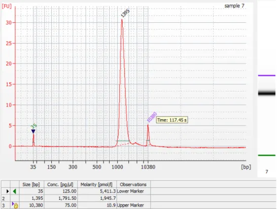
proceeding with sequencing (Fig. 1). After pooling and gel purification, we recommend testing the fragment size distribution via Bioanalyzer before
sequencing (Fig. 1). The front of the fragment size distribution will be the peak of the sequenced fragment size distribution. During assembly, reads from E. coli genomic DNA serve as an internal control to assess library insert size and
sequencing error rates. As each clone is assembled, we align putative overlapping clones to identify differences using Consed34, verifying that the reads in each clone support the difference. Most of these high quality differences will be SFVs that distinguish paralogous amplicons, but some will be genuine errors due to mutations in the BAC or fosmids. With care, it is possible to achieve error rates as low as one in 1,000,000 nucleotides.
A major benefit of our new protocol is that it allows a small team to carry out a SHIMS project that, only a few years ago, would have required the cooperation of a large genome center. A single technician can process 192 clones from frozen library plates to Illumina libraries in a single week, and a bioinformatics specialist can set up a pipeline to automatically process the resulting reads into draft assemblies, identify SFVs, and manually finish complex clone assemblies. It is particularly helpful to have a team member or collaborator with experience in metaphase FISH to help resolve the order and orientation of contigs within the final assembly.
References
1 Lupski, J. R. Genomic disorders: structural features of the genome can lead to DNA rearrangements and human disease traits. Trends Genet 14, 417-‐422, doi:Doi 10.1016/S0168-‐9525(98)01555-‐8 (1998).
2 Stankiewicz, P. & Lupski, J. R. Structural variation in the human genome and its role in disease. Annu Rev Med 61, 437-‐455, doi:10.1146/annurev-‐med-‐ 100708-‐204735 (2010).
3 International Human Genome Sequencing, C. Finishing the euchromatic sequence of the human genome. Nature 431, 931-‐945,
doi:10.1038/nature03001 (2004).
4 Alkan, C., Sajjadian, S. & Eichler, E. E. Limitations of next-‐generation genome sequence assembly. Nat Methods 8, 61-‐65, doi:10.1038/nmeth.1527 (2011). 5 Gordon, D. et al. Long-‐read sequence assembly of the gorilla genome. Science
352, aae0344, doi:10.1126/science.aae0344 (2016).
6 Skaletsky, H. et al. The male-‐specific region of the human Y chromosome is a mosaic of discrete sequence classes. Nature 423, 825-‐837,
doi:10.1038/nature01722 (2003).
7 Bellott, D. W. et al. Mammalian Y chromosomes retain widely expressed dosage-‐sensitive regulators. Nature 508, 494-‐499, doi:10.1038/nature13206 (2014).
8 Bellott, D. W. et al. Convergent evolution of chicken Z and human X chromosomes by expansion and gene acquisition. Nature 466, 612-‐616, doi:10.1038/nature09172 (2010).
9 Hughes, J. F. et al. Strict evolutionary conservation followed rapid gene loss on human and rhesus Y chromosomes. Nature 483, 82-‐86,
doi:10.1038/nature10843 (2012).
10 Hughes, J. F. et al. Chimpanzee and human Y chromosomes are remarkably divergent in structure and gene content. Nature 463, 536-‐539,
doi:10.1038/nature08700 (2010).
11 Mueller, J. L. et al. Independent specialization of the human and mouse X chromosomes for the male germ line. Nat Genet 45, 1083-‐1087,
doi:10.1038/ng.2705 (2013).
12 Soh, Y. Q. et al. Sequencing the mouse Y chromosome reveals convergent gene acquisition and amplification on both sex chromosomes. Cell 159, 800-‐ 813, doi:10.1016/j.cell.2014.09.052 (2014).
13 Bellott, D. W. et al. Avian W and mammalian Y chromosomes convergently retained dosage-‐sensitive regulators. Nat Genet 49, 387-‐394,
doi:10.1038/ng.3778 (2017).
14 Ross, M. T. et al. The DNA sequence of the human X chromosome. Nature 434, 325-‐337, doi:10.1038/nature03440 (2005).
15 Eichler, E. E. Segmental duplications: what's missing, misassigned, and misassembled-‐-‐and should we care? Genome Res 11, 653-‐656,
doi:10.1101/gr.188901 (2001).
16 Sachidanandam, R. et al. A map of human genome sequence variation containing 1.42 million single nucleotide polymorphisms. Nature 409, 928-‐ 933, doi:10.1038/35057149 (2001).
17 Dennis, M. Y. et al. Evolution of human-‐specific neural SRGAP2 genes by incomplete segmental duplication. Cell 149, 912-‐922,
doi:10.1016/j.cell.2012.03.033 (2012).
18 Steinberg, K. M. et al. Single haplotype assembly of the human genome from a hydatidiform mole. Genome Res 24, 2066-‐2076, doi:10.1101/gr.180893.114 (2014).
19 Watson, C. T. et al. Complete haplotype sequence of the human
immunoglobulin heavy-‐chain variable, diversity, and joining genes and characterization of allelic and copy-‐number variation. Am J Hum Genet 92, 530-‐546, doi:10.1016/j.ajhg.2013.03.004 (2013).
20 Kuroda-‐Kawaguchi, T. et al. The AZFc region of the Y chromosome features massive palindromes and uniform recurrent deletions in infertile men. Nat Genet 29, 279-‐286, doi:10.1038/ng757 (2001).
21 Repping, S. et al. High mutation rates have driven extensive structural polymorphism among human Y chromosomes. Nat Genet 38, 463-‐467, doi:10.1038/ng1754 (2006).
22 Lange, J. et al. Intrachromosomal homologous recombination between inverted amplicons on opposing Y-‐chromosome arms. Genomics, doi:10.1016/j.ygeno.2013.04.018 (2013).
23 Lange, J., Skaletsky, H., Bell, G. W. & Page, D. C. MSY Breakpoint Mapper, a database of sequence-‐tagged sites useful in defining naturally occurring deletions in the human Y chromosome. Nucleic Acids Res 36, D809-‐814, doi:10.1093/nar/gkm849 (2008).
24 Lange, J. et al. Isodicentric Y chromosomes and sex disorders as byproducts of homologous recombination that maintains palindromes. Cell 138, 855-‐869, doi:10.1016/j.cell.2009.07.042 (2009).
25 Repping, S. et al. Polymorphism for a 1.6-‐Mb deletion of the human Y chromosome persists through balance between recurrent mutation and haploid selection. Nat Genet 35, 247-‐251 (2003).
26 Repping, S. et al. Recombination between palindromes P5 and P1 on the human Y chromosome causes massive deletions and spermatogenic failure. Am J Hum Genet 71, 906-‐922, doi:Doi 10.1086/342928 (2002).
27 Repping, S. et al. A family of human Y chromosomes has dispersed
throughout northern Eurasia despite a 1.8-‐Mb deletion in the azoospermia factor c region. Genomics 83, 1046-‐1052, doi:10.1016/j.ygeno.2003.12.018 (2004).
28 Rozen, S. et al. Abundant gene conversion between arms of palindromes in human and ape Y chromosomes. Nature 423, 873-‐876 (2003).
29 Rozen, S. G. et al. AZFc deletions and spermatogenic failure: a population-‐ based survey of 20,000 Y chromosomes. Am J Hum Genet 91, 890-‐896, doi:10.1016/j.ajhg.2012.09.003 (2012).
30 Li, G. et al. Comparative analysis of mammalian Y chromosomes illuminates ancestral structure and lineage-‐specific evolution. Genome Res 23, 1486-‐ 1495, doi:10.1101/gr.154286.112 (2013).
31 Sato, K., Motoi, Y., Yamaji, N. & Yoshida, H. 454 sequencing of pooled BAC clones on chromosome 3H of barley. BMC Genomics 12, 246,
doi:10.1186/1471-‐2164-‐12-‐246 (2011).
32 Quinn, N. L. et al. Assessing the feasibility of GS FLX Pyrosequencing for sequencing the Atlantic salmon genome. BMC Genomics 9, 404,
doi:10.1186/1471-‐2164-‐9-‐404 (2008).
33 Rounsley, S., Lin, X. & Ketchum, K. A. Large-‐scale sequencing of plant genomes. Curr Opin Plant Biol 1, 136-‐141 (1998).
34 Gordon, D. & Green, P. Consed: a graphical editor for next-‐generation sequencing. Bioinformatics 29, 2936-‐2937,
doi:10.1093/bioinformatics/btt515 (2013).
35 Bonfield, J. K. & Whitwham, A. Gap5-‐-‐editing the billion fragment sequence assembly. Bioinformatics 26, 1699-‐1703,
doi:10.1093/bioinformatics/btq268 (2010).
36 Gymrek, M., Golan, D., Rosset, S. & Erlich, Y. lobSTR: A short tandem repeat profiler for personal genomes. Genome Res 22, 1154-‐1162,
doi:10.1101/gr.135780.111 (2012).
37 Goodwin, S. et al. Oxford Nanopore sequencing, hybrid error correction, and de novo assembly of a eukaryotic genome. Genome Res 25, 1750-‐1756, doi:10.1101/gr.191395.115 (2015).
38 Berlin, K. et al. Assembling large genomes with single-‐molecule sequencing and locality-‐sensitive hashing. Nat Biotechnol 33, 623-‐630,
doi:10.1038/nbt.3238 (2015).
39 Koren, S. et al. Hybrid error correction and de novo assembly of single-‐ molecule sequencing reads. Nat Biotechnol 30, 693-‐700,
doi:10.1038/nbt.2280 (2012).
40 Madoui, M. A. et al. Genome assembly using Nanopore-‐guided long and error-‐ free DNA reads. BMC Genomics 16, 327, doi:10.1186/s12864-‐015-‐1519-‐z (2015).
41 Tomaszkiewicz, M. et al. A time-‐ and cost-‐effective strategy to sequence mammalian Y Chromosomes: an application to the de novo assembly of gorilla Y. Genome Res 26, 530-‐540, doi:10.1101/gr.199448.115 (2016). 42 McCoy, R. C. et al. Illumina TruSeq synthetic long-‐reads empower de novo
assembly and resolve complex, highly-‐repetitive transposable elements. PLoS One 9, e106689, doi:10.1371/journal.pone.0106689 (2014).
43 Li, R. et al. Illumina synthetic long read sequencing allows recovery of
missing sequences even in the "finished" C. elegans genome. Sci Rep 5, 10814, doi:10.1038/srep10814 (2015).
44 Dong, Y. et al. Sequencing and automated whole-‐genome optical mapping of the genome of a domestic goat (Capra hircus). Nat Biotechnol 31, 135-‐141, doi:10.1038/nbt.2478 (2013).
45 Seo, J. S. et al. De novo assembly and phasing of a Korean human genome. Nature 538, 243-‐247, doi:10.1038/nature20098 (2016).
46 Nagaraja, R. et al. Characterization of four human YAC libraries for clone size, chimerism and X chromosome sequence representation. Nucleic Acids Res 22, 3406-‐3411 (1994).
47 Glenn, T. C. Field guide to next-‐generation DNA sequencers. Mol Ecol Resour
11, 759-‐769, doi:10.1111/j.1755-‐0998.2011.03024.x (2011).
48 Lange, V. et al. Cost-‐efficient high-‐throughput HLA typing by MiSeq amplicon sequencing. BMC Genomics 15, 63, doi:10.1186/1471-‐2164-‐15-‐63 (2014). 49 Martin, M. Cutadapt removes adapter sequences from high-‐throughput
sequencing reads. EMBnet.journal 17, 10-‐12, doi:10.14806/ej.17.1.200 (2011).
50 Langmead, B. & Salzberg, S. L. Fast gapped-‐read alignment with Bowtie 2. Nat Methods 9, 357-‐359, doi:10.1038/nmeth.1923 (2012).
51 Bankevich, A. et al. SPAdes: a new genome assembly algorithm and its applications to single-‐cell sequencing. J Comput Biol 19, 455-‐477, doi:10.1089/cmb.2012.0021 (2012).
52 Sahlin, K., Vezzi, F., Nystedt, B., Lundeberg, J. & Arvestad, L. BESST-‐-‐efficient scaffolding of large fragmented assemblies. BMC Bioinformatics 15, 281, doi:10.1186/1471-‐2105-‐15-‐281 (2014).
53 Salmela, L., Sahlin, K., Makinen, V. & Tomescu, A. I. Gap filling as exact path length problem. J Comput Biol 23, 347-‐361, doi:10.1089/cmb.2015.0197 (2016).
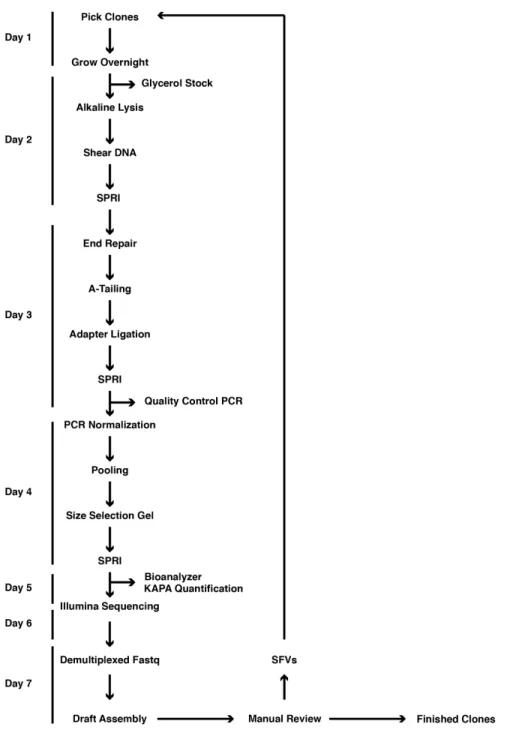
Figure 1 | Overview of SHIMS 2.0 protocol.
A timeline of a single iteration of the SHIMS 2.0 protocol, showing the major protocol steps, with key quality controls on the right. During a single week-‐long iteration, 192 clones are processed in parallel, and the resulting draft clone sequences are used to identify sequence family variants (SFVs) that distinguish paralogous ampliconic sequences. A single technician can proceed from a list of clones to completed Illumina libraries in 5 days. After a 2-‐day long MiSeq run, a bioinformatics specialist assembles demultiplexed fastq sequences into draft clone assemblies and identifies SFVs to select clones for the next iteration.
MATERIALS
REAGENTS• AirPore Tape Sheets (Qiagen, cat. no. 19571)
• Eppendorf Twin.tec Plate (USA Scientific, cat. no. 4095-‐2624Q) • Nunc 96 DeepWell Plate (Thermo Fisher Scientific, cat. no. 278743) • TempPlate Semi-‐Skirt PCR Plate (USA Scientific, cat. no. 1402-‐9700) • TempPlate XP PCR Sealing Film (USA Scientific, cat. no. 2972-‐2100) • 10X ligase buffer + 10 mM ATP (New England Biolabs, cat. no. B0202S) • 100 uM Barcode Adapter (Integrated DNA Technologies)
• Adhesive PCR Plate Seals (Thermo Fisher Scientific, cat. no. AB0558) • Isopropyl Alcohol (VWR, cat. no. BDH1133-‐1LP)
• 96 microTUBE Plate (Covaris, cat. no. 520078) • Costar Assay Plate 96-‐well (Corning, cat. no. 3797)
• Taq DNA Polymerase with Standard Taq Buffer (New England Biolabs, cat. no. M0273L) • T4 Polynucleotide Kinase (New England Biolabs, cat. no. M0201L)
• End Repair Module (New England Biolabs, cat. no. E6050L) • A-‐Base Master Mix (New England Biolabs, cat. no. M0212L) • T4 DNA Ligase (New England Biolabs, cat. no. M0202L) • 100 mM dATP (New England Biolabs, cat. no. N0440S) • Rediload (Invitrogen, cat. no. 750026)
• SeaKem ME Agarose (Lonza, cat. no. 50014)
• 5X Phusion Buffer (New England Biolabs, cat. no. M0530L) • Phusion Enzyme (2U/μL) (New England Biolabs, cat. no. M0530L) • Solexa Primer 1.0 (10uM) (Integrated DNA Technologies)
• Solexa Primer 2.0 (10uM) (Integrated DNA Technologies)
• Thermo Scientific dNTP Set (Thermo Fisher Scientific, cat. no. R0186)
• Library Quantification Kit -‐ Illumina/ABI Prism (Kapa Biosystems, cat. no. K4835) • 12-‐Strip 0.2 ml PCR Tubes (Neptune, cat. no. 3426.12.X)
• MicroAmp Fast Optical 96-‐Well Reaction Plate (Thermo Fisher Scientific, cat. no. 4346906) • MicroAmp Optical Adhesive Film (Thermo Fisher Scientific, cat. no. 431197)
• MiSeq Reagent Kit v3 (Illumina cat. no. MS-‐102-‐2023)
• Seal-‐Rite 1.5 ml Microcentrifuge Tube (USA Scientific, cat. no. 1615-‐5500) • Glycerol (EMD Millipore Corp., cat. no. 356350-‐1000ML)
• Aluminum Adhesive Foil (Bio-‐rad, cat. no. MSF1001) • RNase A (17,500 U) (Qiagen, cat. no. 19101)
• E-‐Gel SizeSelect Agarose Gels, 2% (Invitrogen, cat. no. G661002)
• GE Healthcare Sera-‐Mag SpeedBeads™ Carboxyl Magnetic Beads, hydrophobic ( Thermo Fisher Scientific, cat. no. 09981123)
EQUIPMENT
• 96-‐well Format Plate Magnet (Alpaqua, cat. no. 003011) • 2100 Bioanalyzer (Agilent Technologies, cat. no. G2938A)
• 7500 Fast Real-‐Time PCR System (Thermo Fisher Scientific, cat. no. 4351107) • NanoDrop 1000 Spectrophotometer (Thermo Fisher Scientific, cat. no. ND 1000 ) • SimpliAmp Thermal Cycler (Applied Biosystems, cat. no. A24811)
• Centrifuge 5810 R (Eppendorf, cat. no. 00267023)
• New Brunswick Innova 2300 (Eppendorf, cat. no. M1191-‐0022)
• E-‐Gel Precast Agarose Electrophoresis System (Thermo Fisher Scientific, cat. no. G6465) • DynaMag-‐2 Magnet (Thermo Fisher Scientific, cat. no. 12321D)
• Vortex-‐Genie 2 (Scientific Industries, cat. no. SI-‐0236) • LE220 Focused-‐ultrasonicator (Covaris, cat. no. 500219)
SOFTWARE • cutadapt (https://github.com/marcelm/cutadapt) • flash (https://ccb.jhu.edu/software/FLASH/) • bowtie2 (https://github.com/BenLangmead/bowtie2) • SPAdes (http://cab.spbu.ru/software/spades/) • samtools (https://github.com/samtools/samtools) • BESST (https://github.com/ksahlin/BESST) • Gap2Seq (https://www.cs.helsinki.fi/u/lmsalmel/Gap2Seq/) • Consed (http://www.phrap.org/consed/consed.html) • BLAST+ (ftp://ftp.ncbi.nlm.nih.gov/blast/executables/blast+/LATEST/) REAGENT SETUP
70% (v/v) Ethanol Mix 30 mL of 100% ethanol with 70 mL ddH2O. pCRITICAL 70% ethanol should be prepared on the day of experiment.
1N NaOH Dissolve 40 g of NaOH in 1 L of ddH2O. pCRITICAL 1N NaOH can be prepared in advance and stored at room temperature for up to a year.
18% PEG/1M SPRI solution Dissolve 180 g of PEG 8000 in 750 mL ddH2O then bring the final volume to 1 liter. Shake well to mix until PEG 8000 completely dissolves into solution. pCRITICAL SPRI Solution can be prepared in advance and stored at 4°C for up to a year.
1M Tris-Cl, pH 8.5 Dissolve 121 g Tris base in 800 mL ddH2O. Adjust pH to 8.5 with concentrated HCl then adjust volume with ddH2O to 1 liter. pCRITICAL 1M Tris-‐Cl can be prepared in advance and stored at room temperature for up to a year.
10 mM Tris-Cl, ph 8.5 Mix 0.5 mL 1M Tris-‐Cl with 49.5 mL ddH2O.
80% v/v Glycerol solution Add 400 ml of glycerol in a graduated cylinder fill up to 500 ml with ddH2O. Seal the cylinder with PARAFILM “M”, and mix by inversion. Transfer to a bottle and autoclave for 20 min in liquid cycle.
Solution 1 Dissolve 6.06 g Tris base and 3.72 g Na2EDTA•2H2O in 800 mL ddH2O. Adjust the pH to 8.0 with concentrated HCl, then bring the volume to 1 liter with ddH2O. Add 100 mg RNase A into the final solution. pCRITICAL Solution can be prepared in advanceand stored at 4°C for up to a year. Add fresh RNase A after 6 months.
Solution 2 Dissolve 8 g of NaOH in 950 mL ddH2O. Add 30 mL 20% SDS (w/v) solution. pCRITICAL Solution 2 should be prepared on the day of experiment.
Solution 3 Dissolve 294.5 g potassium acetate in 500 mL ddH2O. Adjust pH to 5.0 with glacial acetic acid. Bring the final volume to 1 liter with ddH2O.
5M NaCl Dissolve 292 g of NaCl in 800 mL of ddH2O. Adjust the volume to 1 L with ddH2O.
SPRI Beads Add 135 g PEG-‐8000 powder into 1 liter bottle. Add 150 mL 5M NaCl, 7.5 mL Tris-‐HCl, 1.5 mL 0.5M EDTA and 450 mL ddH2O. Resuspend stock solution of Sera-‐Mag beads by vortexing. Transfer