Université du Québec
Institut National de la Recherche Scientifique INRS-Institut Armand-Frappier
PLACENTAL MELATONIN AND ITS RECEPTORS : ROLE AND
MECHANISM OF ACTION IN VILLOUS TROPHOBLAST
SYNCYTIALISATION
Par
AHMED SOLIMAN
Baccalauréat en sciences pharmaceutiques
Mémoire présenté pour l’obtention du grade de Maître ès sciences (M.Sc.)
Maîtrise en sciences expérimentales de la santé
Jury d’évaluation
Président du jury et Pr Denis Girard
examinateur interne INRS-Institut Armand-Frappier
Examinateur externe Pre Deborah Sloboda
Université McMaster Directeur de recherche Pre Cathy Vaillancourt
INRS-Institut Armand-Frappier
ii
RÉSUMÉ
L’implication de la mélatonine dans le déroulement de la grossesse et le développement du fœtus a fait l'objet de plusieurs études. Notre laboratoire a montré que le trophoblaste villeux humain à terme, ainsi que les lignées cellulaires trophoblastiques (BeWo et JEG-3), expriment les récepteurs 1 et 2 (MT1, MT2) et les enzymes de synthèse (aralkylamine N-acétyltransférase (AANAT) et hydroxyindole O-méthyltransférase (HIOMT)) de la mélatonine. Cependant, le rôle et les mécanismes d’actions de la mélatonine et de ses récepteurs dans le placenta humain restent encore peu connus. Les objectifs spécifiques de la présente étude sont de déterminer le rôle de la mélatonine placentaire et ses récepteurs au cours de la syncytialisation des primocultures de trophoblaste villeux et déterminer les mécanismes qui régulent la production et la sécrétion de l’hormone gonadotrophine chorionique (β-hCG). Notre hypothèse de recherche est que (1) la mélatonine et ses récepteurs placentaires, jouent un rôle important dans la régulation de la syncytialisation de trophoblaste villeux et (2) réguler la sécrétion de la hCG via l'activation de la voie de signalisation PLC-PKC-MEK/ERK1/2. Les résultats montrent que le système mélatonine (MT1, MT2, AANAT et HIOMT) est exprimé dans les tissus placentaires à tous les trimestres de la grossesse. L’expression des enzymes AANAT et HIOMT est maximale au troisième trimestre de la grossesse. Le récepteur MT1 est significativement plus exprimé au premier trimestre par rapport au deuxième et troisième trimestre de la grossesse tandis que l’expression de MT2 ne varie pas. Au cours de la différenciation des primocultures de cytotrophoblaste villeux en syncytiotrophoblaste, on observe une augmentation et une diminution de l’expression (protéine et ARNm) des récepteurs MT1 et MT2, respectivement. La mélatonine (1mM) augmente la fusion du cytotrophoblaste villeux en syncytiotrophoblaste (21 % vs témoin) ainsi que la production de β-hCG (121% vs témoin). Dans les cellules BeWo, la mélatonine stimule la sécrétion basale, et inhibe la sécrétion stimulée par la forskoline, de β-hCG d'une manière dose dépendante. Ces effets sont médiés par la stimulation des voies PLC-β et ERK1/2. La mélatonine stimule la phosphorylation d’ERK1/2 après 5 min de culture (73% vs témoin), effet qui est renversé pas le luzindole (antagoniste MT1/MT2) mais non par 4-P-PDOT (antagoniste MT2). De plus, la stimulation d’ERK1/2 par la mélatonine est complètement renversée par les siRNA ciblant le récepteur MT1, tandis que ceux ciblant MT2 n’ont pas d’effet. L’ensemble de ces résultats suggère un rôle important de la mélatonine, et de ses récepteurs MT1, dans la régulation de la syncytialisation et la sécrétion de la β-hCG dans le trophoblaste villeux et, par conséquent, dans la grossesse et le bien-être fœtal.
iii
Mots-clés: AANAT / HIOMT (ASMT) / le récepteur de la mélatonine MT1 / le récepteur de la
mélatonine MT2 / syncytiotrophoblaste / cytotrophoblaste villeux / l’hormone gonadotrophine chorionique (β-hCG) / PLC-β /ERK 1/2 / luzindole/ 4-P-PDOT.
iv
ABSTRACT
The role of melatonin in pregnancy well-being and fetal development was the subject of several studies. However, little is known about the function of melatonin and its receptors in human placenta. Our laboratory has demonstrated that human trophoblast at term and trophoblast-like cell lines express melatonin receptors (MT1, MT2 and ROR) and functional melatonin synthesizing enzymes (AANAT and HIOMT). In JEG-3 and BeWo choriocarcinoma cell lines, we have demonstrated that melatonin regulates the secretion of human chorionic gonadotropin (β-hCG) via an action mediated by MT1 and MT2 receptors. However, the role and mechanism of action of melatonin in villous trophoblast development and endocrine function is still obscure. This master’s research aims to fill this gap. The specific objectives for the present proposal are to determine the role of placental melatonin and its receptors on the stages of villous trophoblast syncytialization and the mechanisms that regulate β-hCG production/secretion. Our research hypothesis is that (1) melatonin and its placental receptors, play an important role in the regulation of villous trophoblast syncytialization and (2) regulate β-hCG production via activating PLC-PKC signaling pathway with subsequent activation of MEK/ERK1/2. Our data show that melatonin system is expressed throughout pregnancy (from week 7 to term) in placental tissues. AANAT and HIOMT show maximal expression at the 3rd trimester of pregnancy. MT1 receptor expression is maximal at the 1st trimester compared to the 2nd and 3rd trimesters, while MT2 receptor expression does not change significantly during pregnancy. During the differentiation of villous cytotrophoblast into syncytiotrophoblast, we observed an increased and decreased expression (protein and RNA) of the MT1 and MT2 receptor respectively. Melatonin (1 mM) increased (21% vs control) the fusion of villous cytotrophoblast in syncytiotrophoblast accompanied by an increased production of human chorionic gonadotropin (β-hCG) (121% vs control) via an action mediated by MT1 and MT2 receptors. In BeWo cells, melatonin through a dose-dependent manner stimulates β-hCG basal secretion and inhibits forskolin-stimulated secretion of β-hCG. These effects of melatonin are mediated via the stimulation of PLC-β and ERK1/2 signaling pathways. Moreover, melatonin stimulates ERK1/2 phosphorylation after 5 min of culture (73% vs control), an effect that is reversed by luzindole (specific antagonist of MT1 and MT2), but not by 4-P-PDOT (specific MT2 antagonist). Furthermore, ERK1/2 stimulation by melatonin is completely reversed by small interfering RNA (siRNA) targeting the MT1 receptor, while MT2 targeting siRNA had no effect. Taken together, these results suggest an important role of melatonin and its MT1 receptor in the regulation of villous trophoblast syncytialization and secretion of β-hCG and, consequently, in
v
pregnancy and fetal well-being. Keywords: AANAT / HIOMT (ASMT) / MT1 melatonin receptor / MT2 melatonin receptor / Syncytiotrophoblast/ Villous cytotrophoblast/ Human chorionic gonadotropin (hCG)/ ERK 1/2 / PLC-β/ luzindole/ 4-P-PDOT.
vi
ACKNOWLEDGMENT
First and foremost, I would like to express my deepest appreciation and my sincerest gratitude to my mentor, Dr. Cathy Vaillancourt, for her exceptional guidance, kindness, patience and providing me with an excellent atmosphere for doing research. Likewise, for patiently correcting my writing and financially supporting my research. I attribute the level of my Master’s degree to her inspiration and effort. One simply could not wish for a better or nicer mentor.
Besides my mentor, I would like to thank my thesis juries: Dr. Denis Girard and Dr. Deborah Sloboda for their acceptance to revise my thesis.
A special thanks to all my laboratory colleagues for their assistance, upkeep and the exciting atmosphere of the laboratory, as well as nice times spent with them outside the lab.
I would also like to acknowledge my parents and my sister; they were always supporting me and motivating me with their best wishes.
Finally, I appreciate the financial support from Fondation Armand Frappier and Réseau Québécois en reproduction (RQR-CREATE) Program that sponsored the research discussed in this thesis.
vii
TABLE OF CONTENTS
RÉSUMÉ ... ii
ABSTRACT ... iv
ACKNOWLEDGMENT ... vi
TABLE OF CONTENTS ... vii
TABLE OF FIGURES ... ix
TABLE OF TABLES ... x
LIST OF ABBREVIATIONS ... xi
CHAPTER 1: INTRODUCTION ... 1
Human placenta ... 1
1.1 Role of the placenta ... 1
1.2 Placental structure ... 3
1.2.1 Trophoblast differentiation ... 3
1.3 Chorionic villi: villous trophoblast ... 5
1.3.1 Syncytialisation ... 6
1.3.2 Factors regulating the syncytialization of villous trophoblast ... 10
1.4 Human chorionic gonadotropin (hCG) role and regulation of placentation ... 12
1.4.1 hCG structure ... 12
1.4.2 hCG function ... 14
1.4.3 Luteinizing hormone (LH)/hCG receptor ... 15
viii
2.1 Melatonin synthesis ... 18
2.2 Melatonin receptors ... 19
2.3. Melatonin MT1 and MT2 receptors ... 21
MLT: melatonin ... 25
2.4 Melatonin and pregnancy ... 25
2.6 Melatonin and hCG secretion ... 30
CHAPTER 2: HYPOTHESIS AND OBJECTIVES ... 32
CHAPTER 3 ... 34
RESULTS ... 34
3.1: Placental melatonin system is present throughout pregnancy and regulates villous trophoblast differentiation ... 35
Contribution of the student ... 35
3.2: Mechanisms by which melatonin and its receptors regulate β-hCG secretion ... 71
CHAPTER 4 ... 84
GENERAL DISCUSSION AND CONCLUSION ... 84
REFERENCES: ... 93
ANNEXES ... 123
Review article: Maternal and placental melatonin: actions and implication for successful pregnancies ... 125
ix
TABLE OF FIGURES
Figure 1 : Illustration of human villous trophoblast differentiation. ... 4
Figure 2 : Pathways of human trophoblast differentiation ... 6
Figure 3 : Villous trophoblast turnover (homeostasis) ... 7
Figure 4 : Biochemical and morphological differentiation of villous trophoblast ... 9
Figure 5 : The 3-D structure of hCG. ... 13
Figure 7 : Melatonin synthesis from L-tryptophan ... 19
Figure 8 : Illustration of G-Protein coupled receptors (GPCRs). ... 22
Figure 9 : Illustration of signaling pathways triggered by melatonin MT1/MT2 receptor 23 Figure 10: Suggested model of synergetic action of melatonin and oxytocin ... 27
x
TABLE OF TABLES
Table 1: Major factors modulating the syncytialization of vCTB in STB ... 11 Table 2 : Melatonin receptors role and distribution in human tissues ... 20 Table 3 : Melatonin MT1 and MT2 receptors agonists and antagonists ... 24
xi
LIST OF ABBREVIATIONS
AANAT: Aralkylamine N-acetyltransferase ASMT: Acetylserotonine - O – methytransferase
AFMK: N (1)-acetyl-N (2)-formyl-5-methoxykynuramine AMK: N1-acetyl-5-methoxykynuramine
ATP: Adenosine triphosphate
AKAPs: A-kinase-anchoring proteins AC: Adenylyl cyclase
ANOVA: Analysis of variance BCL-2: B-Cell Lymphoma 2 BCA: Bicinchoninic acid
Caspases: Cysteine aspartase CO2: Carbon dioxide
CSF-1: Colony stimulating factor 1 CTB: Cytotrophoblast
Ca2+: Calcium
cAMP: 3'-5'-cyclic adenosine monophosphate cDNA: Complementary deoxyribonucleic acid
COX-2: Cyclo-oxygenase (also called inducible cyclooxygenase) Cx 43: connexin 43
DMSO: Dimethylsulfoxide
DMEM-HG: Dulbecco's modified Eagle medium-High glucose DNA: Deoxyribonucleic acid
ELISA: Enzyme linked immunosorbent assay ERK 1/2: Extracellular signal regulated kinases EVT: Extravillous trophoblasts
evCTB: Extravillous cytotrophoblast
EGF: Epidermal growth factor, epidermal growth factor ETC: Electron transport chain
xii FSH: Follicle-stimulating hormone (follitropin)
GM-CSF: Granulocyte-macrophage colony-stimulating factor GAPDH: Glyceraldehyde -3- phosphate dehydrogenase GC: Giant Cells
GIT: Gastrointestinal tract GLUT1: Glucose transporter 1 GLUT3: Glucose transporter 3 GPx: Glutathion peroxidase
GPCR: G-protein coupled receptors H/R: Hypoxia/reoxygenation
hCG: Hormone chorionic gonadotropin hCG-H: Hyperglycosylated hCG
HDL: High-density lipoprotein HIF-1: Hypoxia inducible factors 1
HIOMT: Hydroxyindole - O - methyltransferase HLA: Human leukocyte antigen
hPL: Hormone placental lactogen
HPRT1: hypoxanthine phosphoribosyltransferase 1 IUGR: Intrauterine growth restriction
IVF: In vitro fertilization
IGF: Insulin-like growth factor
iNOS: Inducible nitric oxide synthase JNK: C-Jun N-terminal kinase
MAPK: Mitogen-activated protein kinases MMP: Matrix metalloproteinase
mRNA: Messenger ribonucleic acid MT1: Melatonin receptor 1
MT2: Melatonin receptor 2 Na+: Sodium
NF-kB: Nuclear factor-kappa B
xiii PDEs: Phosphodiesterases
pGH: Placental growth hormone PI3K: Phosphatidylinositol - 3 - kinase PKA: Protein kinase A
PKC: Protein kinase C PLA2: Phospholipase A2
PLC-β: Phospholipase C-β PS: Phosphatidylserines
PVDF: Polyvinylidene fluoride polyvinylidene difluoride PTX: Pertussis toxin
LDL: Low-density lipoprotein LH: Luteinizing hormone (lutropin) LIF: Leukemia inhibitory factor RLS: Rate limiting step
RNS: Reactive nitrogen species
ROR: Retinoic acid receptor related orphan RZR: Retinoid Z receptor
ROS: Reactive oxygen species
RT-qPCR: Reverse transcription polymerase chain reaction SD: Standard deviation
SDS-PAGE: Sodium dodecyl sulfate polyacrylamide gel electrophoresis STB: Syncytiotrophoblast
SOD: Superoxide dismutase siRNA: Small interfering RNA SEM: Standard error of the mean
TGFα: Transforming growth factor alpha TNF: Tumor necrosis factor alpha
TSH: Thyroid-Stimulating Hormone (thyrotropin) VEGF: Vascular endothelial growth factor
1
CHAPTER 1: INTRODUCTION
Human placenta
1.1 Role of the placenta
The human placenta is a transitory organ that executes an imperative role in pregnancy well-being and fetal development. It provides a crossing point between the mother and baby where it facilitates oxygen and nutrients exchange as well as fetal waste and toxin elimination. The placenta also functions as an endocrine organ, and secretes specific peptide and steroid hormones throughout pregnancy (al-Lamki et al., 1999, Aplin, 1991, Malassine, 2001, Morrish et al., 1987, Teasdale et al., 1985).It also protects the fetus from being rejected by maternal immune system, via contributing to maternal immune tolerance towards fetal antigens (Le Bouteiller, 2001).
A healthy and functional placenta is compulsory for appropriate fetal growth, and placental efficiency is conserved through the action of several hormones. During early pregnancy, trophoblast cells secrete human chorionic gonadotropin (hCG) into the maternal circulation that ensures continuous progesterone production by the corpus luteum. By the end of the 1st trimester, the yellow body degenerates and progesterone is then produced by the placenta. Human placental lactogen (hPL), a peptide hormone produced by the trophoblast cells, is released into maternal blood and stimulates lipolysis. The latter increases free fatty acids bioavailability which is required for progesterone production. In addition, hPL stimulates the expansion of maternal β-cells and consequently increases insulin production, thus it protects against the development of maternal gestational diabetes mellitus (Handschuh et al., 2007a, Handschuh et al., 2007b, Munro et al., 1983, Newbern et al., 2011).
2
The placenta permits maternal-fetal exchange and the transfer of various nutrients to the baby. Water, fatty acids, glucose, and amino acids are considered the main transferred nutrients (Fuglsang et al., 2004, Graham et al., 1992a, Marin et al., 2003, Newbern et al., 2011). Since glucose intake is very important and considered the main energy source, glucose transporters (GLUTs) are present in both placental and fetal cells. Precisely, GLUT1 transporter is present and numerous in the syncytiotrophoblasts (STB) while GLUT 3 is localized in fetal endothelial cells. Both transporters operate via facilitated diffusion (Smith et al., 1992).
Transporters coupled to Na+ pumps and Na+ independent transporters facilitate amino acids transfer across the placenta. These transporters enable the influx of anionic, cationic and neutral amino acids (including L-tryptophan, the precursor of melatonin) across the STB (Avagliano et al., 2012, Munro et al., 1983, Smith et al., 1992). Placental cholesterol is crucial as it serves as a precursor for estrogen production. The existence of lipid transporters across the placenta permits the transfer of maternal high-density lipoprotein (HDL) and low-density lipoprotein (LDL) to achieve this task (Woollett, 2011).
Placental dysfunctions through placental trauma or altered placental development result in pregnancy pathologies such as premature births, intrauterine growth restrictions (IUGR) and preeclampsia (Toal et al., 2008). Collectively, this declares a healthy placental necessity for fetal growth, survival and pregnancy success.
3
1.2 Placental structure
The human placenta is of hemomonochorial structure that is characterized by an intensive extravillous trophoblast (EVT) invasion in the decidua and maternal myometrium. This type of placenta allows direct contact between maternal blood and trophoblasts and is capable of producing unique hCG, complicating the use of animal model (Crosley et al., 2013, Malassine et al., 2003).
1.2.1 Trophoblast differentiation
Villous cytotrophoblast (vCTB) are the stem cells of villous trophoblast. Throughout the syncytialization process, mononucleated vCTB fuse and differentiate into the multinucleated non-proliferative STB (figure 1) (Vaillancourt et al., 2009a) .
STB performs villus cell external coating and accomplishes a principal role in maternal fetal exchange. STB comprises also several microvilli on its apical membrane that facilitates the exchange between the placental and maternal blood. Additionally, STB lacks human leukocyte antigen (HLA) class I resulting in proper maternal immune tolerance towards the placenta. The syncytialization process is not a common event that occurs only in the syncytiotrophoblast, osteoclasts (bone cell), male and female gametes and skeletal muscle cells (Graham et al., 1992b, Kaufmann et al., 2003, O. Nakamura, 2009, Teasdale et al., 1985).
STB are the main endocrine cell of the placenta. These cells secrete pregnancy-related hormones like hCG, hPL, GH-v (placental growth hormone) and steroids (e.g. estrogens, progesterone). These hormones function in endocrine, paracrine and
4
autocrine fashion, each of which is essential for pregnancy well-being and fetal development (Orendi et al., 2010).
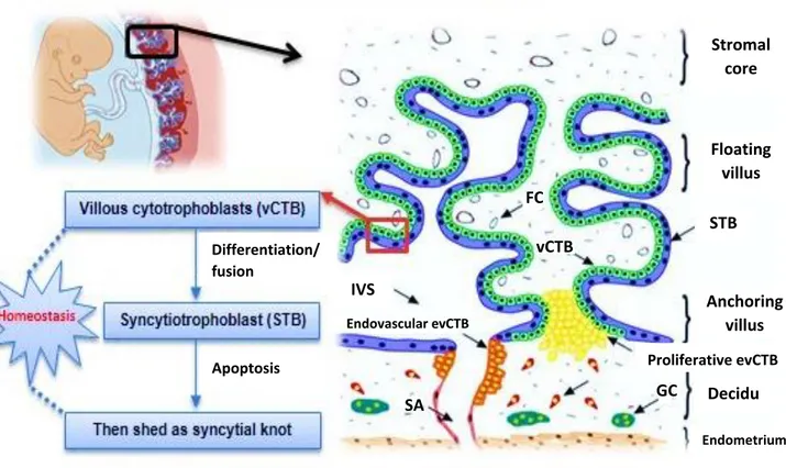
EVT play a significant role in embryo implantation, placentation and in uterine spiral arteries remodeling. The EVT proliferates and invades the decidua and the myometrium to form the interstitial cytotrophoblast. Afterwards, some either migrate into maternal vessels acquiring endovascular trait, or form the giant cells in the decidua
(figure 2) (Handschuh et al., 2009, Handschuh et al., 2007a, Ji et al., 2013).
Figure 1 : Illustration of human villous trophoblast differentiation. Villous cytotrophoblast
(mononucleated) aggregate and fuse into syncytiotrophoblast (multinucleated) in a highly organized process. SA: spiral artery; GC: giant cell; evCTB: extravillous cytotrophoblast; FC: fetal capillary STB: syncytiotrophoblast; vCTB: villous cytotrophoblast; IVS: intervillous space (Sagrillo-Fagundes et al., 2014). Stromal core Floating villus STB Anchoring villus Proliferative evCTB Decidu a Endometrium vCTB FC IVS Endovascular evCTB GC SA Apoptosis Differentiation/ fusion
5
Maternal-fetal circulation is established by the end of the 1st trimester and the remodeling process of maternal spiral arteries is accomplished by the end of 2nd trimester. Endovascular ETVs are responsible for the invasion of the arteries, and substitute the endothelial cells. As a result, the small high resistance muscular arteries are converted into large low resistance fibrous vessels, which facilitate the entry of the blood into trophoblastic cavities (Cartwright et al., 2010, Ji et al., 2013, Kaufmann et al., 2003). The giant cells (GC) are suggested to function as the STB, secreting both hPL and hCG hormones, emphasizing their role in pregnancy maintenance (al-Lamki et al., 1999, Hu et al., 2010, Simmons et al., 2007).
1.3 Chorionic villi: villous trophoblast
After the adhesion of the blastocyst to the endometrium, the trophoblast begins to differentiate along two pathways: vCTB pathway and EVT pathway. During the 1st trimester of pregnancy, trophoblasts differentiate mainly towards the EVT path, allowing the invasion of the blastocyst into the uterus and the establishment of the utero-placental circulation. After the 1st trimester, vCTB pathway becomes the major one, a situation that demonstrates the adaptive and variable capacity of trophoblast differentiation during pregnancy (figure 2) (Graham et al., 1992a, Ji et al., 2013, Kaufmann et al., 2003).
6
Figure 2 : Pathways of human trophoblast differentiation
Human trophoblast differentiates through either the villous or extravillous pathways. In the villous pathway, vCTB (mononuclear) merges and further differentiates to form the STB (multinucleated) which constitutes the outer layer of villus cells. Alternatively, in the extravillous pathway, evCTB proliferates and becomes invasive. Thereafter, some migrate to maternal vessels, and become a vascular cytotrophoblast (vascular evCTB) while other evCTB differentiate into multinucleated giant cells migrating in the decidua. vCTB: villous cytotrophoblast, STB: syncytiotrophoblast, evCTB: extravillous cytotrophoblast (Vaillancourt
et al., 2009a).
1.3.1 Syncytialisation
During the course of pregnancy vCTB differentiate and fuse to maintain STB hemostasis, and preserve its cellular constituents (proteins, nucleic acids, lipids, organelles, etc...) (Le Bellego et al., 2009). STB undergoes apoptosis (both intrinsic and extrinsic) then expel apoptotic yields to the maternal circulation (figure 3). STB renewal process is complex and comprises a large number of factors such as hormones,
7
cytokines, growth factors (Ackerman et al., 2012) or transcription factors (Heazell et al., 2009, Huppertz et al., 2011).
Figure 3 : Villous trophoblast turnover (homeostasis). Villous trophoblast is formed by proliferating
stem cytotrophoblast that exit the cell cycle and differentiate in villous cytotrophoblast. Mononuclear villous cytotrophoblasts fuse and differentiate into a syncytium, the syncytiotrophoblast. Syncytiotrophoblast degeneration occurs through apoptosis to allow its regeneration and thus maintenance of villous trophoblast homeostasis (Lanoix et al., 2012b).
The first evidence of trophoblast in vivo fusion into a syncytium was revealed upon using [3H]-thymidine incorporation assays (Richart, 1961). Further studies on villous trophoblast differentiation were realized in vitro by isolating and purifying vCTB from human placenta, followed by harvesting them in primary culture (Kliman et al., 1986). This technique employs Trypsin and DNase for performing enzymatic digestion then discontinuous Percoll gradient for VTB purification. Afterwards, freshly isolated
8
vCTB demonstrated a successful fusion and differentiation into functional STB (Benirschke et al., 2000, Kliman et al., 1986).
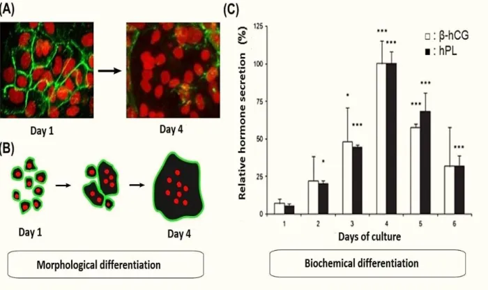
The differentiation process is characterized by morphological and biochemical parameters. Morphological differentiation is defined through the fusion of the mononucleated vCTB into a syncytium, while the biochemical differentiation is characterized by the production of specific hormones such as hCG, placental lactogen (hPL), placental growth hormone (PGH), neuropeptide Y and leptin. The hCG and hPL are the two hormones frequently examined for vCTB biochemical differentiation studies (Jacquemin et al., 1996, Kliman et al., 1986, Morrish et al., 1987, Senaris et al., 1997).
Complete morphological and biochemical differentiation of vCTB into STB - in culture- necessitates the presence of fetal serum (Bischof et al., 2005, Morrish et al., 1987). This process takes place after 48 hours of culture while both survival drop and increased apoptosis are observed after 96 hours of culture (figure 4). Although the two apoptotic pathways (intrinsic and extrinsic) are involved in this process, the intrinsic pathway is predominantly found and is particularly up regulated during variable oxygen concentrations as well as hypoxia and oxidative stress in the placenta (Crocker et al., 2001a, Heazell et al., 2008, Hung et al., 2010, Kar et al., 2007, Lanoix et al., 2013, Longtine et al., 2012).
9
Figure 4 : Biochemical and morphological differentiation of villous trophoblast.
(A) Morphological differentiation of primary cultures of villous trophoblast isolated from human placenta at term. Marking desmoplakin membrane and nuclei by confocal microscopy after one and four days of culture (400X). (B) Schematic of morphological differentiation in vitro. (C) Biochemical differentiation of primary cultures of villous trophoblast from human term placenta. Relative secretion of β-subunit of human chorionic gonadotropin (β-hCG) and placental lactogen (hPL) of a 6 day culture. The data represent the relative secretion ± SEM compared to the secretion at four days of culture (maximum secretion). * P <0.05; *** P <0.001 compared to the first day of culture (Vaillancourt et al., 2009a).
Various obstetric complications, such as miscarriage, intra-uterine growth restriction or preterm delivery could be a result of altered villous trophoblast homeostasis (Evain-Brion, 2001, Jauniaux et al., 2010, John et al., 2012, Norwitz, 2006). For example, a lack of vCTB differentiation is involved in preeclampsia. This condition, affects between 2 to 7% of pregnant women and is characterized by maternal hypertension and proteinuria (Noris et al., 2005). Although the pathophysiology of this complex disease is not yet fully understood, many factors involved in trophoblast
10
differentiation appear to be involved such as: vascular endothelial growth factor (VEGF), β-hCG, and caspase 8 (Ackerman et al., 2012, Ji et al., 2013).
The human placenta, as the other higher primates, is of hemomonochorial type. Since no type of - non primate- animal placenta is comparable to humans and none has the same type of trophoblast cells as the human placenta. Hence, animal models are not suitable for investigating human vCTB differentiation, thus emphasizing that primary cultures of vCTB isolated from human placentas as a model of choice to decrypt villous trophoblast differentiation (Benirschke et al., 2000, Kliman et al., 1986).
1.3.2 Factors regulating the syncytialization of villous trophoblast
Although the mechanisms involved in vCTB syncytialization are complex, some major mechanisms and factors that stimulate or inhibit the fusion process and biochemical differentiation were identified (e.g., growth factors, nucleic acid, kinases, hormones or retroviral particles) (summarized in Table 1) (Evain-Brion, 2001).11
Factors inhibiting the biochemical differentiation (hCG secretion) and fusion are expressed with (-); factors inducing the biochemical differentiation (hCG secretion) and fusion are expressed with (+) FT: First trimester; TT: Third trimester; B: BeWo cells. EGF: Epidermal growth factor, VEGF: vascular endothelial growth factor, CSF: colony stimulating factor, TGF-α: transforming growth factor alpha, ERK1/2: Extracellular signal regulated kinases, PKA: Protein kinase A, p38 mitogen-activated protein kinases, TNF: Tumor necrosis factor alpha. Adapted from (Gauster et al., 2009)
Factor Description (-) (+) Cell type Action Reference
hCG Hormone + TT Biochemical (Shi et al., 1993)
EGF Growth factor + TT Biochemical (Morrish et al., 1987)
VEGF Growth factor + FT, TT Biochemical (Crocker et al.,
2001b)
CSF Growth factor + TT Biochemical (Garcia-Lloret et al., 1994)
TGF-α Growth factor + TT Biochemical (Yang et al., 2003)
ERK1/2 Protein kinase + TT Biochemical
and fusion
(Vaillancourt et al., 2009a)
PKA Protein kinase +
B TT Fusion Biochemical and fusion (Keryer et al., 1998, Knerr et al., 2005, Knofler et al., 1999)
p38 Protein kinase + TT Biochemical
and fusion
(Vaillancourt et al., 2009a)
Syncytin 1 Membrane
protein + TT, B Fusion (Frendo et al., 2003)
Connexin 43 Membrane
protein + TT Fusion (Frendo et al., 2003)
Galectin 3 Membrane
protein + B Fusion (Dalton et al., 2007)
CD98 Transporter of
amino acid + B Fusion (Kudo et al., 2003)
Galectin 3 Membrane
protein + B Fusion (Dalton et al., 2007)
Calcium + TT, B Fusion (Alsat et al., 1996)
TNF Cytokine - TT Biochemical (Leisser et al., 2006)
Hypoxia Low oxygen tension - TT, B Biochemical
and fusion (Alsat et al., 1996)
12
1.4 Human chorionic gonadotropin (hCG) role and regulation of
placentation
1.4.1 hCG structure
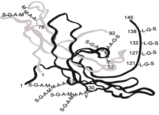
The hCG belongs to a family of glycoprotein hormones, which include luteinizing hormone (LH), thyrotropin (TSH) and follitropin (FSH). HCG consists of α-subunit and a β-subunit which are held together by non-covalent hydrophobic and ionic interactions
(Figure 5). Secretion of hCG is constant all over the course of pregnancy, while it peaks
at the 10th week of gestation. Conferring to sugar side chains, hCG can be present as a regular (hCG) or hyper glycosylated hCG (hCG-H) sharing a common hCG β-subunit amino acid sequence. In early pregnancy, hCG-H is the predominant form of hCG, and is secreted by the evCTB (Cole et al., 2006, Sasaki et al., 2008). hCG-H is a sugar variant of hCG and stimulates cytotrophoblasts invasion and implantation in an autocrine manner. Afterwards the regular hCG becomes the predominant form and is secreted by the differentiated and fused STB (Cole, 2010, Wide et al., 1994).
Measurements of hCG is an indicator commonly used for pregnancy diagnosis. Recently, it has been shown that hCG-H levels quantification permits earlier pregnancy recognition than standard pregnancy tests, as its levels surge quickly during the early stages of pregnancy. Interestingly, ultrasensitive hCG-H assay permits pregnancy detection within 6 days in women undertaking in vitro fertilization (IVF) (Strom et al., 2012). In contrast, inadequate levels of urinary hCG-H (less than 50% of total hCG) have been identified in a large proportion of early pregnancy losses in spontaneously conceived cycles (Sasaki et al., 2008). Moreover, insufficient levels of hCG-H during the late stages of pregnancy are linked to preeclampsia development, suggesting that preeclampsia is associated with abnormal placentation and trophoblast invasion
13
(Bahado-Singh et al., 2002, Uzan et al., 2011). Symptomatic patients of ectopic pregnancy experience low circulating levels of another form of hCG, the disassociated β-subunit (Borrelli et al., 2003). Conversely, down syndrome pregnancy are characterized with elevated hCG-H or hCG free β-subunit levels in maternal serum throughout the 1st trimester (Palomaki et al., 2007).
Blood levels of hCG are elevated in patients with certain types of tumors. The β- subunit is highly secreted in case of choriocarcinoma as well as testicular and ovarian cancers. It’s also higher in patients with germ cell tumors and hydatidiform mole formation. Accordingly, hCG blood levels can be used as a marker for these tumors, as a surveillance tool to monitor the efficiency of the treatment, and for post-treatment monitoring to avoid tumor relapse (Goyal et al., 2014, Lempiainen et al., 2012, Lempiainen et al., 2014, van Cromvoirt et al., 2014).
Figure 5 : The 3-D structure of hCG. L: N-acetylgalactosamine, A : N-acetylglucosamine, S: sialic acid
14
1.4.2 hCG function
hCG plays a crucial role in pregnancy maintenance by preventing the regression of the corpus luteum. Accordingly, hCG sustains progesterone production by the corpus luteum during the early gestation. However, hCG levels drop during the late part of the first trimester and at that time the production of progesterone takes over by the placenta (Robert Hardin Melmed Shlomo Williams, 2011). hCG participates in the vital early pregnancy events such as the implantation and placentation. Its crucial tasks to pregnancy success is via enhancing the receptivity of the endometrium by promoting maternal immunotolerance of the fetus, remodulation of endometrial spiral artery, boosting natural killer population in the uterus and fostering the formation of new blood vessels (Cole, 2010, Licht et al., 2007, Perrier d'Hauterive et al., 2007).
hCG promotes the formation of new blood vessels (angiogenesis), thus increases the blood flow to the placenta and supports nutrient transfer to the fetus (Berndt et al., 2009, Zygmunt et al., 2002). It stimulates the growth and development of the umbilical cord and corresponding uterine growth to facilitate pregnancy progression and fetal development. It also maintains myometrial relaxation (Cole, 2010).
Stimulation of endometrial receptors by hCG result in various downstream molecules activation. For example, several growth factors are triggered such as transforming growth factor beta (TGF-β) and VEGF, which promote angiogenesis (Berndt et al., 2009). Proteinases such as matrix metalloproteinase 9 (MMP-9) is stimulated and is involved in the regulation of tissues remodeling (Riley et al., 2004). Cytokines as leukemia inhibitory factor (LIF) are also activated. However, the exact role of hCG in endometrial functions that is linked to implantation still remains a subject of
15
investigation (Gridelet et al., 2013, Licht et al., 2007, Perrier d'Hauterive et al., 2007). It has been demonstrated that hCG/LH receptors are expressed in several fetal organs, but they are absent in the adult ones. Liver, kidneys, lung and spleen are among those fetal organs (Abdallah et al., 2004, Goldsmith et al., 1983, Rao et al., 2007), this suggests a pivotal role of the hCG in enhancing their differentiation and development. The concentrations of hCG in the fetus are much lower than the levels of maternal circulation. Therefore, placental hCG is suggested to be leaked directly into maternal circulation rather than entering the circulation of the fetus (McGregor et al., 1981).
1.4.3 Luteinizing hormone (LH)/hCG receptor
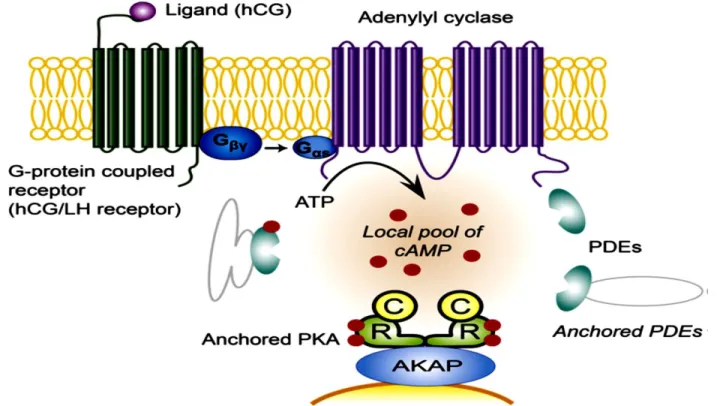
LH/hCG receptor is a member of G-protein coupled receptors family and is expressed in the testis, ovary and the placenta .The main pathway triggered upon its stimulation is cAMP/protein kinase A (PKA) pathway. hCG binding to its receptor activates adenylyl cyclase (AC), phospholipase C (PLC) and K+ channels, which in turn control cellular cAMP, inositol triphosphates (IP3), Ca2+ and other secondary messengers. This process is spatiotemporally controlled (in space and time) by the compensating actions of ACs and phosphodiesterases (PDEs) inside the cell (figure 6) (Conti et al., 1999, Houslay et al., 2003, Lybaert et al., 2013, J. L. Williams et al., 2008).
Under the control of A-kinase-anchoring proteins (AKAPs), PDEs participate in the generation of cAMP localized pool (Dodge et al., 2001, Tasken et al., 2001) and both are present in the placenta. The AC is localized in the cytotrophoblast and syncytiotrophoblast while PDE in the syncytiotrophoblast (Bernatchez et al., 2003, Matsubara et al., 1987, Sato et al., 1971, Stacey et al., 1998). In trophoblasts, the increase in cAMP levels triggers new signal mediators expression. Consequently, this
16
results in an augmented secretion of hCG, hPL, PGH, gonadotropin-releasing hormone (GnRH), syncytin and aromatase initiating estrogen creation (Y. Chen et al., 2013, Depoix et al., 2011, Strauss et al., 1992).Activation of cAMP/PKA signaling pathway stimulates both ERK1/2 and p38 MAPK pathways. Interestingly, this signaling pathway appears to stimulate β-hCG secretion during the syncytialization process in both BeWo and trophoblastic cells (Daoud et al., 2005, Weedon-Fekjaer et al., 2012).
Figure 6 : hCG/LH receptor (G-protein coupled receptor) once stimulated by hCG results in AC activation and cAMP pool increase. Through the compensating actions of ACs and PDEs, AKAPs
organize PKA anchoring in the area of cAMP pools, allowing for distinct control of its stimulation in space and time. AKAPs: A-kinase-anchoring proteins, AC: adenylyl cyclase, PKA: protein kinase A, PDEs: phosphodiesterases (Weedon-Fekjaer et al., 2012).
17
Melatonin
In the early days, melatonin was exclusively considered as the chief product of the pineal gland, that regulates the circadian and circannual cycle. However, further studies revealed that melatonin is also produced and present in various tissues and cells in the human body such as skin, retina, gastrointestinal tract (GIT), ovary, amniotic fluid and placenta (Bubenik, 2002, Lanoix et al., 2008b, A. Slominski et al., 2008, Tosini
et al., 1998, Zawilska et al., 2009). Melatonin is known to protect cell, and its role in
sustaining cell viability belongs to its receptor dependent and independent activities. Melatonin is a highly distributable molecule as it has both hydrophilic and lipophilic characteristics. This could explain the presence of melatonin in high levels at all subcellular components including the nucleus, the cytoplasm and the mitochondria. Melatonin can readily cross all morphological barriers including the placental and blood brain barrier (da Silva et al., 2011, Reiter et al., 2013a, R. M. Slominski et al., 2012, Venegas et al., 2012). Together, this highlights the significance of melatonin physicochemical properties in performing its protective action.
Melatonin is a potent antioxidant with higher activity compared to other antioxidants such as vitamin C and vitamin D (Milczarek et al., 2010). When melatonin interacts with free radicals for their neutralization, a number of melatonin metabolites are produced such as N1-acetyl-5-methoxykynuramine (AMK). Interestingly, melatonin metabolites also have antioxidant properties and some of them maybe more active than melatonin itself (Galano et al., 2013, Reiter et al., 2013a). Melatonin has also an antioxidant action through receptor dependent activity. Specifically, the activation of
18
melatonin receptors MT1 and MT2 induce antioxidant enzymes expression and activity (Lanoix et al., 2012b, Reiter et al., 2014).
Melatonin has also anti-inflammatory properties as it modulates and lowers the expression and activity of the inducible form of nitric oxide synthase (iNOS) and cyclooxygenase (COX-2), respectively (Carrillo-Vico et al., 2005a, Carrillo-Vico et al., 2005b). Inflammatory conditions with increased tumor necrosis factor (TNF) and nuclear factor kappa B (NFĸB) levels reduce N-acetyl transferase arylalkylamine (AANAT) transcription, which is the rate limiting enzyme in melatonin synthesis (Cecon et al., 2011, Fernandes et al., 2006, Markus et al., 2013). Alternatively, the anti-inflammatory properties of melatonin could inhibit NFĸB activation (Negi et al., 2011).
2.1 Melatonin synthesis
Synthesis of melatonin is carried out from the essential amino acid L-tryptophan. First, L-tryptophan is transformed into serotonin, then converted to N-acetyl-5-methoxytryptamine (melatonin) by the consecutive action of the two specific enzymes AANAT followed by hydroxyindole-O-methyltransferase (HIOMT or ASMT, acetylserotonine-O-methytransferase) that methylate the product of AANAT (figure 7) (Maronde et al., 2011, A. Slominski et al., 2008, Stehle et al., 2011, Tan et al., 2013, Wurtman et al., 1968). Melatonin synthesizing enzymes are expressed in both the placenta and placental cell line as BeWo and JEG-3 (Lanoix et al., 2008b).
19
Figure 7 : Melatonin synthesis from L-tryptophan. L-tryprophan is converted into serotonin via the
action of the two enzymes tryptophan 5-hydroxylase and aromatic L-amino acid decarboxylase, respectively. Afterwards, serotonin is transformed to melatonin via the consecutive action of arylalkylamine N-acetyltransferase (AANAT) and hydroxyindole-O-methyltransferase (HIOMT or ASMT) (Konturek et al., 2006).
2.2 Melatonin receptors
Melatonin has been identified to bind the two membrane receptors MT1 (Mel1a) and MT2 (Mel1b) and the nuclear receptor family RZR / ROR (retinoid orphan receptor and retinoid Z receptor) (Smirnov, 2001). Despite the affinity of ROR/RZR receptors for melatonin, there is still a controversy as to whether melatonin is their native ligand (Jetten, 2009, Luchetti et al., 2010, Smirnov, 2001).
20
Melatonin MT1 and MT2 receptors are widely distributed and have been found in most human tissues. Melatonin via the activation of these receptors triggers different biological functions in various tissues (Table 2). Melatonin effects through the stimulation of its receptors could elicit the following actions: reducing the pressure of the cardiovascular system, stimulation of the antioxidant enzymes expression and activity, and the regulation of daily night-day cycle (Reiter, 1993, Rodriguez et al., 2004, Sewerynek, 2002, Tomas-Zapico et al., 2005).
Our group has demonstrated that melatonin receptors MT1, MT2 and RORα (retinoid orphan receptor alpha) are expressed in the villous trophoblast (vCTB and STB) as well as in JEG3 and BeWo cells (Lanoix et al., 2008b).
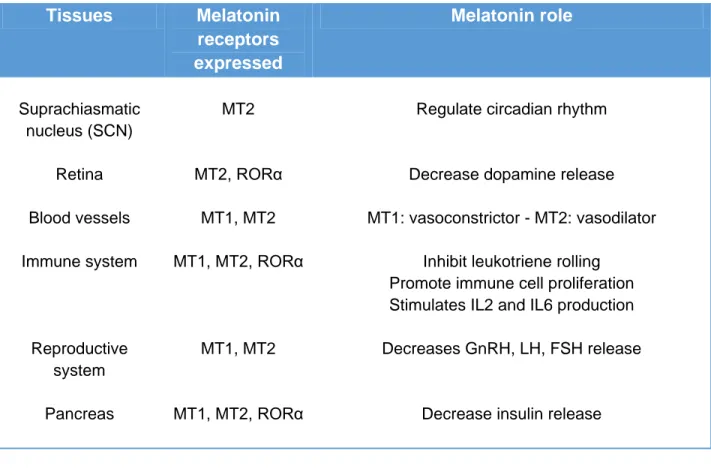
Table 2 : Melatonin receptors role and distribution in human tissues
Table from (R. M. Slominski et al., 2012)
Tissues Melatonin receptors expressed Melatonin role Suprachiasmatic nucleus (SCN) Retina Blood vessels Immune system Reproductive system Pancreas MT2 MT2, RORα MT1, MT2 MT1, MT2, RORα MT1, MT2 MT1, MT2, RORα
Regulate circadian rhythm
Decrease dopamine release MT1: vasoconstrictor - MT2: vasodilator
Inhibit leukotriene rolling
Promote immune cell proliferation Stimulates IL2 and IL6 production
Decreases GnRH, LH, FSH release
21 Skin Gastrointestinal tract Bone Kidneys Placenta Uterus Endometrium MT1, MT2, RORα MT1, MT2 MT1, RORα MT1, MT2 MT1, MT2, RORα MT1, MT2 MT1, MT2
Regulate hair growth, and functions of epidermis Decrease gastric contraction,
peristalsis, and serotonin’s actions Increases bicarbonate, amylase, and CCK release
Increases osteoblastic activity and decrease osteoclastic activity
Protects from inflammation, regulate glomerular filtration
ROS scavenger, decrease apoptosis Myometrial contractility
Trophoblast invasion in early pregnancy
2.3. Melatonin MT1 and MT2 receptors
Melatonin MT1 and MT2 receptors are part of the GPCR family with seven trans-membrane (TM1-TM7) α-helices, 4 intracellular (I1-I4) and 4 extracellular (E1-E4) domains (figure 8) (Dubocovich et al., 2005, R. M. Slominski et al., 2012). MT1 receptor is a 350 amino acid protein while MT2 receptor is composed of 362 amino acids. MT1 and MT2 receptors are structurally similar with 60% homology in their amino acid sequence. Binding sites of both MT1 and MT2 receptors are located in the TM5 immediate vicinity and in the extracellular half of the trans-membrane domain. It has been observed that MT1 receptor possesses a relatively smaller binding site dimension compared to MT2 receptor. This is the principal structural difference that is anticipated to affect both binding selectivity and affinity. Residues at the C-terminal region of TM7 domain is recognized to be a G-protein binding site and also is involved in the interaction with other intracellular proteins. Cysteine residue, for example, appears to
22
mediate the inhibitory effect on AC. C-terminal cytoplasmic domains are also suggested to contain phosphorylation sites for PKA and PKC (Al-Ghoul et al., 1998, Luchetti et al., 2010).
Figure 8 : Illustration of G-Protein coupled receptors (GPCRs). MT1 and MT2 melatonin receptors
consist of seven hydrophobic trans-membrane domains of α-helical structure, joined by four intracellular and four extracellular loops (Luchetti et al., 2010).
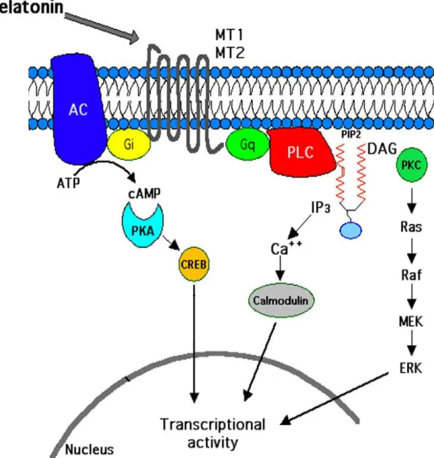
Activation of MT1 and MT2 is involved in the regulation of different signaling pathways depending on the tissues and cells (e.g. cAMP/PKA, PLC, PLA2, ERK 1/2 and K+ /Ca2+ channels) (figure 9) (Dubocovich et al., 2005, Dubocovich et al., 2003, Pandi-Perumal et al., 2008). There are several commercially available compounds that act as either melatonin MT1/MT2 receptors agonists or antagonists (Table 3).
23
Figure 9 : Illustration of signaling pathways triggered by melatonin MT1/MT2 receptor. Stimulation
of melatonin MT1 and MT2 receptors that activates pertussis toxin-insensitive Gq, triggers PLC activation. PLC hydrolyses PIP2 into DAG and IP3. DAG activates PKC, stimulating the Ras/Raf/MEK/ERK signaling pathway while IP3 stimulates Ca2+ signalingby calmodulin kinases (CaMK). Activation of pertussis toxin-sensitive Gi as a result of melatonin MT1 and MT2 receptors stimulation inhibits adenylyl cyclase (AC) activity. This inhibits cAMP/PKA pathway and lowers cAMP levels. PLC: phospholipase, PIP2: phosphatidylinositol 4, 5-bisphosphate, DAG: diacylglycerol, IP3: inositol 1, 4, 5- triphosphate, PKC: protein kinases type C, ERK1/2: Extracellular signal regulated kinases (R. M. Slominski et al., 2012).
24
Table 3 : Melatonin MT1 and MT2 receptors agonists and antagonists
Drug Action Reference
Melatonin receptors agonists
TIK-301
Is a melatonergic agonist developed by Eli Lilly Co. (Indianapolis, IN, USA), It exhibits a high affinity towards MT1 and MT2 receptors. Its affinity for MT1 is similar to that of melatonin (pKi=10.09), but a little higher for MT2 (pKi=10.38).
(Hardeland, 2010, Rivara et al., 2008)
Agomelatine It is the naphtalenic analogue of MLT and is a potent MT1 (pKi=10.21) and MT2 (pKi=9.57) agonist. That is developed by Servier Laboratories, France
(de Bodinat et al., 2010)
Ramelteon
In vitro binding studies revealed that ramelteon’s affinity for MT1 (Ki=0.014 nM) and MT2 (Ki=0.112 nM) receptors is 3 to 16 times greater than for MLT. It is developed by Takeda Pharmaceutical Co, Japan
(Kato et al., 2005)
Tasimelteon
Currently is at phase III clinical trial by Vanda Pharmaceuticals Inc (Washington, DC, USA). It has a slightly lower affinity to MT1 (pKi=9.45) and a moderately higher affinity to MT2 (pKi=9.80) than MLT.
(Hardeland, 2009, Ohta et al., 2013, Rajaratnam et al.,
2009) 6-chloromelatonin
A potent agonist for melatonin MT1 (pKi= 9.10) and MT2 (pKi= 9.77) receptors
(Browning et al., 2000)
Melatonin receptors antagonists
Luzindole A competitive melatonin receptor antagonist, it has 11 to 25 fold higher affinity for the MT2 over the MT1 receptor
(Requintina et al., 2007)
4-P-PDOT A melatonin receptor antagonist with more than 300 fold selectivity for the MT2 receptor compared to the MT1 receptor.
(Nonno et al., 1999, Requintina et al.,
25 S22153
A specific antagonist of MT1 and MT2 melatonin receptors with high affinity MT1 (pKi= 8.7) and MT2 (pK i= 8.4).
(Li et al., 2004, Weibel
et al., 1999)
MLT: melatonin
2.4 Melatonin and pregnancy
Maternal melatonin is able to reach the fetus and modulate its development. Melatonin is also produced by the reproductive system in the ovary, the oocyte and the placenta (Lanoix et al., 2008b, Reiter et al., 2014, Sakaguchi et al., 2013, Tamura et al., 2009).
Alterations in maternal melatonin levels have an impact on its levels in the fetal circulation and may affect gene expression in the fetal central nervous system (Novakova et al., 2010, Okatani et al., 1999, Reiter et al., 2014). In rats, melatonin has been shown to protect the brain against oxidative stress, lipid peroxidation and DNA damage caused by ischemia and reperfusion during the embryonic period (Wakatsuki et
al., 1999). In pregnant rats, pinealectomy triggered maternal circadian cycle
suppression where behavioral alterations in newborns have been observed, which was restored after its induction with melatonin (Bellavia et al., 2006).
Studies suggest that births are more likely to occur during night-time under the influence of melatonin. The explanation of this phenomenon may be due to the interaction between melatonin and oxytocin, a hormone that modulates the uterine contractility. Both hormones activate the same signaling pathways in myometrial cells,
26
PLC and PKC pathways (figure 11) (Sharkey et al., 2010, Sharkey et al., 2009). The importance of this interaction is supported by studies that show that the oxytocin-induced contractility of myometrial cells is increased in vitro after being co-treated with melatonin. This phenomenon is not observed if the cells were treated only with oxytocin, suggesting that the melatonin sensitizes myometrial cells to the pro-contractile signaling of oxytocin (Reiter et al., 2014, Sharkey et al., 2010, Sharkey et al., 2009).
27
Figure 10: Suggested model of synergetic action of melatonin and oxytocin on night-time myometrial contractility at parturition time. Melatonin circadian rhythm and oxytocin level is described
28
through the last trimester of pregnancy, at parturition time and after delivery of human females. Melatonin and oxytocin stimulate respectively MT2 receptor (MT2R) and oxytocin (OTR) receptor to synergistically activate phospholipase (PLC)-β, which hydrolyses phosphatidylinositol 4, 5-bisphosphate (PIP2) into diacylglycerol (DAG) and inositol 1, 4, 5- triphosphate (IP3). DAG activates protein kinases C (PKC)-α, activating the Ras/Raf/MAPK signaling pathway and leading to caldesmon (hCaD) phosphorylation. This increases actin availability for myosin binding and contractility induction. PLCβ stimulates both c-Jun and c-Fos, resulting in enhanced gap junctional communication via connexin 43 activation. Finally, IP3 stimulates Ca2+ release that activates myosin light-chain (MLC) and enhances myometrial contractility (Sagrillo-Fagundes et al., 2014).
2.5 Melatonin and placenta
During the course of pregnancy, maternal circulating melatonin levels progressively increase with a maximum peak at the time of delivery. Pregnant women therefore have significantly higher plasma melatonin concentrations (day and night) compared to non-pregnant women. The placenta is believed to be the source of this increase as melatonin levels return to normal after the delivery of the placenta (Y. Nakamura et al., 2001, Tamura et al., 2008b, Vatish et al., 2010).
It has been demonstrated that the mRNA of melatonin synthesizing enzymes (AANAT and HIOMT) is expressed in placental tissue from first trimester of pregnancy (Iwasaki et al., 2005). Afterwards, our group confirmed the presence of melatonin synthesizing enzymes, and melatonin receptors (MT1, MT2 and RORα) mRNA and protein in normal human term placenta and in human placental choriocarcinoma cell lines. These data suggest that placental melatonin could be the source of the increased circulating melatonin levels observed in pregnant women (Lanoix et al., 2008b).
Our group has shown that preeclampsia is associated with reduced activity and expression of AANAT, as well as the concentration of melatonin and its receptors (MT1
29
and MT2) in the placental tissue (Lanoix et al., 2012a, Lanoix et al., 2012b, Lanoix et
al., 2013). We have also previously shown that melatonin protects trophoblast cells from
apoptosis and oxidative stress-induced by hypoxia-reoxygenation, in vitro (figure 11) (Lanoix et al., 2013).
Figure 11: Potential role of melatonin in healthy and complicated pregnancies. Plasmatic melatonin,
released in a circadian manner, reaches the intervillous space and accesses fetal capillaries (FC) through syncytiotrophoblast (STB) and villous cytotrophoblast (vCTB). In parallel, both STB and vCTB produce melatonin locally that can also reach fetal capillaries and maternal circulation. Both plasmatic and placental melatonin are related with maternal and fetal protective effects observed during pregnancy (Sagrillo-Fagundes et al., 2014).
In summary, the presence of melatonin, its receptors and its synthesizing enzymes in the placental tissue implicate melatonin in maintaining the placenta during
30
pregnancy (Lanoix et al., 2008b). The production of placental melatonin during pregnancy abnormalities is compromised. Based on this theory, recently, several studies have linked certain pregnancy diseases to alterations in its synthesis, in its receptors or its concentrations as in case of preeclampsia and intra uterine growth restriction (IUGR). Consequently, melatonin treatment is suggested to recover the absent protective effects of placental melatonin in those cases (Hobson et al., 2013, Lanoix et al., 2012a, Lanoix et al., 2013, Y. Nakamura et al., 2001, Reiter et al., 2014).
2.6 Melatonin and hCG secretion
Activation of the PKA pathway induces both BeWo and trophoblast cell fusion and hCG secretion (Keryer et al., 1998, Knerr et al., 2005, Knofler et al., 1999). Additionally, forskolin (inducer of AC), induces ERK1/2 and p38-MAPK phosphorylation in BeWo cells, and maximal stimulation is within 5 -10 minutes after forskolin treatment (Delidaki et al., 2011). Our team has demonstrated that 6-chloromelatonin (a melatonin agonist) inhibited forskolin stimulated β-hCG secretion in a dose dependent manner (maximum effect at 10µM MLT), while it has no effect on basal β-hCG secretion in BeWo and JEG-3 cells (Lanoix et al., 2006). Furthermore, BeWo and JEG3 pretreatment with pertussis toxin (PTX; inhibits the activation of Gi/o proteins) reversed the 6-chloromelatonin inhibitory effect on forskolin-stimulated β-hCG secretion. This reveals that 6-chloromelatonin inhibitory effect is mediated, in part, by a PTX-sensitive G protein, Gi/o. Indeed, activation of melatonin MT1 and MT2 receptors through Gi/o
pathway inhibits AC pathway and cAMP accumulation in various mammalian tissues, as reviewed in (Dubocovich et al., 2005, Witt-Enderby et al., 2003). On the other hand, the stimulatory effect of 6-chloromelatonin on basal β-hCG secretion in PTX-pretreated cells
31
could be due to Gq/11 signaling pathway activation. The Gq/11 pathway results in Ca2+
mobilization from intracellular stores and activation of PKC through PLC pathway (Feinman et al., 1986, Lanoix et al., 2006). The specific signaling pathways involved in melatonin effects on β-hCG production in normal trophoblasts as well as in choriocarcinoma cells remains to be explored.
32
CHAPTER 2: HYPOTHESIS AND OBJECTIVES
Our main research hypothesis is that melatonin and its placental receptors, play an important role in the regulation of villous trophoblast development and endocrine function. The objectives for the present project are to determine the role of melatonin and its placental MT1/MT2 receptors on villous trophoblast syncytialization and the mechanisms by which they regulate β-hCG secretion. The two specific objectives and hypotheses are:
Objective 1: Determine the effect of melatonin and its MT1 and MT2 receptors on
villous trophoblast syncytialization (biochemical (β-hCG secretion) and fusion). It has been demonstrated that melatonin has the ability to stimulate cellular differentiation of many cell types. Melatonin can stimulate the final stage of chick retinal cell differentiation, that is crucial for the development of several other cell types (Sampaio Lde et al., 2010) and can decrease the apoptosis process in numerous cell types as CD4+ T cells (Pedrosa et al., 2010) and spermatozoa (Espino et al., 2010). In neural cells, melatonin stimulates the differentiation of pluripotent P19 cells (Oct4+ Sosx2+) into neural stem cells (Oct4- Sox2+) through MT1 receptor and ERK1/2 pathway activation (X. Chen et al., 2014). Moreover, melatonin can stimulate human Saos2 osteoblast-like cell differentiation - a model of osteoblast - and consequently stimulates bone formation, through an action mediated by MT2 receptor and ERK1/2 signaling pathway activation (Matsumura et al., 2014). Likewise, via receptor dependent and independent activities, melatonin triggers mesenchymal stem cells (MSCs) differentiation and survival (Luchetti
et al., 2014). Hypothesis 1: We propose that the differentiation of vCTB into STB is
33
Objective 2: Determine the mechanisms by which melatonin and its MT1/MT2
receptors regulate β-hCG secretion. The MAPK signaling pathway plays a vital role in villous trophoblast morphological and biochemical differentiation. Dysregulation of this pathway could lead to the formation of aberrant STB and consequently lead to obstetric complications. Melatonin stimulates differentiation in various cell types via activating its MT1 and MT2 receptors with subsequent ERK1/2 signaling pathway stimulation. In addition, we observed earlier that melatonin stimulates β-hCG secretion in our in vitro models of human trophoblast; BeWo and JEG-3 cell lines (Lanoix et al., 2006).
Hypothesis 2: We propose that melatonin in a receptor-dependent manner stimulates
34
CHAPTER 3
RESULTS
35
Contribution of the student
The student analyzed all the data, designed and conducted the protein expression of AANAT enzyme throughout pregnancy and wrote the manuscript.
3.1: Placental melatonin system is present throughout
pregnancy and regulates villous trophoblast differentiation
Ahmed SOLIMAN, Andrée-Anne LACASSE, Dave LANOIX, Lucas SAGRILLO-FAGUNDES, Véronique BOULARD, Cathy VAILLANCOURT
36
De: onbehalfof+reiter+uthscsa.edu@manuscriptcentral.com Envoyé: 5 décembre 2014 10:47
À: Vaillancourt, Cathy
Objet: Journal of Pineal Research - Decision on Manuscript ID JPI-OM-11-14-0225 05-Dec-2014
Dear Prof. Vaillancourt:
Manuscript ID JPI-OM-11-14-0225 entitled "Placental melatonin system is present throughout pregnancy and regulates villous trophoblast differentiation" which you submitted to the Journal of Pineal Research, has been reviewed. The comments of the reviewer(s) are included at the bottom of this letter. The reviewer(s) have recommended publication, but also suggest some revisions to your manuscript. Therefore, I invite you to respond to the reviewer(s)' comments and revise your manuscript.
PLEASE NOTE: DO NOT RE-SUBMIT YOUR PAPER UNTIL YOU HAVE RECEIVED THE EDITED VERSION (VIA MAIL) FROM THE EDITOR AND YOU HAVE MADE THE
APPROPRIATE EDITORIAL CHANGES.
To revise your manuscript, log into https://mc.manuscriptcentral.com/jpi and enter your Author Center, where you will find your manuscript title listed under "Manuscripts with Decisions." Under "Actions," click on "Create a Revision." Your manuscript number has been appended to denote a revision.
You will be unable to make your revisions on the originally submitted version of the manuscript. Instead, revise your manuscript using a word processing program and save it on your computer. Please also highlight the changes to your manuscript within the document by using the track changes mode in MS Word or by using bold or colored text. Once the revised manuscript is prepared, you can upload it and submit it through your Author Center. When submitting your revised manuscript, you will be able to respond to the comments made by the reviewer(s) in the space provided. You can use this space to document any changes you make to the original manuscript. In order to expedite the processing of the revised manuscript, please be as specific as possible in your response to the reviewer(s).
IMPORTANT: Your original files are available to you when you upload your revised manuscript. Please delete any redundant files before completing the submission.
Because we are trying to facilitate timely publication of manuscripts submitted to the Journal of Pineal Research, your revised manuscript should be uploaded as soon as possible. If it is not possible for you to submit your revision in a reasonable amount of time, we may have to consider your paper as a new submission. Once again, thank you for submitting your manuscript to the Journal of Pineal Research and I look forward to receiving your revision. Sincerely,
Dr. Russel Reiter
Editor-in-Chief, Journal of Pineal Research
37
Placental melatonin system is present throughout pregnancy and regulates
villous trophoblast differentiation
Ahmed SOLIMAN, Andrée-Anne LACASSE, Dave LANOIX, Lucas SAGRILLO-FAGUNDES, Véronique BOULARD, Cathy VAILLANCOURT *
1INRS-Institut Armand-Frappier and BioMed research Center, Université du Québec, 531 boulevard des Prairies, Laval, QC, H7V 1B7, Canada.
Short title: Placental melatonin system during pregnancy
*
Corresponding author: Cathy Vaillancourt, Ph.D. INRS-Institut Armand-Frappier 531, boul. des Prairies, Building 18
Laval (Québec) H7V 1B7, CANADA T: (450) 687-5010 # 8812
Email: cathy.vaillancourt@iaf.inrs.ca
Keywords: AANAT / HIOMT (ASMT) / MT1 melatonin receptor / MT2 melatonin receptor /
38
ABSTRACT
Melatonin is highly produced in the placenta where it protects against molecular damage and cellular dysfunction arising from hypoxia/re-oxygenation induced oxidative stress as observed in primary cultures of syncytiotrophoblast. However, little is known about melatonin and its receptor in the human placenta throughout pregnancy and their role in villous trophoblast development. The purpose of this study is to determine melatonin synthesizing enzymes; arylalkylamine N-acetyltransferase (AANAT) and hydroxyindole O-methyltransferase (HIOMT), and melatonin receptor (MT1 and MT2) expression throughout pregnancy as well as the role of melatonin and its receptors in villous trophoblast syncytialisation. Our data show that the melatonin generating system is expressed throughout pregnancy (from week 7 to term) in placental tissues. AANAT and HIOMT show maximal expression at the 3rd trimester of pregnancy. MT1 receptor expression is maximal at the 1st trimester compared to the 2nd and 3rd trimesters, while MT2 receptor expression does not change significantly during pregnancy. Moreover, during primary villous cytotrophoblast syncytialisation, MT1 receptor expression increases while MT2 receptor expression decreases. Treatment of primary villous cytotrophoblast with an increasing concentration of melatonin (10 pM -1 mM) increases the fusion index (syncytium formation; 21% augmentation at 1 mM melatonin (vs. vehicle)) and β-hCG secretion (121% augmentation at 1 mM melatonin vs. vehicle). This effect of melatonin appears to be mediated via its MT1 and MT2 receptors. In sum, melatonin synthetic machinery (synthetizing-enzymes and receptors) is expressed in human placenta throughout pregnancy and promotes syncytium formation, suggesting an essential role of this indolamine in placental function and pregnancy well-being.
39
INTRODUCTION
Human placenta is crucial for the growth and the development of the fetus as well as the adaptation of maternal physiology to pregnancy (Le Bouteiller, 2001, Longtine et al., 2011). It mediates the exchange of nutrients and oxygen between the mother and the fetus, and eliminates waste products excreted by the fetus. The functional unit of the placenta is the chorionic villus that is formed by villous cytotrophoblast and is bathed in maternal blood (Aplin, 1991, Malassine, 2001, Rampersad et al., 2011). Throughout the syncytialisation process, mononucleated villous cytotrophoblast cells fuse and differentiate into the multinucleated non-proliferative syncytiotrophoblast. Differentiation of villous cytotrophoblast is described by morphological and biochemical features. Morphological differentiation is characterized by the fusion of villous cytotrophoblast to syncytiotrophoblast. Biochemical differentiation is defined by the activation of genes expressed specifically in the syncytiotrophoblast, coding for secreted hormones such as human chorionic gonadotropin (hCG) (Handschuh et al., 2007a, Jacquemin et
al., 1996, Kliman et al., 1986, Morrish et al., 1987, Mounier et al., 2009, Newbern et al., 2011).
To maintain homeostasis, syncytiotrophoblast goes through apoptosis to be constantly renewed by the underlying villous cytotrophoblast. Syncytialization of villous cytotrophoblast in vitro is completed after approximately four days of culture (Castellucci et al., 1990, Miller et al., 2005, Vaillancourt et al., 2009b). The disruption of syncytiotrophoblast homeostasis (via oxidative stress) contributes to various pregnancy complications such as intrauterine growth restriction and preeclampsia (Burton et al., 2011, Ishihara et al., 2002, Lanoix et al., 2012a).
Melatonin was initially thought to be only produced by the pinealocytes under circadian rhythm control. However, it has been demonstrated that the gastrointestinal tract, skin, reproductive organs and placenta synthesize it as well (Bubenik, 2002, Hardeland et al., 2006,