European Journal of Chemistry
ISSN 2153-2249 (Print) / ISSN 2153-2257 (Online) – Copyright © 2020 The Authors – Atlanta Publishing House LLC – Printed in the USA. This work is published and licensed by Atlanta Publishing House LLC – CC BY NC – Some Rights Reserved.
European Journal of Chemistry
View Journal Online
View Article Online
Synthesis, crystal structure, Hirshfeld surface and interaction energies analysis
of 5-methyl-1,3-bis(3-nitrobenzyl)pyrimidine-2,4(1H,3H)-dione
Koffi Senam Etsè
1,*, Laura Comeron Lamela
1, Guillermo Zaragoza
2and Bernard Pirotte
11 Laboratory of Medicinal Chemistry, Center for Interdisciplinary Research on Medicines (CIRM), University of Liège, Quartier Hôpital B36 Av. Hippocrate 15 B-4000 Liège, Belgium
[email protected] (K.S.E.), [email protected] (L.C.L.), [email protected] (B.P.)
2 Unidade de Difracción de Raios X, RIAIDT, Universidade de Santiago de Compostela, Campus VIDA, 15782 Santiago de Compostela, Spain
[email protected] (G.Z.)
* Corresponding author at: Laboratory of Medicinal Chemistry, Center for Interdisciplinary Research on Medicines (CIRM), University of Liège, Quartier Hôpital
B36 Av. Hippocrate 15 B-4000 Liège, Belgium. e-mail: [email protected] (K.S. Etsè).
10.5155/eurjchem.11.2.91-99.1973 Received: 19 February 2020
Received in revised form: 22 March 2020 Accepted: 24 March 2020
Published online: 30 June 2020 Printed: 30 June 2020
The title compound 5-methyl-1,3-bis(3-nitrobenzyl)pyrimidine-2,4(1H,3H)-dione was obtained by reaction of thymine with 3-nitrobenzylbromide in the presence of cesium carbonate. Characterization of the product was achieved by NMR spectroscopy and its stability was probed in basic condition using UV-Visible analysis. Furthermore, the molecular structure was confirmed by X-ray diffraction analysis. The compound crystallizes in orthorhombic Pna21 space group with unit cell parameters a = 14.9594 (15) Å, b = 25.711 (3) Å, c = 4.5004 (4) Å, V = 1731.0 (3) Å3 and Z = 4. The crystal packing of the title compound
is stabilized by intermolecular hydrogen bond, π···π and C−H···π stacking interactions. The intermolecular interactions were furthermore analyzed through the mapping of different Hirshfeld surfaces. The two-dimensional fingerprint revealed that the most important contributions to these surfaces come from O···H (37.1%), H···H (24%) and H···C/C···H (22.6%) interactions. The interaction energies stabilizing the crystal packing were calculated and were presented graphically as framework energy diagrams. Finally, the energy-framework analysis reveals that π···π and C−H···π interactions energies are mainly dispersive and are the most important forces in the crystal.
Thymine Crystal structure Hirshfeld surfaces Hydrogen bonding Interaction energies
3-Nitrobenzylbromide Cite this: Eur. J. Chem. 2020, 11(2), 91-99 Journal website: www.eurjchem.com
1. Introduction
Heterocyclic compounds containing the pyrimidine ring has wide occurrence in nature [1]. Together with adenine, guanine, cytosine, and uracil, thymine is one of the funda-mental nitrogenous bases observed in the genetic code of the living beings. As nucleobase, thymine is implicated in various important biological processes [2]. Numerous thymine derivatives are described as biologically active compounds. Some of them are reported as efficient to inhibit the highly invasive A431 tumor cell line [3], active against the majority of wild-type of human immunodeficiency HIV-1 mutant viruses [4], inhibitory potency on a mycobacterial enzyme thymidine mono-phosphate kinase (TMPKmt), on the growth of M. smegmatis and M. tuberculosis bacteria [5], susceptibility to phosphorylation by TK1 and TK2 and candidates for appli-cation as boron carriers for boron neutron capture therapy (BNCT) [6]. Among the various substituted thymine derivatives, the N1,N3-benzyl substituted compounds are
recognized to be interesting according to their conformation and the rigidity offered by the different rings. In particular,
nitrobenzyl-substituted pyrimidine derivatives show potency in agrochemical field as herbicides [7], in the medical and pharmaceutical fields as bio-reductive alkylating agents [8] or even as anti-epileptogenic agents [9,10] and photosensitizer for the photodynamic therapy [11].
Our group is continuously interested in the synthesis, full characterization and evaluation of biological activity of benzo-isothiazoles [12] and benzothiadiazines as AMPA receptor antagonist [13-15]. As a part of our works, we recently became interested in the synthesis of new structures based on the thymine heterocycle such as Willardiine [16] derivatives containing the nitrobenzyl substituent. Although some 1,3-bis(nitrobenzyl)thymine compounds are described [8], their molecular structures obtained by X-ray diffraction analysis were not reported at all. In view to develop new family of these interesting compounds, various key precursors are prepared. Herein, we report, the synthesis, the crystal structure, the Hirshfeld surface analysis and the interaction energies of 5-methyl-1,3-bis(3-nitrobenzyl)pyrimidine-2,4(1H, 3H)-dione.
ABSTRACT RESEARCH ARTICLE
Table 1. Crystal data and details of the structure refinement for title compound.
Parameters Compound
Empirical formula C19H16N4O6
Formula weight 396.36
Temperature (K) 100(2)
Crystal system Orthorhombic
Space group Pna21
a (Å) 14.9594(15) b (Å) 25.711(3) c (Å) 4.5004(4) Volume (Å3) 1731.0(3) Z 4 ρcalc (g/cm3) 1.521 μ (mm-1) 0.116 F(000) 824.0 Crystal size (mm3) 0.10 × 0.06 × 0.03 Radiation MoKα (λ = 0.71073)
2Θ range for data collection (°) 5.446 to 56.568
Index ranges -19 ≤ h ≤ 19 -34 ≤ k ≤ 34 -5 ≤ l ≤ 6 Tmin, Tmax 0.911, 0.953 (sin θ/λ) max (Å−1) 0.667 Reflections collected 78246
Independent reflections 4257 [Rint = 0.0736, Rsigma = 0.0265]
Data/restraints/parameters 4257/1/263
Goodness-of-fit on F2 1.068
Final R indexes [I≥2σ (I)] R1 = 0.0367, wR2 = 0.0833
Final R indexes [all data] R1 = 0.0447, wR2 = 0.0883
Largest diff. peak/hole (e Å-3) 0.21/-0.22
Flack parameter -0.1(4)
2. Experimental
All the chemicals and solvents used were of analytical grade from Sigma Aldrich or Abcr. Melting points were determined on a Büchi Tottoli capillary apparatus and are uncorrected. The 1H and 13C NMR spectra were recorded on a
Bruker Avance (500 MHz for 1H; 125 MHz for 13C) instrument
using deuterated dimethyl sulfoxide (DMSO-d6) as the solvent
with tetramethylsilane (TMS) as an internal standard; chemical shifts are reported in δ values (ppm) relative to that of internal TMS. The abbreviations s = singlet, d = doublet, t = triplet, q = quadruplet, m = multiplet, dd = doublet of doublet, qd = quadruplet of doublet, dt = doublet of triplet, tt = triplet of triplet, and bs = broad singlet are used throughout. Elemental analyses (C, H, N, S) were realized on a Thermo Scientific Flash EA 1112 elemental analyzer and were within ±0.4% of the theoretical values for carbon, hydrogen, and nitrogen. This analytical method certified a purity of ≥95% for each tested compound. All reactions were routinely checked by TLC on silica gel Merck 60F254 [17].
2.1. Synthesis of 5-methyl-1,3-bis(3-nitrobenzyl)pyrimidine-2,4(1H,3H)-dione
In a 50 mL one-neck round-bottomed flask equipped with a magnetic bar containing thymine (1 g, 7.93 mmol, 1 equiv.), 3-nitrobenzylbromide (3.44 g, 15.86 mmol, 2 equiv.) and cesium carbonate (3.41 g, 10.46 mmol, 1.3 equiv.), was added 30 mL of dry dimethylformamide. The mixture was stirred at 30 °C for 3 days. At the end of the reaction, the excess of Cs2CO3 was eliminated by filtration and the solution was
diluted with 200 mL of water. The desired compound was extracted with ethyl acetate. The organic fraction was dried over MgSO4 and the solvent was removed under vacuum. The
light-yellow powder was purified by silica-gel column chromatography using an ethyl acetate as eluent giving the desired product. Color: White powder. Yield: 90%. Rf (ethyl
acetate): 0.77. M.p.: 152-154 °C. UV/Vis (DMSO, λmax, nm): 271.
Anal. calcd. for C19H16N4O6: C, 57.58; H, 4.07; N, 14.14. Found:
C, 57.55; H, 4.31; N, 13.97 %. 1H NMR (500 MHz, DMSO-d6, δ, ppm): 8.20-8.16 (m, 2H, Ar-H), 8.13-8.10 (m, 2H, Ar-H), 7.88 (d, 1H, J = 1 Hz, C=C-H-N), 7.79 (d, 1H, J = 5 Hz, Ar-H), 7.73 (d, 1H, J = 5 Hz, Ar-H), 7.66 (t, 1H, J = 7.5 Hz, Ar-H), 7.61 (t, 1H, J = 7.5 Hz, Ar-H), 5.13 (s, 2H, N-CH2), 5.06 (s, 2H, N-CH2), 1.85 (s, 3H, CH3). 13C NMR (126 MHz, DMSO-d6, δ, ppm): 162.92, 151.11, 147.77, 147.64, 140.38, 139.21, 138.79, 34.34, 134.16, 130.13, 129.85, 122.61, 122.29, 122.22, 122.19, 108.50, 50.83, 43.21, 12.51. 2.2. Crystal growth
Monocrystal of the title compound 5-methyl-1,3-bis(3-nitrobenzyl)pyrimidine-2,4(1H,3H)-dione was obtained by slow evaporation at 6 °C of its solution in a 1:1 mixture of acetone and dichloromethane.
2.3. Single crystal X-ray diffraction data collection
For the crystal structure determination, the data were collected by applying the omega and phi scans method on a Bruker D8 Venture PHOTON III-14 diffractometer using INCOATEC multilayer mirror monochromated with MoKα radiation (λ = 0.71073 Å) from a microfocus sealed tube source at 100 K. Computing data and reduction were made with the APEX3 [18]. The structure was solved using SHELXT [19] and finally refined by full-matrix least-squares based on F2 by SHELXL [20]. An empirical absorption correction was
applied using the SADABS program. Software used to molecular graphics: ORTEP for Windows [21]. Software used to prepare material for publication: WinGX publication routines [21] and Mercury [22].
Crystal data, data collection and structure refinement details are summarized in Table 1. All non-hydrogen atoms were refined anisotropically and the hydrogen atom positions were included in the model on the basis of Fourier difference electron density maps. All CH and aromatic CH hydrogen (C-H = 0.95 Å) atoms were refined using a riding model with Uiso(H)
= 1.2 Ueq(C). The methyl hydrogen (C-H = 0.99 Å) atoms were
refined as a rigid group with torsional freedom [Uiso(H) = 1.5
Ueq(C)].
3. Results and discussion 3.1. Synthesis and UV-visible
N N O O NO2 NO2 Br NO2 HN NH O O Cs2CO3 DMF R.t., 72 hr
Figure 1. Synthesis of 5-methyl-1,3-bis(3-nitrobenzyl)pyrimidine-2,4(1H,3H)-dione.
-0.5 0.0 0.5 1.0 1.5 2.0 2.5 3.0 3.5 4.0 4.5 5.0 5.5 6.0 6.5 7.0 7.5 8.0 8.5 .0 f1 (ppm) 3.02 1.99 1.98 1.06 1.02 1.00 1.01 1.00 1.97 1.96 -0.00 1.85 1.85 1.99 2.50 3.32 5.06 5.13 5.53 7.31 7.60 7.61 7.63 7.65 7.66 7.68 7.73 7.73 7.74 7.79 7.79 7.80 7.88 7.88 8.10 8.10 8.10 8.11 8.11 8.13 8.13 8.13 8.16 8.18 8.18 8.20
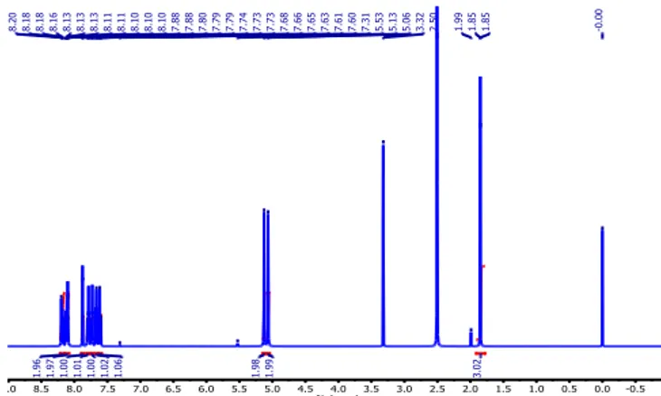
Figure 2. 1H NMR (500 MHz) spectrum of 5-methyl-1,3-bis(3-nitrobenzyl)pyrimidine-2,4(1H,3H)-dione in DMSO-d6 at 298 K.
-1 0 10 20 30 40 50 60 70 80 90 100 110 120 130 140 150 160 170 f1 (ppm) 3 12.51 38.93 39.09 39.26 39.43 39.59 39.69 39.76 39.85 39.93 40.02 43.21 50.83 108.50 122.18 122.21 122.29 122.60 129.85 130.13 134.16 134.34 138.79 139.21 140.38 147.64 147.76 151.10 162.92
Figure 3. 13C NMR (126 MHz) spectrum of 5-methyl-1,3-bis(3-nitrobenzyl)pyrimidine-2,4(1H,3H)-dione in DMSO-d6 at 298 K.
nitrobenzyl bromide in the presence of cesium carbonate as base in dry dimethylformamide as shown in Figure 1. The desired product was isolated as a white powder after silica-gel column chromatography and characterized by NMR.
The proton NMR spectrum of the product is shown in
Figure 2. This spectrum highlight one singlet at δ 1.85 ppm easily attributed to the methyl CH3 group and two singlet at δ
5.13 and 5.06 ppm corresponding to the two different benzyl CH2 hydrogens. Furthermore, the endocyclic CH hydrogen of
the heterocycle can be recognized by the peak at δ 7.8 ppm. Finally, the set of peaks between δ 7.6 and 8.2 ppm, can be attributed to hydrogens of the aromatics rings.
The 13C NMR spectrum (Figure 3) of the title compound
was recorded in DMSO-d6 at 298 K to complete the nuclear
magnetic resonance characterization. The chemical shifts are observed between 180 and -10 ppm. The –CH3 carbon of the
thymine appears at 12.51 ppm. The two benzyl carbon are characterized by two different chemical shifts at 50.83 and 43.21 ppm respectively. This result is logical since the N1
substituted methylene carbon in 5-methyl-1,3-bis(3-nitro benzyl)pyrimidine-2,4(1H,3H)-dione located between two carbonyl groups is more deshielded than it counterpart at the
N3 position. The carbons of the carbonyl groups are observed
at 162.92 and 151.11 ppm, respectively.
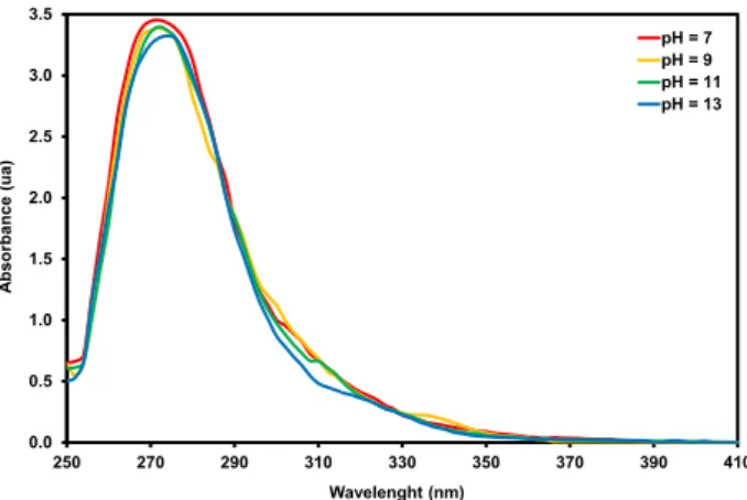
Characterization of the isolated compound by UV-Visible spectroscopy was also realized. Moreover, the stability of the title compound was investigated in alkaline condition (pH ranging from 7 to 13) using UV/Vis spectroscopy. The absor-bance is recorded at room temperature on a HP ChemStation (HP 845X UV-visible Software) with an HP 8453 UV-visible Spectrophotometer (Hewlett Packard, Waldbronn, Germany) fitted with a 1024-element diode-array. Roughly, 100 μM concentration of the compound is used in the experiment and the pH is adjusted using sodium hydroxide and hydrochloride solution. The spectra obtained under neutral pH condition shows a single peak at 271 nm that can be assigned to the π-π* transition in the molecule (Figure 4). In contrary to the effect of base on thymine reported by Kaspar et al. [23], the results revealed that the alkaline condition do not affect the UV/Vis spectrum of the 1,3-bis(3-nitrobenzyl)thymine (Figure 4). This result is logical because in this product, acidic protons are replaced by benzyl substituents that avoid formation of mesomers forms (in case of thymine) leading to good stability of the compound.
0.0 0.5 1.0 1.5 2.0 2.5 3.0 3.5 250 270 290 310 330 350 370 390 410 Abs or ba nc e (ua ) Wavelenght (nm) pH = 7 pH = 9 pH = 11 pH = 13
Figure 4. UV/Vis spectra of 5-methyl-1,3-bis(3-nitrobenzyl)pyrimidine-2,4(1H,3H)-dione at pH ranging 7 to 13.
Figure 5. ORTEP diagram for the title compound with displacement ellipsoids drawn at the 50% probability level.
3.2. Molecular structure
To complete the structural characterization, the exact molecular structure of the title compound was obtained by X-ray diffraction analysis. The structure of 5-methyl-1,3-bis(3-nitrobenzyl)pyrimidine-2,4(1H,3H)-dione is shown in Figure 5.
The compound crystallizes in orthorhombic Pna21 space group. The two nitro bond lengths are identical with C2-N1 and C19-N4 values of 1.472 (3) and 1.470 (3) Å, respectively (Table 2). All the nitro N-O bonds distances are similar (~1.22 Å). Moreover, analysis of the thymine heterocycle highlights close carbonyl C12-O3 and C11-O4 bonds distances with 1.218 (3) and 1.225 (3) Å. The double bond C9-C10 distance [1.344 (3) Å] is typical of those observed in olefin.
The endocyclic C-N bonds of the imide moiety distances are different, especially those of the N2 [1.378 (3) Å], C12-N3 [1.390 (3) Å] and C11-C12-N3 [1.403 (3) Å]. Distortion of the thymine six member ring is observed by measuring selected internal bond angle C9-C10-C11, N2-C12-N3, C12-N2-C9 and C12-N3-C11 giving notable variation with 118.4 (2)°, 115.19 (19)°, 121.79 (17)° and 125.40 (18)°, respectively. The torsion angles C10-C9-N2-C12 [1.2 (4)°] and C10-C11-N3-C12 [1.3 (3)°] are closer and reveal low deviation of the amide bonds from planarity. Furthermore, the weighted average ABS. torsion angle value is 2.7°, suggesting a flattened (planar) conformation of the thymine ring.
3.3. Supramolecular features
The crystal structure packing of the title compound is stabilized by the existence of intermolecular hydrogen bonds.
The H-bond interaction [C14-H14B···O2i, (i) x−1/2, −y+1/2, z]
is established between the benzyl hydrogen atom of one molecule and one oxygen atom of the nitro group of an adjacent molecule with distances H14···O2i of 2.88 Å,
C14B···O2i of 3.453 (3) Å, with an angle C14-H14B···O2i of
144° (Figure 6a).
The molecular conformation revealed that the two benzyl moieties bearing the thymine heterocycle are in the trans disposition with the dihedral angle of 176.23 (8)° (Figure 6b). Such disposition of the benzyl groups allows further stabilization of the crystal packing ensured by π···π and X−H···π interactions. The π···π interactions are established between equivalent aromatic rings of neighboring molecules (See Table 3 and Figure 6b) and C8−H8 with the π electrons of the phenyl ring (C2→C7), all of them along c axis.
3.4. Hirshfeld surface and fingerprint
Hirshfeld surface representation is recognized as a powerful tool for studding intermolecular interactions and molecular packing trends in crystals. This analysis model was used to evaluate intermolecular interactions of the title compound calculated with the program CrystalExplorer 17 [24]. The normalized contact distance, dnorm is obtained by
considering the global relation between the distances of any surface point to the nearest interior (di) and exterior (de) atom
and the Van der Waals radii (rvdw) of the atoms [25,26]. The maps of dnorm, di and de on molecular Hirshfeld
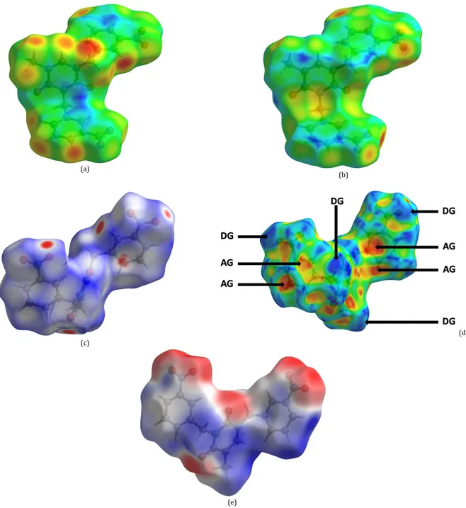
surfaces of the title compound are shown in Figure 7. The three-dimensional dnorm surface mapped over a fixed color
-0.2010 (red) to 1.0774 (blue) highlights intermolecular O···H close contacts as red spots which reveals that the sum of di and
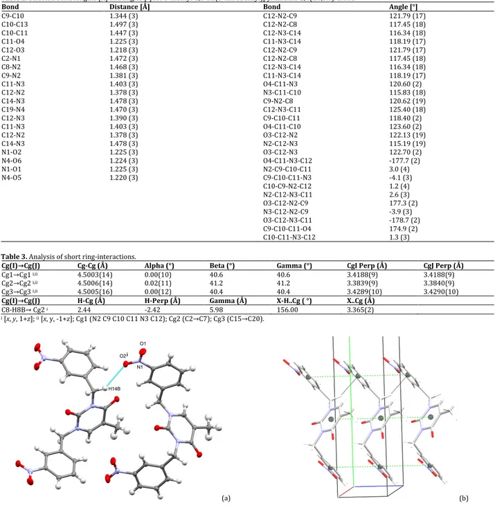
Table 2. Selected bond lengths [Å] and angles [°] for 5-methyl-1,3-bis(3-nitrobenzyl)pyrimidine-2,4(1H,3H)-dione.
Bond Distance [Å] Bond Angle [°]
C9-C10 1.344 (3) C12-N2-C9 121.79 (17) C10-C13 1.497 (3) C12-N2-C8 117.45 (18) C10-C11 1.447 (3) C12-N3-C14 116.34 (18) C11-O4 1.225 (3) C11-N3-C14 118.19 (17) C12-O3 1.218 (3) C12-N2-C9 121.79 (17) C2-N1 1.472 (3) C12-N2-C8 117.45 (18) C8-N2 1.468 (3) C12-N3-C14 116.34 (18) C9-N2 1.381 (3) C11-N3-C14 118.19 (17) C11-N3 1.403 (3) O4-C11-N3 120.60 (2) C12-N2 1.378 (3) N3-C11-C10 115.83 (18) C14-N3 1.478 (3) C9-N2-C8 120.62 (19) C19-N4 1.470 (3) C12-N3-C11 125.40 (18) C12-N3 1.390 (3) C9-C10-C11 118.40 (2) C11-N3 1.403 (3) O4-C11-C10 123.60 (2) C12-N2 1.378 (3) O3-C12-N2 122.13 (19) C14-N3 1.478 (3) N2-C12-N3 115.19 (19) N1-O2 1.225 (3) O3-C12-N3 122.70 (2) N4-O6 1.224 (3) O4-C11-N3-C12 -177.7 (2) N1-O1 1.225 (3) N2-C9-C10-C11 3.0 (4) N4-O5 1.220 (3) C9-C10-C11-N3 -4.1 (3) C10-C9-N2-C12 1.2 (4) N2-C12-N3-C11 2.6 (3) O3-C12-N2-C9 177.3 (2) N3-C12-N2-C9 -3.9 (3) O3-C12-N3-C11 -178.7 (2) C9-C10-C11-O4 174.9 (2) C10-C11-N3-C12 1.3 (3)
Table 3. Analysis of short ring-interactions.
Cg(I)→Cg(J) Cg-Cg (Å) Alpha (°) Beta (°) Gamma (°) CgI Perp (Å) CgJ Perp (Å)
Cg1→Cg1 i,ii 4.5003(14) 0.00(10) 40.6 40.6 3.4188(9) 3.4188(9)
Cg2→Cg2 i,ii 4.5006(14) 0.02(11) 41.2 41.2 3.3839(9) 3.3840(9)
Cg3→Cg3 i,ii 4.5005(16) 0.00(12) 40.4 40.4 3.4289(10) 3.4290(10)
Cg(I)→Cg(J) H-Cg (Å) H-Perp (Å) Gamma (Å) X-H..Cg ( °) X..Cg (Å)
C8-H8B→ Cg2 i 2.44 -2.42 5.98 156.00 3.365(2)
i [x, y, 1+z]; ii [x, y, -1+z]; Cg1 (N2 C9 C10 C11 N3 C12); Cg2 (C2→C7); Cg3 (C15→C20).
(a) (b)
Figure 6. (a) Intermolecular hydrogen bond. (i) x−1/2, −y+1/2, z. (b) Intermolecular interactions in the crystal packing of title compound.
de is shorter than the sum of the Van der Waals radii (Figure
7c). White and blue regions on the surface reveal distances equal and longer than the sum of the Van der Waals radii respectively [25]. On the Hirshfeld surface mapped with shape-index, convex blue regions represent hydrogen donor groups and concave red regions represent acceptor groups (Figure 7d).
The electrostatic potentials for 5-methyl-1,3-bis(3-nitrobenzyl)pyrimidine-2,4(1H,3H)-dione were calculated at B3LYP/6-31G(d,p) level of theory and mapped on the Hirshfeld surfaces (Figure 7e). The map analysis allows a quantitative evaluation of the rich and electron-deficient sites in the molecule. The blue and red regions around the different atoms correspond to positive and negative electrostatic potentials respectively. As shown in
Figure 7e, in the molecule, the electronegative regions are
essentially located around the oxygen atoms and the electropositive regions were observed around the C-H bonds.
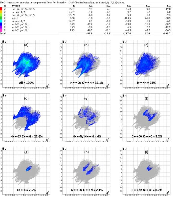
The dispositions adopted by the molecules in the crystal packing lead to close contacts that could be quantified by the mapping of the two-dimensional fingerprint. The fingerprint plot indicates the contributions of interatomic contacts to the Hirshfeld surfaces of the crystal packing. In 1,3-bis(3-nitrobenzyl)thymine, decomposed fingerprint reveals that the most important contributions come from O···H (37.1%), H···H (24%) and H···C/C···H (22.6%) interactions. The high contribution of O···H contacts in the fingerprint could be explained by the presence of several oxygen atoms in the nitro groups and in the thymine cycle. Other interactions are H···N/N···H (4%), C···O/O···C (3.2%), C···C (2.5%) N···O/O···N (2.1%) and C···N/N···C (0.7%) (Figure 8).
(a)
(b)
(c)
(d)
(e)
Figure 7. The Hirshfeld surface of the title compound mapped over: (a) di in the range 0.9306 to 2.5121 Å, (b) de in the range 0.9306 to 2.5121 Å, (c) dnorm in the
range -0.2010 to 1.0774, (d) shape-index (-1.0 to 1.0, DG: Donor Group, AG: Acceptor Group) and (e) electrostatic potential in the range -0.0646 to 0.043 atomic units.
3.5. Energy framework
In view to get more accuracy about molecular interactions, we use Tonto quantum chemistry package for wave-function calculation as an alternative to Gaussian16 quantum chemistry packages [27]. The various intermolecular interactions depicted in the title compound participate to the crystal packing stabilization. Therefore, the intermolecular interac-tion energies for 5-methyl-1,3-bis(3-nitrobenzyl)pyrimidine-2,4(1H,3H)-dione are calculated using CE-B3LYP/6-31G(d,p) energy model available in Crystal-Explorer (CE) [24].
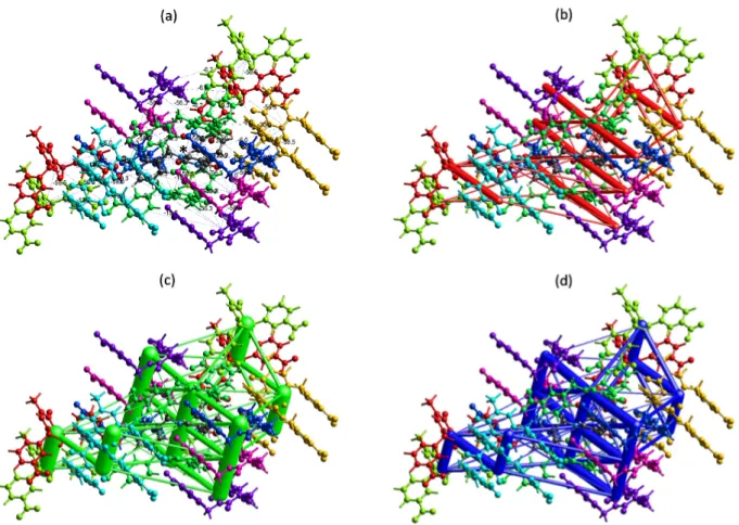
The interaction energies calculations are represented graphically in Figure 9 as framework energy diagrams. The radii of the corresponding cylinders are proportional to the magnitude of interaction energy. The total intermolecular energy Etot (kJ/mol) relative to the reference molecule (in
black) is the sum of energies of four main components,
comprising electrostatic (Eele), polarization (Epol), dispersion
(Edis) and exchange-repulsion (Erep) [28] with scale factors of
1.057, 0.740, 0.871 and 0.618, respectively [29,30]. Table 3
shows information about interaction energies components (E), rotational symmetry operations with respect to the reference molecule (Symop), the centroid-to-centroid distance between the reference molecule and interacting molecules (R) as well as the number of pair(s) of interacting molecules with respect to the reference molecule (N). The results give total interaction energy of -199.7 kJ/mol involving the electrostatic (-83.8 kJ/mol), polarization (-29.8 kJ/mol), dispersion (-217.4 kJ/mol) and repulsion (162.4 kJ/mol). The π···π and C−H···π contacts highlight in Figure 6b are the most stable interactions present in the crystal packing with an interaction energy of -58.5 kJ/mol. Hydrogen bonds with C-H···O interactions energies are in the same energy order with π-X interactions with energy value of -56.3 kJ/mol.
Table 3. Interaction energies in components form for 5-methyl-1,3-bis(3-nitrobenzyl)pyrimidine-2,4(1H,3H)-dione.
N Symop R Eele Epol Edis Erep Etot
2 -x+1/2, y+1/2, z+1/2 14.01 -9.4 -2.3 -16.3 9.8 -19.8 2 -x, -y, z+1/2 13.57 -1.0 -0.5 -9.7 6.2 -6.0 2 -x+1/2, y+1/2, z+1/2 15.39 -6.8 -1.7 -5.4 6.3 -9.3 2 x, y, z 4.50 -1.8 -8.6 -104.5 65.9 -58.5 2 -x, -y, z+1/2 12.97 0.1 -1.4 -10.9 6.9 -6.2 2 x+1/2, -y+1/2, z 8.73 -17.2 -3.2 -23.8 16.9 -30.9 2 x+1/2, -y+1/2, z 8.73 -7.0 -1.8 -6.5 2.7 -12.7 2 x+1/2, -y+1/2, z 7.49 -40.7 -10.3 -40.3 47.7 -56.3 Total -83.8 -29.8 -217.4 162.4 -199.7
Figure 8. The two-dimensional fingerprint plots of title compound: (a) all contacts, (b) H···O/O···H, (c) H···H, (d) H···C/C···H, (e) H···N/N···H, (f) C···O/O···C, (g)
C···C, (h) N···O/O···N and (i) C···N/N···C.
That interaction energy presents the same order electrostatic (E’ele = -40.7 kJ.mol-1) and dispersive (E’dis = -40.3
kJ/mol) character (Table 3). Analysis of the total energy diagram (Figure 9d) revealed close resemblance to the dispersion energy framework (Figure 9c) demonstrating its main contribution in the total forces in the crystal packing.
4. Conclusion
The reaction of thymine with 3-nitrobenzylbromide in the presence of cesium carbonate yielded
5-methyl-1,3-bis(3-nitrobenzyl)pyrimidine-2,4(1H,3H)-dione as a white powder. The NMR and elemental analysis of the product were further completed by X-ray diffraction analysis performed on the isolated monocrystal obtained by slow evaporation. All the interactions present in the crystal packing were analyzed thank to Hirshfeld surface analysis and the two-dimensional fingerprint, highlighting close contacts as well as acceptor and donor groups. Finally, energy-framework calculations were used to analyze and visualize the three dimensional topology of the crystal packing. The results revealed that total interaction energy in the crystal packing is mainly dispersive.
Figure 9. The colour-coded interaction energy framework map of 5-methyl-1,3-bis(3-nitrobenzyl)pyrimidine-2,4(1H,3H)-dione within 3.8 Å of the centering
(marked by an asterisk) molecular cluster (a). Energy framework diagram for separate electrostatic (b), dispersion (c) components of the title molecule and the total interaction energy (d). The energy factor scale is 100 and the cut-off is 5.00 kJ/mol.
Acknowledgements
The authors thank the Université de Liège for a research grant.
Supporting information
CCDC-1960980 contains the supplementary crystal-lographic data for this paper. These data can be obtained free of charge via https://www.ccdc.cam.ac.uk/structures/, or by e-mailing [email protected], or by contacting The Cambridge Crystallographic Data Centre, 12 Union Road, Cambridge CB2 1EZ, UK; fax: +44(0)1223-336033.
Disclosure statement
Conflict of interests: The authors declare that they have no conflict of interest.
Author contributions: All authors contributed equally to this work.
Ethical approval: All ethical guidelines have been adhered. Sample availability: Samples of the compounds are available from the author.
ORCID
Etsè Koffi Sénam
http://orcid.org/0000-0001-8495-4327 Comeron Lamela Laura
http://orcid.org/0000-0002-5940-3002 Zaragoza Guillermo http://orcid.org/0000-0002-2550-6628 Pirotte Bernard http://orcid.org/0000-0001-8251-8257 References
[1]. Lagoja I. M. Chem. Biodivers. 2005, 2, 1-50.
[2]. Honda, T.; Inagawa, H.; Fukushima, M.; Moriyama, A.; Soma, G. Clin.
Chim. Acta. 2002, 322, 59-66.
[3]. Hadj-Bouazza, A.; Teste, K.; Colombeau, L.; Chaleix, V.; Zerrouki, R.; Kraemer, M.; Sainte Catherine, O. Nucleosides Nucleotides Nucleic
Acids 2008, 27, 439-448.
[4]. Balzarini, J.; Baba, M.; De Clercq. E. Antimicrob. Agents Chemother.
1995, 39, 998-1002.
[5]. Adamska, A.; Rumijowska-Galewicz, A.; Ruszczynska, A.; Studzinska, M.; Jablonska, A.; Paradowska, E.; Bulska, E.; Munier-Lehmann, H.; Dziadek, J.; Lesnikowski, Z. J.; Olejniczak, A. B. Eur. J. Med. Chem.
2016, 121, 71-81.
[6]. Bialek-Pietras, M.; Olejniczak, A. B.; Paradowsk, E.; Studzinska, M.; Jablonska, A.; Lesnikowski, Z. J. J. Organomet. Chem. 2018, 865, 166-172.
[7]. Li, G.; Zhu, Y.; Yang, F.; Yang, H. Hecheng Huaxue. 2011, 19, 111-114. [8]. Lin, T. S.; Wang, L.; Antonini, I.; Cosby, L. A.; Shiba, D. A.; Kirkpatrick,
D. L.; Sartorelli, A. C. J. Med. Chem. 1986, 29, 84-89.
[9]. Weaver, D. F.; Guillain, B. M.; Carran, J. R.; Jones, K. US Patent. 2002, No. 7, 501, 429 B2.
[10]. Kim, B. R.; Park, J. Y.; Jeong, H. J.; Kwon, H. J.; Park, S. J.; Lee, I. C.; Ryu, Y. B.; Lee, W. S. J. Enzyme Inhib. Med. Chem. 2018, 33, 1256-1265. [11]. Li, M.; Cai, X.; Zhu, Y.; Liu, K.; Hu, M. Acta Chim. Sinica (Chinese
Edition) 2011, 69, 425-430.
[12]. Etse, K. S.; Dassonneville, B.; Zaragoza, G.; Demonceau, A.
Tetrahedron Lett. 2017, 58, 789-793.
[13]. Manos-Turvey, A.; Becker, G.; Francotte, P.; Serrano, M. E.; Luxen, A.; Pirotte, B.; Plenevaux, A.; Lemaire, C. Chem. Med. Chem. 2019, 14, 788-795.
[14]. Goffin, E.; Drapier, T.; Larsen, A. P.; Geubelle, P.; Ptak, C. P.; Laulumaa, S.; Rovinskaja, K.; Gilissen, J.; Tullio, P.; Olsen, L.; Frydenvang, K.; Pirotte, B.; Hanson, J.; Oswald, R. E.; Kastrup, J. S.; Francotte, P. J. Med.
[15]. Etse, K. S.; Zaragoza, G.; Pirotte, B. Eur. J. Chem. 2019, 10, 189-194. [16]. Dolman, N. P.; More, J. C.; Alt, A.; Knauss, J. L.; Troop, H. M.; Bleakman,
D.; Collingridge, G. L.; Jane, D. E. J. Med. Chem. 2006, 49, 2579-2592. [17]. Drapier, T.; Geubelle, P.; Bouckaert, C.; Nielsen, L.; Laulumaa, S.;
Goffin, E.; Dilly, S.; Francotte, P.; Hanson, J.; Pochet, L.; Kastrup, J. S.; Pirotte, B. J. Med. Chem. 2018, 61, 5279-529.
[18]. Bruker APEX II Bruker AXS Inc., Madison, WI, USA, 2004. [19]. Sheldrick, G. M. Acta Cryst. A 2015, 71, 3-8.
[20]. Sheldrick, G. M. Acta Cryst. C 2015, 71, 3-8. [21]. Farrugia, L. J. J. Appl. Cryst. 2012, 45, 849-854.
[22]. Macrae, C. F.; Bruno, I. J.; Chisholm, J. A.; Edgington, P. R.; McCabe, P.; Pidcock, E.; Rodriguez-Monge, L.; Taylor, R.; Van de Streek, J.; Wood, P. A. J. Appl. Cryst. 2008, 41, 466-470.
[23]. Kaspar, K.; Giessmann, R. T.; Krausch, N.; Neubauer, P.; Wagner, A.; Gimpel, M. Methods Protoc. 2019, 2, 60-72.
[24]. Turner, M. J.; McKinnon, J. J.; Wolff, S. K.; Grimwood, D. J.; Spackman, P. R.; Jayatilaka, D.; Spackman, M. A. CrystalExplorer17, The University of Western Australia. http://hirshfeldsurface. net, 2017. [25]. Spackman, M. A.; Jayatilaka, D. CrystEngComm. 2009, 11, 19-32. [26]. Demircioglu, Z.; Yesil, A. E.; Altun, M.; Bal-Demirci, T.; Ozdemir, N. J.
Mol. Struct. 2018, 1162, 96-108.
[27]. Frisch, M. J.; Trucks, G. W.; Schlegel, H. B.; Scuseria, G. E. , Robb, M. A.; Cheeseman, J. R.; Scalmani, G.; Barone, V.; Mennucci, B.; Petersson, G. A.; Nakatsuji, H.; Caricato, M.; Li, X.; Hratchian, H. P.; Izmaylov, A. F.; Bloino, J.; Zheng, G.; Sonnenberg, J. L.; Hada, M.; Ehara, M.; Toyota, K.; Fukuda, R.; Hasegawa, J.; Ishida, M.; Nakajima, T.; Honda, Y.; Kitao, O.; Nakai, H.; Vreven, T.; Montgomery, J. A. Jr.; Peralta, J. E.; Ogliaro, F.; Bearpark, M.; Heyd, J. J.; Brothers, E.; Kudin, K. N.; Staroverov, V. N.; Kobayashi, R.; Normand, J.; Raghavachari, K. , Rendell, A. , Burant, J. C.; Iyengar, S. S.; Tomasi, J.; Cossi, M.; Rega, N.; Millam, J. M.; Klene, M.; Knox, J. E.; Cross, J. B.; Bakken, V.; Adamo, C.; Jaramillo, J.; Gomperts, R.; Stratmann, R. E.; Yazyev, O.; Austin, A. J.; Cammi, R.; Pomelli, C.; Ochterski, J. W.; Martin, R. L.; Morokuma, K.; Zakrzewski, V. G.; Voth, G. A.; Salvador, P.; Dannenberg, J. J.; Dapprich, S.; Daniels, A. D.; Farkas, O.; Foresman, J. B.; Ortiz, J. V.; Cioslowski, J.; Fox, D. J. Gaussian16. Gaussian Inc. Wallingford, Connecticut, USA, 2016. [28]. Turner, M. J.; Thomas, S. P.; Shi, M. W.; Jayatilaka, D.; Spackman, M. A.
Chem. Commun. 2015, 51, 3735-3738.
[29]. Mackenzie, C. F.; Spackman, P. R.; Jayatilaka, D.; Spackman, M. A.
IUCrJ. 2017, 4, 575-587.
[30]. Tan, S. L.; Jotani, M. M.; Tiekink, E. R. T. Acta Cryst. E 2019, 75, 308-318.
Copyright © 2020 by Authors. This work is published and licensed by Atlanta Publishing House LLC, Atlanta, GA, USA. The full terms of this license are available at http://www.eurjchem.com/index.php/eurjchem/pages/view/terms and incorporate the Creative Commons Attribution-Non Commercial (CC BY NC) (International, v4.0) License (http://creativecommons.org/licenses/by-nc/4.0). By accessing the work, you hereby accept the Terms. This is an open access article distributed under the terms and conditions of the CC BY NC License, which permits unrestricted non-commercial use, distribution, and reproduction in any medium, provided the original work is properly cited without any further permission from Atlanta Publishing House LLC (European Journal of Chemistry). No use, distribution or reproduction is permitted which does not comply with these terms. Permissions for commercial use of this work beyond the scope of the License (http://www.eurjchem.com/index.php/eurjchem/pages/view/terms) are administered by Atlanta Publishing House LLC (European Journal of Chemistry).