Two new organotin(IV)-phosphoryl complexes: crystal structure and Hirshfeld surface analysis
Mehrdad Pourayoubi
1· Anahid Saneei
1· Michal Dušek
2· Soobiyeh Alemi Rostami
1· Aurelien Crochet
3· Monika Kucˇeraková
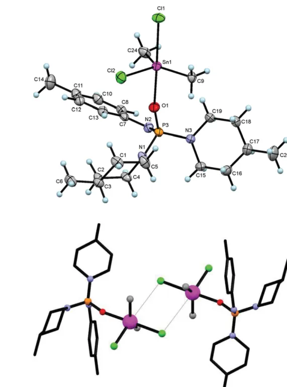
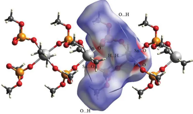
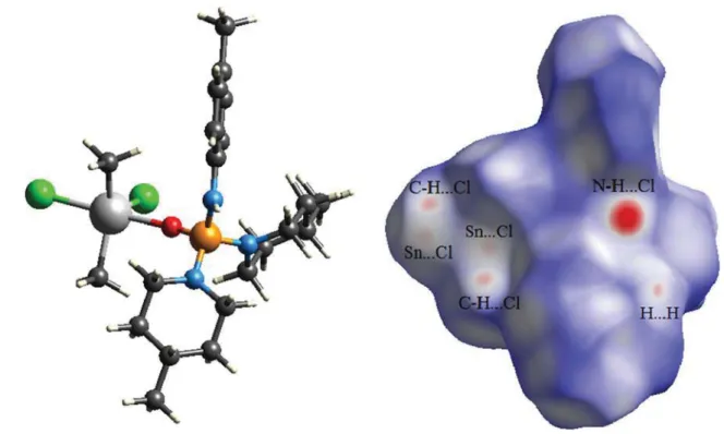
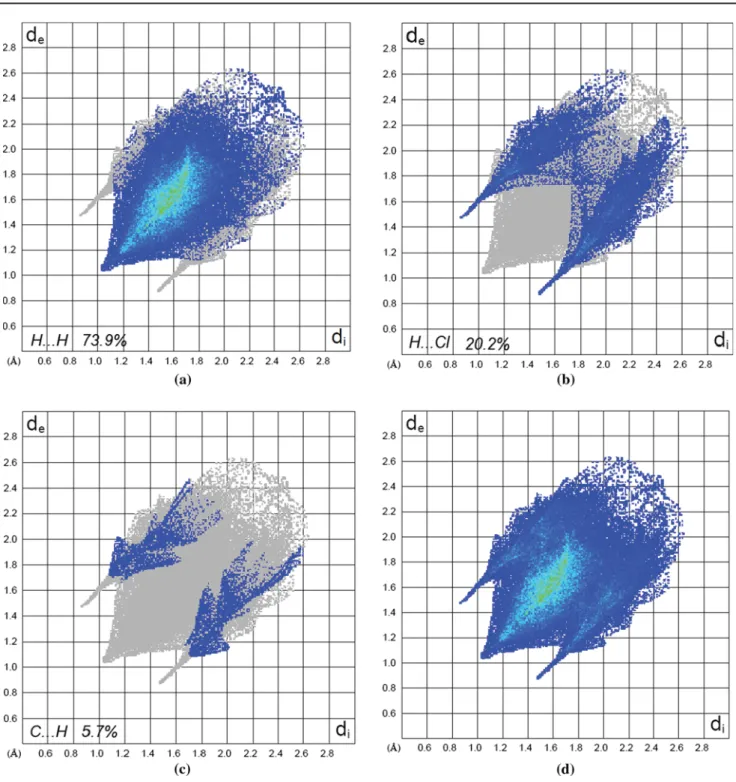
2in the asymmetric unit of (II) receives a second interaction from chlorine atom that, however, has negligible contribu- tion portion in the total interactions (evaluated by finger- print plot) in the crystal. On the other hand, this chlorine affects the geometry at the Sn atom and causes the open C–Sn–C bond angle (of 140.18(24)°). For both structures, the contribution of H…H contacts to the total interaction is decisive, being 59.1 % for (I) and 73.9 % for (II); so that the one distinct spike observed in each of the finger- print plots is related to H…H contacts. The O…H (34.1 %) in (I) and the H…Cl (20.2 %) in (II) are the characteris- tic intermolecular contacts in the corresponding structures, monitoring as two pairs of H-bond spikes for O…H and two sharp spikes for H…Cl. Furthermore, the structure (I) does not show the C…H interaction, whereas (II) includ- ing unsaturated carbon atoms manifests such interaction (5.7 %) as two very short spikes.
Keywords Organotin(IV)-phosphoryl complex · Hirshfeld surface · Crystal structure
Introduction
Tin complexes are of great interest because of their unique properties in medicine, industrial applications and bio- logical systems [1–4]. Among which the tin-phosphoryl complexes have received considerable attention due to the diverse structural features known for phosphoryl-donor ligands [5] and various applications such as anti-bacterial [6], anti-HIV [7] and anti-cancer [8] activities and also agricultural uses [9] and enzyme inhibitory [10] for phos- phoryl compounds.
Up to now, different families of phosphoryl compounds, with different coordination environment at the P atom Abstract Hirshfeld surfaces and two-dimensional finger-
print plots are used to visualize and analyse intermolecular interactions in two new organotin(IV)-phosphoryl com- plexes: [Sn(CH
3)
2[OP(O)(OCH
3)
2]
2]
n(I) and {[(4-CH
3) C
6H
4NH][C
5H
9(4-CH
3)N]
2P(O)}Sn(CH
3)
2Cl
2(II). In (I), the asymmetric unit is composed of the Sn(CH
3)
2[OP(O) (OCH
3)
2]
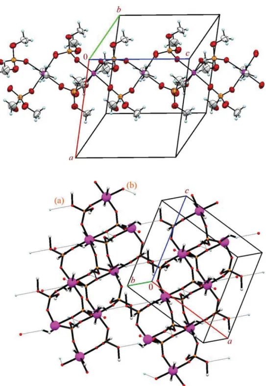
2segment bonded to the neighbouring segment through Sn–O bonds thus constructing a coordination pol- ymer. The tin centre has an Sn[C]
2[O]
4environment with the axial positions occupied by methyl groups and with the oxygen atoms of Sn–O bonds located at the equatorial plane, while the oxygen atoms of OCH
3groups do not take part in the coordination interaction. In (II), the asymmetric unit contains one complete molecule and the tin centre has an Sn[C]
2[Cl]
2O trigonal bipyramid coordination environ- ment, where the phosphoric triamide ligand and one of the chlorine atoms are placed in axial positions. Hirshfeld sur- face analysis on the asymmetric unit of (I) shows a contri- bution of 5.9 % of total interactions in crystal for Sn…O contacts related to involving of four-coordinated Sn site of asymmetric unit with two oxygen atoms of two neighbour- ing phosphates in the coordination polymer. The Sn centre
Electronic supplementary material is available
* Mehrdad Pourayoubi [email protected]
1
Department of Chemistry, Faculty of Sciences, Ferdowsi University of Mashhad, Mashhad, Iran
2
Institute of Physics ASCR, v.v.i., Na Slovance 2, 182 21 Prague 8, Czech Republic
3