A N N A L S O F T H E N E W Y O R K A C A D E M Y O F S C I E N C E S Issue:Thymosins in Health and Disease
Thymosin
β
4 in multiple myeloma: friend or foe
Jo Caers,1,2Eleonore Otjacques,1Dirk Hose,3Bernard Klein,4and Karin Vanderkerken2 1Department of Hematology, CHU University of Li `ege, Li `ege, Belgium.2Laboratory of Hematology and Immunology, Vrije
Universiteit Brussel, Myeloma Center Brussels, Brussels, Belgium.3Nationales Centrum f ¨ur Tumorerkrankungen, Heidelberg,
Germany and Medizinische Klinik V, Universit ¨atsklinikum Heidelberg, Heidelberg, Germany.4Institut National de la Sant ´e et
de la Recherche M ´edicale U475 and Unit for Cellular Therapy, CHU Montpellier, Montpellier, France
Address for correspondence: Jo Caers, M.D., Ph.D., Department of Clinical Hematology, Centre Hospitalier Universitaire de Li `ege, Domaine Universitaire du Sart Tilman, B ˆatiment B 35, B-4000 Li `ege, Belgium. jo.caers@chu.ulg.ac.be
Multiple myeloma (MM) is a malignancy characterized by the accumulation of monoclonal plasma cells in the bone marrow (BM). Because of the known involvement of thymosin β4 (Tβ4) in metastasis of tumor cells, we examined the expression and role of Tβ4 in MM disease. In a large patient population, we demonstrated that Tβ4 expression was significantly lower in myeloma cells compared to normal plasma cells and that patients with a high Tβ4 expression had a longer event free and overall survival. The decreased Tβ4 expression was also found in the murine 5TMM model. To study its function, we overexpressed the Tβ4 gene in 5T33MMvt cells by lentiviral transduction. These cells demonstrated a decreased proliferative capability and an increased sensitivity to apoptosis. Mice injected with Tβ4-overexpressing cells showed a prolonged survival compared to mice injected with controls. In contrast to its role in solid tumors, we found a decreased expression in myeloma cells compared to their normal counterpart and studies with overexpression of the Tβ4 gene indicated a tumor suppressive function of Tβ4 in myeloma development. Keywords: multiple myeloma; thymosin beta 4; plasma cell
Introduction
Thymosin 4 (T4) is a 43 amino-acid small peptide originally isolated from the thymus.1 It
was shown that T4 interacts with G-actin and functions as a major actin-sequestering protein.2
T4 is considered to be a major actin-sequestering molecule, which specifically binds monomeric actin (G-actin), forming a 1:1 complex, or in a tern-ery complex including profilin.3The mechanism by
which Tβ4 influences cell proliferation, migration, and differentiation is generally believed to be linked with maintaining a dynamic equilibrium between G-actin and F-actin, critical for the rapid reorgani-zation of the cytoskeleton. Tβ4 induced cell prolifer-ation, migrprolifer-ation, and differentiation contribute to different physiological and pathological processes, such as angiogenesis,4wound healing,5and cardiac
repair.6
Thymosin β4 in cancer
Multiple studies have indicated that Tβ4 was over-expressed in various tumor tissues and may play
an important role by affecting tumor cell prolifer-ation, migrprolifer-ation, metastasis, and induction of an-giogenesis. The different studies on solid tumors can be summarized as a potential role of Tβ4 in the malignant conversion of a normal cell or in a potential role in metastasis of primary tumor cells. Evidence for this role can be found from the stud-ies in melanoma, sarcoma, and pancreas cancer. In both murine and human melanoma cells, Clark et al. found that Tβ4 levels were increased in tumor cells from metastatic lesions compared with the parental cells isolated from the primary site.7A similar obser-vation was made in a murine fibrosarcoma model, where Tβ4 was one of genes that increased when mRNA from highly tumorigenic and metastatic cells was compared with its weakly tumorigenic precur-sor cell line or normal counterpart.8,9Furthermore, these increased levels of Tβ4 were demonstrated to regulate the migratory and invasive capacities of these cells. A adenoviral-based overexpression of Tβ4 was applied in a human colon cancer cell line and melanoma cell line. These experiments showed an increased growth, motility, and invasive
capacities in vitro10,11and a larger tumor load (size of primary site and number of metastasis in vivo). In addition, Tβ4 overexpression was associated with the stimulation of blood vessel formation in these latter experiments.12 Recently Zhang et al. showed
that Tβ4 expression was elevated in pancreatic can-cer cell lines and clinical tissue specimens, compared with control pancreatic duct cells and surrounding normal pancreatic tissues. Tβ4 was believed to be in-volved in stimulating human pancreatic cancer pro-gression by promoting a proinflammatory cytokine environment and by activating JNK signaling path-way.13In nonsmall cell lung cancer, the Tβ4 gene was found to be upregulated in metastasizing pri-mary tumors compared with non-metastasizing tu-mors and Tβ4 expression had also prognostic value: high expression levels of Tβ4 was associated with significantly worse survival of patients with stage I disease.14
Thymosin β4 in angiogenesis
A first indication for an implication of Tβ4 in an-giogenesis came from its identification in a screen for rapidly induced genes following culture of hu-man umbilical vein endothelial cells (HUVEC) on a basement membrane matrix.15A fivefold induction
of Tβ4 was observed during endothelial cell dif-ferentiation in vitro and transfection of HUVECs with Tβ4 caused an increase in the rate of at-tachment, spreading, and tube formation.15Hynda
Kleinmans’ group also demonstrated that Tβ4 acts as a chemo-attractant for endothelial cells, by stim-ulating directional migration of HUVECs in vitro and endothelial cell migration in vivo in a subcuta-neous Matrigel plug assay.4In addition to
stimulat-ing proliferation, attachment, and differentiation of endothelial cells, Tβ4 was also able to induce tube formation on Matrigel and vascular sprouting and neo-vascularization.16All these studies validated the
involvement of Tβ4 in angiogenesis by promot-ing migration of endothelial cells, but the precise mechanism by which Tβ4 directs cell migration is poorly defined and the role of actin binding versus other receptor-mediated events is still a matter of debate.17
Thymosin β4 in multiple myeloma
Multiple myeloma (MM) is a malignant plasma dis-order characterized by the accumulation of mon-oclonal plasma cells in the bone marrow (BM).
Despite the introduction of novel treatment strate-gies, MM remains an incurable disease.18Migration
and invasion are important processes in the ini-tial homing of the cells to the bone marrow and subsequent spreading to distant sites.19 Moreover,
angiogenesis is one of the hallmarks in MM disease progression.20,21Because of its involvement in these
processes, we were interested in analyzing Tβ4 ex-pression and function in multiple myeloma cells. Thymosin beta 4 was examined in a historical study that analyzed Tβ4 gene expression in lymphoid ma-lignant cells. The authors found high levels of Tβ4 in lymphocytes and in early stages of B-cell differen-tiation, but Tβ4 was absent in MM cells. Since this pattern of Tβ4 gene expression was similar to that of immunologically important molecules (such as HLA class II antigen, Fc receptor, and complement receptor), a relationship between Tβ4 and B-cell dif-ferentiation state was suggested.22This implication
in cellular differentiation was also proposed by a second study that examined Tβ4 expression in lym-phoid and myeloid cells and that showed that Tβ4 gene was regulated in a maturation-related man-ner.23We studied the expression of Tβ4 in human
disease and in the 5TMM mouse model of MM.24
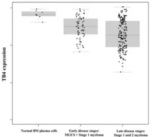
We investigated the Tβ-expression pattern in a large (n= 298) sample of primary MM cells and in nor-mal plasma cell samples from healthy donors as pre-viously reported.25 We found that Tβ4 was lower
expressed in MM cells compared to normal plasma cells (cf. Fig. 1). A similar finding could be seen when plasma cells from monoclonal gammopathy of undetermined significance (MGUS) patients were compared with normal plasma cells. MGUS is a pre-malignant form that may develop into overt MM disease. We subsequently analyzed the survival of MM patients that showed a high Tβ4 expression (termed Tβ4high) compared to patients with a low
Tβ4 expression (termed Tβ4low). All patients were
treated by high dose induction chemotherapy that was followed by an autologous stem cell transplanta-tion. Tβ4highpatients showed a significantly longer median EFS (38 months) than Tβ4lowpatients (26
months). The overall survival tended to differ in favor of Tβ4highpatients.
A similar Tβ4 expression pattern was seen in the 5TMM murine MM model where Tβ4 was decreased compared to normal hematopoietic cells (Figs. 2 and 3A).24Addition of exogenous Tβ4
Figure 1. Shows the micro-array data obtained for the
Tβ4 expression in CD138+sorted BM plasma cells from healthy donors and MM patients. These results were val-idated by quantitative RT-PCR. The premalignant form, MGUS, and Salmon & Durie Stage 1 were considered as early myeloma disease stages and Salmon & Durie Stage 2 and 3 as late disease stages. Figure illustrates the ex-pression in plasma cells from normal healthy volunteers, patients with early disease stage and patients with late disease stage.24
addition of the AcSDKP tetrapeptide had no effect on proliferation. Because of its low expression in 5T33MMvt cells, the Tβ4 gene was overexpressed using a lentiviral expression vector. In a proliferation assay, 5T33MMvtTβ4+ cells showed a significant
decrease in DNA synthesis compared to control cells. 5T33MMvtTβ4+ cells showed a significantly
increased sensitivity to different anti-MM agents such as the NF-B inhibitor Bortezomib, dexam-ethasone, or melphalan (Fig. 3B). The quantifica-tion of G-Actin and F-Actin by Western blotting showed a lowered G-Actin–F-Actin ratio after Tβ4 overexpression. F-Actin is of particular importance in cytoskeleton changes involved in cellular mi-gration and in microtubuli organization control-ling the mitotic spindle. In line with these results, vinca-alkaloids with micro-tubulin inhibitory ac-tivity had more effect on the proliferation capacities of 5T33MMvtTβ4+cells than on control cells. After
intravenous injection of 5T33MMvtTβ4+or control
cells, the survival of mice injected with control cells was significantly shorter: 65.9 days compared to 88.9 days for mice injected with 5T33MMvtTβ4+cells.
In contrast to what is seen in solid tumors, we found in MM cells a decreased expression of Tβ4 compared to their normal counterpart (plasma cells). Tβ4 expression had some prognostic value; patients with a high Tβ4 expression had a better outcome after intensive chemotherapy compared to a low Tβ4 expression. We observed in vitro differ-ences in proliferation and sensitivity to anti-MM agents, which indicated a direct effect on prolifer-ation. Quantitative RT-PCR showed that the Tβ4 gene expression could be correlated to the malig-nant phenotype of 5TMM cells: 5T33MMvt cells are highly proliferative cells with limited migratory capacities (and showed a very low Tβ4 expression),
Figure 2. Tβ4 staining on normal hematopoietic cells, bone marrow endothelial cells, and 5T33MMvv cells. In normal hematopoietic cells, a strong nuclear staining was seen (A). MM disease progression is associated with a neo-vascularization. BM endothelial cells also showed strong positivity for Tβ4 (B). MM cells stained positive intra-cytoplasmatic for Tβ4 (C), which was less intense as in normal hematopoietic cells.
Figure 3.(A) In the 5TMM model, a similar gene expression pattern was observed as in humans. Tβ4 mRNA expression in 5T33MM and 5T2MM invaded BM was lowered compared to normal BM cells. 5T33MMvt cells showed the lowest mRNA expression. Transfection of Tβ4 using a lentiviral vector resulted in overexpression of the Tβ4 gene. (B) 3-H thymidine uptake revealed a decreased DNA synthesis rate in 5T33MMvtTβ4+cells compared to wild-type cells. Incubation with the anti-MM agents bortezomib (5 nM), melphalan (6M), and dexamethasone (250M) had stronger effects on 5T33MMvtTβ4+than on control cells.24
whereas 5T2MM and 5T33MMvv cells have a lower proliferative, but higher migratory index (with Tβ4 expression that was higher in a low proliferative 5T2MM cells). These results may suggest that Tβ4 may be involved in the differentiation status of MM cells, as indicated by the earlier studies on lymphoid and myeloid cells. However, its mechanism of action is unclear and currently under investigation.
In conclusion, our results propose a tumor sup-pressive function of Tβ4 expression in MM with impact on survival. Tβ4 was downregulated in MM cells of patients compared to the normal BM plasma cells and studies with the murine 5T33MM model show a decreased in vitro and in vivo tumor growth for cells overexpressing the Tβ4 gene.
Acknowledgments
The authors would like to thank Hendrik De Raeve for providing the Tβ4 staining on BM sections from
diseased and healthy mice. This work was finan-cially supported by the Fund for Scientific Research-Vlaanderen (FWO-Research-Vlaanderen) and by a grant from the European Commission FP6 to MSCNET. E Ot-jacques is a T´el´evie research assistant at the FNRS.
Conflicts of interest
The authors declare no conflicts of interest.
References
1. Goldstein, A.L. & M. Badamchian. 2004. Thymosins: chemistry and biological properties in health and disease.
Expert Opin. Biol. Ther. 4: 559–573.
2. Hertzog, M. et al. 2004. The beta-thymosin/WH2 do-main; structural basis for the switch from inhibition to promotion of actin assembly. Cell 117: 611–623. 3. Yarmola, E.G. & M.R. Bubb. 2004. Effects of profilin and
demonstrated in vitro and in cell extracts with a novel direct assay. J. Biol. Chem. 279: 33519–33527.
4. Malinda, K.M., A.L. Goldstein & H.K. Kleinman. 1997. Thymosin beta 4 stimulates directional migration of hu-man umbilical vein endothelial cells. FASEB J. 11: 474– 481.
5. Philp, D. et al. 2006. Thymosin beta4 promotes ma-trix metalloproteinase expression during wound repair.
J. Cell Physiol. 208: 195–200.
6. Smart, N. et al. 2007. Thymosin beta4 induces adult epi-cardial progenitor mobilization and neovascularization.
Nature 445: 177–182.
7. Clark, E.A. et al. 2000. Genomic analysis of metastasis reveals an essential role for RhoC. Nature 406: 532–535. 8. Nummela, P. et al. 2006. Thymosin beta4 is a deter-minant of the transformed phenotype and invasiveness of S-adenosylmethionine decarboxylase-transfected fi-broblasts. Cancer Res. 66: 701–712.
9. Kobayashi, T. et al. 2002. Thymosin-beta4 regulates motility and metastasis of malignant mouse fibrosar-coma cells. Am. J. Pathol. 160: 869–882.
10. Wang, W.S. et al. 2004. Overexpression of the thymosin beta-4 gene is associated with increased invasion of SW480 colon carcinoma cells and the distant metasta-sis of human colorectal carcinoma. Oncogene 23: 6666– 6671.
11. Wang, W.S. et al. 2003. Overexpression of the thymosin beta-4 gene is associated with malignant progression of SW480 colon cancer cells. Oncogene 22: 3297–3306. 12. Cha, H.J., M.J. Jeong & H.K. Kleinman. 2003. Role of
thymosin beta4 in tumor metastasis and angiogenesis. J.
Natl. Cancer Inst. 95: 1674–1680.
13. Zhang, Y. et al. 2008. Thymosin beta 4 is overexpressed in human pancreatic cancer cells and stimulates proinflam-matory cytokine secretion and JNK activation. Cancer
Biol. Ther. 7: 419–423.
14. Ji, P. et al. 2003. MALAT-1, a novel noncoding RNA, and thymosin beta4 predict metastasis and survival in
early-stage non-small cell lung cancer. Oncogene 22: 8031– 8041.
15. Grant, D.S. et al. 1995. Matrigel induces thymosin beta 4 gene in differentiating endothelial cells. J. Cell Sci. 108(Pt 12): 3685–3694.
16. Grant, D.S. et al. 1999. Thymosin beta4 enhances en-dothelial cell differentiation and angiogenesis.
Angiogen-esis 3: 125–135.
17. Smart, N., A. Rossdeutsch & P.R. Riley. 2007. Thymosin beta4 and angiogenesis: Modes of action and therapeutic potential. Angiogenesis 10: 229–241.
18. Caers, J. et al. 2008. Multiple myeloma—an update on diagnosis and treatment. Eur. J. Haematol. 81: 329–343. 19. Caers, J. et al. 2008. Unraveling the biology of multi-ple myeloma disease: cancer stem cells, acquired intra-cellular changes and interactions with the surrounding micro-environment. Bull. Cancer 95: 301–313. 20. Vacca, A. et al. 1999. Bone marrow
neovasculariza-tion, plasma cell angiogenic potential, and matrix metalloproteinase-2 secretion parallel progression of hu-man multiple myeloma. Blood 93: 3064–3073. 21. Hose, D. et al. 2009. Induction of angiogenesis by normal
and malignant plasma cells. Blood 114: 128–143. 22. Gondo, H. et al. 1987. Differential expression of
the human thymosin-beta 4 gene in lymphocytes, macrophages, and granulocytes. J. Immunol. 139: 3840– 3848.
23. Shimamura, R. et al. 1990. Expression of the thymosin beta 4 gene during differentiation of hematopoietic cells.
Blood 76: 977–984.
24. Caers, J. et al. 2009. Thymosin {beta}4 has tumor suppressive effects and its decreased expression results in poor prognosis and decreased survival in multiple myeloma. Haematologica 95: 163–167.
25. Mahtouk, K. et al. 2005. Expression of EGF-family recep-tors and amphiregulin in multiple myeloma. Amphireg-ulin is a growth factor for myeloma cells. Oncogene 24: 3512–3524.