Year 2019
Thesis N°269
Impact of the pneumococcal conjugate vaccine
on nasopharyngeal pneumococcal carriage in
feverish infants in Mohamed VI teaching
hospital of Marrakesh
THESIS
PRESENTED AND DEFENDED PUBLICLY ON December 24
PthP, 2019
BY
Miss.
Amal HABCHANE
Born on October 27th, 1991 in Tahannaout
TO OBTAIN A MEDICAL DOCTORATE
KEYWORDS
Nasopharyngeal carriage – Pneumococcus - Feverish infants - PCV10 –
Vaccine serotypes - Non-vaccine serotypes
JURY
Mr.
Mme.
Mr.
Mr.
Mr.
M. BOUSKRAOUI
Professor of Pediatrics
N. SORAA
Professor of Microbiology-Virology
S. ZOUHAIR
Professor of Microbiology-Virology
M. BOURROUS
Professor of Pediatrics
N. RADA
Professor of Pediatrics
PRESIDENT
SUPERVISOR
JUDGES
ﻲﺘﻟﺍ ﻚﺘﻤﻌﻧ ﺮﻜﺷﺃ ﻥﺃ ﻲﻨﻋﺯﻭﺃ ﺏﺭ
″
ﻞﻤﻋﺃ ﻥﺃﻭ َﻱﺪﻟﺍﻭ ﻰﻠﻋﻭ َﻲﻠﻋ ﺖﻤﻌﻧﺃ
ﻲﻧﺇ ﻲﺘﻳﺭﺫ ﻲﻓ ﻲﻟ ﺢﻠﺻﺃﻭ ﻩﺎﺿﺮﺗ ﺎﺤﻟﺎﺻ
ﻦﻴﻤﻠﺴﻤﻟﺍ ﻦﻣ ﻲﻧﺇﻭ ﻚﻴﻟﺇ ﺖﺒﺗ
″
AS A MEMBER OF THE MEDICAL PROFESSION:
I SOLEMNLY PLEDGE to dedicate my life to the service of humanity;
THE HEALTH AND WELL-BEING OF MY PATIENT will be my first consideration;
I WILL RESPECT the autonomy and dignity of my patient;
I WILL MAINTAIN the utmost respect for human life;
I WILL NOT PERMIT considerations of age, disease or disability, creed, ethnic origin, gender,
nationality, political affiliation, race, sexual orientation, social standing, or any other factor to
intervene between my duty and my patient;
I WILL RESPECT the secrets that are confided in me, even after the patient has died;
I WILL PRACTISE my profession with conscience and dignity and in accordance with good
medical practice;
I WILL FOSTER the honor and noble traditions of the medical profession;
I WILL GIVE to my teachers, colleagues, and students the respect and gratitude that is their
due;
I WILL SHARE my medical knowledge for the benefit of the patient and the advancement of
healthcare;
I WILL ATTEND TO my own health, well-being, and abilities in order to provide care of the
highest standard;
I WILL NOT USE my medical knowledge to violate human rights and civil liberties, even
under threat;
I MAKE THESE PROMISES solemnly, freely, and upon my honor.
UNIVERSITE CADI AYYAD
FACULTE DE MEDECINE ET DE PHARMACIE MARRAKECH
Doyens Honoraires : Pr. Badie Azzaman MEHADJI
: Pr. Abdelhaq ALAOUI YAZIDI ADMINISTRATION
Doyen : Pr. Mohammed BOUSKRAOUI
Vice doyen à la Recherche et la Coopération : Pr. Mohamed AMINE
Vice doyen aux Affaires Pédagogiques : Pr. Redouane EL FEZZAZI
Secrétaire Générale : Mr. Azzeddine EL HOUDAIGUI
Professeurs de l’enseignement supérieur
Nom et Prénom Spécialité Nom et Prénom Spécialité
ABKARI Imad Traumato- orthopédie FAKHIR Bouchra Gynécologie- obstétrique
ABOU EL HASSAN Taoufik
Anésthésie-
réanimation FINECH Benasser Chirurgie – générale
ABOUCHADI Abdeljalil Stomatologie et chir maxillo faciale FOURAIJI Karima Chirurgie pédiatrique
ABOULFALAH Abderrahim
Gynécologie-
obstétrique GHANNANE Houssine Neurochirurgie
ABOUSSAIR Nisrine Génétique GHOUNDALE Omar Urologie
ADALI Imane Psychiatrie HACHIMI Abdelhamid Réanimation médicale
ADERDOUR Lahcen Oto- rhino- laryngologie HAJJI Ibtissam Ophtalmologie
ADMOU Brahim Immunologie HAROU Karam Gynécologie- obstétrique
AGHOUTANE El
Mouhtadi Chirurgie pédiatrique HOCAR Ouafa Dermatologie
AIT AMEUR Mustapha Hématologie Biologique JALAL Hicham Radiologie
AIT BENALI Said Neurochirurgie KAMILI El Ouafi El Aouni Chirurgie pédiatrique
AIT BENKADDOUR Yassir
Gynécologie- obstétrique
KHALLOUKI
Mohammed Anesthésie- réanimation
AIT-SAB Imane Pédiatrie KHATOURI Ali Cardiologie
AKHDARI Nadia Dermatologie KHOUCHANI Mouna Radiothérapie
ALAOUI Mustapha Chirurgie- vasculaire
péripherique KISSANI Najib Neurologie
AMAL Said Dermatologie KOULALI IDRISSI Khalid Traumato- orthopédie
AMINE Mohamed Epidémiologie- clinique KRATI Khadija Gastro- entérologie
AMMAR Haddou Oto-rhino-laryngologie KRIET Mohamed Ophtalmologie
AMRO Lamyae Pneumo- phtisiologie LAGHMARI Mehdi Neurochirurgie
ANIBA Khalid Neurochirurgie LAKMICHI Mohamed
Amine Urologie
ASMOUKI Hamid Gynécologie- obstétrique LOUHAB Nisrine Neurologie
ASRI Fatima Psychiatrie LOUZI Abdelouahed Chirurgie – générale
BASRAOUI Dounia Radiologie MADHAR Si Mohamed Traumato- orthopédie
BASSIR Ahlam Gynécologie-
obstétrique MANOUDI Fatiha Psychiatrie
BELKHOU Ahlam Rhumatologie MANSOURI Nadia Stomatologie et chiru maxillo faciale
BEN DRISS Laila Cardiologie MAOULAININE Fadl mrabih rabou Pédiatrie (Neonatologie)
BENCHAMKHA Yassine Chirurgie réparatrice et
plastique MATRANE Aboubakr Médecine nucléaire
BENELKHAIAT
BENOMAR Ridouan Chirurgie - générale MOUAFFAK Youssef Anesthésie - réanimation
BENHIMA Mohamed Amine Traumatologie - orthopédie MOUDOUNI Said Mohammed Urologie
BENJILALI Laila Médecine interne MOUFID Kamal Urologie
BENZAROUEL Dounia Cardiologie MOUTAJ Redouane Parasitologie
BOUAITY Brahim Oto-rhino- laryngologie MOUTAOUAKIL
Abdeljalil Ophtalmologie
BOUCHENTOUF Rachid Pneumo- phtisiologie MSOUGGAR Yassine Chirurgie thoracique
BOUGHALEM Mohamed Anesthésie -
réanimation NAJEB Youssef Traumato- orthopédie
BOUKHANNI Lahcen Gynécologie- obstétrique NARJISS Youssef Chirurgie générale
BOUKHIRA
Abderrahman Biochimie - chimie NEJMI Hicham Anesthésie- réanimation
BOUMZEBRA Drissi Chirurgie
Cardio-Vasculaire NIAMANE Radouane Rhumatologie
BOURRAHOUAT Aicha Pédiatrie NOURI Hassan Oto rhino laryngologie
BOURROUS Monir Pédiatrie OUALI IDRISSI Mariem Radiologie
BOUSKRAOUI
Mohammed Pédiatrie
OULAD SAIAD
Mohamed Chirurgie pédiatrique
CHAFIK Rachid Traumato- orthopédie QACIF Hassan Médecine interne
CHAKOUR Mohamed Hématologie Biologique QAMOUSS Youssef Anésthésie- réanimation
CHELLAK Saliha Biochimie- chimie RABBANI Khalid Chirurgie générale
CHERIF IDRISSI EL
GANOUNI Najat Radiologie RADA Noureddine Pédiatrie
CHOULLI Mohamed
Khaled Neuro pharmacologie RAIS Hanane Anatomie pathologique
DAHAMI Zakaria Urologie RAJI Abdelaziz Oto-rhino-laryngologie
DRAISS Ghizlane Pédiatrie ROCHDI Youssef Oto-rhino- laryngologie
EL ANSARI Nawal
Endocrinologie et maladies
métaboliques
SAMKAOUI Mohamed
Abdenasser Anesthésie- réanimation
EL BARNI Rachid Chirurgie- générale SAMLANI Zouhour Gastro- entérologie
EL BOUCHTI Imane Rhumatologie SARF Ismail Urologie
EL BOUIHI Mohamed Stomatologie et chir
maxillo faciale SORAA Nabila Microbiologie - Virologie
EL FEZZAZI Redouane Chirurgie pédiatrique SOUMMANI Abderraouf Gynécologie- obstétrique
EL HAOURY Hanane Traumato- orthopédie TASSI Noura Maladies infectieuses
EL HATTAOUI
Mustapha Cardiologie TAZI Mohamed Illias Hématologie- clinique
EL HOUDZI Jamila Pédiatrie YOUNOUS Said Anesthésie- réanimation
EL IDRISSI SLITINE
Nadia Pédiatrie ZAHLANE Kawtar Microbiologie - virologie
EL KARIMI Saloua Cardiologie ZAHLANE Mouna Médecine interne
EL KHAYARI Mina Réanimation médicale ZAOUI Sanaa Pharmacologie
EL MGHARI TABIB Ghizlane
Endocrinologie et
maladies ZIADI Amra Anesthésie - réanimation
ELFIKRI Abdelghani Radiologie ZOUHAIR Said Microbiologie
ESSAADOUNI Lamiaa Médecine interne ZYANI Mohammed Médecine interne
FADILI Wafaa Néphrologie
Professeurs Agrégés
Nom et Prénom Spécialité Nom et Prénom Spécialité
ABIR Badreddine Stomatologie et
Chirurgie maxillo facial
HAZMIRI Fatima Ezzahra Histologie – Embryologie -Cytogénéque ADARMOUCH Latifa Médecine Communautaire (médecine préventive, santé publique et hygiène)
IHBIBANE fatima Maladies Infectieuses
AISSAOUI Younes Anesthésie - réanimation KADDOURI Said Médecine interne
AIT BATAHAR Salma Pneumo- phtisiologie LAHKIM Mohammed Chirurgie générale
ALJ Soumaya Radiologie LAKOUICHMI
Mohammed
Stomatologie et Chirurgie maxillo faciale
ATMANE El Mehdi Radiologie MARGAD Omar Traumatologie
-orthopédie
BAIZRI Hicham Endocrinologie et
maladies métaboliques MEJDANE Abdelhadi Chirurgie Générale
BELBACHIR Anass Anatomie- pathologique MLIHA TOUATI Mohammed Oto-Rhino - Laryngologie
BENJELLOUN HARZIMI
Amine Pneumo- phtisiologie NADER Youssef
Traumatologie - orthopédie
BENALI Abdeslam Psychiatrie OUBAHA Sofia Physiologie
BSISS Mohamed Aziz Biophysique RBAIBI Aziz Cardiologie
CHRAA Mohamed Physiologie SAJIAI Hafsa Pneumo- phtisiologie
DAROUASSI Youssef Oto-Rhino - Laryngologie SALAMA Tarik Chirurgie pédiatrique
EL AMRANI Moulay
Driss Anatomie SEDDIKI Rachid Anesthésie - Réanimation
EL HAOUATI Rachid Chirurgie
Cardiovasculaire SERGHINI Issam Anesthésie - Réanimation
EL KHADER Ahmed Chirurgie générale TOURABI Khalid Chirurgie réparatrice et
plastique EL MEZOUARI El
Moustafa Parasitologie Mycologie ZARROUKI Youssef Anesthésie - Réanimation
EL OMRANI
Abdelhamid Radiothérapie ZEMRAOUI Nadir Néphrologie
FAKHRI Anass Histologie- embyologie cytogénétique ZIDANE Moulay Abdelfettah Chirurgie Thoracique
GHAZI Mirieme Rhumatologie
Professeurs Assistants
Nom et Prénom Spécialité Nom et Prénom Spécialité
ABDELFETTAH Youness
Rééducation et Réhabilitation Fonctionnelle
ELOUARDI Youssef Anesthésie réanimation
ABDOU Abdessamad Chiru Cardio vasculaire ELQATNI Mohamed Médecine interne
AIT ERRAMI Adil Gastro-entérologie ESSADI Ismail Oncologie Médicale
AKKA Rachid Gastro - entérologie FDIL Naima Chimie de Coordination
Bioorganique
ALAOUI Hassan Anesthésie - Réanimation FENNANE Hicham Chirurgie Thoracique
AMINE Abdellah Cardiologie GHOZLANI Imad Rhumatologie
ARABI Hafid
Médecine physique et réadaptation
fonctionnelle
HAJJI Fouad Urologie
ARSALANE Adil Chirurgie Thoracique HAMMI Salah Eddine Médecine interne
ASSERRAJI Mohammed Néphrologie Hammoune Nabil Radiologie
AZIZ Zakaria Stomatologie et
chirurgie maxillo faciale JALLAL Hamid Cardiologie
BAALLAL Hassan Neurochirurgie JANAH Hicham Pneumo- phtisiologie
BELARBI Marouane Néphrologie LAHLIMI Fatima Ezzahra Hématologie clinique
BELFQUIH Hatim Neurochirurgie LAHMINI Widad Pédiatrie
BELGHMAIDI Sarah OPhtalmologie LALYA Issam Radiothérapie
BELHADJ Ayoub Anesthésie -Réanimation LOQMAN Souad
Microbiologie et toxicologie environnementale
BELLASRI Salah Radiologie MAHFOUD Tarik Oncologie médicale
BENANTAR Lamia Neurochirurgie MILOUDI Mohcine Microbiologie - Virologie
BENNAOUI Fatiha Pédiatrie MOUNACH Aziza Rhumatologie
BOUCHENTOUF Sidi
Mohammed Chirurgie générale NAOUI Hafida Parasitologie Mycologie
BOUKHRIS Jalal Traumatologie -
orthopédie NASSIH Houda Pédiatrie
BOUTAKIOUTE Badr Radiologie NASSIM SABAH Taoufik Chirurgie Réparatrice et Plastique
BOUZERDA Abdelmajid Cardiologie NYA Fouad Chirurgie Cardio - Vasculaire
CHETOUI Abdelkhalek Cardiologie OUERIAGLI NABIH
Fadoua Psychiatrie
CHETTATI Mariam Néphrologie OUMERZOUK Jawad Neurologie
DAMI Abdallah Médecine Légale RAISSI Abderrahim Hématologie clinique
DOUIREK Fouzia Anesthésie- réanimation REBAHI Houssam Anesthésie - Réanimation
EL- AKHIRI Mohammed Oto- rhino- laryngologie RHARRASSI Isam Anatomie-patologique
EL AMIRI My Ahmed Chimie de Coordination
bio-organnique SAOUAB Rachida Radiologie
EL FADLI Mohammed Oncologie médicale SAYAGH Sanae Hématologie
EL FAKIRI Karima Pédiatrie SEBBANI Majda
Médecine Communautaire (médecine préventive, santé publique et hygiène)
EL HAKKOUNI Awatif Parasitologie mycologie TAMZAOURTE Mouna Gastro - entérologie
EL HAMZAOUI Hamza Anesthésie réanimation WARDA Karima Microbiologie
EL KAMOUNI Youssef Microbiologie Virologie ZBITOU Mohamed
Anas Cardiologie
ELBAZ Meriem Pédiatrie ZOUIZRA Zahira Chirurgie Cardio-
vasculaire
First praise is to ALLAH, the Almighty and the Greatest
of all. I would like to thank ALLAH for giving me the
opportunity, determination, and strength to complete this
thesis.
To my dear father El Houssaine HABCHANE
Thank you for believing in me and supporting me endlessly. Without
what you have done and still do for me, none of who I am today would
have existed. I hope you are proud of me, and I will be working hard to
always make you so.
To the most precious present, Allah has gifted me with, my mother
Fatima OUHAMMOU.
You are my love and escape. None of what I do would ever allow me to
repay all that you have sacrificed for me, but I will always do my best to
make you proud.
To my best friends: my lovely sisters Saida and Aziza.
To the two people around whom I can just be myself. Thank you for being
there for me, through my better and my worse. Thank you for believing
in me and supporting me. Thank you for being who you are.
To my little brother Omar,
the troublemaker yet the smart and determined spirit of the family,
thank you for always helping me in your unique lovely way.
To the memory of my grandfather Omar HABCHANE,
May Allah bless your soul and grant you the highest levels of Jannah.
To my maternal grandparents and paternal grandmother,
Thank you for your love and prayers.
To my aunt Fatima,
To all my uncles and aunts
To my cousins, Amina, Fatima and Khadija OUHAMMOU,
thank you for all your love, help and support.
To my little angel cousins: Hind, Khalil and Brahim.
To all my paternal and maternal cousins
To the Sisters’ Group:
My lovely best friend Fdiwa, now Dr. Fadoua Ijim, I’m so proud of you.
My dear Dooja, Khadija Ben Laaguid Sbaai.
My dear Chamita, Chaimae Haidala.
My dear Islamo, Islam El Aaskri.
Thank you for all the precious memories, great moments, love and
support you gave me. I love you all.
To my C.H.I.N.G.U Shin Hye, Asma Chafi,
thank you for being always there for me. Thank you for all the precious
memories and new experiences I lived with you. Thank you for being so
lovely and special.
To my childhood friend Bouchra El Hachimi,
although we don’t communicate as we used to, you will always be in my
heart. Thank you.
To my newly made friends whom I feel I have known forever; Fatima
Zahra Chikli and Ayoub Ajddig, thank you.
To the memory of Halima Habil,
may ALLAH bless your soul. You’ll never be forgotten
To all my friends and colleagues:
Kaoutar Boustati, Mariem Hindi, Hayat Ibourk, Rabab Ghalim,
Ouidad Elbaz, Raymond Klevor, Marj-Zohour Haida, Fadfad El Batoul,
Rokaya Iharti, Ghizlane Ezzahar, Samira Idmanga, Fatima Ezzahra
Idhajoub, Oumayma Jamil, Meriem Jalami, Sanae Irifi, Khaoula
Hormatallah, Aicha Halmaoui, Oussama Halloumi, Mouad Gourti,
Mohammed Haddou…
To Professor M. Fourtassi,
my role model, who helped me in many ways and to whom I’m forever
grateful.
To Benny and Choco
To all my teachers, from kindergarten to the faculty of medicine of
Marrakesh
To all the students of the faculty of medicine of Marrakesh, the medical
and paramedical staff of Mohamed VI teaching hospital of Marrakesh,
Mohammed VI hospital of El-Haouz and the health center of Tahannaout
To every person who once helped or touched me in one way or another
throughout my life, and who I failed to mention
To my dear Master and thesis president,
Professor Mohammed BOUSKRAOUI, Dean of the Faculty of Medicine of
Marrakesh and professor of Pediatrics;
thank you for granting me this great honor by agreeing to preside over
this honorable jury. You have always been an example of great human
and professional qualities. Your seriousness, competence and sense of duty
have always been an inspiration for me and the rest of all your students.
Please accept through this work the expression of my sincere gratitude
and my deep consideration and respect.
To my dear Master and thesis supervisor,
Professor Nabila SORAA, Professor of Microbiology and Virology;
I would like to express my sincere gratitude and my deep respect for
trusting me to conduct this study. I also would like to let you know how
grateful I am for your guidance, enormous help, precious advice and most
of all for your patience and understanding. Your kindness and your
human and professional qualities deserve all admiration, and they make
of you a role model for me and for all your students and trainees. I owe
you this research experience, and I hope that I have been up to your
expectations.
To my dear Master and thesis judge,
Professor Said ZOUHAIR, Professor of Microbiology and Virology;
thank you for honoring us with your presence and your interest in our
thesis topic. Thank you for your participation in the development of this
work. Allow me to express my admiration for your professional qualities.
Please accept the expression of my high esteem, consideration and deep
respect
To my dear master and thesis judge,
Professor Mounir BOURROUS, Professor of Pediatrics;
thank you for honoring us with your presence and your interest in our
thesis topic. Thank you for your valuable participation in the
development of this work. Allow me to express my admiration for your
professional qualities. Please accept the expression of my high esteem,
consideration and deep respect.
To my dear master and thesis judge,
Professor Noureddine RADA, Professor of Pediatrics;
it is a great honor for us that you have agreed to be a member of this
honorable jury. Your professional skills and your human qualities have
always been an example to us all. Please find here the expression of my
respect and admiration.
And to all of those who participated, one way or another, in
accomplishing this work, please accept my endless gratefulness.
List of tables
Table I : Specific antisera tested for serogroups 6 and 9
Table II : Rate Ratio calculation parameters
Table III : Characteristics of S. pneumoniae feverish carrier infants in the Marrakesh region
Table IV : Pneumococcal carriage risk factors univariate analysis results in febrile infants in
Marrakesh
Table V : Simpson’s index of diversity in the pre and post-vaccination periods
Table VI : Distribution of vaccine and non-vaccine serotypes before and after PCV’s
introduction in Marrakesh
Table VIII : Rates of nasopharyngeal carriage of Streptococcus pneumoniae in children at the
List of Figures
Figure 1 : Nasopharyngeal swabbing technique in infants
Figure 2 : Brain and Heart Infusion broth
Figure 3 : Encapsulated Gram-positive diplococci
Figure 4 : Streptococcus pneumoniae colonies surrounded by a greenish halo on blood
agar, reflecting the alpha-type hemolysis
Figure 5 : S. pneumoniae with optochin sensitivity
Figure 6 : a) PastorexTM meningitidis (Bio-Rad) Streptococcus pneumoniae identification
reagent, b) Positive agglutination
Figure 7 : penicillin-susceptible pneumococcal strain
Figure 8 : 1) A drop of the reagent + a drop of PBS (Phosphate Buffered Saline), 2) Positive
agglutination aspect, 3) Negative agglutination aspect
Figure 9 : Schematic representation of the Quellung reaction results
Figure 10 : Overall prevalence of Streptococcus pneumoniae nasopharyngeal carriage in
sampled infants
Figure 11 : Comparison of risk factors associated with NP S. pneumoniae carriage between
the feverish carrier and non-carrier infants in Marrakesh
Figure 12 : Impact of immunization status on vaccine (VS) and non-vaccine serotype (NVS)
carriage
Figure 13 : Distribution of isolated vaccine serotypes in feverish infants in the region of
Marrakesh
Figure 14 : Distribution of isolated non-vaccine serotypes in feverish infants in the region of
Marrakesh
Figure 15 : Prevalence of Pneumococci with reduced susceptibility to penicillin (PRSP) and
penicillin-susceptible pneumococci (PSP) carried by feverish infants in the region of Marrakech.
Figure 16 : Distribution of vaccine serotypes according to their penicillin susceptibility (PRSP: Pneumococcus with reduced susceptibility to penicillin – PSP: Penicillin susceptible pneumococcus)
Figure 17 : Distribution of non-vaccine serotypes according to their penicillin susceptibility
(PRSP: Pneumococcus with reduced susceptibility to penicillin – PSP: Penicillin susceptible pneumococcus)
Figure 18 : Streptococcus pneumoniae’s different ways of progression in the human body.
Figure 19 : Distribution of cases of invasive pneumococcal disease for children <5 years, by
months of age for children in a developing country (South Africa) and in an industrialized country (United States)
Figure 20 : Streptococcus pneumoniae visualized as encapsulated Gram-positive diplococci
Figure 21 : Streptococcus pneumoniae colonies: note the central depression
Figure 22 : Streptococcus pneumoniae colonies with a mucoid aspect
Figure 23 : MIC determination using amoxicillin and ceftriaxone strips
List of Abbreviations
S.p : Streptococcus pneumoniae
NP : Nasopharynx/ Nasopharyngeal
WHO : World Health Organization
PCV : Pneumococcal Conjugate Vaccine
CDC : Centers for Disease Control and Prevention
BHI : Brain and Heart infusion
CAN : Colistin and Nalidixic Acid
CO2 : Carbon Dioxide
TSB : Trypticase-Soy Broth
OXA1 : 1 µg oxacillin disk
EUCAST : European Committee on Antimicrobial Susceptibility Testing
RR : Rate Ratio VS : Vaccine Serotype VE : Vaccine Efficacy OR : Odds Ratio CI : Confidence Interval NVS : non-vaccine serotypes
PCR : Polymerase Chain Reaction
PRSP : Pneumococcus with Reduced Susceptibility to Penicillin
PSP : Penicillin-Susceptible Pneumococcus
HIV : Human Immunodeficiency Virus
PspA/C : Pneumococcal surface proteins A/C
IPD : Invasive pneumococcal disease
Non-IPD : Non-Invasive pneumococcal disease
Cbp A : choline-binding protein A
MIC : Minimum Inhibitory Concentration
QRDR : Quinolone Resistance Determination Region
PPSV : Pneumococcal Polysaccharide Vaccine
USA : United States of America
Gavi : Global alliance for vaccines and immunization
cpsA : Capsular Polysaccharide Synthesis A
INTRODUCTION 1
PATIENTS & METHODS 4
I. Study characteristics 5 1. Study type 5 2. Target population 5 3. Study location 5 4. Inclusion criteria 5 5. Exclusion criteria 6
II. Work methodology 6
1. Data collection 6
2. Microbiological analysis 7
3. Statistical analyses 13
RESULTS 15
I. Nasopharyngeal Streptococcus pneumoniae carriage prevalence in feverish infants
in the region of Marrakesh 16
II. Characteristics of Streptococcus pneumoniae carriers 16
III. Univariate analysis of nasopharyngeal pneumococcal carriage risk factors in febrile
infants in Marrakesh 17
IV. Impact of immunization status on serotype carriage 19
V. Distribution of isolated Streptococcus pneumoniae serotypes in sampled infants.20
1. Vaccine serotype distribution 20
2. Non-vaccine serotype distribution 20
VI. Effect of PCV10’s introduction on the diversity of carried serotypes 21
VII. Effect of PCV10’s introduction on vaccine serotypes’ carriage 22
VIII. Penicillin susceptibility of S. pneumoniae serotypes isolated in carriage in feverish
infants in Marrakesh 23
1. Prevalence of S. pneumoniae serotypes with reduced susceptibility to
penicillin…...23 2. Distribution of isolated serotypes according to their penicillin susceptibility
profile 23 DISCUSSION 26 I. Generalities 27 1. History 27 2. Taxonomy 27 3. Epidemiology 27 4. Microbiological aspects 30 5. Pathogenicity 34 6. Microbiological diagnosis 36 7. Antibiotic resistance 37 8. Prophylaxis 38
1. Nasopharyngeal Streptococcus pneumoniae carriage prevalence in feverish
infants in the region of Marrakesh 41
2. Risk factors associated with pneumococcal nasopharyngeal carriage 42
3. Distribution of isolated Streptococcus pneumoniae serotypes in sampled
infants………43
4. Effect of PCV10’s introduction on the diversity of carried serotypes 45
5. Penicillin susceptibility of isolated S. pneumoniae serotypes 46
CONCLUSION 47
ABSTRACTS 49
ANNEX 56
Impact of the pneumococcal conjugate vaccine on nasopharyngeal pneumococcal carriage in feverish infants in Mohamed VI teaching hospital of Marrakesh
- 1 -
Impact of the pneumococcal conjugate vaccine on nasopharyngeal pneumococcal carriage in feverish infants in Mohamed VI teaching hospital of Marrakesh
- 2 -
Streptococcus pneumoniae (S.p) is a commensal bacterium that colonizes the human superior airways, especially the nasopharynx (NP), during the early months of life. It is responsible for high rates of morbidity and mortality among children, by being the first cause of invasive bacterial infections in children aged three months to two years (pneumonia, bacteremia, meningitis, arthritis, and mastoiditis) and the second cause of acute otitis media (1).
Pneumococcal infections are always preceded by a generally asymptomatic S.pneumoniae
nasopharyngeal carriage that reaches its highest peak during early childhood. S. p’s spread is
conditioned by the virulence of the strain, which is related to both the bacterial capsule and the immunity status of the carrier.
In 2005, the World Health Organization (WHO) estimated that pneumococcal infections caused the death of 1.6 million people worldwide, of which, 700000 to 1 million were children younger than five years of age (2). Thus, these infections are a major pediatric health problem; through both the severity of their invasive forms (meningitis, bacteremia) and the elevated frequency of the non-invasive forms. Moreover, the prevalence of antibiotic-resistant pneumococcal strains has been steadily increasing in recent years, making therapeutic strategies more complicated. Therefore, vaccination remains the best preventive mean.
A study had been conducted before the implementation of the Pneumococcal Conjugate Vaccine (PCV) in the region of Marrakesh, in order to assess the nasopharyngeal pneumococcal carriage rate in children less than two years old, the strains’ distribution and their adaptation to the commercialized vaccines. This study reported an overall nasopharyngeal carriage rate of 45.8% and the most carried strains were 19F, 6, 14, 23, 18 and 9A (3).
The implementation of the PCVs in the national immunization programs worldwide had a positive impact on reducing the nasopharyngeal carriage of vaccine serotypes as well as their transmission to the non-vaccinated individuals; this indirect effect is called the “herd effect”. Thanks to this effect, the prevalence of invasive pneumococcal infections caused by these vaccine strains also decreased. In spite of this vaccine serotype reduction, there has been an increase in the rate of non-vaccine serotypes. This serotype replacement might be responsible
Impact of the pneumococcal conjugate vaccine on nasopharyngeal pneumococcal carriage in feverish infants in Mohamed VI teaching hospital of Marrakesh
- 3 -
for more invasive pneumococcal infections and more antibiotic resistance. Thus, long term surveillance is needed.
Due to the observed pneumococcal invasive infections and the increase in the rate of
drug-resistant strains, the Moroccan Health Ministry has expended the immunization against S.p
in children less than 2 years old via its national immunization program. PCV13 was the first to be introduced in October 2010 in a 2+1 schedule. Then it has been replaced by PCV10 in July 2012 in the same schedule (4). Ever since this introduction, no surveillance or evaluation study of the vaccine effects has been conducted.
This study is a prospective cross-sectional study concerning feverish infants seen at the Pediatric Emergency Department of the Mother-Child hospital in Mohammed VI teaching hospital of Marrakesh. The infants were sampled by nasopharyngeal swabbing, over a period of 3 months (February to April 2017).
The main aim of this study is to provide epidemiological monitoring after the implementation of the PCV in the Moroccan immunization program and to assess its impact on the nasopharyngeal pneumococcal carriage in the region of Marrakesh. The specific goals are:
- To determine the overall S. pneumoniae nasopharyngeal carriage rate in feverish
infants.
- To analyze the pneumococcal nasopharyngeal carriage risk factors in the target
population (age, gender, mode of daycare, number of siblings…)
- To serotype the isolated pneumococcal strains in order to evaluate the impact of the
PCV and detect the emerging serotypes.
- To detect the S. pneumoniae strains with reduced susceptibility to penicillin by using 1
µg oxacillin disks.
Impact of the pneumococcal conjugate vaccine on nasopharyngeal pneumococcal carriage in feverish infants in Mohamed VI teaching hospital of Marrakesh
- 4 -
Impact of the pneumococcal conjugate vaccine on nasopharyngeal pneumococcal carriage in feverish infants in Mohamed VI teaching hospital of Marrakesh
- 5 -
I.
Study characteristics
1.
This is a prospective cross-sectional study, which lasted 3 months: February to April 2017.
Study type
2.
Feverish infants aged 2 to 18 months, seen at the Pediatric Emergency Department of the Mother-Child hospital in Mohammed VI teaching hospital of Marrakesh.
Target population
3.
The samples’ collection took place in the Pediatric Emergency Department of the Mother-Child hospital in Mohammed VI teaching hospital of Marrakesh.
Swabs’ bacteriological analysis, strains’ identification and penicillin susceptibility tests were performed in the Microbiology laboratory of Ar-Razi hospital in Mohammed VI teaching hospital of Marrakesh.
Isolated strains’ serogrouping was carried out in the Microbiology laboratory of the Avicenna Military Hospital.
The typing by PCR and swelling of the capsule reaction were done in collaboration with the Microbiology laboratory of Ibn-Rushd teaching hospital, Casablanca.
Study location
4.
The infants who had been included in this study had to:
Inclusion criteria
- Be aged 2 to 18 months.
Impact of the pneumococcal conjugate vaccine on nasopharyngeal pneumococcal carriage in feverish infants in Mohamed VI teaching hospital of Marrakesh
- 6 -
5.
The infants who had taken antibiotics in the 7 days preceding the sampling were excluded from this study.
Exclusion criteria
II.
Work methodology
1.
Data collection
1.1. QuestionnaireEpidemiological and clinical data were collected using a questionnaire (Annex) that focused on:
- Socio-demographic data: gender, mode of daycare, age, number of siblings.
- Antecedents: Number of received PCV doses, taking antibiotic treatment.
- Clinical features: Fever
1.2. Sampling
The sampling was performed by nasopharyngeal swabbing, using sterile swabs. The latter were introduced perpendicularly to the face, at the level of the middle nasal concha, until resistance was perceived (Figure 1). Then these simple and non-traumatic samples were rapidly carried to the microbiology laboratory of Ar-Razi hospital.
Impact of the pneumococcal conjugate vaccine on nasopharyngeal pneumococcal carriage in feverish infants in Mohamed VI teaching hospital of Marrakesh
- 7 -
Figure 1: Nasopharyngeal swabbing technique in infants (5)
2. Microbiological analysis
S. pneumoniae identification was executed following the Centers for Disease Control and Prevention (CDC) recommendations (6).
2.1. Culturing
The collected swabs were put in a Brain and Heart infusion broth (BHI); a nutrient-rich liquid growth medium (Figure 2). Then they were sowed on a pneumococcus selective medium (Columbia Agar + CAN (Colistin and Nalidixic Acid) + 5% of blood)). Thereafter, they were incubated in a stove at 37°C, under 5% of CO2, during 24 to 48 hours.
Impact of the pneumococcal conjugate vaccine on nasopharyngeal pneumococcal carriage in feverish infants in Mohamed VI teaching hospital of Marrakesh
- 8 - 2.2. Strain identification
The pneumococcal strains were identified based on cultural, morphologic, biochemical and antigenic characteristics (hemolysis, optochin sensitivity, agglutination test).
a. Morphology
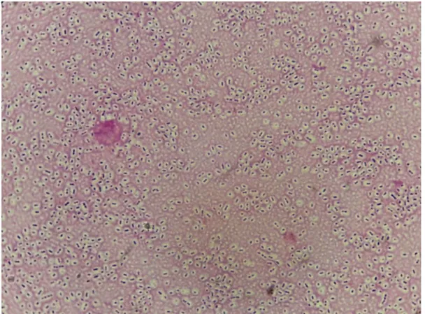
On Gram’s stain, pneumococci are Gram-positive cocci. They appear to be encapsulated lanceolate 8-shaped or candle-flame-shaped diplococci (Figure 3).
Figure 3: Encapsulated Gram-positive diplococci b. Search for hemolysis
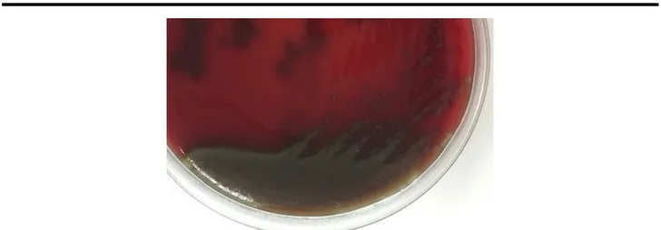
Hemolysin expression is favored by a CO2-rich or even an anaerobic incubation
atmosphere. S. pneumoniae is generally characterized by alpha-type hemolysis. This type of
hemolysis is recognized by the visualization of a greenish halo that surrounds the pneumococcal
Impact of the pneumococcal conjugate vaccine on nasopharyngeal pneumococcal carriage in feverish infants in Mohamed VI teaching hospital of Marrakesh
- 9 -
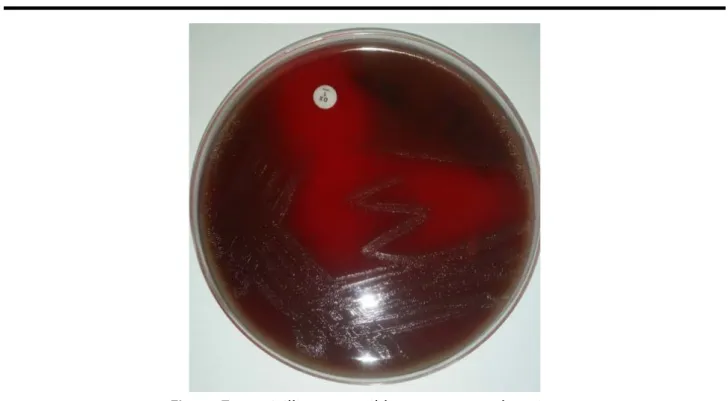
Figure 4: Streptococcus pneumoniae colonies surrounded by a greenish halo on blood agar,
reflecting the alpha-type hemolysis c. Optochin sensitivity test
S. pneumoniae is the only streptococcus that is sensitive to optochin, thus, the optochin sensitivity test is an essential criterion for pneumococcal identification.
The interpretation is done by measuring the diameter of the inhibition zone around the optochin disk. When the diameter is equal to or more than 14 mm, the strain is identified as pneumococcus (Figure 5). As for the strains with a diameter of less than 14 mm, further testing
should be done for the identification of S. pneumoniae.
Impact of the pneumococcal conjugate vaccine on nasopharyngeal pneumococcal carriage in feverish infants in Mohamed VI teaching hospital of Marrakesh
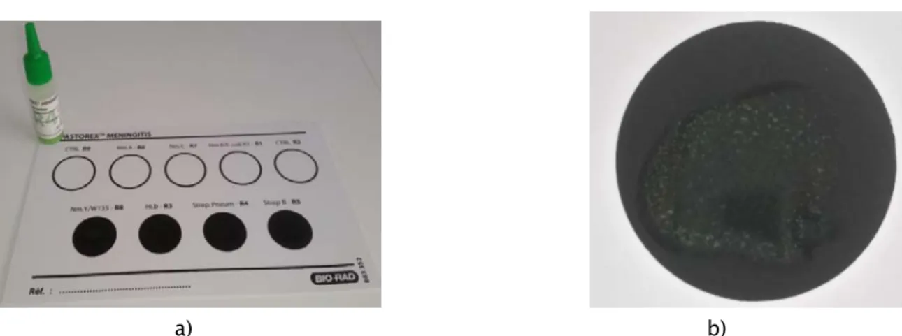
- 10 - d. Agglutination test
S.p identification can also be done by detecting capsular antigens using latex particles
sensitized with specific antibodies. When the matching antigen exists, the latex particles agglutinate heavily, while they remain in homogeneous suspension when it doesn’t.
a) b)
Figure 6: a) PastorexTM
2.3. Storage
meningitidis (Bio-Rad) Streptococcus pneumoniae identification reagent,
b) Positive agglutination
Every strain confirmed to be a Streptococcus pneumoniae was stored for further tests.
The storage was done from pure and fresh S.p colonies into cryo-tubes containing preservation
media (TSB: Trypticase-Soy Broth) plus 15% to 20% of glycerol. The tubes were stored at -80°C. 2.4. Pneumococcal penicillin susceptibility
Research for strains with reduced penicillin susceptibility was performed using 1 µg oxacillin (OXA1) disks, following EUCAST (European Committee on Antimicrobial Susceptibility Testing) recommendations (8).
A strain is said to be susceptible to penicillin G, and therefore to beta-lactams if the diameter of the inhibition zone around the OXA1 disk is more than or equal to 20 mm (Figure 7). While it is considered to be with reduced susceptibility to penicillin G, and thus to beta-lactams if this diameter is less than 20 mm.
Impact of the pneumococcal conjugate vaccine on nasopharyngeal pneumococcal carriage in feverish infants in Mohamed VI teaching hospital of Marrakesh
- 11 -
Figure 7 : penicillin susceptible pneumococcal strain. 2.5. Serogrouping by agglutination
Although serotyping and serogrouping of the pneumococcal isolates in patient specimens are not recommended on day-to-day practice, they become necessary in epidemiological studies aiming to monitor vaccine impact.
Strain serogrouping was performed using latex agglutination method, which is based on antigen-antibody reactions.
S. pneumoniae strain serogrouping was executed using Statens Serum Institut antiserums (from ImmuLexTM Pneumotest, Copenhagen, Denmark).
The type or group identification was first done via a test using the nine polyvalent antiserums from A to I until the acquisition of positive agglutination. After that, the strain was tested against antiserums from P to T until the visualization of agglutination. The type or group was read on the double-entry chessboard that comes with the kit.
The results were interpreted with the naked eye. The reaction is positive when large agglutinates appear in 5 seconds or less (Figure 8).
Impact of the pneumococcal conjugate vaccine on nasopharyngeal pneumococcal carriage in feverish infants in Mohamed VI teaching hospital of Marrakesh
- 12 -
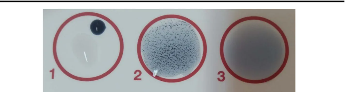
Figure 8: 1) A drop of the reagent + a drop of PBS (Phosphate Buffered Saline), 2) Positive agglutination aspect, 3) Negative agglutination aspect
2.6. Serotyping by molecular biology
Serotyping using molecular biology was executed in cooperation with the Microbiology Laboratory of Ibn-Rushd teaching hospital of Casablanca, according to the protocol and recommendations of pneumococcus molecular typing published by the CDC (9).
2.7. Serotyping using the capsular swelling reaction
This technique was used for serotyping strains belonging to serogroups 6 and 9.
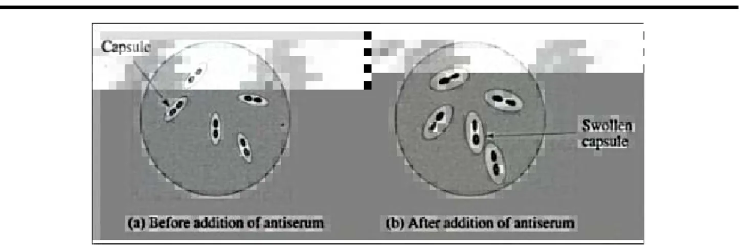
The Quellung reaction, first described by Neufeld (10), consists of a change in the refractive index of the pneumococcal capsule, due to an interaction of this capsule with a type-specific antibody. This change makes the capsule swollen and clearly visible.
The antisera that were used in this reaction were serotype-specific antisera from Staten Serum Institut (Copenhagen, Denmark) (Table I).
Table I: Specific antisera tested for serogroups 6 and 9
Serogroups Tested specific antisera
6 6A – 6B
9 9N/V – 9V
The results were examined under a phase-contrast microscope. The reaction is positive when a bright and clearly visible halo surrounding the bacterium capsule is visualized (Figure 9).
Impact of the pneumococcal conjugate vaccine on nasopharyngeal pneumococcal carriage in feverish infants in Mohamed VI teaching hospital of Marrakesh
- 13 -
Figure 9: Schematic representation of the Quellung reaction results (11)
3. Statistical analyses
Raw data exploitation was performed using the Microsoft Excel 2007 program. Statistical analyses were carried out using the SPSS 20.0 software.
Simpson’s diversity index was calculated in order to evaluate the change in the serotype diversity in the bacterial population, before and after the implementation of the vaccine.
The Chi-square test was used to compare the serotype distribution before and after the introduction of the PCV.
The Rate Ratio (RR) of the vaccine serotype (VS) carriage is the ratio of the VS prevalence in vaccinated children to the VS prevalence in non-vaccinated children (Table II).
The PCV10 efficacy against the vaccine serotype carriage was calculated using the following equation:
VE: Vaccine Efficacy.
VE = (1 – RR) × 100
RR = (a/N1) / (c/N2)
Impact of the pneumococcal conjugate vaccine on nasopharyngeal pneumococcal carriage in feverish infants in Mohamed VI teaching hospital of Marrakesh
- 14 -
Table II: Rate Ratio calculation parameters
S. pneumoniae carriers Total
Vaccine serotypes Non-vaccine serotypes
Vaccinated children a B N1
Non-vaccinated children c C N2
a: Number of vaccinated children that are vaccine serotype carriers b: Number of vaccinated children that are non-vaccine serotype carriers c: Number of non-vaccinated children that are vaccine serotype carriers d: Number of non-vaccinated children that are non-vaccine serotype carriers N1= a + b
Impact of the pneumococcal conjugate vaccine on nasopharyngeal pneumococcal carriage in feverish infants in Mohamed VI teaching hospital of Marrakesh
- 15 -
Impact of the pneumococcal conjugate vaccine on nasopharyngeal pneumococcal carriage in feverish infants in Mohamed VI teaching hospital of Marrakesh
- 16 -
I. Nasopharyngeal
Streptococcus pneumoniae
carriage prevalence in
feverish infants in the region of Marrakesh
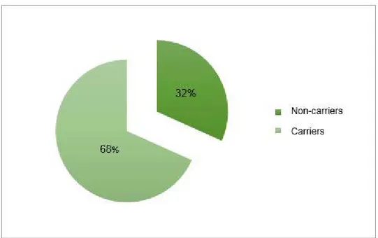
A total of 183 swabs were collected from feverish infants seen at the Emergency Department of Mohammed VI teaching hospital. Out of which, 125 strains were isolated, giving an overall nasopharyngeal pneumococcal carriage rate of 68.3% (Figure 10).
Figure 10: Overall prevalence of Streptococcus pneumoniae nasopharyngeal carriage in sampled
infants
II. Characteristics of
Streptococcus pneumoniae
carriers
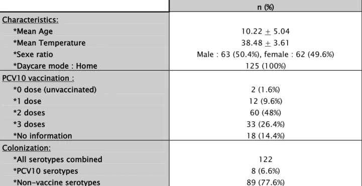
The average age of the feverish infants colonized by S. pneumoniae was 10.22 months
(±5.04 months), with extremes ranging from 2 to 20 months. The sex ratio was 1.03. And the mean temperature detected in these infants was 38.48°C (±3.61°C).
Concerning immunization coverage, 84% of the carriers had received at least one dose of the vaccine, 14.4% had no information about their vaccination history, while only two infants were unvaccinated.
Impact of the pneumococcal conjugate vaccine on nasopharyngeal pneumococcal carriage in feverish infants in Mohamed VI teaching hospital of Marrakesh
- 17 -
The main characteristics of the carrier infants are shown in Table III.
Table III: Characteristics of S. pneumoniae feverish carrier infants in the Marrakesh region
n (%) Characteristics:
*Mean Age
*Mean Temperature *Sexe ratio
*Daycare mode : Home
10.22 + 5.04 38.48 + 3.61 Male : 63 (50.4%), female : 62 (49.6%) 125 (100%) PCV10 vaccination : *0 dose (unvaccinated) *1 dose *2 doses *3 doses *No information 2 (1.6%) 12 (9.6%) 60 (48%) 33 (26.4%) 18 (14.4%) Colonization:
*All serotypes combined *PCV10 serotypes *Non-vaccine serotypes
122 8 (6.6%) 89 (77.6%)
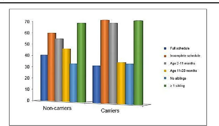
III. Univariate analysis of nasopharyngeal pneumococcal carriage risk
factors in febrile infants in Marrakesh
The comparison of the risk factors associated with nasopharyngeal pneumococcal carriage between the carriers and non-carriers showed (Figure 11):
No difference between the two groups concerning the number of siblings
A different distribution according to age and immunization status: 66.2% of the carrier infants were aged 2 to 11 months and 68.86% had an incomplete vaccination schedule (The third dose was not yet received).
Impact of the pneumococcal conjugate vaccine on nasopharyngeal pneumococcal carriage in feverish infants in Mohamed VI teaching hospital of Marrakesh
- 18 -
Figure 11: Comparison of risk factors associated with NP S. pneumoniae carriage between the
feverish carrier and non-carrier infants in Marrakesh
According to the statistical analysis of these factors, only the association between the immunization status and NP pneumococcal carriage was statistically significant (OR = 1.50). Therefore, febrile infants with incomplete vaccination schedule had a higher risk of being
colonized by S.p than those fully vaccinated (2 + 1) (Table IV).
This analysis did not show any significant difference between the two groups (colonized and non-colonized infants) concerning other factors (age, gender, number of siblings more than 1 and antibiotic treatment) (p = 0.05).
Given that all the sampled infants had a household daycare mode, daycare mode was not considered as a risk factor in this study.
Impact of the pneumococcal conjugate vaccine on nasopharyngeal pneumococcal carriage in feverish infants in Mohamed VI teaching hospital of Marrakesh
- 19 -
Table IV: Pneumococcal carriage risk factors univariate analysis results in febrile infants in Marrakesh
Carriage risk factors S. pneumoniae carriage p OR CI 95 %
Siblings ≥ 1 (n= 123) 84 (68.3%) 0.996 0.998 0.514-1.938
Antibiotic treatment (n= 71) 48 (67.6%) 0.820 0.928 0.485-1.774
PCV10 doses: Incomplete schedule (n= 101) 73 (72.3%) 0.263 1.50 0.736-3.062
Male (n= 95) 63 (66.3%) 0.358 0.746 0.398-1.396
Age : 2 to 11 months (n= 113) 82 (72.6%) 0.130 0.611 0.322-1.159
OR: Odds Ratio CI: Confidence Interval
IV. Impact of immunization status on serotype carriage
Statistical analysis of the impact of the number of administrated vaccine doses on the vaccine and non-vaccine serotype carriage showed a highly significant difference (p<0.001) between the different immunization statuses. In fact, the decrease of vaccine serotype carriage was more important in infants who received the third PCV dose (Figure 12).
Figure 12: Impact of immunization status on vaccine (VS) and non-vaccine serotype (NVS) carriage
Impact of the pneumococcal conjugate vaccine on nasopharyngeal pneumococcal carriage in feverish infants in Mohamed VI teaching hospital of Marrakesh
- 20 -
V. Distribution of isolated
Streptococcus pneumoniae
serotypes in
sampled infants
1. Vaccine serotype distribution
The vaccine serotypes that were carried by the sampled infants accounted for a low percentage of 6.6%. The isolated serotypes were 19F (2 cases), 1 (2 cases) and one case for each of the following serotypes: 14, 23F, 6B and 9V (Figure 13).
Figure 13: Distribution of isolated vaccine serotypes in feverish infants in the region of Marrakesh
2. Non-vaccine serotype distribution
This study concluded to an increase in the rate of the non-vaccine serotype carriage in the enrolled infants. This illustrates the serotype replacement phenomenon; vaccine serotypes have been replaced by non-vaccine serotypes. The isolated non-vaccine serotypes were: 6A (6.4%), 15A/15F (5.6%) and 3.2% for each of serotypes 20, 15B/C, 23B and 13 (Figure 14).
For non-typable and non-vaccine strains, PCR is needed to define their serotypes. 0 1 1 2 2 1 14 19F 23F 6B 9V Number of isolates Serotypes
Impact of the pneumococcal conjugate vaccine on nasopharyngeal pneumococcal carriage in feverish infants in Mohamed VI teaching hospital of Marrakesh
- 21 -
Figure 14: Distribution of isolated non-vaccine serotypes in feverish infants in the region of Marrakesh
*NV: Non-vaccine serotypes other than serotypes 3, 6A, 8, 10, 11A/11D, 12F, 13, 15A/15F, 15B/15C, 17F, 19A, 20, 22F/22A, 23A, 23B, 31, 33F/33A, 34, 35A and 35F. *Non-typable: Strains which hadn’t agglutinated with any antiserum.
VI. Effect of PCV10’s introduction on the diversity of carried
serotypes
Simpson’s index of diversity was calculated before and after the implementation of the PCV10 in order to measure the serotype diversity in carriage. This index was significantly higher (p<0.001) in the post-vaccination period (Table V), reflecting a great serotype diversity compared to the pre-vaccination period.
0 5 10 15 20 25 30 2 8 1 1 3 2 4 7 4 2 3 4 1 1 4 1 3 2 3 2 17 27 N u m b e r o f i s o la te s Non-vaccine serotypes
Impact of the pneumococcal conjugate vaccine on nasopharyngeal pneumococcal carriage in feverish infants in Mohamed VI teaching hospital of Marrakesh
- 22 -
Table V: Simpson’s index of diversity in the pre and post-vaccination periods
Before vaccination* After vaccination p
Simpson’s index of diversity 0,83 0,92 < 0,001
*The index was calculated based on the results of the pneumococcal carriage study conducted in Marrakesh before the introduction of the vaccine (3).
VII. Effect of PCV10’s introduction on vaccine serotypes’ carriage
There was a clear change in the vaccine and non-vaccine serotype distribution after the implementation of the PCV. Before the introduction of the vaccine, the vaccine and non-vaccine serotype carriage rates were 53.9% and 34.2%, respectively. While in the post-vaccine period, the vaccine serotype proportion has significantly decreased (p<0.001) to 6.6% and the non-vaccine serotype percentage has increased to 77.6% (Table VI).The calculated vaccine serotype Rate Ratio (RR (VS)) was less than 1 (0.117), which confirms the contribution of the vaccine in reducing the vaccine serotype carriage rate, with vaccine efficacy (VE) reaching 88.23% (Table VI).
Table VI: Distribution of vaccine and non-vaccine serotypes before and after PCV’s introduction in Marrakesh
Population Pneumococcal carriage Total P RR (VS) VE
VS NVS
Vaccinated 8 (6,6%) 95 (77,6%) 103 < 0,001 0,117 88,23%
Impact of the pneumococcal conjugate vaccine on nasopharyngeal pneumococcal carriage in feverish infants in Mohamed VI teaching hospital of Marrakesh
- 23 -
VIII. Penicillin susceptibility of
S. pneumoniae
serotypes isolated in
carriage in feverish infants in Marrakesh
1. Prevalence of S. pneumoniae serotypes with reduced susceptibility to
penicillin
According to the penicillin susceptibility screening test that was systematically
performed, 33.6% of the S.p serotypes isolated in carriage in the sampled infants were
pneumococci with reduced susceptibility to penicillin (PRSP) (Figure 15).
Figure 15: Prevalence of Pneumococci with reduced susceptibility to penicillin (PRSP) and penicillin-susceptible pneumococci (PSP) carried by feverish infants in the region of Marrakech.
2. Distribution of isolated serotypes according to their penicillin susceptibility
profile
The study of the serotypes according to their penicillin susceptibility profile revealed the following results:
Only two of the 8 isolated vaccine serotypes were PRSP, the serotypes in question were serotypes 14 and 6B (Figure 16).
Impact of the pneumococcal conjugate vaccine on nasopharyngeal pneumococcal carriage in feverish infants in Mohamed VI teaching hospital of Marrakesh
- 24 -
As for non-vaccine serotypes, 38 out of 102 strains were PRSP (which accounts for 82.6% of the overall PRSP serotypes), non-typable and penicillin-resistant strains were very common in this study (21 resistant strains out of 27 non-typable strains). (Figure 17).
Figure 16: Distribution of vaccine serotypes according to their penicillin susceptibility (PRSP: Pneumococcus with reduced susceptibility to penicillin
Impact of the pneumococcal conjugate vaccine on nasopharyngeal pneumococcal carriage in feverish infants in Mohamed VI teaching hospital of Marrakesh
- 25 -
Figure 17: Distribution of non-vaccine serotypes according to their penicillin susceptibility (PRSP: Pneumococcus with reduced susceptibility to penicillin
Impact of the pneumococcal conjugate vaccine on nasopharyngeal pneumococcal carriage in feverish infants in Mohamed VI teaching hospital of Marrakesh
- 26 -
Impact of the pneumococcal conjugate vaccine on nasopharyngeal pneumococcal carriage in feverish infants in Mohamed VI teaching hospital of Marrakesh
- 27 -
I. Generalities
1. History
Streptococcus pneumoniae was isolated for the first time in 1881, simultaneously by two microbiologists: George M. Sternberg in the United States and Louis Pasteur in France. They both independently described roughly lancet-shaped pairs of cocci in human saliva after injecting it into rabbits (12).
By 1886, this microorganism was being referred to as Pneumococcus by Fraenkel, because of its tendency to cause pulmonary disease. Then it was renamed Diplococcus
pneumoniae in 1920, before being finally given its present name -Streptococcus pneumoniae -
in 1974, primarily on the basis of its characteristic growth as chains of cocci in liquid media (12).
2. Taxonomy
Streptococcus pneumoniae (commonly known as Pneumococcus) belongs to the Streptococcaceae family, genus Streptococcus. This genus includes over 40 species grouped into
six major groups: The Pyogenic group, the Anginosus group, the Mitis group, the Salivarius
group, the Bovis group, and the Mutans group (13). According to the Lancefield Classification,
S.p belongs to non-groupable species (14).
3. Epidemiology
3.1. Reservoir and transmission
S. pneumoniae is a commensal bacterium of the human upper airways, more precisely, the nasopharynx. (15).
Impact of the pneumococcal conjugate vaccine on nasopharyngeal pneumococcal carriage in feverish infants in Mohamed VI teaching hospital of Marrakesh
- 28 -
Pneumococci are transmitted from human to human through respiratory droplets (16). The bacteria enter the nasal cavity, attach to the nasopharyngeal epithelial cells and might then either remain as a colonizer or spread to other organs, such as the middle ear, sinuses or down to the lungs via bronchi. Thus, it can potentially cross the mucosal barrier to enter the bloodstream, from which it can cross the blood-brain barrier and cause meningitis (17). (Figure18).
Figure 18: Streptococcus pneumoniae’s different ways of progression in the human body (17).
3.2. Pneumococcal carriage risk factors
Many previous studies have concluded to the presence of several factors according to which the rate of pneumococcal nasopharyngeal carriage varies. These factors include age less than two years, for that’s when the registered rates were higher (18). Siblings ≥ 1, smoking environment, breastfeeding for less than two months and poor socio-economic conditions are also associated with pneumococcal colonization (3) as well as attending daycare centers, where promiscuity favors the transmission of the bacterium (19), and the winter season (20). Viral respiratory infections, especially with syncytial and influenza viruses increase the risk of both pneumococcal carriage and infections (18). A Finnish study has also reported the association of
Impact of the pneumococcal conjugate vaccine on nasopharyngeal pneumococcal carriage in feverish infants in Mohamed VI teaching hospital of Marrakesh
- 29 -
the dietary factor which includes high consumption of sweet pastries and jam with an increased
risk of S.p colonization (21).
3.3. Epidemiological aspects
Streptococcus pneumoniae infections are a major source of morbidity and mortality worldwide.
The World Health Organization estimates that invasive pneumococcal infections are responsible for the death of almost 500 000 children aged less than five years old, most of which are in developing countries (22). Moreover, hospitalizations for pneumococcal pneumonia (23), as well as medical management of acute otitis media (24), represent a considerable economic burden, especially in pediatrics. It should be noted that invasive pneumococcal infections are also common in the elderly and patients with underlying immunosuppressive diseases such as HIV infection and chronic liver disease (25).
The rate of pneumococcal infections and the deaths caused by them varies according to the socio-economic status of each country; it is higher in developing countries with major mortality prevalence in Sub-Saharan Africa and Southern Asia (61% of overall mortality) (26). Furthermore, the timing of disease onset in children also differs between low-income and high-income countries. In developing countries, most pneumococcal infections and deaths among children <5 years of age occur in the first year of life, with a peak in disease incidence before 6 months of age; in developed countries, these infections peak closer to 12 months of age with about half of the episodes occurring by 18 months (27). (Figure19).
Impact of the pneumococcal conjugate vaccine on nasopharyngeal pneumococcal carriage in feverish infants in Mohamed VI teaching hospital of Marrakesh
- 30 -
Figure 19: Distribution of cases of invasive pneumococcal disease for children <5 years, by months of age for children in a developing country (South Africa) and in an industrialized
country (United States) (27).
In Morocco, the implementation of PCV13 in 2010 then PCV10 in 2012 significantly reduced the incidence of invasive pneumococcal infections in infants < 2 years of age, from 34.6 to 13.5 per 100 000 inhabitants, respectively before and after the vaccine introduction (28).
4. Microbiological aspects
4.1. Morphology and structureOn Gram’s stain, pneumococci have a characteristic morphology: they are Gram-positive, lancet-shaped, encapsulated (Bright halo surrounding the bacteria) and 8-shaped or “candle-flame-shaped” diplococci (15). (Figure 20). They can also grow in chains (29).
Impact of the pneumococcal conjugate vaccine on nasopharyngeal pneumococcal carriage in feverish infants in Mohamed VI teaching hospital of Marrakesh
- 31 -
Figure 20: Streptococcus pneumoniae visualized as encapsulated Gram-positive diplococcic
4.2. Growth characteristics
a. Growth media
As a demanding bacterium, S. pneumoniae requires growth factors for its culture.
Therefore, Soy-Trypticase and Columbia agar with 5% sheep blood are commonly used for culturing pneumococci. Chocolate blood agar to which a vitamin complex has been added is also a favorable medium for pneumococcal growth (15). Mueller Hinton agar with blood is used for
antibiotic susceptibility testing (15). In liquid media, S.p can grow in Brain and Heart infusion
broths (BHI).
b. Growth conditions
Pneumococci are anaerobic or facultative aerobic bacteria. The optimal conditions for their growth are a carbon-dioxide-enriched atmosphere (5 to 10%) or even an anaerobic atmosphere, a temperature ranging from 35 to 37°C and a pH=7.8 (6.5 – 8.3) (30), for an incubation period of 24 to 48 hours.
Impact of the pneumococcal conjugate vaccine on nasopharyngeal pneumococcal carriage in feverish infants in Mohamed VI teaching hospital of Marrakesh
- 32 - c. Colonies’ aspect
S. pneumoniae colonies usually measure 0.5 to 1.5 mm and are surrounded by greenish halos showing incomplete hemolysis and transformation of hemoglobin into biliverdin (Alpha-type hemolysis). They are opaque or grayish, convex and with regular edges. Another feature
characterizing S. pneumoniae colonies is the central umbilicus-like depression that is caused by
the pneumococcal autolysin (Figure 21). Serotype 3 colonies often have a mucoid aspect, due to the excessive development of the capsule (Figure 22). (15).
Figure 21 : Streptococcus pneumoniae colonies : note the central depression (29).
Impact of the pneumococcal conjugate vaccine on nasopharyngeal pneumococcal carriage in feverish infants in Mohamed VI teaching hospital of Marrakesh
- 33 - 4.3. Biochemical characteristics
Pneumococci, as Streptococcaceae family members, produce lactic acid by glucose
fermentation (Homofermentative). They are catalase-negative, oxidase-negative and anaerobic-aero tolerant (15).
4.4. Antigenic characteristics
One of the major factors of pneumococcal virulence is the capsule. It is made up of polysaccharide macromolecules, and its antigenic structure allows pneumococcal strains serotyping (Lund’s Danish Classification). Currently, over 90 serotypes have been described (31) (Table VII)
These serotypes have different propensities concerning pathogenicity and antibiotic resistance (32).
S.p possesses antigens other than its polysaccharide capsular antigens: The
species-specific substance (C) which is a polysaccharide consisting of teichoic acid, the R antigen that is a protein and usually is masked by the capsular antigens, and the M antigen which is a protein type-specific antigen (30).
Table VII: Lund’s Danish Classification of Streptococcus pneumoniae (31)
1, 2, 3, 4, 5, 6A, 6B, 7F, 7A, 7B, 7C, 8, 9A, 9L, 9N, 9V, 10F, 10A, 10B, 10C, 11F, 11A, 11B, 11C, 11D, 12F, 12A, 12B, 13,
14, 15F, 15A, 15B, 15C, 16F, 16A, 17F, 17A, 18F, 18A, 18B, 18C, 19F, 19A, 19B, 19C, 20, 21, 22F, 22A, 23F, 23A, 23B,
24F, 24A, 24B, 25F, 25A, 27, 28F, 28A, 29, 31, 32F, 32A, 33F, 33A, 33B, 33C, 33D, 34, 35F, 35A, 35B, 35C, 36, 37, 38,
Impact of the pneumococcal conjugate vaccine on nasopharyngeal pneumococcal carriage in feverish infants in Mohamed VI teaching hospital of Marrakesh
- 34 -
5. Pathogenicity
5.1. Virulence factors
S. pneumoniae has many virulence factors. They can be on the surface of the intact bacterium (Capsule, PspA…) or be expressed after its destruction or lysis (pneumolysin…). These factors are responsible for inflammatory reactions that can sometimes be very deleterious for the host via complement activation (33).
a. Capsule
The pneumococcal capsule is the first discovered and the most important virulence factor. It is the outermost element of the bacterium. In vivo, it allows the growth of pneumococci and considerably hinders phagocytosis by acting as a physical barrier and preventing phagocyte receptors from being in contact with complement components C3b that have eventually attached to the bacterial wall. The capsule is also able to electrostatically repulse the phagocytes that are negatively charged, like the capsular polysaccharides. And it protects the surface proteins from circulating antibodies.
The strains’ virulence and invasiveness vary depending on the serotype, in other words, depending on the amount of produced capsule and its composition (33).
b. Pneumolysin
Pneumolysin, a thiol-activated toxin, is located in the cytoplasm. It is released in the outside under the action of LytA, a bacterial autolysin.
Pneumolysin has a cytotoxic activity. It inhibits the beating of the cilia involved in the mucociliary clearance of the bronchi and destroys the bronchial epithelium. It is responsible for a decrease in the bactericidal activity of monocytes and neutrophils. It also causes the inhibition of lymphocyte proliferation and reduction of antibody synthesis (33).
There is very little difference in the sequence of pneumolysin from one serotype to another which could be useful for the development of a pneumococcal protein vaccine (34).
Impact of the pneumococcal conjugate vaccine on nasopharyngeal pneumococcal carriage in feverish infants in Mohamed VI teaching hospital of Marrakesh
- 35 - c. Other virulence factors
c.1. Pneumococcal surface proteins (Psp A and C)
They facilitate pneumococcal systemic invasion by inhibiting the alternative complement pathway. Moreover, PspA, unlike PspC, is able to attach to lactoferrin, a human iron-sequestering glycoprotein, and thus provides enough iron for bacterial growth (33).
c.2. Other factors expressed after bacterial lysis
Wall components, especially teichoic and lipoteichoic acids and phosphorylcholine, can also be responsible for triggering inflammatory reactions (33).
c.3. Pili
Studies on the role of pili in the pathogenesis of pneumococcal infections are still few to date. However, in vitro, it has been shown that pili are involved in the process of pneumococcal adhesion to the pulmonary epithelial cells (35), as well as in the invasion and colonization (36).
5.2. Infections caused by Streptococcus pneumoniae
Although pneumococcal carriage is asymptomatic, it can lead to respiratory or even systemic infections (37). These infections can be invasive (Invasive pneumococcal disease; IPD) or non-invasive (non-IPD). The development of the pneumococcal disease is conditioned by many factors, the most important of which are the strain’s virulence, the immunity status, especially the humoral immunity, and the presence of respiratory viral infections (16).
a. Non-invasive pneumococcal diseases
They are mucosal infections of the respiratory epithelium that are spread by contiguity, such as acute otitis media, sinusitis and pneumonia (16).
In fact, these infections are favored by surface proteins such as choline-binding protein A (Cbp A) and neuraminidase Nan A which cause a decrease in mucus viscosity and favor bacterial adhesion. During intercurrent infections by respiratory viruses, this mechanism is amplified under the effect of neuraminidase of viral origin (1).