Science Arts & Métiers (SAM)
is an open access repository that collects the work of Arts et Métiers Institute of
Technology researchers and makes it freely available over the web where possible.
This is an author-deposited version published in: https://sam.ensam.eu Handle ID: .http://hdl.handle.net/10985/11219
To cite this version :
Thierry PALIN-LUC, E. SELLIER, F. D'ERRICO, M. VANHAEREN - Elastomer and resin replicas for sem observation of metallic materials - Experimental Techniques - Vol. 26, n°3, p.33-37 - 2002
Any correspondence concerning this service should be sent to the repository Administrator : [email protected]
N . . . . . . . . . . . . . . . . . . . . . . . . . . . . . . . . . . . . . . . . . . . . . . . . . . . . . . . . . . . . . . .
ELASTOMER AND RESIN REPLICAS FOR SEM
OBSERVATION OF METALLIC MATERIALS
Table 1—Products Used
ELASTOMER NAME COMPANY NAME AND ADDRESS
Provil Novo Light Heraeus Kulzer
D-41538 Dormagen, Germany
Impregum F ESPE
Dental-Medizin GmbH & Co. KG,
D- 82229 Seefeld, Germany Colte`ne Pre´sident Light Body Colte`ne AG,
Feldwiesenstrasse 20, CH-9450 Altstatten, Switzerland
T
he replica technique is often used to study damage evolution at the surface of specimens or industrial components and understand the physical phenom-ena responsible for fatigue crack initiation before failure. Replicas are usually made from acetate cellulose film. This paper presents an alternative technique generally used by archaeologists to study lithic use-wear and bone modification. A mold is made from a dental elastomer (sili-con based impression material) and a positive replica is made by casting epoxy resin in the mold. Comparative SEM analysis of damaged metallic specimens and their resin rep-licas show that this technique provides a good resolution and preserves details up to 0.5 m. This easy and low cost method allows a systematic study of micro-crack growth.Introduction
Studying damage evolution of materials under cyclic loading is an important challenge for scientists and engineers since most structures load in this way. In the laboratory, the rep-lica technique is used to observe damage evolution at the surface of specimens and components after different steps of loading, i.e., different cycles. Observation of replicas made after several cycles allows the monitoring of fatigue crack initiation as well as fatigue crack growth.1,2As the first crack
initiation site cannot be known in advance and looking for a small fatigue crack on a specimen surface is a waste of time, it is impractical to monitor small fatigue crack growth with-out a replication technique or in situ SEM observation. With an efficient replication technique, initiation sites, number of micro-cracks, crack length and growth can be quantified and their evolution versus the number of load cycles studied. This is of major importance to understand the physical phe-nomena responsible for fatigue damage of materials. This paper presents a replication method inspired by a tech-nique used by archaeologists3 to replicate bone and stone
objects for SEM study. A dental elastomer is used to replicate the surface under analysis and an epoxy resin to make a three-dimensional cast. These products are described and it is explained below how to use them to obtain the best re-sults. The cleaning procedure of the original specimen sur-face before molding and the preparation of replicas for SEM analysis are described, and the potential of the method dis-cussed. The products are relatively cheap and easy to use.
MATERIALS AND METHOD
Specimen and Elastomer Preparation
The metallic specimens observed with SEM and duplicated are made of spheroidal graphite cast iron FGS800-2 (Afnor standard); details about this material and the specimens are given in Ref. 4.
T. Palin-Luc is with LAMEFIP-ENSAM, Talence Cedex, France. E. Sellier is affil-iated with CREMEM-Universite Bordeaux 1, Talence Cedex, France. F. d’Errico and M. Vanhaeren are affiliated with IPGQ (UMR 5808 CNRS), Talence Cedex, France.
SEM analysis of metallic specimens requires perfectly clean surfaces. For our application we have adopted a cleaning procedure similar to that described in Refs. 3 and 5. Traces of grease or oil are removed from the specimen with trichlor-ethylene or acetone and a piece of gauze. Paper handkerchief is avoided because of streaking risk. If the contamination of the surface is high and the size of the specimen compatible with its use, an ultrasonic cleaning may be very effective. As suggested by Rose3, the ultrasonicator is run for periods of
one minute until the surface is clean. The final cleaning is achieved by projecting trichlorethylene onto the specimen which is subsequently dried with a hair-dryer. On line com-pressed air should be avoided since it may contaminate the specimen surface with oil particles. Direct contact of the specimen with fingers is to be avoided during the last stage of cleaning. Before replication, specimens are stored in air-tight boxes or plastic bags to isolate them from contaminants in the air.
The following three elastomers were used to test the quality of their replicas for microscopic analysis of metallic speci-mens: Provil Novo Light, Impregum F and Colte`ne Pre´sident Light Body. The corresponding companies and addresses are given in Table 1. These elastomers are usually used in den-tistry to mold teeth.
Clear instructions on how to prepare these elastomers are given by each supplier in a technical note accompanying the product. Proportion of elastomer and catalyst should be care-fully respected to produce high quality molds. At room tem-perature, the mixing time of the two constituents is approx-imatively 30 seconds. Though very simple to use, this replication technique requires some precautions. Since the viscosity of these elastomers is low, the flow of elastomer out of the molded area should be limited.6This is achieved in
34
EXPERIMENTAL TECHNIQUES May/June 2002 N . . . . . . . . . . . . . . . . . . . . . . . . . . . . . . . . . . . . . . . . . . . . . . . . . . . . . . . . . . . . . . . . . . . . . .ELASTOMER AND RESIN
REPLICAS FOR OBSERVATION
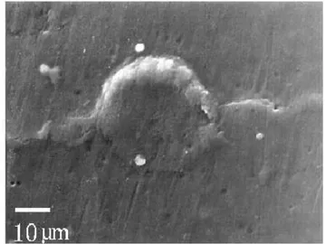
Fig. 1: Elastomer replica made of the Colte`ne Pre´sident Light body product and its ring of Prestocil before casting
Fig. 2: Positive resin replica made from a Provil Novo Light replica
Fig. 3: Positive resin replica made from an Impregum F replica
Fig. 4: Positive resin replica made from a Colte`ne President Light Body replica
Next, the specimen is put horizontally and the molding ma-terial applied with a syringe. The elastomer is immediately flattened with a flexible painting knife in order to eliminate air bubbles at the contact of the replicated surface. The ob-ject and the elastomer are left under a desk light for five minutes to facilitate the hardening of the product. Once this delay is over, the replica is separated from the original by hand. Finger contact with the replicated area and pollution by dust particles must be avoided during this operation. The removal of the elastomer is much easier than with acetate cellulose film because of the flexibility of the elastomer. Com-plex shapes can be duplicated this way. However one should be careful not to bend nor twist the replica too much as this may create micro-cracks in the replica and make it unsuit-able for observation. The replica is stored in an airtight box for 48 hours before casting it, which allows gas to be given off by the elastomer.
Casting
To facilitate SEM analysis of the replicated surface, a posi-tive replica is made by casting resin into the elastomer. This technique allows us to observe the specimen surface easily, count its defects and identify its constituents. It provides the actual morphology of the specimen surface thus avoiding the tedious and tiring exercise of reconstructing mentally posi-tive features from their negaposi-tive imprints. Furthermore, an effective metallization of the elastomer is difficult to per-form.
Positive replica is made by using an epoxy resin as suggested by Rose.3 We choose the resin according to the following
specifications: (i) the positive must be removed from the elas-tomer without altering its microscopic features, (ii) it must show a minimum amount of shrinkage and distortions, and (iii) it should represent a very accurate reproduction of the negative replica. The resin used in the frame of this work, which matches these requirements, is the RBS resin used with the RBS catalyst (T2L Chimie S.A., Z.I. la Plaine, F-11500 Quillan, France).7This low cost transparent resin has
a low viscosity (2300 mPa.s at 25⬚C) which is an important factor for detail rendering.
N
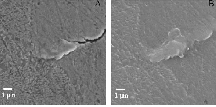
Fig. 5: Comparison between the specimen (A) and the elastomer replica (B)
Fig. 6: Micro-crack and the three phases of the spheroidal graphite cast iron: graphite nodule, ferrite and pearlite matrix. (A) on a specimen, (B) on an elastomer positive replica. The white mark on the replica (B) is a dust particle
N . . . . . . . . . . . . . . . . . . . . . . . . . . . . . . . . . . . . . . . . . . . . . . . . . . . . . . . . . . . . . . . . . . . . . .
ELASTOMER AND RESIN
REPLICAS FOR OBSERVATION
To reinforce the walls of the elastomer replicas and to retain the resin, a ring of another more rigid elastomer is made (Fig. 1) using the Prestocil silicon-based dental elastomer (Dental De´pot Oce´an, Chemin de Bragues, F-33170 Gradig-nan, France). This is a two component silicon based elasto-mer paste also used in dentistry. It has a mixing time of approximately 30–45s and a working time of approxi-matively 1-1mn 30s. It partially hardens after 4 minutes; complete hardening occurs after 24 hours. The resin can then be cast into the mold (elastomer replica and ring of Prestocil).
Preparation of the epoxy resin is performed according to the manufacturer’s technical data sheet. The most important point for good quality positive replicas is to mix it thor-oughly. As noticed by Rose3 and d’Errico6, air-bubbles are
incorporated in the epoxy resin during mixing and, after casting, may remain stuck to the elastomer surface resulting in voids on the positive. To avoid this, we use the casting technique described by d’Errico6to study lithic use-wear and
bone damages. The epoxy resin is gradually poured into the mold by dropping the resin from a height of approximatively one meter. The viscosity of the resin and its density lead to the creation of a thin thread of resin. Air bubbles with a diameter larger than this thread disappear. To be fully cured, the resin is left 24 hours at 23–25⬚C. Before removing carefully the positive replica from the mold one must check that the resin is not tacky to the touch. The positive replica has to be kept far from dust particles and not to be touched with fingers. We recommend storing replicas in an airtight box.
SEM OBSERVATION
Preparation of resin replica for SEM observation does not substantially differ from the one used to analyze other kinds of samples with this equipment. Since observation with SEM requires conductive samples, resin replicas are coated under vacuum with gold-palladium. Then they are mounted on stubs. A bridge of silver lac is put at the interface replica-support to further improve electrical conductivity. We obtain good quality images observing the replicas at a voltage of 15 kV and taking micrographies as secondary electron im-ages (SEI). Observing positive resin replicas is easier and more reliable than analyzing negative elastomers. In our ex-perience, metal-coating under vacuum of the latter produces micro-cracks, which are often difficult to distinguish from load-induced damages and thus constitute a potential source of mistake.
RESULTS AND DISCUSSION
As clearly demonstrated by Figs. 2, 3 and 4, the replicas made with Colte`ne light body elastomer provide the best res-olution. These differences do not necessarily depend on the quality of the elastomer. They may be due to incompatibility between elastomer and resin. This might well be the case for Impregum F since, as shown in Fig. 3, the surface of its resin replica is covered with elastomer particles which have been torn off by the removal from the mold. In the following, elastomer replicas have been made of Colte`ne.
Figure 5, which compares the same detail on the specimen and the replica made of Colte`ne elastomer and RBS resin, allows us to fully appreciate the excellent resolution pro-vided by the technique tested in the present study. Both im-ages retain details up to 0.5m, i.e., the features required for SEM observation and quantification of damages on met-als. This indicates that we can, by this means, reliably follow and record damage evolution of such materials under cyclic loadings. The three phases of the spheroidal graphite cast iron,4for example, may be easily identified on Figure 6. The
pearlite micro structure of the matrix, the two micro-cracks, and the ferrite around the graphite nodule are as clear on the actual specimen (A) as on the replica (B).
Artifacts on the replica may be successfully avoided by care-fully following the replication protocol described above. Fig-ure 6B shows pollution by a dust particle. FigFig-ure 7A illus-trates the consequence on the replica of an insufficient drying of the trichlorethylene used to clean the specimen surface. Evaporation of the trichlorethylene kept in a crack produces a groove-like artifact which is absent on the replica produced with an elastomer taken after a complete drying of the specimen (Fig. 7B).
If used with some precautions, the technique we propose here is particularly appropriate to follow damage on the sur-face of specimens or components under cyclic loading. Due to the low stiffness of the elastomer, removing the negative replica from the duplicated surface is easier than with ace-tate cellulose films, the material usually used for duplicating metal surfaces.1,2 Elastomer can be successfully used even
when the replicated surface has a complex morphology, which often produces the tear of the acetate cellulose peels. Furthermore, the products are low cost and easy to use.
CONCLUSION AND PROSPECTS
We have tested the reliability for mechanics studies of a technique currently used by anthropologists and archaeolo-gists to investigate micro damage on archaeological materi-als [8]. This low cost and easy to use technique provides the resolution required for many laboratory observations of damage initiation and growth on metals (details up to 0.5m). Due to the low stiffness of the dental elastomer used to make the negative replica, surfaces with a complex shape can be replicated. We have demonstrated the rele-vance of this technique to analyze damage growth at the surface of spheroidal graphite cast iron specimens under cy-clic loadings. Future studies must check the reliability of this technique on other metals such as steels, aluminium alloys, and titanium alloys. Results presented in the frame-work of this study, however, are promising.
References
1. Hua, C.T., and Socie, D.F., ‘‘Fatigue Damage in 1045 Steel Un-der Constant Amplitude Biaxial Loading,’’ Fatigue Engng Mater.
Struct., 7(3), 165–179 (1984).
2. Cle´ment, P., Angeli, J-P., and Pineau, A., ‘‘Short Fatigue Crack Behaviour in Nodular Cast Iron,’’ Fatigue Engng Mater. Struct.,
. . . . . . . . . . . . 3. Rose, J.J., ‘‘A Replication Technique for Scanning Electron Mi-croscopy: Applications for Anthropologists,’’ American J. Physical
Anthropology, 62, 255–261 (1983).
4. Palin-Luc, T., Lasserre, S., and Be´rard, J-Y., ‘‘Experimental Investigation on the Significance of the Conventional Endurance Limit of a Spheroidal Graphite Cast Iron,’’ Fat. Fract. Engng. Mater.
Struct., 21(3), 192–200 (1998).
5. Bromage, T.G., ‘‘The Scanning Electron Microscopy Replica Technique and Recent Applications to the Study of Fossile Bone,’’
Scanning Microscopy, 1(2), 607–613 (1987).
6. D’Errico, F., ‘‘Traces d’Usure sur l’Industrie Lithique : Appro-che Me´thodologique et Proposition d’Une Technique,’’
L’Anthropologie ( Paris), 89(4), 439–456 (1985).
7. D’Errico, F., ‘‘Nouvelles Observations sur Deux Pie`ces en Silex et un Objet en Os de la Grotte du Vallonet (Alpes-maritimes),’’
L’Anthropologie ( Paris), 92(2), 615–628 (1988).
8. D’Errico, F., ‘‘The Use of Resin Cast for the Study of Use-wear,’’ Scanning Electron Microscopy in Archaeology, Olsen, S.L., editor, British Arch. Reports Int. series, pages 155–167, 452 Oxford (1988).䡵