HAL Id: tel-00717997
https://tel.archives-ouvertes.fr/tel-00717997
Submitted on 15 Jul 2012HAL is a multi-disciplinary open access archive for the deposit and dissemination of sci-entific research documents, whether they are pub-lished or not. The documents may come from
L’archive ouverte pluridisciplinaire HAL, est destinée au dépôt et à la diffusion de documents scientifiques de niveau recherche, publiés ou non, émanant des établissements d’enseignement et de
Control of ligand-receptor interaction by tuning the
molecular environment
Valentina Lo Schiavo
To cite this version:
Valentina Lo Schiavo. Control of ligand-receptor interaction by tuning the molecular environment. Biological Physics [physics.bio-ph]. Université de la Méditerranée - Aix-Marseille II, 2011. English. �tel-00717997�
A
IX-M
ARSEILLEU
NIVERSITÉF
ACULTÉ DE SCIENCES DEL
UMINYControl of ligand-receptor interaction by
tuning the molecular environment
Doctoral Thesis
Valentina Lo Schiavo
Member of jury:
Dr. Olivier Thoumine, reviewer
Dr. Frédéric Pincet, reviewer
Dr. He Hai-Tao, examiner
Prof. Philippe Dumas, examiner
Prof. Pierre Bongrand, advisor
Dr. Laurent Limozin, co-advisor
Abstract.
Cell adhesion is a fundamental biological process mediated by specific molecular bonds formed by ligands and receptors attached to surfaces. Formation and rupture of these bonds depend on kinetic, mechanical and structural factors. The goal of this work was to observe how the ICAM-1 (Inter-Cellular Adhesion Molecule 1) – anti ICAM-1 interaction can be modified by modification in i) the multivalency of the molecules involved in the bond ii) the topography of the surface and iii) on the mobility of the ligands. The main technique used for this purpose was the laminar flow chamber, complemented by single-particle tracking in fluorescence.
The study on multivalency effects, using monomeric and dimeric ICAM-1, was performed in the absence or the presence of mechanical force, revealing the higher stability of divalent bonds. Also, a force- and time- strengthening dependence was found and described with a two-parameter function, showing, for divalent bonds, an intermediate behaviour between parallel and successive rupture of monovalent bonds. The adhesion frequency of monovalent and divalent bonds exhibit different values accounted for by the difference in length of these molecules.
Adhesion experiments were performed varying the topography of the substrate at the nanoscale for the investigated molecules. A comparison of bond kinetics on these surfaces did not show differences either in attachment or in rupture.
In the flow, the contact time between molecules is controlled by convection of microspheres. Recent results show that there is a minimal time required to form the bond (Robert et al. 2011). To test this prediction, ligands were anchored to supported lipid bilayer (SLB) to investigate how the diffusion can modify the adhesion. Experimentally, the adhesion frequencies of the bonds showed similar behaviour for fixed and fluid SLB. However, 2D numerical simulation predicted an effect on bond formation even at low ligand diffusion. The diffusion seemed to play a role in bond dissociation, strongly limiting the dissociation on the fluid bilayer. This effect can be explained by the possible presence of multiple bonds due to ligand accumulation at the contact area.
Laminar flow chamber and single-particle tracking allowed us to better understand the mechanisms of adhesion and the behaviour of interacting ICAM-1
molecules at single molecule level, when the molecular environment was modified. Similar work can be performed on other adhesion molecules in order to gain a wider knowledge of the adhesion mechanisms, or on TCR – pMHC bonds which are extremely important in immune response.
Resumé.
L'adhésion cellulaire est un processus biologique fondamental contrôlé par des liaisons moléculaires spécifiques entre ligands et récepteurs attachés à des surfaces. La formation et la rupture de ces liens dépendent de facteurs cinétiques, mécaniques et structurels. Le but de ce travail était d'observer comment l'interaction ICAM-1 (Inter-Cellular Adhesion Molecule 1) - anti ICAM-1 pouvait être modifiée en jouant i) sur la multivalence de molécules impliquées dans la liaison ii) sur la topographie de surface et iii) sur la mobilité des ligands. A cette fin, on a principalement utilisé une chambre à flux laminaire, complété par une détection de molécule unique par fluorescence.
L'étude sur les effets de multivalence, utilisant des monomères et dimères d'ICAM-1, a été réalisée en absence ou en présence d'une force mécanique, montrant la plus grande stabilité des liaisons divalentes. En outre, un renforcement avec la force et le temps a été trouvé et décrit avec une fonction à deux paramètres, montrant, pour les liaisons divalentes, un comportement intermédiaire entre rupture parallèles et successives des liaisons monovalentes. La fréquence d'adhésion des liaisons monovalentes et divalentes présente différentes valeurs causées par la différence de longueur de ces molécules.
Les expériences d'adhésion ont été effectuées en variant la topographie du substrat à l'échelle nanométrique pour les molécules étudiées. Une comparaison des cinétiques de liaisons sur ces surfaces ne montrent pas de différences soit dans la formation ou dans la rupture.
Dans l'écoulement, le temps de contact entre les molécules est contrôlé par la convection de microsphères. Des résultats récents montrent qu'un temps minimum est requis pour former la liaison (Robert et al. 2011). Pour tester cette prédiction, les ligands sont ancrés à une bicouche lipidique (SLB) pour étudier comment la diffusion peut modifier l'adhésion. Expérimentalement, les fréquences d'adhésion des liaisons ont
montré un comportement similaire pour les SLB fixes et fluides. Toutefois, une simulation numérique 2D prédit un effet sur la formation de la liaison, même lorsque la diffusion des ligands est faible. Il semblerait que la diffusion joue un rôle dans la dissociation de la liaison, limitant fortement la dissociation de la bicouche fluide. Cet effet peut être expliqué par la présence éventuelle de liaisons multiples dues à l'accumulation des ligands sur la surface de contact.
La chambre à flux laminaire et le suivi de particule individuelle a permis de mieux comprendre les mécanismes d'adhésion et le comportement de l'interaction des molécules d'ICAM-1 au niveau de molécule individuelle, lorsque l'environnement moléculaire a été modifiée. Des travaux similaires peuvent être effectuées sur d'autres molécules d'adhésion afin d'atteindre une connaissance beaucoup plus large des mécanismes d'adhésion, ou sur les liaisons entre TCR et pMHC qui sont extrêmement importantes dans la réponse immunitaire.
Acknowledgements
First of all, I offer my most sincere gratitude to my supervisors, Prof. Pierre Bongrand and Dr. Laurent Limozin who have supported me throughout my thesis with their immense knowledge and professionalism. This thesis would not have been possible without their patience and their passion.
I would like to show my gratitude to Dr. Philippe Robert who showed me the secrets of flow chamber. His brilliant ideas were important during scientific discussions and his jokes were well appreciated too. I am sure he will be so funny even with the little Alice!
I would like to thank Dr. Kheya Sengupta who introduced me to the Langmuir-Blodgett-Schaefer world and read the manuscript correcting all the mistakes due to my 'Italian English'. Many thanks to Dr. Zohar Mishal who kindly performed AFM measurements on my samples and strongly encouraged me during the hardest moments. I will remember him when I will win the Nobel Prize and I will be called “Martyr of Science”.
I owe my deepest gratitude to Dr. Pierre-Henri Puech who has made available his support both at scientific and personal levels. His suggestions and technical computer skills made my thesis work easier, while his kindness and availability were precious in a country whose language was totally unknown to me. I wait for a new painting for the day of the defence!
I am also indebted to many of my colleagues for their support, overall Dr. Roxane Fabre, my “sister”. I cherish our free time together and hope that we will be able to stay in touch despite the large distances between us. “See you later alligator, in a while crocodile!”.
I thank my parents for supporting me throughout all my studies at University, from Messina to Marseilles, via Rome. I know it was not easy for them letting their daughter follows her dreams far from them, but they have never hampered me (well, my dad tried it a bit, but without results!).
Many thanks to my Italian friends, Pippo, Ciccio, Piero and Crispilla who have always been with me, in spite of the distances between us. I am grateful to Sergy (Sergio & Angy, just married!)...too many things to say...just thanks for existing! (Is my personal b&b moving from Rome to San Francisco?).
Table of contents
Introduction
...11. Cellular adhesion...3
1.1 Main functions...3
1.2 Cellular adhesion and diseases...5
2. Physical approach...6
2.1 From biology to biophysics...6
2.2 Cellular model vs acellular model...7
2.3 Models for ligand and receptor...8
3. Single molecule studies...13
3.1 Role and importance of proteins interaction in cell adhesion...13
3.2 Advantages of single-molecule study...14
3.3 Affinity and kinetics parameters necessary to describe a bond...15
3.4 Description of bond formation and kon...17
3.5 Description of bond rupture and koff...19
4. Single molecule methods...21
4.1 Laminar flow chamber...22
4.1.1 Principles...22
4.1.2 Measure of kinetics of single bonds...25
4.2 Atomic Force Microscopy (AFM)...27
4.3 Biomembrane Force Probe (BFP)...29
5. Importance of molecular environment in adhesive interactions...33
5.1 Regulation of cell adhesion by the cytoskeleton...34
5.2 Role of glycocalyx in modulating cell adhesion...34
5.3 Effect of valency on ligand-receptor interaction...35
5.4 Influence of surface topography...37
5.5 Effect of ligand lateral diffusion on bond kinetics...38
6. Main objectives...40
Materials and methods
...437. Functionalization of surface...45
7.1 Constraints imposed by single molecule experiments...45
7.2 Cleaning of slides...46
7.3 Grafting of immobile ligands on glass...48
7.3.1 ICAM-1 vs anti ICAM-1 on smooth substrates ...48
7.3.2 ICAM-1 vs anti ICAM-1 on rough substrates...51
7.3.3 pMHC vs anti HLA...51
7.4 Grafting of mobile ligands on glass...52
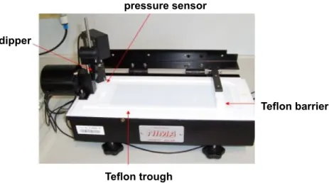
7.4.1 Langmuir-Blodgett-Schaefer technique...53
7.4.2 ICAM-1 – anti ICAM-1 functionalization...57
7.4.3 pMHC – anti HLA functionalization...59
8. Laminar flow chamber...61
8.1 Beads functionalization...62
8.2 Realization of experiments...65
8.3 Acquisition of video signal...66
8.4 Data storage and analysis...67
9. Experiments in fluorescence...70
9.2 TIRF microscopy...71
9.3 Quantum dots...73
9.4 Measurement of ligand density on surface...73
9.4.1 Intensity measurements...74
9.4.2 Single-particle counting...74
9.5 Measurement of diffusion coefficient...76
9.5.1 Continuous Photobleaching for lipid diffusion...77
9.5.2 SPT for ligand diffusion...79
Results and discussions
...8310. Study of effect of divalency...85
10.1 Kinetics at zero force...86
10.2.2 Effect on kon...89
10.2.2 Effect on koff...90
10.3 Discussion...94
11. Study of the effect of surface topography...96
11.1 AFM images of surface topography...96
11.2 Measurements for ligand density...97
11.2.1 Intensity measurement...98
11.2.2 Single-particle counting...99
11.3 Flow chamber experiments...100
11.4 Discussion...103
12. Study of effect of mobility...105
12.1 Results for lipid diffusion...105
12.2 Results for ligand diffusion coefficient...106
12.2.1 pMHC – anti HLA interaction...107
12.2.2 Fc-ICAM-1 – anti ICAM-1 couple...109
12.3.1 Effect on on-rate...112
12.3.2 Effect on off-rate...115
12.4 Discussion...116
Conclusions and perspectives
...121Chapter 1
Cellular adhesion
1.1 Main functions
Cell adhesion is the process by which a cell binds to a surface. The surface can be a membrane of another cell or the extracellular matrix (ECM) or some an inanimate surface. This process has been studied extensively in embryonic cells of higher organisms, where species and tissue specificity of adhesion has been shown (Benoit et al., 2000). Adhesion is a common feature in the life of most organisms. To accomplish adhesion, special protein called cell adhesion molecules (ligands and receptors) serve as linkes that hold the cell to a surface. These proteins can generally be found on the surface of a cell's membrane. There are several different kinds of cell adhesion proteins, and most work towards the general purpose of binding a cell to a surface. These proteins generally have three principle parts:
1. The intercellular domain that is able to interact with and bind to a cell's cytoskeleton which is a protein polimeric structure within the cell's cytoplasm that maintains cellular structure and shape.
2. The transmembrane domain that is able to interact with and bind to the cell's plasma membrane.
3. The extracellular domain that binds with molecules outside of the cell, such as other cell adhesion proteins or the extracellular matrix.
The extracellular matrix (ECM) is commonly involved in cell adhesion because it provides structure and organization to large groups of cells and must be physically connected to them to do so. It regulates and directs chemical communication between cells, in order to avoid that cells receive too many unnecessary stimuli.
Interactions between two cell surfaces may be quite specific, involving certain types of cell-surface protein molecules, or in general, involving production of the ECM that surrounds the cell (Springer, 1994). Most phases of cell development and function are highly dependent on adhesive interactions: cellular recognition, generation and maintaining of form or pattern, migration, regulation and differentiation (Hynes, 2002).
Different kinds of cells in an organism must be bound together for a variety of different purposes. Cell adhesion processes differ by organism type. It is a common process in eukaryotic organisms and is used for many purposes such as binding some specialized cells to blood cells. Adhesion also occurs in prokaryotes such as bacteria when they bind to a host before infecting it. Even viruses use cell adhesion. Indeed, they bind to the cells they overrun. Prokaryotic microorganisms, differently from eukaryotes, adhere to surfaces forming biofilms. When their adhesion is addressed to the cells of higher plants and animals it causes diseases (Arciola et al., 2005).
1.2 Cellular adhesion and diseases
Adhesive interactions are involved in many different pathologies including cardiovascular diseases. In that case, they regulate thrombus formation, making possible the infiltration of leukocyte, the migration and proliferation of some muscle cells. This processes lead to an inability of the deposition of fibrotic tissue (Hillis & Flapan, 1998). Cell adhesion also plays a critical role in many other disease processes: atherosclerosis, acute coronary syndromes, reperfusion injury and allograft vasculopathy (Jang et al., 1994). Atherosclerosis is an important cardiovascular disease (it is probably the first
cause of death in "rich" countries), where monocyte/endothelium adhesion may be an important early event. In neurology, neural cell adhesion proteins play important roles in neural development and are involved in various neurological diseases (Uyemura et al., 1994). In the brain, connection between cell surface adhesion proteins and neurotransmitter receptors with the cytoskeleton proteins are important in neuronal cell migration, synapse formation and synapse plasticity. Dysfunction of cell adhesion molecules may contribute to several psychiatric disorders, and development of brain pathology such as multiple sclerosis and Alzheimer disease (Cotman et al., 1998). Finally, in oncology, it is known that cancer progression is a process in which some adhesion molecules play a pivotal role in the development of recurrent, invasive, and distant metastasis. Evidence indicates that alterations in the adhesion properties of neoplastic cells are fundamentally involved in the development and progression of cancer. Loss of intercellular adhesion allows malignant cells to escape from their site of origin, degrade the ECM, and finally, invade and metastasize.
In addition to this, adhesion molecules regulate or strongly contribute to many physiological functions including signal transduction, cell growth, differentiation, site-specific gene expression, morphogenesis, immunologic function, cell motility, wound healing, and inflammation (Okegawa et al., 2002; Harington & Syrigos, 2000)
Novel therapy development requires the knowledge of cells' adhesive properties. Indeed, cell adhesion to artificial substrates is of fundamental importance in a number of therapeutic and diagnostic techniques such as new bone formation and osseointegration in orthopedic and dental implants, cell recruitment on tissue scaffolds, the operation of biosensors and cell based sensors, and the differentiation of stem cells (Decuzzi & Ferrari, 2010).
Chapter 2
Physical approach
2.1 From biology to biophysics
Although biology and physics are different sciences, nowadays they are becoming much closer. Physicists are increasing their interest in the properties of biological matter, since many processes involving the kinetics of molecular motors, the folding of biomolecules or the viscoelastic properties of the cell are important subjects to study from a physical point of view. On the other hand, biologists are interested in the physical techniques and methods.
Single-molecule techniques have been largely developed by physicists, providing a lot of quantitative information about molecular processes that have to be analysed using statistical methods. These methods attract the attention of molecular biologists and biochemists because they offer complementary tools to investigate problems of their interest. “This gives rise to an unprecedented excitement between physicists and biologists, who are joining efforts and expertise to accomplish common scientific goals” (Ritort, 2006).
Cell adhesion is a process which involves couplings between biochemistry, structural mechanics and surface physics. Therefore, it represents a perfect example of the relationship among physical mechanisms and biological effects. In the last years, the
structure and the biomechanics of the cell was better investigated and understood through important advances in experimental techniques, theoretical models and computational methods. In order to have a more detailed insight of the molecular mechanisms involved in cell adhesion and regulation of cell dynamics, as well as for technological applications, quantitative analysis and modelling of these systems is indispensible. (Orsello et al., 2001).
2.2 Cellular model vs acellular model
There are two approaches used to better understand the mechanisms which underpin cell adhesion:
1. the first one involves the study of cellular models. This approach is closer to the real system. However, it results in complex data since there are a lot of parameters which come into play. Properties related to the interaction between proteins which mediate cell adhesion were often studied with a cellular model. Indeed, the influence of contact time in this kind of interactions (Rinker et al., 2001), or the molecular orientation and length (Huang et al., 2004), or the role of cell-surface topography (Williams et al., 2001; Wu et al., 2007), or the influence of lateral ligand mobility and receptor clustering on cell attachment (Thid et al., 2007) were investigated in presence of cells.
2. The second approach implies the use of an acellular model which reproduces the system under study, simplifying it by focusing just on the properties and components involved in the investigated process. However, in this situation, certain characteristics and properties of cells, such as the presence of microvilli or the cellular motors, which may be important for adhesion, are difficult to take into account. This kind of modelling was used for the present work, where the molecules under study where attached on surfaces such as glass slides or on microspheres.
2.3 Models for ligand and receptor
The study of ligand-receptor interaction was carried out using two pairs of antigen-antibody as models: the main and more studied one here was the ICAM-1 – anti ICAM-1 couple and the second one was represented by pMHC – anti HLA.
ICAM-1. ICAM-1 (Inter-Cellular Adhesion Molecule 1) also known as CD54
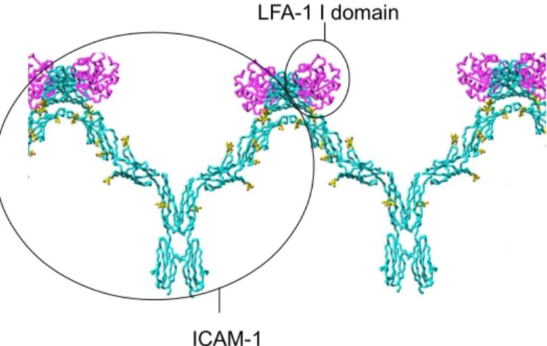
(Cluster of Differentiation 54) is a molecule of the immunoglobulin superfamily proteins which include antibodies and T-cell receptors. ICAM-1 is a protein of 115 kDa and is 28 nm long; it is composed of 5 immunoglobulin domains in which the binding sites for integrin LFA-1 are present. It possesses an amino-terminus extracellular domain, a single transmembrane domain, and a carboxy-terminus cytoplasmic domain. The dominant secondary structure of the protein is the beta-sheet, leading researchers to hypothesize the presence of dimerization domains within ICAM-1. Indeed, it plays the role of ligand for the LFA-1 (Fig. 2.1) and MAC-1 integrins, receptors found on leukocytes and macrophages respectively. When leukocytes are activated, they bind to endothelial cells via ICAM-1/LFA-1 and then transmigrate into tissues in processes such as extravasation and the inflammatory response. Because of these binding characteristics, ICAM-1 has classically been assigned to the function of intercellular adhesion. Different cells, including endothelial cells, express ICAM-1. In response to different pro-inflammatory mediators, such as tumour necrosis factor alpha (TNFα) and interleukin-1 (IL-1), the expression of ICAM-1 can reach a value 40 times higher than the normal level (Dustin et al., 1986).
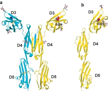
Figure 2.1 In this drawing, ICAM-1 D1–D5 molecules and D4–D4 dimers come together through D1–
D1 contacts (D=domain). The W-shaped tetramers can further propagate into a band-like one-dimensional cluster on the antigen-presenting cell surface. The LFA-1 I domain (magenta) binds to ICAM-1 D1 at the opposite face of D1–D1 dimerization. The glycans on ICAM-1 are in yellow (Carson, 1997)
pMHC. The major histocompatibility molecules (MHC), also referred to as
HLA molecules in humans, play a vital role in the immune system and autoimmunity. Indeed, their function consists of alerting the immune system if foreign material is present inside a cell. MHC molecules, which are anchored on the cell membrane, display small pieces of their structure or “antigens” to T cells. The antigens may be “self” (coming from a protein of the organism itself), or foreign ("nonself"), originating from bacteria, viruses, etc. T cell surface receptors (TCR) are able to recognise the 8 nm long MHC-peptide (presented on the cell surface) through binding interactions, giving rise to the activation of the immune cell that leads to the development of an immune response against the presented antigen. The design of the pMHC-TCR interaction is such that T cells ignore the self-peptides while reacting appropriately to the foreign peptides.
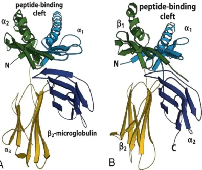
There are two general classes of MHC molecules (Fig. 2.2): class I, which are found on all nucleated cells and present peptides to cytotoxic T cells, and class II that are found on certain immune cells, namely macrophages, B cells and dendritic cells. However, MHC class I and MHC class II differ significantly in the method of peptide presentation. Both types of molecules present antigenic peptides to T lymphocytes, which are responsible for the specific immune response leading to the destruction of the
ICAM-1
pathogen producing those antigens. However, class I and II molecules correspond to two different pathways of antigen processing, and are associated with two different systems of immune defence.
MHC is not an adhesion molecule, but works with ICAM-1 in inflammatory response. The main role of MHC is in allowing the presentation of pMHC to T lymphocytes.
Fig. 2.2 Schematic representation of the MHC I and II extracellular domains coming from
crystallographic results. A: MHC class I. The molecule is composed of three globular domains: α1, α2, α3.
La microglobulin β2 is not covalently bound to the α chains. The two α helices form the peptide binding
site. B: MHC class II. The molecule is composed of two transmembranes molecules α et β formed by two globular domains: α1, α2 , et β1 et β2. Again, the two α helices form the peptide binding site (Murphy
et al., 2008)
Antibody. An antibody is a 150 kDa-protein. All antibodies belong to the
immunoglobulin family of proteins produced in plasma cells. There are 5 classes of immunoglobulins (abbreviated as Ig). IgG are Y-shaped. The two arms of the “Y” have antigen binging sites, and the other end recognizes other structures. The length of an antibody is ~16 nm. It is involved in immune response, its role is to identify and neutralize bacteria, viruses, or other pathogens. The "Y" shape of antibodies is composed of basic structural units forming four chains: two large heavy chains (~55kDa) and two smaller light chains (~25kDa). In each tip of the "Y" of an antibody there is a region, also known as hypervariable region, which is able to recognize
specifically a particular part, named epitope, of the foreign target (antigen). In this way, the two molecules can bind each other with a very high precision (Fig. 2.3). Although the general structure of all antibodies is similar, thanks to the extreme variability of the hypervariable region, many antigen binding sites can be recognized. Antibodies are used extensively as diagnostic and research reagents. Nowadays, their importance in therapeutic tool to treat disease is recognized. Indeed, antibodies are employed for analysis, purification, and mediation of physiological responses (Lipman et al., 2005).
Figure 2.3 Antibody structure. In blue the two heavy chains, while the light one are in pink. The
hypervariable region is in light blue and pink, at the tip of the "Y", showing the antigen binding site.
The role of antibodies in the immune response are:
1. binding to an epitope on an antigen with the arms (monovalent antibody fragment (Fab1) domain) of the Y. Each Fab1 domain has a binding site, making
the antibody at least bivalent.
2. The tail of the Y (Fc domain) gives to the antibody the biological functions of killer cell activation and activation of the phagocytosis (Lipman et al., 2005). When the immune response to an antigen is stimulated, multiple non-identical B cells are activated against the specific epitope on that antigen. This leads to a production of a large number of antibodies (polyclonal antibodies) with different specificities and epitope affinities. Polyclonal antibodies are largely used for biological research, such as immunoprecipitation, enzyme linked immunosorbent assays (ELISA), diagnosis of disease, etc.
identical B cells belonging to the same clone.
In 1975 Kohler and Milstein developed a technology to generate monoclonal antibodies of a desired specificity, by fusing immortal heteromyleoma cells with B-cells, The resulting cell, called a hybridoma is an immortal cell able to produce monoclonal antibody. Monoclonal antibodies can be produced to bind almost any substance, becoming then an important tool in biochemistry, molecular biology and medicine.
The anti ICAM-1 and anti HLA that were used in this study are monoclonal antibodies reacting with ICAM-1 molecules and histocompatibility antigen respectively. The first binds the domain 1 of ICAM-1, meaning the integrin binding site, while the pMHC binding site for anti HLA depends on the type of anti HLA: the mouse anti HLA A2 recognise the α2 helix and the turn of one of the underlying β strands, while the rat
Chapter 3
Single molecule studies
As already stated, cell adhesion is a fundamental biological process that is mediated by specific interactions between adhesion receptors and their ligands on cell surfaces or in ECM. In this chapter I will enter more in detail into this interactions explaining their importance, their advantages and the kinetics of bonds.
3.1 Role and importance of proteins interaction in cell
adhesion
Cell adhesion molecules mediate adhesive interactions by forming specific bonds between proteins. In addition, they often link directly to protein complexes which mediate interactions with the cytoskeleton and signal transduction pathways. Consequently, these cell adhesion and signalling complexes also help to obtain extracellular and signal transduction information within cells (Yamada, 2003).
In cell-to-cell adhesion, an adhesion receptor binds to a target protein which can be a "counter-receptor" or a complex carbohydrate on a protein anchored to the plasma membrane. In cell-to-matrix interactions, a plasma membrane adhesion protein such as an integrin, can bind to an ECM protein that is itself considered to be an adhesive protein. Consequently, adhesion molecules or receptors can be grouped in two main
groups: i) the first one is anchored on plasma membrane frequently as a transmembrane protein; this type of molecule consists of a hydrophobic transmembrane domain, a cytoplasmic domain or tail and an extracellular domain containing domains or sites for interactions; ii) the second class of adhesion molecules consists of proteins that are classified as cell surface or ECM proteins (fibronectins, laminins, etc.) and contain domains or sites involved in cellular adhesion.
It has become clear that cell adhesion molecules also play a critical role in cellular signalling. In this case, these proteins cluster together or bind other proteins in order to enhance their enzymatic function on the substrates which are going into contact. In the immune system, an example of these type of bonds is the interaction between T lymphocytes and the antigen-presenting cell.
The mechanical function of adhesion proteins often implies the application of forces on the bond. For example, in case of leukocytes adhesion to endothelium, there is a reinforcement of the interaction between selectins when a force is applied (Marshall et al., 2003). This property seems to enhance the adhesion of leukocytes to the walls of blood vessels, where the strong force acting on the bond is due to the bloodstream (Hammer, 2005).
3.2 Advantages of single-molecule study
Single-molecule studies are central to biophysical research because they allow us to enter into the details of molecular processes. Indeed, with single-molecule studies it is possible to measure kinetics of biomolecular reactions or time-dependent processes being able to follow the movement, and spatially and temporally localise individual molecules. A problem with multivalent attachments is that the relationship between molecular properties and attachment and detachment kinetics are dependent on a number of environmental parameters that are not easy to control. Single molecule properties are "intrinsic".
There are several advantages of studying individual ligand–receptor pairs instead of ensembles of molecules:
• A lot of molecules have the tendency to aggregate when their concentration is sufficiently high. In the experimental conditions of single molecule studies the number of molecules involved in a process is not so high and/or cooperative and clustering effects are minimized. Consequently, one can easily know the number of molecules involved in reactions (Weisel et al., 2003).
• It is possible to reveal the structural and functional heterogeneity of seemingly identical molecules.
• Single molecule studies allow us to apply forces on single molecules and observe their response under the imposed constraint. Indeed, directly quantifying the magnitudes and working distances of forces in ligand–receptor interactions gives an insight into the relationship between molecular structure and the thermodynamics of bond dissociation (Bongrand, 1999).
• At any given time, a single molecule exists in a particular conformational state within a particular environment. Observing only population averages can hide important dynamic or mechanistic features of biological molecules. Watching individual events and distributions rather than observing average values may reveal rare but physiologically important functional fluctuations (Merkel, 2001; Hinterdorfer et al., 2001).
“Single-molecule analysis requires statistical data so that the observed behaviour of minor, unusual molecules is not overestimated. However, as single-molecule studies deal with small numbers of molecules, sampling noise is an inevitable problem of these analyses compared with conventional biochemical analyses” (Sako, 2003).
3.3 Affinity and kinetics parameters necessary to describe a
bond
In order to well describe cellular adhesion processes, the knowledge of the average lifetimes as well as kinetic association and dissociation rates of ligand-receptor interactions that mediate this processes is required. To understand the mechanisms
governing the sensitivity, specificity, and regulation of cell adhesion, it is therefore necessary to be able to accurately characterize the kinetics of ligand-receptor interactions. However, it has been demonstrated (Seifert, 2000; Sulchek et al., 2006) that the kinetics and mechanics of multivalent attachment rupture depend on parameters such as receptor and surface topography, lateral mobility, length and flexibility of membrane anchors. Therefore, the study of ligand-receptor interaction is rather complex.
The interaction between ligand and receptor during adhesion processes can be characterized in terms of binding affinity. In general, high affinity ligand binding means that the binding sites are well occupied giving rise to the physiological response. In this situation, the concentration of ligand necessary to elicit a response is quite low. Conversely, low-affinity ligand binding implies the necessity of a high concentration of the ligand so that the binding sites are occupied and the physiological response to the ligand is achieved.
The affinity between a ligand and a receptor is commonly described in terms of the concentration of ligand at which half of the receptor binding sites are occupied, known as the dissociation constant (Kd ). Ligand-receptor affinities are influenced by
non-covalent intermolecular interactions between the two molecules such as those mediated by hydrogen bonds, electrostatic interaction, hydrophobic and Van der Waals forces. The formation of a ligand-receptor complex (LR) can be described by a two-state process:
L+R LR (3.1)
in which a complex LR is formed by the L and the R subunits. The kinetic constants kon
and koff account for the forward and reverse rates according to the following equation:
d[LR]/dt = kon [L][R] - koff [LR] (3.2)
where [R], [L] and [LR] represent molar concentrations of the receptor, ligand and complex, respectively, usually expressed in mole/litre. The forward and reverse rates allow us to classically describe the interactions between proteins at the non-equilibrium state: kon describes the bond formation and koff the bond rupture. The bond lifetime is
then dependent on the off-rate. The thermodynamic definition for the reaction (3.1) follows the relationship :
∆G°=GL°+GR°-GLR° (3.3)
where the quantity ∆G° is the standard Gibbs free energy of the reaction and GL°,GR°
and GLR° are the Gibbs free energies of reactants (L and R) and product (LR). Under
pure thermodynamic reaction control, when the equilibrium has been reached, the ratio of product concentrations will equal the equilibrium constant Keq and therefore be a
function of the difference in Gibbs free energies:
ln([L]eq[R]eq/[LR]eq )=ln Keq=-∆G°/RgT (3.4)
where "eq" means that we are dealing with equilibrium concentrations, Rg is the perfect
gas constant and T is the absolute temperature. It is readily found that, whatever the initial conditions, the system will tend to an equilibrium state following the Guldberg-Waage law (Atkins & de Paula, 2002). When applied to reactions in solution, this is usually written as:
[L]eq[R]eq/[LR]eq = koff/ kon = Kd = 1/Ka (3.5)
where Ka is called the affinity constant (in litre/mole) and Kd is the dissociation constant
measured in mole/litre (Bongrand, 1999). The smaller the dissociation constant, the more tightly bound the ligand is, or the higher is the affinity between ligand and receptor. The dissociation constant for a particular ligand-receptor interaction can significantly change with environmental conditions (e.g. temperature, pH and salt concentration). The effect of different environmental conditions is to modify the strength of the intermolecular interaction between the ligand-receptor pair.
3.4 Description of bond formation and k
onThe rate of binding soluble (three-dimensional binding) or surface-attached (two-dimensional binding) ligands influences the efficiency of cell surface receptors. The conventional formalism used to describe 3D reactions cannot be used to account for (2D) interactions between surface-attached molecules. This point is considered in the well known Bell paper (Bell, 1978), which describes the association between two
molecules. Before binding, a diffusion-limited phase is necessary to bring the reactive sites into contact:
L + R LR C (3.6)
The first step describes the initial contact between the molecules L and R with the formation of a transient complex LR which precedes the final complex C. In this first relation, the velocity of bond formation between proteins depends on the distance between them and consequently, on the membrane diffusion of the two surfaces where proteins are anchored (2D diffusion). The formation of the final complex C, described by the second step, can be expressed by the forward rate kon. To describe the kinetics of
the interaction which leads to the formation of the product C, it results then necessary knowing that the bond lifetime and the force resistance are critical parameters which play an important role.
Many authors emphasized the importance of the association rate and its suitability to account for molecular interactions. Indeed, the receptor efficiency was demonstrated to be dependent on the association rate in a variety of situations: integrin activation (Vitte et al., 2004), antigen-antibody interaction (Foote & Milstein, 1991) or tethering of leukocyte to the vessel walls mediated by selectins (Lawrence & Springer, 1991; Dwir et al., 2000). The probability of bond formation is dependent on the encounter time te and it can be classically expressed as follows:
P(te)~1-exp(-konte) (3.7)
Nevertheless, the association rate does not completely describe the bond formation under all conditions. In some cases a minimum duration time t0 is necessary to form the
bond (Robert et al., 2009), modifying the expression for the bond formation probability as follows:
P(te)~erfc(t0/te)1/2 (3.8)
In the case of ICAM-1 – anti ICAM-1 interaction, a value for the minimal contact time was estimated being t0=6 ms (Robert et al., 2011).
3.5 Description of bond rupture and k
offWhen an interaction between proteins takes place, the bond lifetime can be described by the reverse rate koff. This parameter is a function of the force which is
applied on the bond, as already expressed by Bell (Bell, 1978):
koff(F) = koff(0)exp(xF/kBT) (3.9)
where koff(F) is the dissociation rate when a force F is applied, koff(0) is the off-rate in
absence of force, x represents the interaction range with a dimension of length, kB is the
Boltzmann constant and T is the absolute temperature.
However, this simple framework is not sufficient for many interactions. During the last decades, important progress was made in measuring interactions between surface-attached adhesion receptors in presence of forces at the single-molecule level, leading the following conclusions:
i. In the simplest cases, the dissociation rate of a ligand-receptor bond increases exponentially in presence of a disruptive force, as predicted by Bell (Chen & Springer, 2001; Dudko et al., 2008). Bond rupture could be modelled following the prediction of Kramers (Kramers, 1940), namely as the passage of a single potential energy barrier in an unidimensional reaction path.
ii. in many cases, such as streptavidin-biotin (Merkel et al., 1999), antigen-antibody (Pierres et al., 1995) or integrin-ligand (Zhang et al., 2002) interaction, bond rupture seemed to involve the passage of several energy barriers, implying the presence of a multiplicity of bound states for a given ligand-receptor couple. This might explain the time-dependent strengthening of several bonds, including streptavidin-biotin (Pincet & Husson, 2005).
iii. The studies of selectins behaviour while they rolls on leukocytes, led to the discovery of “catch bonds”, in which the dissociation lifetime increases with tensile force applied to the bond. Examples were provided by Marshall (Marshall et al., 2003) in the case of P-selectin-PSGL-1 interaction and Thomas (Thomas et al., 2002) for lectin-sugar bond. However, a recent work of Zhu (Zhu et al., 2008) provided experimental evidence that a disruptive force might
strangely decrease the lifetime of L-selectin ligand interaction, as predicted by eq. (3.9), although this association was considered catch-bond. Indeed, the catch-bond phenomenon is not fully understood yet. A possible feature is that bond rupture may not follow an unidimensional path but an alternative rupture path by deforming a multidimensional energy landscape (Evans et al., 2004).
Chapter 4
Single molecule methods
Many biological reactions are extremely complex and their comprehension through the use of conventional biochemical techniques is difficult. Studying one biological macromolecule at a time clearly allows us to look at these molecules in action. But, at present, although single-molecule techniques applied on biological processes are growing very fast, they are still difficult to master. It is possible to make a distinction between these techniques based on the detection and manipulation of individual molecules (Ha & Selvin, 2008).
• In a first class of techniques where the manipulation of molecules is allowed, we have laminar flow chamber, force application via atomic force microscopy (AFM), biomembrane force probe (BFP) to cite the most representative ones. The last two techniques are force-based methods, which can give an estimation of the frequency of adhesive events and the force necessary to break a bond. The laminar flow chamber is a technique working at constant force and able to measure the frequency of adhesive events and the bond lifetime.
• In a second class, which includes techniques able to detect and follow in real time (but not manipulate) individual molecules, in addition to imaging by AFM, there are predominantly optical techniques such as single-molecule fluorescence (SMF) and semiconductor quantum dot emission, to cite the most common. Single-molecule fluorescence allows us to detect and localize molecules with
about 1.5-nm precision (Yildiz et al., 2003).
4.1 Laminar flow chamber
4.1.1 Principles
Laminar flow chamber is a widely used tool in cell adhesion studies either at the cell scale or at the single molecule scale. It is a versatile tool in understanding the mechanisms of proliferation, adhesion, and metastasis of cancer cell. Many researchers used it to investigate the dynamic adhesion of cells under a definite shear stress (Ling et al. 2003; Rinker et al., 2001). Kaplanski et al. (Kaplanski et al., 1993) monitored the motion of human white blood cells along a surface coated with activated endothelial cells in presence of a very low shear rate. In particular, some studies have been carried out to study leukocyte receptor-ligand interactions (Taite et al., 2006), interactions between selectins or integrins and their ligands (Alon et al., 1995; Wiese et al., 2009; Wayman et al., 2010), or biotin-streptavidin interaction (Agarwal et al., 2009; Pierres et al., 2002).
The laminar flow chamber is a technique that allows us to quantify the physical parameters involved in the interaction between biological molecules on surfaces. This technique provides a method to measure the association and dissociation kinetics of individual bonds. Additionally, it is possible to investigate the behaviour of the bond when a force is applied on it.
The laminar flow chamber is an enclosed space where a flux is imposed by flowing a liquid at a desired velocity. When the flux is sufficiently slow the flow become laminar and the flux velocity is parallel to the chamber floor. On the walls of the chamber this velocity is zero, and far from them its value is given by a first order of approximation (Pierres et al., 1995):
v(z)=Gz (4.1)
expressed in s-1. The force T exercised by the flux per surface unit is given by:
T=µ x G (4.2) If the viscosity µ is expressed in Pa/s, T is expressed in N/m² The shear rate in a chamber of dimension L x l x H and for a given flux Q will be:
G=6Q/lH² (4.3)
A model for cell movement in a flow can be described by using microspheres. When the sphere is many radii distant from the walls, its velocity is equal to the flux velocity, but when it comes close to the walls the thin layer of flux between the microsphere and the walls resists to a deformation making the microsphere slowing down.
When a microsphere with a radius a is fixed to the floor of the chamber, it is subjected to an hydrodynamic force f which tends to detach it and to a torque force Γ. These are given by:
f≈32µa²G (4.4)
Γ≈4µa3G (4.5)
Because of a lever effect, the tensile force F exerted by the flux on the bond holding the microsphere will be:
F≈(f+Γ/a)(a/2λ)1/2 (4.6)
where λ is the length of the bond (Pierres et al., 1995).
It has to be remembered that this modelling is not perfect for describing cells. Indeed, a force of friction has to be added to describe cell movements due to asperities and protrusions on cell surface which give an elongated shape to cells compare to spheres (Tissot et al., 1992).
Figure 4.1 Principle of flow chamber. The flux velocity v is parallel to the axe Ox of the chamber. Close
to the wall, v is a linear function of the z coordinate. The derivative dv=dz is the shear rate; a is the radius of the particle in the flow (Robert, thesis 2009).
The aim of a flow chamber experiment is to detect bonds between molecules attached on the microsphere surface and on the chamber bottom surface. The weak hydrodynamic traction (tens of pN) exerted on the bond after bead stops, together with the thermal stress causes the rupture of the bond. The flow chamber allows us to measure the bond lifetime, by analysing the duration of bead arrest. During a flow chamber experiment, a huge number of microsphere trajectories and arrests are recorded and analysed. If the bond rupture is a simple monophasic reaction:
P(t) = exp(-koff t) (4.7)
The curve obtained by plotting P versus t on a semi-log scale is a straight line, the slope of which is the dissociation rate, koff. In real life, survival curves (P versus t) have non
zero curvature and the analysis of these curves can give information on the rupture mechanism and the energy landscape (Pierres et al., 2002).
It is possible to define a spatial frequency of arrests as a ratio between the total number of arrests and the total distance covered by the microspheres after their sedimentation in the chamber. When beads reach the equilibrium after sedimentation on the chamber, the particle distance h from the surface follows a Boltzmann distribution. If the interaction force between bead and surface is negligible, the mean value <h> follows:
h=kBT/mg (4.8)
where kB is the Boltzmann constant, T is the temperature of the system, m is the mass of
a particle and g is the acceleration of gravity. The temporal frequency of arrests is defined as the ratio between the total number of arrests and the total duration of bead displacements.
4.1.2 Measure of kinetics of single bonds
Measure of kon. The flow chamber is able to provide information on bond formation by
measuring a related parameter, the adhesion frequency. However, it must be emphasized that single bond formation is more difficult to study than bond dissociation for at least two reasons:
• Studying bond formation requires us to generate multiple intermolecular contacts and to perform many checks to determine the proportion of contacts. • Bond formation is more dependent on molecular environment than bond rupture.
It can depend on the topography of the surfaces where ligands and receptor are attached (Brunetti et al., 2010; Gentile et al., 2010), on the valency of molecules (Haun & Hammer, 2008), on the molecular orientation and length (Huang et al., 2004) and on the lateral mobility of the molecules involved in the interaction (Chan et al., 1991). An exact knowledge of the conditions of bond formation is usually hard to reach and strong approximations are required to derive molecular association rates from adhesion frequencies measured on surface-attached molecules.
Laminar flow chambers can easily determine the average frequency of particle or cell arrest per unit length of trajectory or per unit time of observation. When interpreting the results, one has to observe that (Pierres et al., 2008):
1. if binding efficiency is high, the adhesion frequency represents the number of encounters between active molecules. Thus, it represents a geometrical rather than a kinetic parameter. In this case, the adhesion frequency per unit length should be weakly affected by limited variations of the flow.
2. Conversely, when binding probability is proportional to the encounter time (the time during which the molecules are close enough to form the bond), adhesion frequency per unit time should be weakly affected by limited variations of the flow. Adhesion frequencies are highly dependent on the definition of binding events and any detailed analysis requires a correction to make arrest detection independent of the shear rate.
Measure of koff. Flow chamber is an interesting tool to investigate the bond rupture of
ligand-receptor interactions. The primary output of data processing is related to all binding events together with their duration. This allows straightforward derivation of detachment curves displaying the logarithm of the proportion of binding events lasting for a time t as a function of t. If binding events are mediated by single bonds with
which is equal to the dissociation rate koff. However, non linearity is a frequent
occurrence as a possible consequence of different phenomena:
• additional bond formation may occur after initial particle arrest, resulting in progressive strengthening of attachment (Kaplanski et al., 1993);
• a single bond may display multiphasic behaviour with a time-dependent strengthening due to the passage of sequential barriers on the energy landscape (Pincet & Husson, 2005);
• particle-to-surface attachment may be mediated by several bond species with different dissociation rates.
In order to avoid some of these problems, one has to be sure of measuring single-molecule interaction. For this reason, the density of ligands on the coverslip or receptors on the bead surface has to be sufficiently low. In this situation, the formation of multiple bonds is unlikely. To test this condition, different concentrations of molecules are exploited during flow chamber experiment: if the frequency of arrests is proportional to the molecule density and the dissociation constant koff does not change, the bond
lifetime is related to the same type of events, meaning that the observed arrests are due to single molecule bonds.
The pioneering studies with laminar flow chamber were made on selectin-mediated adhesion (Kaplanski et al., 1993; Alon et al., 1995), followed by more recent studies (Sarangapani et al., 2004; Wayman et al., 2010). In the last decade, with laminar flow chamber the koff was measured in case of monocyte adhesion to vascular
endothelium (Rinker et al., 2001) and streptavidin-biotin interaction (Pierres et al., 2002).
4.2 Atomic Force Microscopy (AFM)
Nowadays, the most widespread single-molecule technique is atomic force microscopy (AFM). The AFM is based on the principle that a very soft cantilever with a tip that is moved to a surface, can sense the roughness of the surface and deflect by an amount which is proportional to the distance between the tip and the surface.
Figure 4.2 Atomic Force Microscope (AFM) – A laser is reflected off the back of a cantilever with a
sharp tip and detected by a photodiode detector. AFM produces a topographical image. When the cantilever tip is deflected due to the forces between the tip and surface, the laser is reflected differently and the detector senses the difference in topography.
(http://npl-web.stanford.edu/user/files/www/afm0.jpg)
The most important application of the AFM is imaging, where it can work in various modes: contact mode, tapping mode and jumping mode. For example, in the tapping mode the tip is made to oscillate close to the sample surface. The amplitude of the oscillation is recorded and controlled by a feedback mechanism that keeps such amplitude constant. When passing over a bump the amplitude decreases, so the distance between tip and surface is increased to keep the amplitude of oscillation constant. On the contrary, when passing over a depression the tip is moved to the surface. This mode has the advantage that the transverse motion of the tip along the surface is not influenced by shearing and frictional forces, avoiding damage to the sample and noisy interference effects. A map of the distance of the tip from the sample provides an accurate topographic image of the surface (González-García et al., 2010; Peressadko et al., 2005). AFM has been also used to characterize the surface treatments (Eske & Galipeau, 1999). Other modes are preferable depending on the particular system.
Figure 4.3 Attachment of living cells by means of receptor-ligand interactions. By applying a repulsive
contact force between the cantilever-mounted cell and a target cell at the bottom of a Petri dish, and then retracting the cantilever from the target cell (right schematics), specific cell-cell adhesion forces can be measured. Scale bar, 20 μm. (Hinterdorfer & Dufrêne, 2006)
The AFM is also used to manipulate and exert mechanical force on individual molecules. A surface is coated with the molecules to be manipulated and the AFM tip is coated with molecules that can bind to the ones on the substrate. By moving the tip to the substrate, a contact with one of the molecules adsorbed on the substrate is made. The motion of the tip is controlled by a piezoelectric stage which allows to reach a sub-nanometer resolution. Retraction of the tip at constant speed allows to measure its deflection in real time, providing a measure of the force acting on the molecule as a function of its extension. The AFM covers forces in the 20 pN–10 nN range, depending on the stiffness of the cantilever. AFM measurements in force mode were performed on avidin-biotin bonds (Moy et al., 1994; Florin et al., 1994; Lee et al. 1994), antigen-antibody (Hinterdorfer et al., 1996) or cadherins (Baumgartner et al., 2000).
Although AFM is a very versatile and powerful tool, some points of caution are warranted for manipulating single molecules (Neuman & Nagy, 2008):
• the presence of undesired interactions between tip and substrate (van der Waals, electrostatic and adhesion forces) and the non-specificity of the attachments that often occur between tip and substrate;
• the difficulty in controlling the specific location of the attachment between the tip and the molecule. Spatial and force resolution in the AFM are limited by thermal fluctuations. Consequently, the signal-to-noise ratio for the force is small for values of just a few tens of pN (weak interactions), showing the
limitations of AFM. In contrast, AFM is ideal to investigate strong to covalent interactions.
4.3 Biomembrane Force Probe (BFP)
The biomembrane force probe (BFP) is a technique developed by Evans and collaborators (Evans et al., 1991). The principle of the BFP is similar to the AFM one for force measurement. it consists of approaching with two micropipettes with cells, or lipid vesicles, or microspheres, covered with suitable receptor and ligand molecules.
Figure 4.4 Image of Biomembrane Force Probe tool. A red blood cell is aspired by a micropipette (left)
and a functionalized microsphere is fixed on its surface. A lymphocyte T is aspired by another micropipette (right). The micropipette on the left is displaced by a piezoelectric device, in order to make the microsphere in contact with the lymphocyte T. The red blood cell works like a spring and its deformations are measured after allowing the contact. P.-H. Puech, 2008.
As shown in Fig. 4.4, a micropipette aspires a red blood cell which works like a spring. A microsphere functionalized with a receptor is fixed on the red blood cell. Another microsphere or a cell functionalized with the suitable ligand is attached on the second micropipette. The two micropipettes are mounted on micromanipulators which allow their displacement using a piezo translator stage. After the contact between the two microspheres is made and bonds are formed, the first pipette is pulled out under microscopic control. The applied force results in the red blood cell deformation and it increases linearly when the velocity of the moving micropipette is constant. A spherical shape of the cell is recovered when the last bond is ruptured. Thus, the experimental parameter which is measured with this technique is the unbinding force rather than the bond lifetime (Evans et al., 1991). The interest of this procedure is that the stiffness of
the spring depends on the red blood cell surface tension and it may be varied in a wide range by controlling the sucking pressure applied through pipettes. The cell can indeed be subjected to a distractive force ranging from less than 1 to 100 pN.
The BFP can be used to apply a very wide range of loading rates (Merkel et al., 1999) and it has been mainly used to study molecular interactions at single molecule level, such as E-cadherins (Perret et al., 2004; Bayas et al., 2006) bonds or P- and L-selectin-PSGL-1 (Evans et al., 2004; Evans et al., 2001).
4.4 Single Particle Tracking (SPT)
Single-particle tracking (SPT) exploits the fact that the location of an isolated particle can be measured with a higher accuracy than the Rayleigh limit (typically around 200 nm). By attaching fluorescent molecules to proteins it is possible to detect the light emitted by this fluorophore and follow its trajectory. Fluorophores are excited from their ground state by absorbing light from an external light source. In this situation, a valence electron jumps into a higher energy excited state. When this electron returns to its ground state, a quantum of light is emitted.
Single-particle tracking is a powerful tool to study:
1. ligand density on surfaces. Measuring the density of surface-attached ligands is an important issue for single-molecule studies. This parameter can influence the absorption of biological molecules, the activation of cells (Kim et al., 1996), the kinetics of bond because of the possibility to observe cooperative and multivalent bonds. As previously explained, for single-molecule experiment it is fundamental to check the density of ligand on the surface, since this parameter is firmly important in the estimation of the kon.
2. Ligand lateral diffusion by following its trajectory. Protein within the cell membrane are expected to undergo Brownian motion, but if interactions with other membrane constituents or peripheral structures occur, a deviation from this behaviour is registered. The diffusive motion of molecules of interest at the surface of living cells or artificial membranes can be followed after labelling
them. In this frame, the diffusion dynamics of glycine receptors (Dahan et al., 2003), the confinement of immunoglobulin receptors (Simson et al., 1995) and confinement and jumps of a G-protein-coupled-receptor (Meilhac et al., 2006) where measured.
3. The kinetics of ligand-receptor bonds. In the absence of mechanical constraint, the molecules have a probability to unbind due to thermal fluctuations at the molecular interface. A quantitative approach to quantify the bond lifetime is to detect and follow single fluorescent ligands or fluorescent nanocrystals (quantum dots) bound to ligands, interacting with receptors. When the fluorescent signal disappears, the molecule has detached and the statistics of these events gives the bond lifetime, as found for cadherins (Baumgartner et al., 2003) (koff~1 s) and neurexin-neuroligin (Saint-Michel et al., 2009), biotin-avidin
(Wayment & Harris, 2009) or TCR-pMHC (Huppa et al., 2010) interactions. Thoumine and co-workers (Thoumine et al., 2008) observed the detachment from the cell surface of quantum dots conjugated to adhesion proteins (synCAM) and they calculated the koff of the bond being on the order of 0.1min-1.
A drawback related to SPT is the ever-presence of the noise during all the steps (Saxton, 2008): labelling, localization, connection of the dots and interpretation of the connected dots. In fluorescence labelling, the main sources of noise are dark states of the label and background fluorescence. Labelling is mainly an experimental problem, solved by using appropriate fluorophores. Problems related to the connection of the dots and to the interpretation of the trajectories may be solved statistically, by using algorithms which are able to take into account the merging and splitting of dots and the causes of their motion (pure diffusion, anomalous sub-diffusion, confined motion and directed motion).
Two works, which were focused on this problem, present improved algorithms to well detect and interpret trajectories from SPT images, introducing useful statistical approaches. The studies revolved round the possibility to track CD36 receptors, evaluate the lifetimes of clathrin-coated pits (Jaqaman et al., 2008) and map the mobility of the epidermal growth factor receptor on the cell surface (Sergé et al., 2008), by creating algorithms that are able to increase the density of particles that can be
tracked.
Fig. A single imaged fluorophore can be modelled by a two-dimensional Gaussian to determine its
position with nanometer accuracy. The three-dimensional peak to the left shows the recorded intensity for each pixel as a coloured surface. A corresponding contour map is shown to the right (Walter et al., 2008).
Chapter 5
Importance of molecular environment in adhesive
interactions
Ligand-receptor interactions and consequently adhesion processes are rather influenced by different factors and structures that surround the pair. This means that modification in the molecular environment may lead to changes in the kinetics and dynamics of bonds.
Huang and collaborators showed that the orientation and length of adhesion receptors such as P- and E-selectins influence their two-dimensional forward rate without consequently affecting the reverse rate and the stability of binding (Huang et al., 2004). A predominant role of environmental factors, such as surface topography and accessibility of active molecules to regions of contact, in determining forward rates of bond formation at cell interfaces was also demonstrated (Waugh & Lomakina, 2009). Thus, association rates of adhesion bonds may be strongly influenced by steric barriers between the surfaces and the reactive molecules in the contact region. Waugh's team postulated the necessity of available molecules and formation of “reaction zones” at the interface of adhesion sites that precedes bond formation (Waugh & Lomakina, 2009). The study of simple systems such as single-molecule interactions on acellular surfaces is the basis for the investigation of more complicated system with interacting cells. When these ''simple'' interactions are well understood, it would be possible to put back molecules in a ''biomimetic'' context, to mimic and study more complicated mechanisms involved in cell-adhesion.
5.1 Regulation of cell adhesion by the cytoskeleton
The cytoskeleton is a dynamic three-dimensional structure that fills the cytoplasm and is made out of proteins. This structure acts as both muscle and skeleton, for movement and stability. The primary types of fibres comprising the cytoskeleton are microfilaments, microtubules, and intermediate filaments. It plays an important role in regulation, activation and adhesion.
As an example, integrin adhesion receptors link the ECM to the actin cytoskeleton and transmit biochemical signals and mechanical force across the plasma membrane. Integrin-mediated cell adhesion is enabled by cytoskeletal linkages. Inside-out signals to integrins originate from diverse plasma membrane receptors which presumably regulate integrins. This signalling is then modulated by cytoskeletal proteins that allow the activation of integrin-regulatory molecules and the control of their nearness to integrin cytoplasmic tails (Calderwood et al., 2000). In the case of T cells, it is known that to become activated, they must efficiently recognize antigen-presenting cells or target cells through several complex cytoskeleton-dependent processes, including integrin-mediated adhesion. A regulated cytoskeletal framework provides to recruit molecules that regulate adhesion and necessary for T-cell development, activation and proliferation (Billadeau et al., 2007).
5.2 Role of glycocalyx in modulating cell adhesion
Glycocalyx is defined as a set of extracellular polymeric materials (glycoproteins), glycolipids and sugar residuals with a variable thickness. Only identical twins have chemically identical glycocalices; everyone else is unique. The glycocalyx is used by the organism to discriminate between its healthy cells and foreign organisms, transplanted tissues, or diseased cells. The glycocalyx also includes some cell-adhesion molecules that enable cells to adhere to each other, thus playing an important role on cell-adhesion (Robert et al., 2006), and guide the movement of cells during embryonic development.