Regulatory Peptides 67 (1996) 39-45
Pwms
Cytokine production by human thymic epithelial cells: control by the
immune recognition of the neurohypophysial self-antigen
H. Martens a, B. Malgrange b, F. Robert a, C. Charlet a, D. De Groote a, D. Heymann c,
A. Godard c, J.P. Soulillou c, G. Moonen b, V. Geenen a’”
a
Institute of Pathology CHU-B23, Lab of Radio-Immunology and Neuroendocrine-Immunology, University of Li2ge, B-4000 Li2ge-Sart Tilman, Belgium b Department of Human Physiology and Pathophysiology, University of Li2ge, 17, place Delcour, B-4020 Lii2ge,Belgium‘ INSERM U437, Institut de Transplantation et de Recherche en Transplantation, CHU-H&el Dieu, Bid, J. Monnet, F-44035 Nantes Cedex 01, France
Received 8 Febmary 1996; accepted 14 May 1996
Abstract
Oxytocin(OT) has been shownto be the dominantpeptideof the neurohypophysialfamilyexpressedby thymicepithelialand nurse cells(TEC/TNC) invmious
species.
Thymic OT is not secreted but, after translocation of a hybrid neurophysin/MHC class I protein,is integratedwithin the plasmamembraneof TEC, thus allowing its presentation to pre-T cells. In order to further demonstrate that thymicOTbehaves like a membrane antigen, we assessed the effect of rnAbs toOT on cytokineproductions by cultures enriched in human TEC. 75–85% pure TEC cultures were prepared tlom human thymic fragments. Using immunofluorescence and confocal microscopy, ir-OT, ir-interleukin-1~ (IL-1P), ir-interleukin-6 (IL-6) and ir-leukemia inhibitory factor (LIF) could be detected in these TECcultures. ir-OT was restricted to TEC, while some ir-IL-6 and ir-LIF were also seen in occasional fibroblasts. In basal conditions, ir-IL-6 and ir-LIF (but not ir-OT and ir-IL-l@) were detected in the supematants of human TEC cultures. MAbs to OT induced a marked increase of ir-IL-6 and ir-LIF secretion in TEC cultures. No significant effect was observed using mAbs against vasopressin, mouse immunoglobulins, or control ascitic fluid controls. These data show that OT is fully processed and recognized by specific mAbs at the outer surface of TEC plasma membrane. They further support that thymic OT behaves as the self-antigen of the nenrohypophysial family.
Keywords: Tbymus; Neurohypophysial peptide family; Self-antigen; T-cell self-education
1. Introduction
The epithelial component of thymic stroma plays an important role in positive and negative selection of T lymphocytes, [1–7]. The thymic repertoire of neuroen-docrine-related polypeptide precursors has been proposed to recapitulate at the molecular level the dual physiologic role played by the thymus in T-cell development [8–10]. With a special regard to the neurohypophysial peptide family, our group as well as others have shown that thymic epithelial and nurse cells (TEC/TNC) synthesize oxytocin (OT) and its precursor-associated protein, OT-neurophysin [11,12]. Thymic OT plays a dual role in T-cell selection. First, following its interaction with cognate receptors ex-pressed on T cells [13,14], OT acts as a cryptocrine signal involved in an accessory pathway of T-cell positive
selec-“ Corresponding author.
tion [15,16]. Second, OT has been suggested to be the prototype neurohypophysial self-antigen presented to im-mature T cells by the thymic major histocompatibility complex (MHO system.
Although ir-OT concentrations in the human thymus are much higher than those of ir-vasopressin (VP), both neuro-hypophysial (proOT and proVP) genes are transcribed in the human and murine thymus [17,18]. Thymic OT is not secreted but translocated in TEC plasma membrane in association with a membranal 55-kDa neurophysin/MHC class I hybrid protein [19]. A functional analogy was observed in the binding and transport of OT between neurophysin in the hypothalamo-neurohypophy sial axis [20] and the neurophysin domain in the thymic microenviron-ment. Presenting OT in such a manner to pre-T cells is thought to lead to the central T-cell tolerance in neurohy-pophysial family members [21]. This model of T-cell education to the neurohypophysial self-antigen offers se-lective advantages such as avoiding MHC class I allelic 0167-01 15/96/$15.00 Copyright 01996 Elsevier Science B.V. All rights reserved.
40 H. Martens et al. /Regulatory Peptides 67 (1996) 39-45
restriction and presenting the overall confirmational struc-ture of the neurohypophysial peptide family.
TEC also express transcripts and proteins of various cytokines, such as interleukin 1 (IL-1) [22], interleukin 6 (IL-6) and leukemia inhibitory factor (LIF) [23] which were also found in the supernatants of TEC cultures. We previously showed ir-IL-1(3to be colocated with ir-OT and its neurophysin in thymic sections, more specifically in TEC recognized with cytokeratin (CK) antibodies [24].
In the immune system, T-cell activation and cytokine production usually follow the recognition and binding of Abs to antigenic markers expressed on the surface of T-cell membranes. In an attempt to unravel the physio-logical significance of the colocalization of OT with other polypeptides in TEC, we investigated the effect of anti-OT monoclinal antibodies (mAbs) upon IL-1P, IL-6 and LIF secretion by cultured human TEC cultures.
2. Material and methods 2.1. Antibodies
Anti-human cytokeratin mAb MNF 116 (lot 33) and rabbit polyclonal Abs anti-cytokeratin A575 (lot 31D) were obtained from Dako (Denmark). 033 and 013 are anti-OT mAbs; 033 is directed against the cyclic part of OT and 013 is directed against the C-terminal part of OT [25]. BER 312 is an anti-VP mAb [26]. Anti-IL-6 mAb (clone 4B2) was obtained from Medgenix Diagnostics (Belgium). Anti-LIF is a specific antiserum kindly pro-vided by A. Godard [27]. Second antibodies were
fluores-cein (FITC)-conjugated Affinipure goat anti-mouse IgG (lot 28970), hereafter referred to as second antibody A; FITC-conjugated Affinipure goat anti-mouse IgM (lot 17708), hereafter referred yo as second antibody B; and rhodamine (TRITC)-conjugated Affinipure F(ab’)2 frag-ment goat anti-rabbit IgG (H + L) (lot 27272),
he=fter
referred as second antibody C. All conjugated Abs were obtained from Jackson (Pennsylvania).2.2, TEC cultures
Thymus fragments were obtained from children (6 months to 3 years old) undergoing corrective cardio-vascular surgery for congenital cardiopathies. Surgical fragments were pre-cut and washed by sedimentation at 1 X g, twice for 5 min in HBSS (Flow, 19-101-49), then minced with scissors and washed again. The small frag-ments ( <2 mm) were then transferred in Eagle’s mini-mum essential medium containing D-VdiUe(Gibco) and supplemented with 2 mmol/1 L-glutamine, 10 mmol/1 HEPES, 100 U/ml penicillin, 100 p,g/ml streptomycin and 1096 heat-inactivated fetal calf serum (FCS; Gibco), hereafter referred as complete TEC medium. D-Valine was used to reduce the growth of fibroblasts [28,29]. Fragments
were then allowed to attach to T-75 flasks in the same medium in a humidified atmosphere at 37°C and 5% C02, and complete TEC medium was replaced every 3–4 days. On day 17, the explants and adherent cells were detached by treatment with Puck’s-modified solution containing trypsin and EDTA (Gibco) and filtered through nylon gaze to eliminate fragment residues. Cells were counted and seeded at 6000 cells/well on 10 well/immunofluo-rescence microplates, and at 50000 cells/well in 12-well culture plates. The 10% FCS in complete medium was replaced at this point by 1% Ultroser G-steroid free (Gibco), hereafter designed as US-SF-TEC medium, for cytokine assays. Ultroser G was used in order to avoid possible artefacts reflecting the presence of cytokines in FCS, and to further reduce fibroblast growth [30].
2.3. Immunojhorescence procedures
Immunofluorescence microplates were kept for three days in FCS-TEC medium to allow plating of the TEC and then stained for immunofluorescence after a 7-rein, –20°C methanol fixation [24]. Plates were first incubated for 30 min at room temperature with 10Yonormal goat serum to prevent non-specific binding of goat second antibody to mouse or rabbit immunoglobulins. Single or double im-munostaining was performed with the following reagents: anti-human cytokeratins mAb MNF 116 diluted at 1:50 followed by second antibody A; anti-OT mAb 033, diluted 1:100 followed by second antibody B; anti-IL-6 mAb diluted 1:10 followed by second mAb A diluted 1:40; LIF antiserum diluted 1:50, followed by second anti-body C. In some experiments MNF 116 was replaced by rabbit polyclonal anti-cytokeratin A575 diluted 1:200 fol-lowed by second antibody C diluted 1:100. Extinction controls were performed with second antibody alone, or after the first non-related antibody. In the double immuno-staining studies, the first step was always performed with first mAb followed by the appropriate second antibody prior to immunostaining with polyclonal antiserum. After the final wash, the plates were mounted with glycerol/gelatin medium.
2.4. Confocal microscopy
Immunostained cells were imaged using a Bio-Rad MRC 100 laser scanning confocal unit mounted on an Axiovert 135 Zeiss inverted microscope. Images were collected with an 40 X objective (aperture 1.3, plan Neo-fluar) or an oil immersion 63 X objective (aperture 1.4, plan Apochromat). An Argon-Krypton ion laser source was utilized for fluorescein (488 nm) and for rhodamine (568 nrn). Kalman filtering was used to integrate the signal collected over four frames to decrease the background noise. Photographs were taken using a camera mounted on a FVM 1702 Lucius and Bauer film recorder.
Fig. 1. Colocafization of ir-OT, iELfF, ir-IL6 and ir-ILli3 in human TEC after 21-day cultnre. First COhUUU, immunofluorescence staining with mti-OT (green) mAb 033 and anti-LIF (red) antisemm. lower frame is a superimposed image of OT and LIF irmnunostaining. yellow-orange indicates an overlap of ir-OT and ir-LIF. Note the positive staining with anti-LIF of fibroblast-like cells that are wea.fdy labeled with anti-OT. Second column, immunoffuorescence with mti-cytokeratirr (CK) polyclonal A575 (green) md mti-~6 (red). Superimposed image shows ir-~6 in TW but ~SO in CK negative cells. Third COhmUI, immnnofluorescence staining of ~C cultires with mti-CK ~b m 116 (green) md ~b mti-ILIB (red). Superimposition shows fiat the major Pm of the CK network is associated with ir-ILl(3. Bar is 10 ~m.
42 H. Martens et al. /Regulatory Peptides 67 (1996) 39-45
2.5. Cytokine assays
One ml of US-SF-TEC medium, with or without addi-tives, was added on days 17, 21, 24 and 28 and super-natants collected for assay on days 21, 24, 28 and 31. Each experiment was repeated at least three times in duplicate. Collected medium was stored at –20”C for cytokine measurements. Additives were anti-OT IgM 033 ascitic fluid, anti-OT IgG 013 ascitic fluid, anti-VP BER-312 ascitic fluid, mouse ascitic fluid depleted of IgG by three fast-flow protein-A affinity absorption, and 5 mg/ml mouse IgG (Sigma; lot 38F-8912), each at 1% (v/v).
Cytokine levels were measured using IL-1P, IL-6 and LIF EASIAS (Medgenix Diagnostics) according to pub-lished methods [31,32]
2.6. Statistical analyses
Values obtained from multiple experiments are reported as percentage (%) of basal value at each time point. Significance levels were determined by Mann-Whitney U-test.
3. Results
3.1. Cytokeratin (CK), ir-OT, ir-LIF, ir-IL-6, ir-IL-lfi immunojluorescence in hurnun TEC after 21-day culture (Fig. 1)
A
The staining obtained with anti-CK mAb demonstrates the dominant epithelial phenotype of cultured TEC. Count-ing of mAb-stained cells versus total cultured cells showed that 75–85% cells were authentic TEC (CK-positive) at day 21, while 15–25% exhibited a fibroblast-like morphol-ogy. All cultured cells were stained with LIF anti-serum, with a slightly different pattern according to the cell of origin: bright filaments were stained in epithelial cell, while a speckled and diffuse staining pattern was observed in fibroblast-like cells (Fig. 1, first column). Ir-OT was found in very close association with the CK network. Epithelial cells appeared to be brightly stained with anti-IL-6, while anti-IL-6 staining was weaker in CK-negative cells (Fig. 1, second cohunn). Ir-IL-1~ was also detected in the cytoplasm of cultured human TEC associated with the major part of the CK network.
TEC and fibroblast-like cells were found to contain both ir-IL-6 and ir-LIF, while ir-OT was almost exclu-sively detected in epithelial (CK-positive) cells. Moreover, the combined use of confocal microscopy and immuno-fluorescence showed that anti-LIF and anti-OT outlined a common filament network in epithelial cells, while LIF immunoreactivity was much more less marked in fibrob-last-like cells.
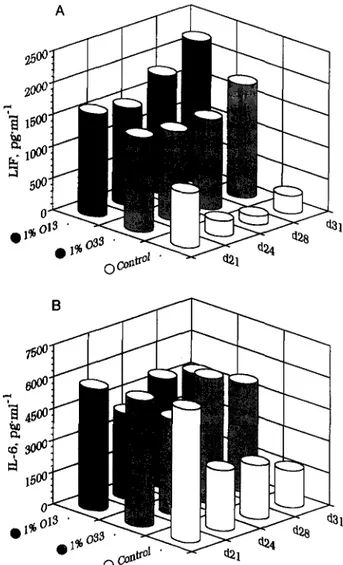
Fig. 2. Production of LIF and IL6 by human TEC: influence of mAb to OT 013 and 033. TEC supematrmts tested for LIF (A) and IL6 (B) content at time indicated, from day 21 to day 31. White columns are for control culture, grey are for lYo 033 added-culture and black are for lYo 013. Each column is the mean of quadmplicate measurements, with C.V. less than 5%.
3.2. Production of IL-6 and LIF by human TEC: Influence
of mAbs to neurohypophysial peptides
IL-1P was not detected in our experimental conditions, even in 5-fold concentrated medium, in either basal or mAb-stimulated TEC supematants. As the detection limit of the IL-1P EASIA is 50 pg/ml, IL-1P production by 50000 cultured TEC is under 10 pg/ml. On the other hand, under basal conditions cultured TEC produce 500– 1000 pg\ml ir-LIF, as well as 6–40 rig/ml ir-IL-6. LIF production showed a pronounced and sustained increase over basal when anti-OT 033 and 013 mAbs were added to TEC cultures (Fig. 2A). IL-6 production was sustained by the addition of anti-OT mAbs, in contrast with the rapid decrease under basal conditions (Fig. 2B).
3.3. Controls
LIF and IL-6 production by human TEC were not affected by anti-VP mAb BER-312, by IgG-starved ascitic
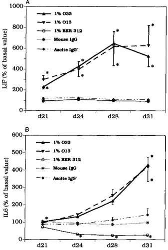
+ 8-.. —8-..—&- .. —..— .. —..— 0 I B 600 - 1%033 500 I +- 1%013 z ~ 1% BER 31’2 T* I d21 d24 d28 d31
Fig. 3. Production of LIF and IL6 by human TEC: control experiments. Only mAbs to OT (013 and 033) induced a marked increase of both LIF (A) and IL6 (B) production by human TEC. Anti-VP mAb BER-312, murine IgG, or murine ascitic fluid without IgG had no significant effect upon IL6. Anti-VP induced a significant inhibition of IL6 production. Each point is the mean of four experiments+ SD in triplicate. * P <0.001, by Mann-Whitney U-test.
fluid, or by mouse IgG (Fig. 3A). In terms of IL-6 production, a slight but significant inhibitory effect of anti-VP BER-312 was observed (Fig. 3B). There was no significant difference between the positive effects of anti-OT 013 and 033.
4. Discussion
The co-localization of ir-OT, IL-1~, IL-6 and LIF in cultured human TEC has been shown by combined im-munofluorescence and confocal microscopy. OT and LIF staining in epithelial cells was closely related to the cytok-eratin network on double immunofluorescence with anti-cytokeratin MNF 116 mAb. This finding suggests that the cytoskeleton of human TEC could be implicated in the processes of membrane translocation and secretion for this cell type. The results also are in close accord with previous ultrastructural analyses showing ir-OT to be diffusely lo-cated in the cytoplasm of murine TEC [12]. Cultured human TEC thus express both OT and cytokirtes, and the
colocalization demonstrated confirms previous observa-tions on human thymic secobserva-tions [24]. Anti-OT staining of cultured TEC adds further evidence that OT is the domi-nant neurohypophysial-related peptide expressed in thymic epitheliums.
A particular finding of the present study is that anti-OT mAbs strongly stimulate LIF and IL-6 secretion by cul-tured human TEC. The absence of EGF in US-SF might explain why ir-IL-6 is below detection limits under basal conditions, since biologically active and ir-IL-6 have been detected in human TEC culture in EGF-supplemented serum-free media, but not in DMEM or other EGF-defi-cient media [33]. Moreover, this cytokine has been shown to be regulated by EGF at a post-transcriptional level [34]. Colic et al reported IL-I(3 in the supematants from rat TEC cultures [35], but Shoham et al found IL-1P below detection limits in murine and human TEC cultures [36,37]. Thus, species differences or culture methods could explain some of the discrepancies in the data previously reported. The specificity of TEC activation by anti-OT mAbs has been carefully established by a variety of control experi-ments. It is not clearly due to a non-specific effect of Ig or ascitic fluid, nor did the anti-VP mAb exert any significant effect on LIF production, whereas it slightly inhibited ir-IL-6 production. As OT and VP differ by only two amino acids, the specific increase of cytokine production by TEC upon antigen recognition confirms that OT is the dominant self-antigen of the neurohypophysial family ex-pressed by TEC. Both 033 and 013 stimulate cytokine production by TEC, although 013 was previously shown not to stain human thymic sections [24,38]. In processing of the OT-precursor, OT must be cleaved from neuro-physin before 9gly-OT amidation. The absence of TEC labelling by 013 may thus reflect incomplete processing of the OT precursor within the TEC cytoplasm. In contrast, the biological effects observed with 013 argue for pre-sentation of fully-processed OT at the level of the TEC membranes.
The marked stimulating effect of artti-OT mAbs on TEC cytokine production may be explained by induction following immune recognition of the neurohypophysial-re-lated antigen presented at the level of the TEC membrane. Altogether, these data strongly support a membrane local-ization of OT. The positive effects observed with two OT mAbs directed against different epitopes of the OT molecule strongly suggest that a fully processed OT is presented at the TEC plasma membrane. Somehow like the transduction following the binding of a signal to its recep-tor, the immunological recognition of OT leads to TEC activation reflected here by the secretion of IL-6 and LIF. It will be also of interest to determine whether Abs against other thymic self-antigens such as neurokinin A [39] and insulin-like growth factor II [40,41] are also able to affect TEC secretory activity. At the present stage, this study supports our model on the dual physiological role played
44 H. Martens et al. /Regulatory Peptides 67 (1996) 39–45
in T-cell differentiation by the thymic repertoire of neu-roendocrine-related polypeptides [8,9].
Acknowledgements
V.G. is a Research Associate and Brigitte Malgrange is Senior Research Assistant of the National Fund of Scien-tific Research (Belgium). We thank Prof. J. Urbain (Laboratory of Physiology, Free University of Brussels, Rhode-Saint-Gen?se, Belgium) who provided us with hy-bridomas producing anti-OT mAbs. These studies were supported by the Research Fund of Lii5geUniversity Medi-cal School, by the Fondation L60n Fr6d&icq of the Li3ge University Hospital, by the Fund for Scientific Medical Research of Belgium (grant No. 3.4562.90; grants T616vie No. 7.4611.91, 7.4548.93 and 7.4532.95), by the Associa-tion contre le Cancer (Belgium), and by the European Science Foundation (Strasbourg).
References
[1] Haynes, B.F., The human thymic microenvironment, Adv. Irnmunol., 36 (1984) 87-142.
[2] Sprent, J., Lo, D., Gao, E.-K. and Ron, Y., T cell selection in the thymus, Irmmrnol. Rev., 101 (1988) 173–190.
[3] Muller, K.-P. and Kyewski, B.A., T cell receptor targeting to thymic corticat epitheliaf cells in vivo induces survival, activation and differentiation of immature thymocytes, Eur. J. Jmrmrnol., 23 (1993)
1661-1670.
[4] Jenkinson, E.J., Anderson, G. and Owen, J.J.T., Studies on T cell maturation on defined thymic stromat cell populations in vitro, J. Exp. Med., 176 (1992) 845–853.
[5] Hugo, P., Kappler, J.W. and Marrack, P.C., Positive selection of TcR afpha beta thymocytes: is cortical thymic epitheliumsan obliga-tory participant in the presentation of major histocompatibility com-plex protein?, Irnmunol. Rev., 135 (1993) 133–155.
[6] Bonomo, A. and Matzinger, P., Thymus epithelimn induces tissue-specific tolerance, J. Exp. Med., 177 (1993) 1153–1 164.
[7] Yukmanovic, S., Jameson, S.C. and Bevan, M.J., A thymic epithe-lial cell line induces both positive and negative selection in the thymus, Int. Immunol., 6 (1994) 239–246.
[8] Geenen, V., Martens, H., Robert, F., De Groote, D. and Fmnchi-mont, P., The thymic education of developing T cells in self neumendocrine principles, J. Endocrinol.Invest.,H (1992)621–629.
[9] Martens, H., Goxe, B. and Geenen, V., The thyrnic repertoire of
neuroendocrine self-peptides: Physiological implications in T-cell life and death, Immunol. Today, 17 (1996) 312–317.
[10] Kelly, P.J., Blalock, J.E., Chrousos, G.P., Yu-Lee, L. and Geenen, V., Neuroendocrine hormones and the immune system. In A. Cuello and B. Collier (Eds.), Pharmacological Sciences: Perspectives for Research and Therapy in the Late 1990s., Birkhauser, Basel, 1995, pp. 365-372..
[11] Geenen, V., Legros, J.J., Framchimont, P., Baudrihaye, M., De-fresne, M.-P. and Boniver, J., The neuroendocrine thymus: Coexis-tence of oxytocin and neurophysin in the human thymus, Science, 232 (1986) 508-511.
[12] Wiemarm, M. and Ehret, G., Subcellular localization of immunore-active oxytocin within thymic epithelial cells of the mate mouse, Cell Tissue Res., 273 (1993) 79-87.
[13] Elands, J., Resink, A. and de Kloet, E.R., Neurohypophysial
hor-mone receptors in the rat thymus, spleen, and lymphocytes, En-docrinology, 126 (1989) 2703-2710.
[14] Martens, H., Robert, F., Legros, J.J., Geenen, V. and Fmnchimont, P., Expression of functional neurohypophysiat peptide receptors by marine immature and cytotoxic T cell lines, Prog. NeuroEndocrin-Immnnol., 5 (1992) 31-39.
[15] Funder, J.W., Paracrine, cryptocrine, acrocrine, Mol. Cell. En-docrinol., 70 (1990) C21-C24.
[16] Geenen, V., Robert, F., Martens, H., Benhida, A., Degiovanni, G., Defresne, M.-P., Boniver, J., Legros, J.J., Martial, J. and Fmnchi-mont, P., Biosynthesis and paracrine\cryptocrine actions of “self” neurohypophysii+related peptides in the thymus, Mol. Cell. En-docrinol., 76 (1991) C27-C31.
[17] Geenen, V., Legros, J.-J., Francfrimont, P., Defresne, M.-P. Boniver, J., Ivell, R. and Richter, D., The thymus as a neuroendocrine organ. synthesis of vasopressin and oxytocirr in human thymic epitheliums., Ann. NY Acad. Sci., 496 (1987) 56-66.
[18] Geenen, V., Vandersmissen, E., Martens, H., Goxe, B., Kecha, O., Legros, J.-J., Lefebvre, P.J., Benhida, A. Rentier-Delrue, F. and Martiaf, J.A., Cellular and molecular aspects of thymic T-cell educa-tion to nenrohypophysial principles. In T. Saito, K. Kurokawa and S. Yoshida (Eds.), Neurohypophysis. Recent progress of vasopressin and oxytocin research. Proceedings of the First Joint World Congress of Neurohypophysis and Vasopressin, Excerpta Medica Intemationaf Congress Series 1098, Elsevier, Amsterdam, 1995, pp. 309-319. [19] Geenen, V., Vandersmissen, E., Cormann-Goffin, N., Martens, H.,
Legros, J.J., Degiovanni, G., Benhida, A., Martiaf, J. and Franchi-mont, P., Membrane translocation and relationship with MHC class I of a human thymic neurophysin-hke protein, Thymus, 22 (1993) 55-66.
[20] Gainer, H. and Wray, S., Cellular and molecular biology of oxytocin and vasopressin. In E. Knobil and J.D. Neill (Eds.), The Physiology of Reproduction, 2nd Edn., Raven Press, New York, 1993, pp. 1099–1129.
[21] Geenen, V. and Kroemer, G., The multiple ways to cellular immune tolerance, Irmmrnol. Today, 14 (1993) 573–575.
[22] Le, P.T., Tuck, D.T., Dirrarello, C.A., Haynes, B.F. and Singer, K.H., Human thymic epithelial cells produce interleukin-1, J. Im-munol., 138 (1987) 2520–2526.
[23] Le, P.T., Lazorick, S., Whichard, L.P., Yang, Y.C., Clarck, S.C., Haynes, B.F. and Singer, K.H., Human thymic epithelial cells produce IL-6, granrrlocyte-monocyte-CSF, and leukemia inhibitory factor, J. Immmrol., 145 (1990) 3310–3315.
[24] Robert, F., Geenen, V., Schoenen, J., Burgeon, E., De Groote, D., Defresne, M.-P., Legros, J.J. and Fmnchimont, P., Co-localization of immunoreactive oxytocin, vasopressin and interleukin-1 in human thymic epithelial neuroendocrine cells, Brain Behav. Irnmun., 5 (1991) 102-115.
[25] Burgeon, E., Chapleur, M., Schoenen, J., Remichius, D., Legros, J.J., Geenen, V. and Robert, F., Monoclinal antibodies to oxytocin: Production and characterization, J. Neuroimmunol., 31 (1991) 235-244.
[26] Robert, F.R., Leon-Henri, B.P., Chapleur-Chateau, M.M., Girr, M.N. and Burlet, A.J., Comparison of three immunoassay in the screen-ing and characterization of monoclinal antibodies against arginine-vasopressin, J. Neuroirnrmrnol., 9 (1985) 205–220.
[27] Coupley, L., Berrada, L., Gascan, H., Godsrrd, A. and Praforrm, V., High titre antibodies obtained by intralympfmode immunization with low amounts of antigen, Cytokine, 5 (1993) 564–569.
[28] Gilbert, S.F. and Migeon, B.R., ~Valine as a selective agent for normal human and rodent epitheliat cells in culture, Cell, 5 (1975) 11–17.
[29] Small, M., Barr-Nea, L. and Aronson, M., Culture of thymic epithe-lial cells from mice and age-related studies on the growing cells, Eur. J. Irnmunol., 14 (1984) 936–942.
vitro culture of human thymic epithelial cells in serum-free media, APMIS, 97 (1989) 926-934.
[31] De Groote, D., Zarrger14,P.F., Gevaert, Y., Fassotte, M.F., Beguin, Y., Noizat-Pirenne, F., Pirenne, J., Gathy, R., Lopez, M., Dehart, I., Igot, D., Baudrihaye, M., Delacroix, D. and Franchimont, P., Direct stimulation of cytokines (IL-1P, TNFa, IL-6, IL-2, IFNy and GM-CSF) in whole blood: I. Comparison with isolated PBMC stimulation, Cytokine, 4 (1992) 239–248.
[32] De Groote, D., Fauchet, F., Jadoul, M., Dehart, I., Gevaert, Y., Lopez, M., Gathy, R., Franssen, J.D., Radoux, D., Franchimont, P., Soulillou, J.P., Jacques, Y. and Godard, A., An Elisa for the measurements of human leukemia inhibitory factor in biological fluids and culture supematants, J. Immunol. Methods, 167 (1994) 253–261.
[33] Andersen, A., Pedersen, H., Bendtzen, K. and Ropke, C., Effects of growth factors on cytokine production in serum-free cultures of human thymic epithelial cells, Scand. J. Immunol., 38 (1993) 233– 238,
[34] Le, P.T., Lazorick, S., Whichard, L.P., Haynes, B.F. and Singer, K.H., Regulation of cytokine production in the human thymus: epidermal growth factor and transforming growth factor-a regulate mRNA levels of interleukir-la (IL-IcY.),IL-1P and IL-6 in human thymic epithelial cells at a post-transcriptional level, J. Exp. Med., 174 (1991) 1147–1157.
[35] Colic, M., Pejnovic, N., Katanranovski, M., Stojarrovic, N., Terzic, T. and Dujic, A., Rat thymic epithelial cells in culture constitutively secrete IL-1 and IL-6, Int. ImmunoL, 3 (1991) 1165–1174.
[36] Eshel, I., Savion, N. and Shoham, J., Analysis of thymic stromal cell subpopulations grown in vitro on extracellular matrix in defined medium. IL Cytokines activities in murine thymic epithelial and mesenchymal cell culture supernatants, J. ImmunoL, 144 (1990) 1563–1570.
[37] Meilin, A., Shoharn, J. and Sharabi, Y., Analysis of thymic stromal cell subpoptdations grown in vitro on extracelhdar matrix in defined medium. IV. Cytokines secreted by human thymic epithelial cells in culture and theiractivities on murine thymocytes and bone marrow cells, Immunology, 77 (1992) 208–213.
[38] Robert, F., Martens, H., Cormarm, N., Benhida, A., Schocnen, J. and Geenen, V., The recognition of hypothalamo-neurohypophysial func-tions by developing T cells, Dev. Immunol., 2 (1992) 131–140. [39] Ericsson, A., Genen V., Robert F., Legros, J.J., Vrindts-Gevaert, Y.,
Franchimont, P., Brene, S. and Persson, H., Expression of prepro-tachykinin A and neuropeptide Y mRNA in the thymus, Mol. Endocrinol., 4 (1990) 1211–1218.
[40] Geenen, V., Achour, I., Robert, F., Varrdersmissen, E., Sodoyez, J.C., Defresne, M.-P., Boniver, J., L&bvre, P.J. and Franchimont, P., Evidence that insulin-like growth factor 2 (IGF2) is the dominant thymic peptide of the insulin superfamily, Thymus, 21 (1993)
115–127.
[41] Kooijman, R., van BurrI-Offers, S.C., Scholtens, L.E., Schuurman, H.J., Van den Brande, L.J. and Zegers, B.J.M., T cell development in insulin-like growth factor-II transgenic mice, J. Immunol., 154 (1995) 5736-5745.