HAL Id: dumas-01753118
https://dumas.ccsd.cnrs.fr/dumas-01753118
Submitted on 5 Jun 2018
HAL is a multi-disciplinary open access
archive for the deposit and dissemination of
sci-entific research documents, whether they are
pub-lished or not. The documents may come from
teaching and research institutions in France or
abroad, or from public or private research centers.
L’archive ouverte pluridisciplinaire HAL, est
destinée au dépôt et à la diffusion de documents
scientifiques de niveau recherche, publiés ou non,
émanant des établissements d’enseignement et de
recherche français ou étrangers, des laboratoires
publics ou privés.
with high-dose topical corticosteroids for a bullous
pemphigoid
Nolwenn Ropars
To cite this version:
Nolwenn Ropars. Evolution of bone mineral density in patients treated with high-dose topical
corti-costeroids for a bullous pemphigoid. Life Sciences [q-bio]. 2016. �dumas-01753118�
ANNEE 2016
THÈSE D'EXERCICE / UNIVERSITÉ DE RENNES 1
FACULTÉ DE MEDECINE
sous le sceau de l’Université Européenne de Bretagne
THÈSE EN VUE DU
DIPLÔME D'ÉTAT DE DOCTEUR EN MEDECINE
Présentée par
Nolwenn ROPARS
née le 25 janvier 1988 à Brest
Evolution de la densité minérale
osseuse chez des patients traités
par dermocorticothérapie
prolongée pour une pemphigoïde.
Evolution of bone mineral density
in patients treated with high-dose
topical corticosteroids for a
bullous pemphigoid.
Thèse soutenue à RENNES
le 26/04/2016
devant le jury composé de :
Pascal GUGGENBUHL
Professeur-CHU Rennes / Président du jury
Alain DUPUY
Professeur-CHU Rennes / Directeur de thèse
Fabrice BONNET
Professeur-CHU Rennes / Juge
Lise BOUSSEMART
Maître de conférences-CHU Rennes / Juge
Monica DINULESCU
PROFESSEUR DES UNIVERSITES - PRATICIENS HOSPITALIERS
ANNE-GALIBERT Marie Dominique Biochimie et biologie moléculaire
BELAUD-ROTUREAU Marc-Antoine Histologie; embryologie et cytogénétique BELLISSANT Eric Pharmacologie fondamentale; pharmacologie clinique; addictologie BELLOU Abdelouahab Thérapeutique; médecine d'urgence; addictologie BELOEIL Hélène Anesthésiologie-réanimation; médecine d'urgence BENDAVID Claude Biochimie et biologie moléculaire
BENSALAH Karim Urologie BEUCHEE Alain Pédiatrie
BONAN Isabelle Médecine physique et de réadaptation
BONNET Fabrice Endocrinologie, diabète et maladies métaboliques; gynécologie médicale BOUDJEMA Karim Chirurgie générale
BOUGET Jacques Thérapeutique; médecine d'urgence; addictologie BOURGUET Patrick
Professeur des Universités en surnombre Biophysique et médecine nucléaire BRASSIER Gilles Neurochirurgie
BRETAGNE Jean-François Gastroentérologie; hépatologie; addictologie BRISSOT Pierre
Professeur des Universités en surnombre Gastroentérologie; hépatologie; addictologie CARRE François Physiologie
CATROS Véronique Biologie cellulaire CHALES Gérard
Professeur des Universités émérite Rhumatologie
CORBINEAU Hervé Chirurgie thoracique et cardiovasculaire CUGGIA Marc Biostatistiques, informatique médicale et
technologies de communication DARNAULT Pierre Anatomie
DAUBERT Jean-Claude
Professeur des Universités émérite Cardiologie
DAVID Véronique Biochimie et biologie moléculaire DAYAN Jacques
Professeur des Universités associé Pédopsychiatrie; addictologie DE CREVOISIER Renaud Cancérologie; radiothérapie
DECAUX Olivier Médecine interne; gériatrie et biologie du vieillissement; addictologie
DELAVAL Philippe Pneumologie; addictologie DESRUES Benoît Pneumologie; addictologie DEUGNIER Yves
DONAL Erwan Cardiologie
DRAPIER Dominique Psychiatrie d'adultes; addictologie DUPUY Alain Dermato-vénéréologie
ECOFFEY Claude Anesthésiologie-réanimation; médecine d'urgence EDAN Gilles Neurologie
FERRE Jean Christophe Radiologie et imagerie Médecine FEST Thierry Hématologie; transfusion
FLECHER Erwan Chirurgie thoracique et cardiovasculaire FREMOND Benjamin Chirurgie infantile
GANDEMER Virginie Pédiatrie
GANDON Yves Radiologie et imagerie Médecine GANGNEUX Jean-Pierre Parasitologie et mycologie
GARIN Etienne Biophysique et médecine nucléaire GAUVRIT Jean-Yves Radiologie et imagerie Médecine GODEY Benoit Oto-rhino-laryngologie
GUGGENBUHL Pascal Rhumatologie GUIGUEN Claude
Professeur des Universités émérite Parasitologie et mycologie GUILLÉ François Urologie
GUYADER Dominique Gastroentérologie; hépatologie; addictologie HOUOT Roch Hématologie; transfusion
HUGÉ Sandrine
Professeur des Universités associé Médecine générale HUSSON Jean-Louis
Professeur des Universités en surnombre Chirurgie orthopédique et traumatologique JEGO Patrick Médecine interne; gériatrie et biologie du
vieillissement; addictologie JEGOUX Franck Oto-rhino-laryngologie JOUNEAU Stéphane Pneumologie; addictologie
KAYAL Samer Bactériologie-virologie; hygiène hospitalière KERBRAT Pierre Cancérologie; radiothérapie
LAMY DE LA CHAPELLE Thierry Hématologie; transfusion
LAVIOLLE Bruno Pharmacologie fondamentale; pharmacologie clinique; addictologie
LAVOUE Vincent Gynécologie-obstétrique; gynécologie médicale LE BRETON Hervé Cardiologie
LE GUEUT Maryannick Médecine légale et droit de la santé LE TULZO Yves Réanimation; médecine d'urgence LECLERCQ Christophe Cardiologie
LEGUERRIER Alain Chirurgie thoracique et cardiovasculaire LEJEUNE Florence Biophysique et médecine nucléaire
LEVEQUE Jean Gynécologie-obstétrique; gynécologie médicale LIEVRE Astrid Gastroentérologie; hépatologie; addictologie MABO Philippe Cardiologie
MALLEDANT Yannick Anesthésiologie-réanimation; médecine d'urgence MEUNIER Bernard Chirurgie digestive
MICHELET Christian Maladies infectieuses; maladies tropicales MOIRAND Romain Gastroentérologie; hépatologie; addictologie MORANDI Xavier Anatomie
MORTEMOUSQUE Bruno Ophtalmologie
MOSSER Jean Biochimie et biologie moléculaire MOULINOUX Jacques Biologie cellulaire
MOURIAUX Frédéric Ophtalmologie ODENT Sylvie Génétique
OGER Emmanuel Pharmacologie fondamentale; pharmacologie clinique; addictologie
PERDRIGER Aleth Rhumatologie PLADYS Patrick Pédiatrie
POULAIN Patrice Gynécologie-obstétrique; gynécologie médicale RAVEL Célia Histologie; embryologie et cytogénétique RIFFAUD Laurent Neurochirurgie
RIOUX-LECLERCQ Nathalie Anatomie et cytologie pathologiques ROBERT-GANGNEUX Florence Parasitologie et mycologie
SAINT-JALMES Hervé Biophysique et médecine nucléaire
SEGUIN Philippe Anesthésiologie-réanimation; médecine d'urgence SEMANA Gilbert Immunologie
SIPROUDHIS Laurent Gastroentérologie; hépatologie; addictologie SOMME Dominique Médecine interne; gériatrie et biologie du
vieillisement; addictologie SULPICE Laurent Chirurgie générale TARTE Karin Immunologie
TATTEVIN Pierre Maladies infectieuses; maladies tropicales THIBAULT Ronan Nutrition
THIBAULT Vincent Bactériologie-virologie; hygiène hospitalière THOMAZEAU Hervé Chirurgie orthopédique et traumatologique
TORDJMAN Sylvie Pédopsychiatrie; addictologie VERGER Christian
Professeur des Universités émérite Médecine et santé au travail
VERHOYE Jean-Philippe Chirurgie thoracique et cardiovasculaire VERIN Marc Neurologie
VIEL Jean-François Epidémiologie, économie de la santé et prévention VIGNEAU Cécile Néphrologie
VIOLAS Philippe Chirurgie infantile
WATIER Eric Chirurgie plastique, reconstructrice et esthétique; brûlologie
MAITRES DE CONFERENCES DES UNIVERSITES - PRATICIENS HOSPITALIERS
AME-THOMAS Patricia Immunologie
AMIOT Laurence Hématologie; transfusion
BARDOU-JACQUET Edouard Gastroentérologie; hépatologie; addictologie BEGUE Jean-Marc Physiologie
BOUSSEMART Lise Dermato-vénéréologie CABILLIC Florian Biologie cellulaire
CAUBET Alain Médecine et santé au travail DAMERON Olivier Informatique
DE TAYRAC Marie Biochimie et biologie moléculaire DEGEILH Brigitte Parasitologie et mycologie DUBOURG Christèle Biochimie et biologie moléculaire
DUGAY Frédéric Histologie; embryologie et cytogénétique EDELINE Julien Cancérologie; radiothérapie
GALLAND Françoise Endocrinologie, diabète et maladies métaboliques; gynécologie médicale
GARLANTEZEC Ronan Epidémiologie, économie de la santé et prévention GUILLET Benoit Hématologie; transfusion
HAEGELEN Claire Anatomie
JAILLARD Sylvie Histologie; embryologie et cytogénétique LAVENU Audrey Sciences physico-chimiques et technologies pharmaceutiques LE GALL François Anatomie et cytologie pathologiques
LE RUMEUR Elisabeth Physiologie
MAHÉ Guillaume Chirurgie vasculaire; médecine vasculaire MARTINS Raphaël Cardiologie
MASSART Catherine Biochimie et biologie moléculaire MATHIEU-SANQUER Romain Urologie
MENARD Cédric Immunologie MENER Eric Médecine générale MILON Joëlle Anatomie
MOREAU Caroline Biochimie et biologie moléculaire MOUSSOUNI Fouzia Informatique
MYHIE Didier Médecine générale PANGAULT Céline Hématologie; transfusion RENAUT Pierric Médecine générale
RIOU Françoise Epidémiologie, économie de la santé et prévention ROBERT Gabriel Psychiatrie d'adultes; addictologie
ROPARS Mickaël Anatomie SAULEAU Paul Physiologie
TADIÉ Jean-Marc Réamination; médecine d'urgence TATTEVIN-FABLET Françoise Médecine générale
TURLIN Bruno Anatomie et cytologie pathologiques
VERDIER Marie-Clémence Pharmacologie fondamentale; pharmacologie clinique; addictologie
Remerciements
A Monsieur le Professeur Pascal Guggenbuhl, je vous remercie d’avoir accepté de présider cette thèse. Merci aussi pour votre aide et votre réactivité dans nos différents échanges.
A Monsieur le Professeur Alain Dupuy, je vous remercie d’avoir accepté de diriger cette thèse. Merci pour votre enseignement et votre encadrement durant ces années d’internat.
A Monsieur le Professeur Fabrice Bonnet, je vous remercie d’avoir accepté de juger de cette thèse.
A Madame le Docteur Lise Boussemart, je te remercie d’avoir accepté de juger cette thèse. Merci aussi pour ton enseignement pendant ce dernier semestre.
A Madame le Docteur Monica Dinulescu, je te remercie d’avoir accepté de juger cette thèse. Merci aussi pour ton enseignement durant ces années d’internat.
A famille, mes parents ma grand-mère, mes frères, ma belle sœur, Flamm. Merci pour votre soutien constant, votre énergie et votre présence bienveillante depuis toujours.
A Clairette, mon bibi, pour tous ces moments de rigolade depuis le berceau et ceux à venir.
A Cécilou ma colloc, pour ces belles années d’externat à Brest puis d’internat à Robelin que nous avons partagées et que nous partagerons encore.
A Clémence, co interne puis chef exemplaire. Pour ton amitié, ton soutien, ton enseignement, ta présence sans faille.
A mes amis de Brest: Chloé, JH, Elise, Coco, Thibault, en souvenir de nos réunions au 15K, de ces pistes dévalées à coup de bicyclettes, de tous ces bon moments et ceux à venir.
Aux « vieux copains », PJ et VA: Claire, Vincent, Benoit, Hélène, Ludovic, Léna, Pierrot, Charlotte, Antoine, Charlotte, Thibault, Manuel, Clément, Sarah, Eric, Alice. Pour votre amitié si solide qui dure depuis si longtemps et durera encore.
A « la famille rennaise », Marie, Céline, Berthier, Meynard, Cabaret, Pauline et Julien, pour les nombreuses réunions de familles et procès auxquels nous avons participé, pour ceux à venir.
A mes co-internes/chef actuels: Karine, Ayse, Raphaelle, Claire, Anne Clémence, Catherine, Julien, Maxence, merci de m’avoir supportée dans cette dernière ligne droite…
Et mes anciens co-internes/chefs : Florence, Sophie, Lisa, Marie, Alicia, Laetitia, Cécile, Annabelle, Morgane, Lalie, Elsa, Solène, Aurélie, François, Arnaud.
A Juju d’anapath, pour ta relecture attentive.
A tout le personnel du service, médecins, secrétaires (mention spéciale à Patricia pour les alertes aux bulles), infirmières et aides soignantes pour vos bons soins aux patients et ces kilos de crèmes appliqués…
A Olivier, merci pour ton soutien essentiel, ton coatching efficace, ta confiance, ton optimisme, ta bonne humeur et tes attentions quotidiennes… Merci d’être la.
TABLE DES MATIERES
ABSTRACT ... 11
INTRODUCTION ... 12
MATERIALS AND METHODS ... 13
RESULTS ... 15
DISCUSSION ... 16
CONCLUSION... 18
REFERENCES BIBLIOGRAPHIQUES... 20
GLOSSAIRE... 24
F
IGURE1
F
LOW CHART... 25
F
IGURE2
R
EPRESENTATION OF LUMBAR SPINE,
FEMORAL NECK AND HIPBMD
CHANGE,
BASELINEBMD,
AND CUMULATIVE DOSE OF CLOBESTASOL PROPIONATE. ... 26
F
IGURE3
C
ORRELATION BETWEEN LUMBAR SPINE VARIATION(
G/
CM2)
AND DOSE OF CLOBETASOL PROPIONATE(
G/
KG/
DAY)... 27
T
ABLE1
B
ASELINE CHARACTERISTICS... 28
T
ABLE2
B
ASELINE BONE STATUS... 29
T
ABLE3
C
HARACTERISTICS OF PATIENTS WITHBMD
CHANGE AND PATIENTS WITHOUTBMD
CHANGE... 30
ABSTRACT
Introduction: High dose topical corticosteroids may have same systemic side effects as systemic
corticosteroids. Bone side effects are poorly known with topical corticosteroids. We studied evolution of Bone Mineral Density (BMD) in patients with Bullous Pemphigoid (BP) treated with high dose of clobetasol propionate in a retrospective monocentric cohort.
Material and Methods: BMD measurements and biological analysis were performed before and after
topical corticosteroid treatment in patients with BP from January 2014 to June 2015. Tubes of clobetasol propionate were count every month. The objective was to describe changes in BMD and biological markers according to the dose of clobetasol propionate in patients with BP.
Results: 29 patients had BP, 22 patients had at least one assessment, and 9 patients had two
assessments. 6 patients had a BMD variation > 0.03 g/cm2. There was no correlation with dose of clobetasol propionate. There was no biological change before and after treatment. Limitations were missing data, small size of the sample, and number of patients excluded because high mortality of BP.
Conclusion: These results suggest larger studies to analyse links between high dose of topical
INTRODUCTION
In France, first-line treatment for Bullous Pemphigoid (BP), an auto-immune bullous dermatosis, currently lies on prolonged use of high dose superpotent topical corticosteroid (1).
Use systemic corticosteroids was considered the mainstay treatment for BP patients (2,3), until a large French multicenter prospective randomized trial (4) showed that clobetasol propionate cream was more effective than systemic corticosteroids in disease control on day 21, and presented with a lower rate of severe systemic side effects. Another French multicenter randomized prospective trial (5) compared two regimens of topical corticosteroids and showed that a shorter treatment (4 months vs 12 months) using lower doses of clobetasol propionate (10-30 gram (g) vs 40 g per day) was as effective as the standard regimen with fewer life-threatening side effects. Consequently, clobetasol propionate cream has become the BP treatment of reference in France.
Systemic absorption following cutaneous application of corticosteroid cream ranges between 0.05 % and 0.3 % (6) and may cause systemic side effects. Cases of iatrogenic Cushing (7), adrenal suppression (8–13) or hyperglycemia (14–16), have been reported following cutaneous application caused by improper and prolonged usage of super potent topical corticosteroid. These side effects are well described with systemic glucocorticoids. Few cases of topical corticosteroids-related bone side effects (17–22) have been reported. Osteoporosis is one of the most frequent and clinically relevant side effect of long-term systemic glucocorticoid therapy (23) and not described with topical corticosteroid. Osteoporosis is a disease characterized by a reduce in bone strength, leading to an increased risk of fracture and related increases in morbidity and mortality (24). Bone strength is estimated by measuring bone mineral density (BMD). Cross-sectional studies have evidenced low values for BMD in patients with atopic dermatitis (25,26) and psoriasis (22,27). However, low BMD may be due to absorption of topical corticosteroids or to chronic inflammation.
The aim of the present study was to evaluate the change of BMD during high dose topical corticosteroids treatment in patients with BP.
MATERIALS AND METHODS
We performed a retrospective monocentric review of newly diagnosed BP who attended the dermatological department in Rennes University Hospital from January 2014 to June 2015. For all BP patients, the diagnosis of active disease was established on the basis of the presence of blisters on the skin as well as the typical immunopathological criteria such as linear deposition of IgG and C3 at the basement membrane zone on direct immunofluorescence and circulating levels of anti BP180 and/or anti BP230 autoantibodies by ELISA.
Patients were treated with an initial dose of clobetasol propionate cream ranging from 20 to 40 g per day. The clobetasol propionate cream was tapered over 4 months according to the following protocol driven by SFD (Société Française de Dermatologie) guidelines: clobetasol propionate cream every day the first month, every other day the second month, twice a week the third month and once a week the fourth month. If there were less than ten blisters, the treatment was the same but with only 20 g per application. Osteoporosis prophylaxis was not routinely given.
In our Dermatology department, all newly BP underwent physical, biological examinations (complete blood cell counts, blood electrolyte, blood sugar, serum creatinine, thyroid stimulating hormone (TSH), parathyroid hormone (PTH), morning serum cortisol, electrophoresis, and markers of bone metabolism: serum calcium (adjusted with albumin), urinary calcium, serum phosphate, 25-hydroxyvitamin D (25-OHD), total alkaline phosphate, C-terminal telopeptid of type 1 collagen (CTx), N-terminal propeptide of type I procollagen (P1NP)) and a BMD measurement at the initial visit and after treatment (about 6 months later). Biological samples (blood electrolytes, blood sugar and creatinine) were performed every month.
Every month, at each follow up visit, the patients underwent physical examination and the number of new bullae that appeared daily was recorded, together with the number of empty tubes of clobetasol propionate that had been used. In this article, clobetasol propionate is expressed in grams: 10 g of clobetasol propionate correspond to one tube of topical corticosteroids containing 5 mg of clobetasol propionate.
BMD was measured in all patients at the lumbar spine, hip and femoral neck using Dual energy X-ray Absorptiometry (DXA)(Hologic QDR 4500 Bone Densitometer) in a single laboratory. The BMD are expressed in gram per square centimeter after which the Standard Derivation (SD) was calculated. Data was expressed as T-score (number of SDs from the mean value of the young adult of the same sex reference population). DXA was reported by one investigator who was aware of the patient’s diagnosis but not of the frequency, amount or duration of clobetasol propionate used. We considered a meaningful BMD change above 0.03 g/cm2 between both DXA at any site (28,29). Otherwise, bone density was considered stable. WHO Fracture Assessment Fracture tool (or Frax-tool) was calculated for all patients with necessary data according to recommendations (30) of the French Society of Rheumatology and the Groupe de Recherche et d’Information sur les Osteoporoses (we considered no familial history of osteoporosis).
Fracture risk factors were collected: history of osteoporosis with fracture, history of prolonged systemic corticosteroids, prolonged immobilization, current smoker, alcohol consumption, BMI < 19 kg/m2. Parental hip fracture, calcium intake, ages and treatment of menopauses could not be evaluated in this study. We retrospectively collected data from the medical records.
We compared change of BMD and blood analysis before and after the treatment with clobetasol propionate. The objective of this retrospective cohort was to describe changes in BMD and biological markers according to the dose of topical corticosteroids.
The statistical analysis was done with SAS software (version 9.4). Continuous variables were expressed by mean and standard deviation and categorical variables were expressed by the number of patients and percentages. Comparisons between M0 and M6 were realized using Wilcoxon signed rank test.
RESULTS
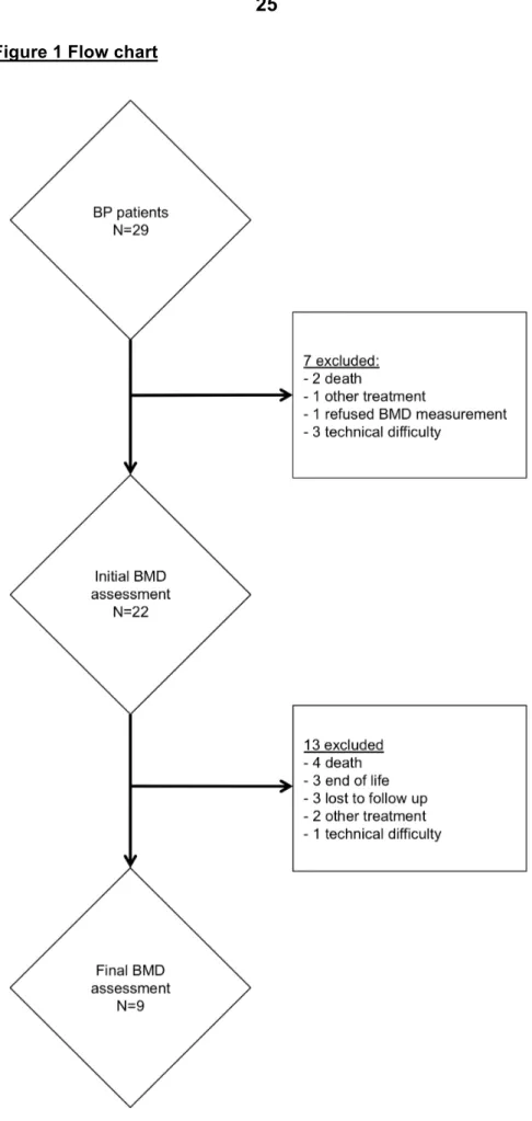
A total of 29 patients with BP attended the dermatological department of Rennes University Hospital between January 2014 and June 2015. Among the 29 patients, 22 had at least one BMD measurement and biological examination. Among them, 9 patients had 2 BMD measurements and biological examinations (flow chart figure 1).
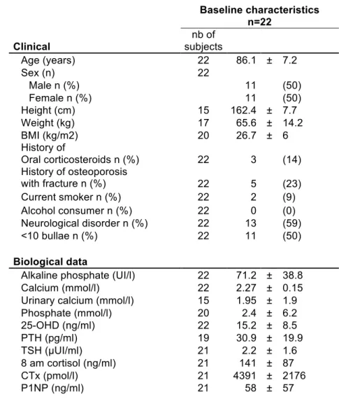
Table 1 shows clinical and biological baseline characteristics of patients. All patients had more than
70 years. Eighteen of them had at least one additional risk factor of fracture. The median BMI was 26.7 ± 6.0 kg/m2. Thirteen patients were bedridden. Five patients had history of osteoporosis with fracture. Among these 5 patients, 1 had bisphosphonate treatment, 1 had calcium plus vitamin D treatment and 3 had vitamin D treatment alone. There was no patient with rheumatoid arthritis or with secondary osteoporosis. Frax-tool was calculated for patients without systemic corticosteroids, but could be calculated for only ten of the nineteen patients. For the other patients femoral necks BMD, weights or heights were missing. Among the ten patients, only two patients had an indication of osteoporosis treatment.
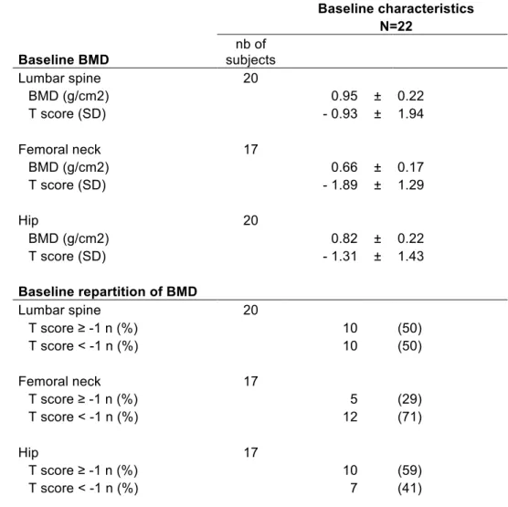
Baseline BMD measurements are presented in table 2.
Among the 22 patients, 11 patients had baseline < 10 bullae per day. Thirteen patients could not be evaluated and 9 patients were evaluated after treatment. Mean cumulative dose of clobetasol propionate was 2070 ± 922 g. Five patients had an initial treatment with 30 g per day and 4 patients had 20 g per day. One patient (patient 1) was evaluating at 12 months. He was treated with a cumulate dose of 7220 g of clobetasol propionate. Six of the 9 patients with two assessments were in clinical remission at 6 months. Four patients still have clobetasol propionate treatment at 6 months. Three of these 4 patients had lower cortisol measurements after 6 months. Only patient 27 had a collapsed cortisol with no clinical symptom. He was treated with 2270 g ± 929 g of clobetasol propionate. There was no fracture during or after treatment.
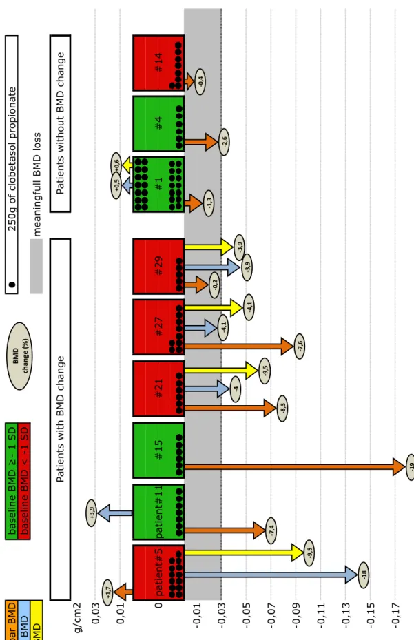
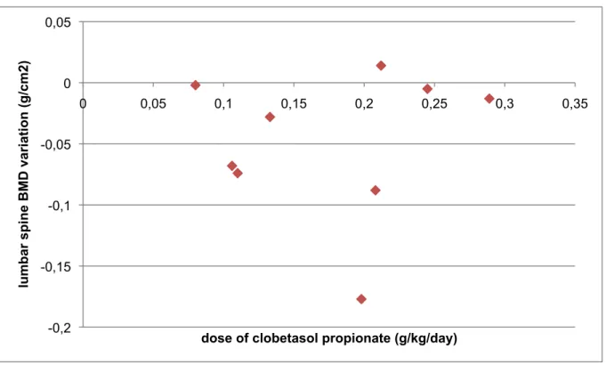
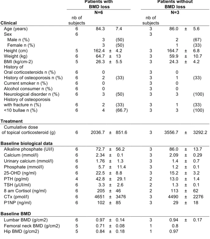
Nine patients had initial and final evaluations. There was no significant biological variation before and after treatment especially in blood sugar, serum calcium, urinary calcium, serum phosphate, 25-OHD, alkaline phosphate, CTx and P1NP for the nine patients with two assessments. Figure 2 presents schematically all variations for the 3 sites measurements together with the cumulative dose of clobetasol propionate. Among the 9 patients with both an initial and final BMD evaluations, 6 had a bone density loss and 3 were stable, without bone density loss. The mean bone loss at 6 months was - 5.0 ± 6.4% at the lumbar spine, - 4.8 ± 4.5% at the hip and - 4.6 ± 7.5% at the femoral neck. Clinical and biological characteristics of patients with or without bone density loss are compared in table 3. P1NP was higher with no significant difference between patients with and without bone loss. Calcium and 25-OHD were higher in patients with bone density loss compared to patients without. Paradoxically, the mean cumulative dose of clobetasol propionate per patient was lower in patients with bone density loss, compared to the patients without bone density loss, and no obvious correlation between clobetasol propionate dose and bone density variation was observed (figure 3 for lumbar site measurements, and data not shown for other sites).
DISCUSSION
In our study, among 9 BP patients treated with high dose of superpotent topical corticosteroids, 6 had BMD loss. No obvious correlation with cumulative or daily dose of clobetasol propionate was observed.
BMD decrease with systemic corticosteroid has been well described (31,32). Significant decrease is observed within the first six months (5 to 15%) then 2% per year in lumbar spine and femoral neck (33). This BMD decrease depends on the daily dose (34) and duration of corticosteroid therapy, rather than the cumulative dose. BMD decline is reversible when corticosteroid treatment is stopped (35–37). Current recommendations for prevention treatment of osteoporosis are to treat patients according Frax-tool and to treat all patients over 70 years having systemic corticosteroids for more than 3 months (38).
In immunobullous disease, change of BMD with systemic corticosteroids was – 0,6% in lumbar spine and + 1% in femoral neck at six months in a prospective study (39) on 14 patients supplemented with calcium and vitamin D. An association between pemphigus and osteoporosis reported independently of corticosteroid use has been reported (40). Immunobullous disease by itself might be associated with bone fractures (41), although the respective roles of corticosteroid and low vitamin D level are not easily disentangled.
Examples of low BMD have already been reported to be associated with atopic dermatitis (25,26) and psoriasis (27), with difficulties in assessing the respective responsibilities of corticosteroids and chronic inflammation. Cases of femoral head necrosis (17–21) and bone fractures (22) in patients with no other risk factor than topical corticosteroids have also been reported. The role of topical corticosteroid remains unclear (42–47). Systemic absorption is as low as 0.05% to 0.3% (6) have been shown to lead to systemic side effects such as cushing, adrenal suppression or hyperglycemia. BP treatment in France may represent a specific situation because of the singularity of the therapeutic scheme with high dose of superpotent corticosteroid during 4 months (1).
In our study, 6 of the 9 patients with both BMD measurements had a BMD loss but relations with cumulative or daily dose of topical corticosteroid were unclear. Only one of the six patients had a collapsed cortisol. Diminution of cortisol during treatment with topical corticosteroids was already reported (8–13) especially with a dose of clobetasol propionate over 50 g per week (8) and return to normal after stopping treatment if less than 50 g per week is applied (8,48).
In our study, patients with BP had multiple risk factor of fracture, in addition to age. There was no obvious relationship between BMD change and baseline BMD status, or risk factors for osteoporosis. Even if no significant variation were found between both groups, patients with higher dose of P1NP had a BMD change. There were more at risk of osteoporosis due to a high bone turnover, according to other study (49,50).
In our study a high mortality rate was observed. The relevance of implementing a systematic prevention of osteoporosis in BP patients is questionable because of the high mortality rate in older BP patients (51–53).
Our study has several limitations. First because of the retrospective nature of the study, data missing partly hampered the analysis. However, the amount of clobetasol propionate was precisely collected and was available for all the 9 patients with both BMD measurements. Second, our sample size was small, and made smaller by a high attrition rate in a population with a high mortality rate; the sample size was large enough, however, to evidence a decrease in BMD in 6 of the 9 patients, and we were able to describe the bone status in 22 patients with BP. Last, we had no information on the pre-treatment dynamics of bone loss in our patients, this piece of information would have been of great value to fully interpret the specific role of dermocorticosteroid in BMD decrease, by allowing a comparison between the periods before and during dermocorticosteroid therapy.
CONCLUSION
Our results suggest that larger studies would be interesting to analyze the links between high dose of clobetasol propionate and osteoporosis in patients with bullous pemphigoid. It seems necessary to continue systematic evaluation of risk factor of osteoporosis for patients with bullous pemphigoid and to propose prevention on case-by-case basis.
REFERENCES
1. Bernard P, Bedane C, Prost C, Ingen-Housz-Oro S, Joly P. Recommandations des centres de référence des maladies bulleuses auto-immunes pour le diagnostic et la prise en charge de la pemphigoïde bulleuse. Ann Dermatol Vénéréologie. 2011 Mar;138(3):247–51.
2. Wood AJ, Fine J-D. Management of acquired bullous skin diseases. N Engl J Med. 1995;333(22):1475–84.
3. Korman NJ. Bullous pemphigoid. The latest in diagnosis, prognosis, and therapy. Arch Dermatol. 1998 Sep;134(9):1137–41.
4. Joly P, Roujeau J-C, Benichou J, Picard C, Dreno B, Delaporte E, et al. A comparison of oral and topical corticosteroids in patients with bullous pemphigoid. N Engl J Med. 2002 Jan 31;346(5):321–7.
5. Joly P, Roujeau J-C, Benichou J, Delaporte E, D’Incan M, Dreno B, et al. A comparison of two regimens of topical corticosteroids in the treatment of patients with bullous pemphigoid: a multicenter randomized study. J Invest Dermatol. 2009 Jul;129(7):1681–7.
6. Mizuchi A, Miyachi Y, Tamaki K, Kukita A. Percutaneous absorption of betamethasone 17-benzoate measured by radioimmunoassay. J Invest Dermatol. 1976 Aug;67(2):279–82.
7. Tempark T, Phatarakijnirund V, Chatproedprai S, Watcharasindhu S, Supornsilchai V, Wananukul S. Exogenous Cushing’s syndrome due to topical corticosteroid application: case report and review literature. Endocrine. 2010 Dec;38(3):328–34.
8. Carruthers JA, August PJ, Staughton RC. Observations on the systemic effect of topical clobetasol propionate (Dermovate). Br Med J. 1975;4(5990):203–4.
9. Munro DD. The effect of percutaneously absorbed steroids on hypothalamic-pituitary adrenal function after intensive use in in-patients*. Br J Dermatol. 1976;94(s12):67–76.
10. Turpeinen M, Salo OP, Leisti S. Effect of percutaneous absorption of hydrocortisone on adrenocortical responsiveness in infants with severe skin disease. Br J Dermatol. 1986 Oct;115(4):475–84.
11. Weston WL, Fennessey PV, Morelli J, Schwab H, Mooney J, Samson C, et al. Comparison of hypothalamus-pituitary-adrenal axis suppression from superpotent topical steroids by standard endocrine function testing and gas chromatographic mass spectrometry. J Invest Dermatol. 1988 Apr;90(4):532–5.
12. Kerner M, Ishay A, Ziv M, Rozenman D, Luboshitzky R. Evaluation of the pituitary-adrenal axis function in patients on topical steroid therapy. J Am Acad Dermatol. 2011 Jul;65(1):215–6. 13. Böckle BC, Jara D, Nindl W, Aberer W, Sepp NT. Adrenal Insufficiency as a Result of
Long-Term Misuse of Topical Corticosteroids. Dermatology. 2014;228(4):289–93.
14. Gomez EC, Frost P. Induction of glycosuria and hyperglycemia by topical corticosteroid therapy. Arch Dermatol. 1976 Nov;112(11):1559–62.
15. Sobngwi E, Lubin V, Ury P, Timsit F-J, Gautier J-F, Vexiau P. Adrenal insufficiency and diabetes mellitus secondary to the use of topical corticosteroids for cosmetic purpose. Ann Endocrinol. 2003 Jun;64(3):202–4.
16. van der Linden MW, Penning-van Beest FJ, Nijsten T, Herings RM. Topical Corticosteroids and the Risk of Diabetes Mellitus. Drug Saf. 2009;32(6):527–37.
17. El Maghraoui A, Tabache F, Bezza A, Ghafir D, Ohayon V, Archane MI. Femoral head osteonecrosis after topical corticosteroid therapy. Clin Exp Rheumatol. 2001 Apr;19(2):233.
18. Kubo T, Kojima A, Yamazoe S, Ueshima K, Yamamoto T, Hirasawa Y. Osteonecrosis of the femoral head that developed after long-term topical steroid application. J Orthop Sci. 2001;6(1):92–4.
19. Kane D, Barnes L, Fitzgerald O. Topical corticosteroid treatment: systemic side-effects. Br J Dermatol. 2003 Aug;149(2):417.
20. Kabata T, Shimanuki K, Shimanuki K, Tsuchiya H. Osteonecrosis of the femoral head and glaucoma caused by topical corticosteroid application. Mod Rheumatol Jpn Rheum Assoc. 2011 Dec;21(6):706–9.
21. Takahashi H, Tsuji H, Honma M, Ishida-Yamamoto A, Iizuka H. Femoral head osteonecrosis after long-term topical corticosteroid treatment in a psoriasis patient. J Dermatol. 2012 Oct;39(10):887–8.
22. Gönül M, Gönül E. A case of multiple bone fractures due to the use of topical corticosteroid therapy for psoriasis. J Dermatol Treat. 2014 Jun 20;1–2.
23. Henneicke H, Gasparini SJ, Brennan-Speranza TC, Zhou H, Seibel MJ. Glucocorticoids and bone: local effects and systemic implications. Trends Endocrinol Metab. 2014 Apr;25(4):197– 211.
24. Bliuc D, Nguyen ND, Milch VE, Nguyen TV, Eisman JA, Center JR. Mortality risk associated with low-trauma osteoporotic fracture and subsequent fracture in men and women. Jama. 2009;301(5):513–21.
25. Aalto-Korte K , Turpeinen M. Bone mineral density in patients with atopic dermatitis. Br J Dermatol. 1997;136(2):172–5.
26. Haeck IM, Hamdy NAT, Timmer-de Mik L, Lentjes EGWM, Verhaar HJJ, Knol MJ, et al. Low bone mineral density in adult patients with moderate to severe atopic dermatitis. Br J Dermatol. 2009 Dec;161(6):1248–54.
27. Nymann P, Kollerup G, Jemec GB, Grossmann E. Decreased bone mineral density in patients with pustulosis palmaris et plantaris. Dermatol Basel Switz. 1996;192(4):307–11.
28. Ravaud P, Reny JL, Giraudeau B, Porcher R, Dougados M, Roux C. Individual smallest detectable difference in bone mineral density measurements. J Bone Miner Res. 1999;14(8):1449–56.
29. Baim S, Wilson CR, Lewiecki EM, Luckey MM, Downs RW, Lentle BC. Precision assessment and radiation safety for dual-energy X-ray absorptiometry: position paper of the International Society for Clinical Densitometry. J Clin Densitom Off J Int Soc Clin Densitom. 2005;8(4):371–8. 30. Briot K, Cortet B, Thomas T, Audran M, Blain H, Breuil V, et al. 2012 update of French guidelines for the pharmacological treatment of postmenopausal osteoporosis. Joint Bone Spine. 2012 May;79(3):304–13.
31. Laan RF, van Riel PL, van de Putte LB, van Erning LJ, van’t Hof MA, Lemmens JA. Low-dose prednisone induces rapid reversible axial bone loss in patients with rheumatoid arthritis. A randomized, controlled study. Ann Intern Med. 1993 Nov 15;119(10):963–8.
32. McKenzie R, Reynolds JC, O’Fallon A, Dale J, Deloria M, Blackwelder W, et al. Decreased bone mineral density during low dose glucocorticoid administration in a randomized, placebo controlled trial. J Rheumatol. 2000 Sep;27(9):2222–6.
33. Lafage-Proust MH, Boudignon B, Thomas T. Glucocorticoid-induced osteoporosis: pathophysiological data and recent treatments. Joint Bone Spine. 2003;70(2):109–18.
34. van Staa TP, Leufkens HG, Abenhaim L, Zhang B, Cooper C. Oral corticosteroids and fracture risk: relationship to daily and cumulative doses. Rheumatol Oxf Engl. 2000 Dec;39(12):1383–9.
35. Staa T van, Staa T van, Staa T van, Leufkens HGM, Cooper C. The epidemiology of corticosteroid-induced osteoporosis: a meta-analysis. Osteoporos Int. 2002;13(10):777–87.
36. Rizzato G, Montemurro L. Reversibility of exogenous corticosteroid-induced bone loss. Eur Respir J. 1993;6(1):116–9.
37. Reid IR. Glucocorticoid osteoporosis–mechanisms and management. Eur J Endocrinol. 1997;137(3):209–17.
38. Briot K, Cortet B, Roux C, Fardet L, Abitbol V, Bacchetta J, et al. 2014 update of recommendations on the prevention and treatment of glucocorticoid-induced osteoporosis. Joint Bone Spine. 2014 Dec;81(6):493–501.
39. Tee S-I. Prevention of Glucocorticoid-Induced Osteoporosis in Immunobullous Diseases With Alendronate: A Randomized, Double-blind, Placebo-Controlled Study. Arch Dermatol. 2012 Mar 1;148(3):307.
40. Wohl Y, Dreiher J, Cohen AD. Pemphigus and osteoporosis: a case-control study. Arch Dermatol. 2010;146(10):1126–31.
41. Marzano AV, Trevisan V, Eller-Vainicher C, Cairoli E, Marchese L, Morelli V, et al. Evidence for vitamin D deficiency and increased prevalence of fractures in autoimmune bullous skin diseases: Vitamin D and prevalence of fractures in bullous diseases. Br J Dermatol. 2012 Sep;167(3):688– 91.
42. Vestergaard P, Olsen ML, Paaske Johnsen S, Rejnmark L, Toft Sørensen H, Mosekilde L. Corticosteroid use and risk of hip fracture: a population-based case–control study in Denmark. J Intern Med. 2003;254(5):486–93.
43. Vestergaard P, Rejnmark L, Mosekilde L. Fracture risk associated with systemic and topical corticosteroids. J Intern Med. 2005;257(4):374–84.
44. Van Velsen SGA, Knol MJ, van Eijk RLA, de Vroede MA, de Wit TC, Lam MGEH, et al. Bone mineral density in children with moderate to severe atopic dermatitis. J Am Acad Dermatol. 2010 Nov;63(5):824–31.
45. Van Velsen SGA, Haeck IM, Knol MJ, Lam MGEH, Bruijnzeel-Koomen CAFM. Two-year assessment of effect of topical corticosteroids on bone mineral density in adults with moderate to severe atopic dermatitis. J Am Acad Dermatol. 2012 Apr;66(4):691–3.
46. Mu Z, Zhang J. The effect of topical corticosteroids on bone mineral density in adults with atopic dermatitis. J Am Acad Dermatol. 2013 Jan;68(1):181–2.
47. Haeck I, van Velsen S, de Bruin-Weller M, Bruijnzeel-Koomen C. Bone Mineral Density in Patients with Atopic Dermatitis. In: Ring J, Darsow U, Behrendt H, editors. Chemical Immunology and Allergy [Internet]. Basel: KARGER; 2012 [cited 2016 Mar 28]. p. 96–9. Available from: http://www.karger.com/doi/10.1159/000331893
48. Van Velsen SGA, Haeck IM, Bruijnzeel-Koomen CAFM. Percutaneous absorption of potent topical corticosteroids in patients with severe atopic dermatitis. J Am Acad Dermatol. 2010 Nov;63(5):911–3.
49. Rogers A, Hannon RA, Eastell R. Biochemical markers as predictors of rates of bone loss after menopause. J Bone Miner Res Off J Am Soc Bone Miner Res. 2000 Jul;15(7):1398–404. 50. Gielen E, O’Neill T, Pye S, Adams J, Ward K, Wu F, et al. Bone turnover markers predict hip
bone loss in elderly European men: results of the European Male Ageing Study (EMAS). Osteoporos Int. 2015 Feb;26(2):617–27.
51. Doffoel-Hantz V, Sparsa A, Marin B, Durox H, Bonnetblanc J-M, Bédane C. Profil évolutif des patients atteints de pemphigoïde bulleuse au cours de la première année de traitement. Ann Dermatol Vénéréologie. 2009 May;136(5):407–11.
52. Cortés B, Khelifa E, Clivaz L, Cazzaniga S, Saurat JH, Naldi L, et al. Mortality Rate in Bullous Pemphigoid: A Retrospective Monocentric Cohort Study. Dermatology. 2012;225(4):320–5. 53. Joly P, Baricault S, Sparsa A, Bernard P, Bédane C, Duvert-Lehembre S, et al. Incidence and
GLOSSAIRE
BMD Bone Mineral Density BMI Body Mass Index BP Bullous Pemphigoid
CTx C-terminal telopeptid of type I collagen DXA Dual energy X-ray Absorptiometry
P1NP N-terminal propeptide of type I procollagen PTH Parathyroid hormone
SD Standard Derivation
SFD Société Française de Dermatologie TSH Thyroid Stimulating Hormone WHO World Health Organization 25-OHD 25-hydroxyvitamin D
Figure 2 Representation of lumbar spine,
femoral neck and hip BMD change,
baseline BMD, and cumulative dose of clobestasol
propionate.
Lu m ba r B M D Ne ck B M D H ip B M D m ea n in gf u ll B M D l o ss g/ cm 2 pa ti en t# 5 pa ti en t# 1 1 #1 5 #2 1 #2 9 -0,07 ba se lin e B M D ! - 1 S D 2 5 0 g o f cl o be ta so l pr o pi o n ate ba se lin e B M D < -1 S D Pa ti en ts w ith B M D ch an ge Pa ti en ts w ith o u t B M D ch an ge #1 #4 #1 4 -0,01 -0,03 0,01 0,03 -0,05 #2 7 0 -0,09 -0,11 -0,13 -0,15 -0,17 !" #$ % &'#(% &$#)% *+, %% -. /0 12 %345 % &"'% &6#7% &'#(% &$#8% &)#"% &9#:% &7#'% &"#7% !9 #8 % &:#8% &9#)% &)% !9 #( % !7 #' % &)#"% &7#'% &"6%
Figure 3 Correlation between lumbar spine variation (g/cm2) and dose of clobetasol
propionate (g/kg/day)
-0,2 -0,15 -0,1 -0,05 0 0,05 0 0,05 0,1 0,15 0,2 0,25 0,3 0,35 lu m b ar s p in e B MD v ar ia ti o n (g /c m 2)Table 1 Baseline characteristics
Baseline characteristics n=22 Clinical nb of subjects Age (years) 22 86.1 ± 7.2 Sex (n) 22 Male n (%) 11 (50) Female n (%) 11 (50) Height (cm) 15 162.4 ± 7.7 Weight (kg) 17 65.6 ± 14.2 BMI (kg/m2) 20 26.7 ± 6 History of Oral corticosteroids n (%) 22 3 (14) History of osteoporosis with fracture n (%) 22 5 (23) Current smoker n (%) 22 2 (9) Alcohol consumer n (%) 22 0 (0) Neurological disorder n (%) 22 13 (59) <10 bullae n (%) 22 11 (50) Biological dataAlkaline phosphate (UI/l) 22 71.2 ± 38.8 Calcium (mmol/l) 22 2.27 ± 0.15 Urinary calcium (mmol/l) 15 1.95 ± 1.9 Phosphate (mmol/l) 20 2.4 ± 6.2 25-OHD (ng/ml) 22 15.2 ± 8.5 PTH (pg/ml) 19 30.9 ± 19.9 TSH (µUI/ml) 21 2.2 ± 1.6 8 am cortisol (ng/ml) 21 141 ± 87 CTx (pmol/l) 21 4391 ± 2176 P1NP (ng/ml) 21 58 ± 57
Table 2 Baseline bone status
Baseline characteristics N=22 Baseline BMD nb of subjects Lumbar spine 20 BMD (g/cm2) 0.95 ± 0.22 T score (SD) - 0.93 ± 1.94 Femoral neck 17 BMD (g/cm2) 0.66 ± 0.17 T score (SD) - 1.89 ± 1.29 Hip 20 BMD (g/cm2) 0.82 ± 0.22 T score (SD) - 1.31 ± 1.43 Baseline repartition of BMD Lumbar spine 20 T score ≥ -1 n (%) 10 (50) T score < -1 n (%) 10 (50) Femoral neck 17 T score ≥ -1 n (%) 5 (29) T score < -1 n (%) 12 (71) Hip 17 T score ≥ -1 n (%) 10 (59) T score < -1 n (%) 7 (41)Table 3 Characteristics of patients with BMD change and patients without BMD
change.
Patients with BMD loss Patients without BMD loss N=6 N=3 Clinical nb of subjects nb of subjects Age (years) 6 84.3 7.4 3 86.0 ± 5.6 Sex 6 3 Male n (%) 3 (50) 2 (67) Female n (%) 3 (50) 1 (33) Height (cm) 5 162.4 ± 4.2 3 164.7 ± 6.8 Weight (kg) 6 64.7 ± 13 3 59.9 ± 10.7 BMI (kg/cm-2) 5 26.3 ± 5.5 3 24.3 ± 4.2 History of Oral corticosteroids n (%) 6 0 3 0 History of osteoporosis n (%) 6 2 (33) 3 1 (33) Current smoker n (%) 6 0 3 0 Alcohol consumer n (%) 6 0 3 0 Neurological disorder n (%) 6 3 (50) 3 3 (100) History of osteoporosis with fracture n (%) 6 2 (33) 3 1 (33) <10 bullae n (%) 6 4 (66.7) 3 3 (100) Treatment Cumulative dose of topical corticosteroid (g) 6 2036.7 ± 851.6 3 3556.7 ± 3292.2Baseline biological data
Alkaline phosphate (UI/l) 6 72.7 ± 56.2 3 86.0 ± 13.7 Calcium (mmol/l) 6 2.34 ± 0.1 3 2.09 ± 0.29 Urinary calcium (mmol/l) 6 1.76 ± 1.3 3 1.4 ± 0.7 Phosphate (mmol/l) 6 5.7 ± 11.4 3 1.2 ± 0.1 25-OHD (ng/ml) 6 22.5 ± 8.8 3 15.2 ± 3.2 PTH (pg/ml) 4 42.8 ± 29.1 2 13.0 ± 1.4 TSH (µUI/ml) 6 3.3 ± 2.6 2 1.3 ± 0.1 8 am Cortisol (ng/ml) 6 205 ± 46 2 113 ± 62 CTx (pmol/l) 6 4651 ± 3476 3 4490 ± 2276 P1NP (ng/ml) 6 102 ± 85 3 29 ± 18 Baseline BMD Lumbar BMD (g/cm2) 6 0.97 ± 0.14 3 0.94 ± 0.17 Femoral neck BMD (g/cm2) 5 0.71 ± 0.08 1 0.8 Hip BMD (g/cm2) 5 0.84 ± 0.18 1 0.97
U.F.R. DE MEDECINE DE RENNES
ROPARS Nolwenn – Evolution de la densité minérale osseuse chez des patients traités par dermocorticothérapie prolongée pour une pemphigoïde.
Evolution of bone mineral density in patients treated with high-dose topical corticosteroids for a bullous pemphigoid.
28 feuilles, 3 graphiques, 3 tableaux, 30 cm – Thèse : (Médecine); Rennes 1; 2016 ; N° Résumé français
Introduction Les dermocorticoïdes (DC) utilisés à forte dose, peuvent avoir des effets secondaires
systémiques (cushing, hyperglycémie) identiques à ceux observés avec la corticothérapie générale mais leurs effets secondaires osseux sont mal connus. Nous avons étudié l’évolution de la densité minérale osseuse (DMO) chez des patients traités par DC pour une pemphigoïde bulleuse (PB).
Matériel & Méthodes Une ostéodensitométrie et un bilan biologique étaient réalisés avant et après
traitement par DC pour les patients suivis pour une PB au CHU de Rennes.
Résultats 29 patients ont eu une PB, 22 patients ont eu au moins une évaluation et 9 patients ont eu
2 évaluations. Nous avons observé une variation de la DMO supérieure à 0,03 g/cm2 chez 6 des 9 patients. Il n’a pas été noté de corrélation avec la dose de DC.
Conclusion Ces résultats incitent à proposer des études de plus grande envergure pour l’analyse des liens entre DC et DMO chez les patients ayant une PB.
Rubrique de classement : Dermatologie - Thérapeutique
Mots-clés : Pemphigoïde Bulleuse ; Dermocorticothérapie ; Densité Minérale Osseuse Mots-clés anglais MeSH : Bullous Pemphigoid; Topical Corticosteroids; Bone Mineral Density
Président : Monsieur le Professeur Pascal GUGGENBUHL
JURY Assesseurs : Monsieur le Professeur Alain DUPUY (directeur de Thèse) Monsieur le Professeur Fabrice BONNET
Madame le Docteur Lise BOUSSEMART Madame le Docteur Monica DINULESCU
![[PDF] Les diagrammes UML | Télécharger PDF](data:image/gif;base64,R0lGODlhAQABAIAAAP///wAAACH5BAEAAAAALAAAAAABAAEAAAICRAEAOw==)