EFFECT OF BIOACTIVE MATERIALS MODIFIED WITH CHONDROITIN SULFATE ON HUMAN MSC
BY
Jessica Elizabeth DE LA TORRE TORRES
THESIS PRESENTED TO
ÉCOLE DE TECHNOLOGIE SUPÉRIEURE IN PARTIAL FULFILLMENT FOR
A MASTER'S DEGREE WITH THESIS IN ENGINEERING CONCENTRATION IN HEALTHCARE TECHNOLOGY
M.Sc.A
MONTREAL, JULY 6, 2016
This Creative Commons license allows readers to download this work and share it with others as long as the author is credited. The content of this work may not be modified in any way or used commercially.
BY THE FOLLOWING BOARD OF EXAMINERS
Mrs. Sophie Lerouge, Thesis Supervisor
Mechanical engineering department at École de technologie supérieure
Mrs. Nicola Hagemeister, President
Automated production engineering department at École de technologie supérieure
Mr. Shant Der Sarkissian, Member of the jury Surgery department at Université de Montréal
THIS THESIS WAS PRENSENTED AND DEFENDED
IN THE PRESENCE OF A BOARD OF EXAMINERS AND THE PUBLIC ON JUNE 29, 2016
First of all, I would like to thank God, for giving me such a great opportunity in my life to grow and learn. I would also like to thank my research director Sophie Lerouge for welcoming me into her laboratory. I am very grateful for her kindness, patience and help which made possible this project.
Moreover, I would like to thank my colleagues at LBeV which helped me with advices and accompanied me during my project. I have learned a lot from all of them and they have helped me to grow professionally and personally. I would like to particularly thank Marion Marie, Caroline Ceccaldi, Pauline Lequoy and Yasaman Alinejad for guiding me during my project and sharing their scientific expertise with me.
Last but not least, I would like to thank my family: my mother Irene Torres, my father Edmundo De La Torre and my sister Jackeline De La Torre for believing, encouraging and supporting me every single step of the process. Thanks to them I have achieved every single goal in my life including this project.
Jessica Elizabeth DE LA TORRE TORRES SUMMARY
Biomaterials functionalization by addition of biomolecules is an interesting approach for enhancing cell-biomaterial interactions and therefore improve their bioactivity. The global objective of this project is to enhance the bioactivity of biomaterials such as implantable devices and 3D scaffolds by adding extracellular matrix components and therefore facilitate the adhesion, growth and survival of human mesenchymal stem cells (hMSC) in biomaterials for tissue repair and tissue engineering applications.
In this project chondroitin sulfate (CS) and growth factors were studied for their effect on hMSC in biomaterials. First, the effect of these biomolecules was tested in solution. Then, two kinds of biomaterials were created: bioactive surfaces for enhancing bioactivity of implantable devices and bioactive hydrogels which can be used as 3D scaffolds for cell encapsulation and delivery.
A pro-survival effect of the growth factors studied in this project (epidermal growth factor, vascular endothelial growth factor and fibroblast growth factor) was not observed when tested in solution, therefore the project further focused on CS effect only. Interestingly, CS did not affect cell growth in media containing serum, while inducing cell detachment from substrate in serum free conditions.
For the bioactive surfaces construction, CS was grafted to either an amine-rich plasma-polymerized coating created on polyethylene terephthalate (PET) films (further referred as LP) or to commercial cell culture plates functionalized with amino groups. The bioactive surfaces were characterized by different techniques such as contact angle, atomic force microscopy, Orange II dye and Toluidine Blue O dye colorimetric assays (for amino group and CS quantification respectively) and finally, cell culture experiments (adhesion, growth and survival). Results confirmed the presence of CS grafted on both substrates. Commercial amine plates grafted almost five times more CS compared to LP. This rendered the surface antifouling for proteins and cells as confirmed by protein adsorption and cell culture assays. Cell culture assays on bioactive surfaces based on LP demonstrated improved cell adhesion and growth when compared to tissue culture plates or bare PET films in serum containing conditions.
Chitosan based hydrogels containing CS at a concentration of 500 µg/ml resulted in a cohesive hydrogel which supported hMSC viability up to 7 days. However increasing CS concentration to high level such as 10000 µg/ml led to decrease of cell viability after 4 or 7 days, probably due to lack of porosity and water since the hydrogel precipitates upon formation and expulses water.
In conclusion, this work demonstrated that CS immobilization can enhance the biological interactions of hMSC with biomaterials used for implantable devices such as PET. Further studies are needed to evaluate the possible effect of CS on hMSC differentiation and phenotype. Hydrogels with CS could be very interesting for tissue engineering applications such as cartilage formation.
Keywords: Biomaterials, mesenchymal stem cells, tissue engineering, bioactivity, chondroitin sulfate
Jessica Elizabeth DE LA TORRE TORRES RÉSUMÉ
La fonctionnalisation de biomatériaux par ajout de biomolécules est une approche intéressante pour améliorer les interactions cellule-biomatériau, et ainsi accroître leur bioactivité. L’objectif de ce projet est d’améliorer la bioactivité de matériaux tels que les implants et les matrices 3D en ajoutant des composantes de la matrice extracellulaire, afin de faciliter l’adhésion, la croissance et la survie des cellules souches mésenchymateuses humaines (hMSC) sur les biomatériaux pour des applications comme la réparation de tissus et l’ingénierie tissulaire.
Dans ce projet, les effets du sulfate de chondroïtine et des facteurs de croissance sur les hMSC ont été étudiés. Tout d’abord, l’effet de ces molécules a été étudié en solution. Deux types de biomatériaux ont ensuite été obtenus : des surfaces bioactives pour une meilleure bioactivité des implants d’une part, et des hydrogels bioactifs pouvant être utilisés comme matrices 3D pour l’encapsulation et l’implantation de cellules d’autre part.
Les facteurs de croissance étudiés au cours de ce projet (facteur de croissance épidermique, facteur de croissance endothélial vasculaire et facteur de croissance des fibroblastes) n’ont pas montré d’effet pro-survie en solution, ainsi le projet s’est ensuite concentré sur les effets du CS seulement. Curieusement, le CS n’a pas affecté la croissance cellulaire en milieu avec sérum, mais a conduit à un détachement des cellules de leur substrat en conditions sans sérum.
Pour obtenir des surfaces bioactives, le CS a été greffé sur deux types de substrats : des films de PET sur lesquels une couche mince, polymérisée par plasma et riche en amines, a été déposée (que l’on nommera LP), et des plaques commerciales de culture cellulaire, fonctionnalisées par des groupements amines. Les surfaces bioactives furent caractérisées par différentes techniques : angle de contact, microscope à force atomique, tests colorimétriques par Orange II et Toluidine Bleue O (pour la quantification d’amines et du CS, respectivement), et tests cellulaires (adhésion, croissance et survie). La présence de CS greffé a été confirmée sur les deux types de substrats. Les plaques commerciales aminées ont permis le greffage de cinq fois plus de CS que les revêtements LP, avec pour conséquence l’obtention d’une surface antiadhésive pour les protéines et les cellules, comme l’ont confirmé les tests d’adsorption de protéine et de culture cellulaire. Les tests de culture cellulaire menés sur les surfaces bioactives obtenues sur les substrats LP, ont montré une adhésion et une croissance cellulaire accrues, en comparaison aux plaques de culture et les films de PET, en présence de sérum.
Les hydrogels de chitosane contenant 500 µg/ml de CS ont conduit à des hydrogels cohésifs permettant la survie des hMSC jusqu’à 7 jours. Toutefois, l’augmentation de la quantité de CS à des hauts niveaux de concentration tels que 10000 µg/ml, a entrainé une décroissance de la survie cellulaire après 4 ou 7 jours, probablement à cause d’un manque de porosité ainsi que d’eau, entrainé par la précipitation des hydrogels lors de leur formation.
En conclusion, ce travail a montré que l’immobilisation de CS permet d’améliorer les interactions biologiques entre les hMSC et les biomatériaux utilisés dans des dispositifs implantables, comme le PET. D’autres études sont nécessaires pour évaluer l’effet possible du CS sur la différentiation des hMSC et leur phénotype. Les hydrogels contenant du CS pourraient être dignes d’intérêt pour des applications d’ingénierie tissulaire comme la formation de cartilage.
Mots clés: Biomatériaux, cellules souches mésenchymateuses, ingénierie tissulaire, bioactivité, sulfate de chondroïtine
INTRODUCTION ...1
CHAPTER 1 LITERATURE REVIEW ...3
1.1 Biomaterials and tissue engineering ...3
1.2 MSC for tissue engineering and cell therapy ...5
1.3 Problematic of biomaterials and tissue engineering ...7
1.4 Factors explaining lack of healing and survival of cells in biomaterials ...8
1.5 Endovascular aneurysm repair (EVAR) example ...13
1.6 Bioactive biomaterials ...14
1.6.1 Physicochemical and biological modifications of biomaterials ... 14
1.6.2 Bioactive molecules for biomaterials ... 18
1.7 Literature review conclusions ...24
CHAPTER 2 OBJECTIVES AND HYPOTHESES ...25
2.1 General objective ...25
2.2 Specific objectives ...25
2.3 Hypotheses underlying the project ...25
CHAPTER 3 MATERIALS AND METHODS ...27
3.1 Preparation of bioactive surfaces with chondroitin sulfate ...27
3.1.1 Amine rich plasma polymerization ... 28
3.1.2 EDC NHS covalent chemistry immobilization ... 29
3.2 Physicochemical characterization of bioactive surfaces ...30
3.2.1 Contact angle measurement ... 30
3.2.2 AFM ... 32
3.2.3 CS grafting potential by Toluidine Blue ... 33
3.2.4 Amino group surface density by Orange II ... 35
3.2.5 Measure of protein adsorption ... 37
3.3 Preparation of a chitosan hydrogel with chondroitin sulfate ...38
3.3.1 Materials for hydrogels preparation ... 38
3.3.2 Chitosan solution ... 39
3.3.3 Gelling agent solutions ... 39
3.3.4 Preparation of the hydrogels ... 40
3.3.5 Rheological testing of hydrogels ... 41
3.4 Effect of CS containing solution, surfaces and hydrogels on MSC ...42
3.4.1 Cell types ... 42
3.4.2 Methods of characterization of the cellular response ... 43
3.4.2.1 Alamar blue ... 43
3.4.2.2 Crystal violet staining ... 44
3.4.3 Effect of growth factors and CS in solution... 44
3.4.4.1 Cell culture on LP based bioactive coatings ... 45
3.4.4.2 Cell culture on amine plate based bioactive coatings ... 46
3.4.5 Cell culture in 3D chitosan hydrogels ... 46
3.5 Statistical Analysis ...47
CHAPTER 4 RESULTS ...49
4.1 Effect of biomolecules in solution ...49
4.1.1 Effect of Growth factors ... 49
4.1.2 Effect of Chondroitin sulfate ... 52
4.1.2.1 CS effect on hMSC ... 52
4.1.2.2 CS effect on VSMC ... 56
4.2 Effect of bioactive surfaces ...58
4.2.1 Physicochemical characterization of bioactive surfaces ... 58
4.2.1.1 Contact angle- wettability ... 58
4.2.1.2 AFM ... 59
4.2.1.3 CS grafting potential by Toluidine blue ... 61
4.2.1.4 Amino group density by Orange II ... 62
4.2.2 Adhesion and growth of hMSC on LP based bioactive coatings ... 64
4.2.2.1 Adhesion ... 64
4.2.2.2 Growth ... 67
4.2.3 Survival of hMSC on LP based bioactive coatings ... 70
4.2.4 Adhesion and growth of hMSC on amine plate based bioactive coatings ... 72
4.2.4.1 Adhesion and growth with different CS grafted densities ... 72
4.2.4.2 Antifouling effect of CS analyzed by protein adsorption ... 73
4.3 Effect of CS in chitosan hydrogels (3D scaffolds) ...74
CHAPTER 5 GENERAL DISCUSSION, LIMITS AND PERSPECTIVES ...81
CONCLUSION ...93
APPENDIX I EGF AND CS EFFECT ON RAT MSC ...95
Figure 1.1 Tissue engineering key factors ...3
Figure 1.2 MSC potential for tissue engineering through different mechanisms of action ...6
Figure 1.3 Interaction between ECM and cells ...9
Figure 1.4 Cell interactions with a substrate through protein ligands ...10
Figure 1.5 Schematic representation of a stent graft implanted in an abdominal aortic aneurysm during EVAR procedure ...14
Figure 1.6 Different structures of chondroitin sulfate ...19
Figure 3.1 Experimental steps achieved during the project ...27
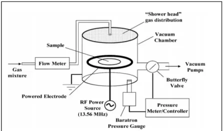
Figure 3.2 Schematic view of low pressure plasma reactor used in the fabrication of LP ...29
Figure 3.3 Illustration of contact angles formed by drops on solid surfaces ...31
Figure 3.4 AFM principle ...32
Figure 3.5 Toluidine Blue O structure ...34
Figure 3.6 Orange II molecule ...36
Figure 3.7 Hydrogels preparation methods ...41
Figure 3.8 Cell-containing hydrogel inside a well ...47
Figure 4.1 Effect of GF on hMSC viability after 4 and 7 days in low serum (LS; 2%) or serum free (SF) medium (cell source =Stem cell technologies). Results are expressed as percentage of alamar blue fluorescence signal compared to signal after 24h adhesion in complete medium (mean ± SD; N=1, n=4) ...50
Figure 4.2 Effect of EGF on hMSC viability after 3 and 7 days in serum free (SF) medium (cells source = Texas A&M Institute for Regenerative Medicine). Results are expressed as percentage of alamar blue fluorescence signal compared to signal after 24h adhesion in complete medium (N=2, n=4) ...51
Figure 4.3 Effect of VEGF on hMSC viability after 3 and 5 days in serum free (SF) medium (cells source = Lonza). Results are expressed as percentage of alamar blue fluorescence signal compared to signal
after 24h adhesion in complete medium (N=1, n=4) ...52 Figure 4.4 Effect of CS in solution on hMSC viability after 4 and 7 days in
normal serum (NS), low serum (LS) or serum free (SF) medium (cell source =Stem cell technologies). Results are expressed as percentage of alamar blue fluorescence signal compared to signal after 24h adhesion in complete medium
(mean ± SD; N=1, n=4 each) ...53 Figure 4.5 Effect of CS in solution on hMSC viability after 3 and 7 days
in normal serum (NS) and serum free (SF) medium
(cells source = Texas A&M Institute for Regenerative Medicine). Results are expressed as percentage of alamar blue fluorescence signal compared to signal after 24h adhesion in complete medium
(N=2, n=4) (*p<0.05) ...54 Figure 4.6 hMSC after 5 days in SF and SF+CS. The round individual
morphology may indicate that cells are about to detach from
the surface ...54 Figure 4.7 Dose response of CS on hMSC viability after 3, 5 and 7 days in
serum free medium (SF). Results correspond to alamar blue fluorescence signal normalized to the signal after 24h adhesion in complete medium. Arrows indicate the conditions where cell
detachment was observed (N=2, n=4) ...56 Figure 4.8 Dose response of CS on VSMC viability after 3, 5 and 7 days
in serum free medium (SF). Results correspond to alamar blue fluorescence signal normalized to the signal after 24h adhesion in complete medium. Arrows indicate the conditions where cell
detachment was observed (N=2, n=4) ...57 Figure 4.9 Contact angle measurement of bare PET, PET coated by LP and
by LP+CS (mean ± SD; N=7, n=3) Significant difference was
observed between each surface (*p<0.05) ...59 Figure 4.10 AFM topographic (a.c) and phase (b,d) images of LP (a,b) and
LP+CS (c,d) (10µm x 10µm) ...60 Figure 4.11 AFM height measurements 3D image for LP+CS sample
Figure 4.12 TBO surface densities on PET, LP, LP+CS, AP and AP+CS
(N=4, n=4) (*p<0.05) ...61
Figure 4.13 TBO bounded on LP and LP+CS before desorption of the dye ...61
Figure 4.14 Grafting of CS at different concentrations on AP (N=4, n=4) ...62
Figure 4.15 Orange II surface densities on LP and AP (N=4, n=4) (*p<0.05) ...63
Figure 4.16 Adhesion of hMSC on LP based bioactive surfaces (10,000 cells/surface) (cells source = Lonza). Results are expressed as percentage of alamar blue fluorescence signal compared to the signal of PET after 24h (N=2, n=4) (*p<0.05) ...65
Figure 4.17 Adhesion of hMSC on LP based bioactive surfaces (15,000 cells/surface) (cells source = Lonza). Results are expressed as percentage of alamar blue fluorescence signal compared to the signal of PET after 24h (N=4, n=4) (* p<0.05 with all other surfaces; & p<0.05 with PCP and PET) ...65
Figure 4.18 Adhesion of hMSC on LP based bioactive surfaces (cells source = Texas A&M Institute for Regenerative Medicine). Results are expressed as percentage of alamar blue fluorescence signal compared to the signal of PET after 24h (N=8, n=4) (*p<0.05) ...66
Figure 4.19 Representative images of hMSC adhesion (cells source = Texas A&M Institute for Regenerative Medicine) at 24h in PCP, PET, LP and LP+CS ...67
Figure 4.20 Growth of hMSC on LP based bioactive surfaces (10,000 cells/ surface) (cells source = Lonza). Results are expressed as percentage of alamar blue fluorescence signal compared to the signal of PET after 24h (N=1, n=4, representative of 2 independent experiments) (* p<0.05 with all other surfaces at the same time point; & p<0.05 with PCP and PET) ...68
Figure 4.21 Growth ratio per surface type of hMSC on LP based bioactive surfaces (10,000 cells/surface) (cells source = Lonza). Results are expressed as percentage of alamar blue fluorescence signal compared to the initial signal (at 24h) on each surface (N=1, n=4, representative of 2 independent experiments) (* p<0.05; + confluency reached) ...68 Figure 4.22 Growth of hMSC on LP based bioactive surfaces (cells source =
expressed as percentage of alamar blue fluorescence signal
compared to the signal of PET after 24h (N=8, n=4) (*p<0.05) ...69 Figure 4.23 Representative images of hMSC growth (cells source = Texas
A&M Institute for Regenerative Medicine) at 4d in PCP, PET,
LP and LP+CS ...70 Figure 4.24 Survival of hMSC (cell source = Lonza lot) after 3d or 7d in serum
free medium. Results are expressed as percentage of alamar blue fluorescence signal compared to the signal of PET after 24h in
complete medium (N=3,n=4) (*p<0.05) ...71 Figure 4.25 Representative image of hMSC (cell source = Lonza) remaining
on LP after 7 days in serum free medium ...71 Figure 4.26 Morphology of hMSC (cell source = Stem cell technologies lot)
when seeded on AP and AP+CS prepared using a concentration
of 1% (w/v) of CS ...72 Figure 4.27 Adhesion and growth of hMSC (cell source = Stem cell technologies
lot) on amine plate (AP) based bioactive coatings prepared using different percentages of CS in the grafting solution. Bare AP and PCP are used as controls. Results are presented as alamar blue fluorescence signal (N=1, n=4, representative of 2 independent
experiments) ...73 Figure 4.28 Protein adsorption on amine plate based bioactive coatings prepared
using different percentages of CS in the grafting solution. Results are presented as fluorescence intensity units (N=1, n=4,
representative of 2 independent experiments) (*p<0.05) ...74 Figure 4.29 Evolution of the storage modulus, G’, at different time
points at 37°C for chitosan-based hydrogels with 1% or 0% of CS
(N=1, n=4) ...75 Figure 4.30 Photograph of chitosan-based hydrogels with 1% or 0% CS after
1h of incubation at 37°C ...75 Figure 4.31 Viability of hMSC entrapped in chitosan hydrogels with 1%
or 0% CS after 24h, 4 and 7 days in alpha MEM medium
(cells source = Texas A&M Institute for Regenerative Medicine). Results are expressed as percentage of alamar blue fluorescence signal compared to signal at 24h in hydrogels with 0% CS
Figure 4.32 Viability of hMSC entrapped in chitosan hydrogels with 1% or 0% CS after 24h, 4 and 7 days in NutriStem XF medium (cells source = Texas A&M Institute for Regenerative Medicine). Results are expressed as percentage of alamar blue
fluorescence signal compared to signal at 24h in hydrogels
with 0% CS (N=3, n=3) ...77 Figure 4.33 Evolution of the storage modulus, G’, at different time
points at 37°C for chitosan-based hydrogels with 0.05% or 0% of
CS (N=1, n=3) ...78 Figure 4.34 Photograph of chitosan-based hydrogel with 0.05% CS after
1h of incubation at 37°C ...78 Figure 4.35 Viability of hMSC entrapped in chitosan hydrogels with 0.05%
or 0% CS after 24h, 4 and 7 days in NutriStem XF medium (cells source = Texas A&M Institute for Regenerative Medicine). Results are expressed as percentage of alamar blue fluorescence signal compared to signal at 24h in hydrogels with 0% CS
AP Commercial Amine Plates
BMP Bone morphogenic protein
BMP-2 Bone morphogenic protein 2 CH Chitosan
CM Complete medium
CS Chondroitin 4-sulfate
ECM Extracellular matrix
EDC N-(3-dimethylaminopropyl)-N’-ethylcarbodiimide hydrochloride EGF Epidermal growth factor
EVAR Endovascular aneurysm repair FBS Fetal bovine serum
FGF Fibroblast growth factor GAG Glycosaminoglycans
GF Growth Factor
HCL Hydrochloric acid
hMSC Human mesenchymal stem cells IGF-I Insulin-like growth factor I
LBeV Laboratory of Endovascular Biomaterials
LP Low-pressure plasma polymerized nitride ethylene
LS Low serum
NaOH Sodium Hydroxide NHS N-hydroxysuccinimide OPA o-phthaldehyde
PAAm Polyallylamine PBS Phosphate buffered saline
PCP Polystyrene culture plate
PET Polyethylene terephthalate
PLA Poly(lactic acid)
PMDS Poly(dimethylsiloxane) rMSC Rat mesenchymal stem cells
SD Standard deviation
SDS Sodium dodecyl sulfate
SF Serum free medium
SHC Sodium hydrogen carbonate SPD Sodium phosphate dibasic SPM Sodium phosphate monobasic TBO Toluidine Blue O
TRITC Tetramethylrhodamine isothiocyanate VEGF Vascular endothelial growth factor
VSMC Vascular Smooth muscle cell VSMC Vascular smooth muscle cells XPS X-ray photoelectron spectroscopy
Pa Pascals
Sccm Standard cubic centimeters per minute W Watts
INTRODUCTION
Biomaterials are materials that intend to interact with biological systems for different purposes, such as replacing or enhancing a body part or function, and they are widely studied nowadays for a variety of particular applications. In the growing field of tissue engineering biomaterials are being used as scaffolds for cells to induce tissue repair and regeneration. Additionally, stem cell research has increased exponentially in the last years due to their potential in tissue engineering. Therefore, the combination of biomaterials with stem cells may provide an excellent template for tissue repair. However one of the main problems biomaterials face is the lack of biological interactions with cells. The lack of biological interactions may prevent cell survival adhesion and growth, thus impairing tissue regeneration.
Biomaterials functionalization by addition of biomolecules is an interesting approach for enhancing cell-biomaterial interactions. The addition of extracellular matrix (ECM) molecules which are normally present in the body and regulate cell behavior is promising for promoting biological interactions with biomaterials. Addition of specific ECM molecules to biomaterials may induce an adequate cell response by communicating with cells through specific ligands and inducing signaling pathways as they do in the body.
The general objective of this project is to study the potential of extracellular matrix components such as chondroitin sulfate and growth factors to enhance the bioactivity of biomaterials such as implantable devices and 3D scaffolds. More particularly we will investigate whether these molecules facilitate the adhesion, growth and survival of hMSC, which play a major role in tissue repair and tissue engineering applications.
Biomaterials’ study will be divided in two settings: implantable devices in which biomolecules can be covalently immobilized by surface modification techniques and 3D scaffolds where biomolecules can be incorporated inside the biomaterial. Commercial plates functionalized with amino groups and Polyethylene terephthalate (PET) a commonly used
polymer in biomaterials were used to create bioactive surfaces for implantable devices and chitosan based hydrogels were used to create a bioactive 3D scaffold model. Prior to testing the biomolecules incorporated to the biomaterials, their effect in solution on hMSC was tested.
Chapter I will describe biomaterials and tissue engineering fields, the potential of mesenchymal stem cells (MSC) for tissue engineering and the problematic biomaterials field faces. A literature review on bioactive materials and the different techniques for enhancing bioactivity will also be included in this chapter. Chapter II and III will respectively present the objectives of the project and the methodology used for preparing the biomaterials and characterizing their physicochemical properties and biological response. Chapter IV will present the results, which were divided in three main sections: the effect of biomolecules in solution, the effect of bioactive surfaces and the effect of CS in 3D scaffolds (hydrogels) on hMSC. Results will be discussed in chapter IV, where the limits and perspectives of this project will also be addressed.
CHAPTER 1 LITERATURE REVIEW 1.1 Biomaterials and tissue engineering
A biomaterial is defined by the Consensus Conference of the European society for biomaterials as “material intended to interface with biological systems to evaluate, treat, augment or replace any tissue, organ or function of the body” (Merolli et Joyce, 2009). Tissue engineering evolved from the field of biomaterials development and refers to the practice of combining scaffolds, cells and biologically active molecules into functional tissues (NIH, 2015). In the growing field of tissue engineering, biomaterials are used as scaffolds for cells to induce tissue repair and regeneration (Figure 1.1). The economic activity within the tissue engineering sector has grown exponentially, with increasing numbers of products entering the market place and into clinical trials. The sales of regenerative biomaterials worldwide already exceed US$240 million per year (Lysaght, Jaklenec et Deweerd, 2008).
Figure 1.1 Tissue engineering key factors Taken from (Vats et al., 2003)
As previously mentioned, in the case of this project, biomaterials will be separated in two kinds: implantable devices and scaffolds for tissue engineering. Implantable devices refers to biomaterials such as prosthesis that intend to replace a body part or function. Scaffolds refers to 3D matrix supports which can be made to attract cells from surrounding tissues (cell homing) or include cells (cell therapy). Such scaffolds are important since when transplanted alone, more than 90% of transplanted cells are lost in the first days after injection (Rodrigues, Griffith et Wells, 2010; Zhang et al., 2001). The cell death is related with the lack of interaction with a substrate and to the environment at the injured tissue where cells are injected. Multiples mechanisms are implied in this early cell death including hypoxia, ischemia (i.e. lack of blood flow), anoikis (i.e. cell death due to lack of ECM interactions), inflammation and oxidative stress (Azarnoush et al., 2005). Injecting cells within a biomaterial or scaffold which promotes adequate cell response may provide the matrix support needed to enhance cell survival after transplantation.
A promising example of a biomaterial for cell therapy are hydrogels, which are water-swollen polymeric materials that maintain a distinct three-dimensional structure. These scaffolds deliver the cells to the desired site in the patient’s body and provide a space/support for new tissue formation. A variety of tissues are being engineered using this approach including arteries, bladder, skin, cartilage, bone, ligament and tendon (Lee et Mooney, 2001; O'Brien, 2011). Among the numerous synthetic and natural polymer-based hydrogels used as scaffolds for tissue engineering in the last decades, chitosan is an interesting natural biodegradable candidate. At the Laboratory of Endovascular Biomaterials (LBeV) injectable chitosan based thermosensitive hydrogels with enhanced mechanical properties for cell therapy have been developed (Assaad, Maire et Lerouge, 2015). These hydrogels represent a potential engineered construct for MSC delivery since they are injectable at room temperature and gel upon reaching body temperature (37°C). L929 mouse fibroblasts encapsulated within the hydrogels had shown increased viability and growth and hMSC viability has been sustained within these hydrogels (Ceccaldi et al., submitted 2015). Additionally the hydrogels supported CD8 T lymphocytes proliferation which makes them promising for their potential use in immunotherapy (Monette et al., 2016).
1.2 MSC for tissue engineering and cell therapy
A particularly interesting cell type for tissue engineering and cell therapy is mesenchymal stem cells (MSC). MSC are pluripotent stromal cells that have the potential to give rise to cells of diverse lineages. MSC can be found in all post-natal tissues and are characterized for their self-renewal capacity and differentiation into tissues of mesodermal origin (Abdi et al., 2008). MSC were identified in 1960s as bone cells capable of osteogenic differentiation (Friedenstein, Piatetzky et Petrakova, 1966). Nowadays, MSC are known for their capacity to differentiate into adipogenic, chondrogenic and osteogenic lineages. MSC are commonly extracted from bone marrow and adipose tissue but can be isolated from many other locations such as skin, liver and kidneys (Hoogduijn et Dor, 2011).
According to the international society for cellular therapy, in addition to the differentiation potential, MSC must also be plastic- adherent in standard culture conditions and possess a specific antigen expression analyzed by flow cytometry (positive CD105, CD73 and CD90, negative CD45, CD34, CD14, CD19 and HLA class II) (Dominici et al., 2006).
MSC are arising as a potential therapeutic tool due to their regenerative and immunomodulatory properties, low immunogenicity and because they are easily accessible and expandable in culture (Schuleri, Boyle et Hare, 2007). Figure 1.2 illustrates the potential of MSC for tissue engineering trough different mechanisms of action. They have the capacity to recruit at injured tissues and promote tissue repair (Pittenger et al., 2002). Their protection from tissue injury was thought to be due to tissue regeneration however recent approaches suggest that the beneficial effects also come from immunomodulation, since they secrete trophic factors that stimulate other cell lines (Abdi et al., 2008; Hoogduijn et Dor, 2011). It has been demonstrated that MSC strongly supress T-lymphocyte proliferation due to production of soluble growth factors, preventing inflammatory response and stimulating other cell lines for tissue regeneration (Di Nicola et al., 2002; Franquesa et al., 2012).
Figure 1.2 MSC potential for tissue engineering through different mechanisms of action
Taken from (Nguyen et al., 2015)
Tissue engineering with MSC is advantageous since as mentioned before, it is possible to induce innate regenerative capacity through signaling. A major challenge in biomaterials field is being able to engineer a scaffold with cells in such a way that it provides by itself regenerative signals to the surrounding cells. This will present an enormous advantage since prolonged in vitro culture prior to implantation is not required (O'Brien, 2011).
The paracrine effect (effect directed through secreted factors and signalling) of MSC has been studied through several cell transplantation assays and co-culture studies. MSC have been tested for tendon regeneration and meniscal regeneration with promising results (Pittenger et al., 2002) and cells transplanted into animal hearts have shown improved myocardial function (Robey et al., 2008). However MSC transplantation faces a huge problem since most of the cells are lost after a couple of weeks or even days of being transplanted (Discher, Mooney et Zandstra, 2009; Rodrigues, Griffith et Wells, 2010), either due to poor cell retention or cell death.
Combining biomaterials with MSC can help increase their retention at the desired site. However still into biomaterials there is an inability of MSC to resist cell death in engineered constructs after implantation (Deschepper et al., 2013). Cell death is mainly due to lack of biological interactions with the construct and nutrients deprivation in 3D scaffolds, as further detailed in section 1.4. Therefore biological interactions with the biomaterials need to be enhanced in order to promote cell survival as further explained in the next sections. Several examples of bioactive biomaterials with MSC are detailed in section 1.6.2.
Finally, it is important to consider that MSC cell culture present certain limitations such as a replicative senescence phenotype which end up in growth arrest and loss of cell multipotency after several passages, as well as an enormous donor variation in growth properties and differentiation potential (Siddappa et al., 2007). In order to avoid senescence and loss of multipotency cells are generally tested at early passages (<10) but donor variation still limits standardization of therapeutic tools.
The origin of the variability in growth and differentiation potential has been studied. Factors including methods of isolation, age and gender of the donor have been investigated for their implication (Phinney et al., 1999). Results indicate that variability may be due to several factors such as: bias in the sampling method, differences in MSC isolation methods, the tissue where MSC were extracted and the existence of distinct subpopulations within a tissue-derived primary culture (Hass et al., 2011; Phinney et al., 1999). As an example MSC from neonatal tissues possess increased proliferative capacity in comparison to MSC populations obtained from adult tissues (Hass et al., 2011).
1.3 Problematic of biomaterials and tissue engineering
One of the most important aspects in the biomaterials’ field is the biocompatibility. Biomaterials should be harmless, nontoxic and they should not induce a pro-inflammatory response impairing healing. Also, the biomaterial mechanical properties should be consistent with the anatomical site into which it is going to be implanted and mimic the properties of
the body part that is replacing (O'Brien, 2011). In the case of tissue engineering and tissue repair, biocompatibility also mean that cells (either in situ or implanted with the materials) must adhere, function normally, migrate and proliferate in or on the biomaterial.
Unfortunately in many cases there is a lack of cell growth on implants: cells do not adhere to the surface of the implants due to a lack of biological interactions and this impairs the performance of the biomaterial, the healing around it and may cause further complications as further explained in section 1.4 and exemplified in section 1.5.
In the case of 3D scaffolds with cells, a big problem is the lack of cell survival inside the scaffold. Scaffolds for tissue engineering purposes present impaired cellular proliferation and cell death mainly due to insufficient nutrient and oxygen supply within the scaffolds (Bergemann et al., 2015). Additionally, in most constructs, the absence of a functional microenvironment, which interacts with cells to elicit a specific cellular response, has hampered the potential for clinical applications and the success of tissue engineering within the scaffolds (Vats et al., 2003). The factors explaining the lack of cell survival in biomaterials will be further detailed in the next section.
1.4 Factors explaining lack of healing and survival of cells in biomaterials
The main factor that may lead to inadequate cell survival and response around an implant or in a scaffold is the lack of biological interactions between the material and cells. Moreover, in many cases, the biological environment where biomaterials are implanted may also impair cell survival. Additionally, in the case of 3D scaffolds nutrients and oxygen deprivation at the interior of the scaffolds impairs cell survival. These factors will be further detailed in this section.
Lack of biological interactions
In the body, cells are normally in constant communication with the extracellular matrix (ECM) which is a constantly renewed complex network made of glycoproteins,
glycosaminoglycans, proteoglycans proteins and degradation enzymes produced by cells. It is extremely important for cells since components found in the ECM bind via integrin and other receptors in the cells to transduce survival signals (Figure 1.3). When cells lose their interaction with the ECM, adhesion-related survival signals are lost and cell death may be triggered. The ECM also serves for storing growth factors (proteins that enhance cell proliferation) and to maintain hydration and filtration of ions. One of the recent approaches used in cell transplant is the co-delivery of ECM molecules in order to improve the survival of transplanted cells (Jacob et al., 2001; Robey et al., 2008).
Figure 1.3 Interaction between ECM and cells Taken from (Kim, Turnbull et Guimond, 2011)
In the case of biomaterials, the lack of interactions such as the ones present in the ECM can lead to inadequate cell survival and response, for the following reason:
Whenever a biomaterial is in contact with a biological environment, the first event that takes place is adsorption of proteins (albumin, fibronectin etc.) from surrounding fluids. At the beginning, usually the small proteins which are more abundant in the body (e.g. albumin) are the first ones to adsorb. Later on, however, these proteins are eventually replaced by other less abundant proteins but with a higher affinity to the biomaterial surface. This replacement is referred as Vroman effect (Schmidt, Waldeck et Kao, 2009; Vroman et al., 1977).
The affinity to the surface is determined by the surface properties of the biomaterial such as rugosity, chemical composition, porosity, hydrophobic or hydrophilic character and charge. For example surfaces with topographic features and bigger surface area provide additional sites for protein interactions and surfaces which present functional species (amino, carbonyl, carboxyl, and aromatic groups) that may interact by affinity with certain proteins. The protein adsorption is done through hydrophobic, ionic or electrostatic bonds. The type, concentration and conformation of the adsorbed proteins will further determine the cell response (Dee, Puleo et Bizios, 2003; Schmidt, Waldeck et Kao, 2009; Von Recum, 1998).
Cells will interact with the protein layer ligands (peptide units which are active sites of the proteins) through their different receptors (integrins, growth factor receptors, cadherines etc.) as illustrated in Figure 1.4. These interactions will trigger a signalling pathway in the cell determining its response (adhesion, proliferation etc.) (Schoen et Mitchell, 2013). Thus, adhesive proteins such as laminin, vitronectin or fibronectin will promote cell adhesion if they adsorb properly on a biomaterial surface (Schmidt, Waldeck et Kao, 2009). Unfortunately, in many cases surface properties of biomaterials are inadequate for promoting optimal biological interactions, due to their inability to promote appropriate protein adsorption and reproduce interactions that are normally present in the ECM (Dee, Puleo et Bizios, 2003).
Figure 1.4 Cell interactions with a substrate through protein ligands Taken from (Schoen et Mitchell, 2013)
Depending on the surface properties of the biomaterials the proteins will adsorb differently, in some cases the protein can unfold whenever adsorbed and therefore denaturation may take place. Denaturation is a rearrangement of the tridimensional structure of the proteins and may cause that the active site or ligand of the protein (which serves to communicate with cells) will no longer be available. As an example, hydrophobic or charged surfaces may induce unfolding of the protein packed structure due to interactions of hydrophobic groups within the protein structure and the biomaterial (Stefani, 2008). If the protein ligands are not available, communication between cells and the protein layer will be impaired leading to a lack of biological interactions.
In the case of MSC it has been reported that their adhesion to polymer surfaces and scaffolds is mediated through fibronectin and vitronectin adhesive proteins, as assessed by integrin expression and adhesion blocking studies (Chastain et al., 2006; Danmark et al., 2012). Additionally fibronectin has been identified as regulator of MSC migration, which induce MSC recruitment at sites of vascular remodeling (Veevers-Lowe et al., 2011). It has also been reported that MSC synthetize ECM proteins such as collagen type I, collagen type IV, laminin and fibronectin to mediate their adhesion, growth and multi-lineage differentiation inside scaffolds (Kollmer et al., 2012). Adhesive motif RGD is a fibronectin amino acid sequence (tripeptide L-arginyl-glycyl-L-aspartic acid; Arg-Gly-Asp) recognized by integrins, which is widely investigated for its effectiveness on prompting cell attachment, survival, migration and differentiation. On MSC it has shown to mediate cell-matrix interactions and prevent anoikis (Benoit et al., 2007). Biomaterials modifications which could enhance adsorption of the previous ECM adhesive proteins can potentially increase MSC interactions. Section 1.6 further details examples of biomaterials modifications for MSC.
Biological environment
The biological environment where biomaterials are implanted has an important role in the cell survival. In many cases biomaterials are implanted in injured tissue which presents an unfavorable environment for cell growth due to inflammation. At injured tissue site, immune response cells produce free radicals and cytokines which can directly damage cells, initiate
pro-apoptotic cascades (signalling pathways that trigger cell death) and prevent healing (Robey et al., 2008).
Nutrients and oxygen deprivation in 3D scaffolds
Studies on cell ingrowth into three-dimensional implants had showed difficulties such as impaired cellular proliferation and survival due to a restriction of medium diffusion in the scaffold, followed by insufficient nutrient and oxygen supply (hypoxic environment) (Bergemann et al., 2015). Hypoxia can create a potentially lethal environment for cells and limit cellular respiration and growth (Malda, Klein et Upton, 2007).
Scaffolds need high porosity, high surface area and structural strength in order to assure cell migration and nutrients diffusion. Pore channels need to provide space for cells retention, nutrients and oxygen diffusion as well as for cell interactions to take place (Vats et al., 2003). Currently the main limitation of cell encapsulation is to entrap cells within a scaffold with an appropriate diffusion coefficient (Loh et Choong, 2013). Super porous hydrogels (pores ranging from 100-600µm) have been developed in tissue engineering in order to improve diffusion properties, allowing cells to attach and proliferate following cell seeding (Keskar et al., 2009; Loh et Choong, 2013).
Homogeneous oxygen diffusion in 3D cell scaffolds is a main challenge for tissue engineering. Uneven oxygen supply impede uniform cellular growth on scaffolds, especially on central regions, where oxygen concentration might drop to negligible values after short periods of in vitro culture (Volkmer et al., 2008). Cell viability is correlated with local oxygen concentration inside 3D scaffolds, the lower oxygen, the more cell viability is affected (Bergemann et al., 2015). Perfusion cell culture modules developed in vitro have improved oxygen concentration at center regions of scaffolds, however shear stress caused by the perfusion flow impedes cell vitality (Bergemann et al., 2015).
In the particular case of MSC, they can withstand hypoxic conditions at in vitro culture but eventually hypoxic environment can lead to cell apoptosis in vivo. (Das et al., 2010; Yew et
al., 2013). It has been suggested that MSC are primarily affected by nutrient deprivation, however when long term hypoxia is combined with serum deprivation, massive MSC cell death is induced (Potier et al., 2007). Therefore a 3D scaffold which can allow nutrients and oxygen diffusion properly is primordial for MSC survival. Scaffold neovascularization has been the subject of tremendous work since it is generally recognized that cells aside from more than a few hundred microns from a blood vessel supply will show decreased viability (Gauvin et al., 2011). Additionally, hypoxic preconditioning of MSC and over expression of pro-survival genes can reduce hypoxia-induced cell death (Das et al., 2010).
1.5 Endovascular aneurysm repair (EVAR) example
A good example of the influence of both lack of biological interactions and biological environment on the lack of healing is the case of EVAR. This treatment aims at preventing the rupture of an abdominal aortic aneurysm (i.e. irreversible dilatation of the aorta due to atherosclerosis). It consists in implanting a stent graft via catheter to exclude blood flow (and pressure) from the aneurysmal sac (Parodi, Palmaz et Barone, 1991) (Figure 1.5). However aneurysmal wall presents inflammatory cells such as macrophages and lymphocytes T and B, which release cytokines that induce apoptosis of vascular cells, as well as a production of proteases which degrades the extracellular matrix (Ailawadi, Eliason et Upchurch, 2003; Henderson et al., 1999). In addition, the materials used in stent graft design, such as polyethylene terephthalate (PET) or polytetrafluoroethylene (PTFE), do not promote cell adhesion, migration or survival due to a lack of cell-surface interactions (Gigout et al., 2011; Lerouge et al., 2007). The alterations at the injured site and the lack of biological interactions with the material impair healing around the stent graft, characterized by poor tissue growth on the external surface of the implant, leading to clinical complications such as endoleaks (i.e. leakage of blood flow to the aneurism) and migration of the prosthesis (Ghouri et Krajcer, 2010).
Figure 1.5 Schematic representation of a stent graft implanted in an abdominal aortic
aneurysm during EVAR procedure Taken from (Cook Medical, 2016)
In order to enhance the healing around an implantable device such as a stent graft, the interactions between cell-biomaterial need to be improved. Good interactions between cells and biomaterials could lead to a better colonization in vivo and therefore improved healing.
1.6 Bioactive biomaterials
As mentioned before, it is primordial to develop a bioactive material which can improve the cell viability after implantation. This section describes the physicochemical and biological modifications that can be done to biomaterials in order to enhance their biological interactions and some examples of bioactive molecules that may enhance bioactivity.
1.6.1 Physicochemical and biological modifications of biomaterials
The recent approaches try to modify the current materials used in medicine (ceramics, synthetic polymers, natural polymers and composites) by using biological, mechanical and physicochemical methods to enhance their bioactivity. First the adequate material which
fulfill the physical and mechanical properties desired (resistance, porosity, durability, flexibility etc.) is selected and afterwards physicochemical modifications and/or functionalization by addition of biomolecules can be done to enhance its biological performance (Chu et al., 2002).
Physicochemical surface modifications
Biomaterials surface modifications are classified mainly in three categories: chemically or physically altering the atoms, compounds or molecules in the existing surface (chemical modification, etching, mechanical roughening etc.), overcoating the existing surface with a material having a different composition (coating, grafting, thin film deposition etc.) and creating surface textures or patterns. Usually surface modifications of biomaterials are thin, about 3-10nm, in order to avoid altering the mechanical properties of the material. Some examples of surface modifications are non-covalent coatings such as Langmuir-Blodgett film deposition and solvent coating, covalently attached coatings such as photografting, plasma deposition and chemical grafting or modifications of the original surface by ion beam etching and plasma etching (Ratner et Hoffman, 2013).
In the case of this project we are particularly interested by plasma polymerization technique. The LBeV laboratory, in collaboration with Professor Wertheimer at Ecole Polytechnique, has developed a plasma polymerized coating rich in primary amine groups (Ruiz et al., 2010; Truica-Marasescu et Wertheimer, 2008). Positively charged amino groups are well known to promote cell adhesion by attracting negatively charged proteins and possibly interacting directly with the negatively charged cell membrane. The plasma depositions can be created on a variety of materials and it has been proven that this coating enhance cell adhesion in different cell lines such as vascular smooth muscle cells (VSMC), human umbilical vein endothelial cells (HUVEC) and human fibroblasts from embryonic lung tissue (Gigout et al., 2011; Lerouge et al., 2007). Plasma polymerized coatings have also been used for modulating MSC behavior, as further detailed in the subsection on modifications of biomaterials for MSC.
Functionalization by addition of biomolecules
In order to enhance bioactivity, a variety of biomolecules such as enzymes, affinity proteins, cell receptor ligands and drugs have been immobilized on and within biomaterials (Hoffman et Hubbell, 2013). Some of the major methods for immobilizing biomolecules are: physical adsorption (through van der waals and electrostatic interactions or affinity recognition), physical entrapment (within microcapsules, hydrogels and mixtures), covalent attachment (through soluble polymer conjugates, conjugates on solid surfaces and conjugates within hydrogels) and attachment by the use of crosslinkers. Immobilization can be short term or long term depending on the affinity interactions, for example a covalent bond is more stable than an electrostatic interaction. In some cases such as in drug delivery, short term immobilizations of the biomolecules is needed, while in the case of adhesion peptides or adhesion proteins, the biomolecules are meant to remain attached or entrapped permanently (Hoffman et Hubbell, 2013).
In order to covalently bind a biomolecule to a biomaterial surface, reactive groups or spacer groups with end group chemistries are needed (e.g. –OH, -COOH –NH2). Polymeric biomaterials are especially interesting for immobilizing since their surfaces may contain reactive groups or these can be created and used to covalently link biomolecules. Plasma methods are commonly used for the generation of chemically reactive surfaces for biomolecules immobilization (Hoffman et Hubbell, 2013; Sarra-Bournet et al., 2006; Siow et al., 2006). In the case of LBeV laboratory the previously mentioned amine-rich plasma coating has been used to covalently link chondroitin sulfate to enhance its bioactivity as detailed in section 1.6.2. Other examples of biomolecules incorporated to biomaterials for particularly enhancing interactions with MSC will be described next.
Modifications of biomaterials for MSC
Several physicochemical surface modifications and addition of biomolecules have been investigated for enhancing MSC interaction with biomaterials. According to literature, polymers surface modifications to create reactive groups or coatings as well as ECM
molecules incorporation are the main techniques used to modulate MSC behavior on biomaterials. Some examples are cited next.
Surface modifications by plasma polymerization techniques have been used for creating amine-rich coatings which have shown to improve MSC adhesion and prevent chondrocyte hypertrophy by preventing collagen type X expression (Mwale et al., 2006; Mwale et al., 2011; Rampersad et al., 2011). The previous amine-rich surfaces are promising for tissue engineering of cartilage and disc tissues with MSC.
Recent studies have described that chondrogenesis of MSC is promoted on glass slides by the presence of surface hydroxyl and carboxyl groups whereas amine and thiol surface groups stimulate osteogenesis (Curran, Chen et Hunt, 2005; 2006). Additionally, –CH3 silane modified glass substrates have shown to improve MSC expansion (Curran et al., 2011). Small molecule chemical functional groups such as amino or phosphate groups have shown to control differentiation of MSC encapsulated in PEG hydrogels (Benoit et al., 2008).
Photoreactive polymer-modified surfaces with polyallylamine (PAAm) have shown to support MSC adhesion and proliferation while enhancing chondrogenic differentiation (Guo et al., 2008). Surfaces coated with positively charged Poly(L-lysine) prompted MSC adhesion, spread, proliferation as well as chondrogenic differentiation by inducing sox 9 aggregan and collagen expression (Lu et al., 2009). Also, poly(lactic acid) (PLA) modified by synthesizing a diblock copolymer with PEG has shown improved osteoblast differentiation (Lieb et al., 2003). Poly(dimethylsiloxane) (PMDS) surfaces coated with polydopamine have shown to contribute to the stability of MSC adhesion, proliferation and multipotency (Chuah et al., 2015).
In the case of stem cells, ECM molecules and adhesive protein motifs are widely used to induce cell adhesion, proliferation and differentiation (Roy, 2010). Fibronectin or collagen type I covalently immobilized on poly(dimethylsiloxane) (PMDS) surfaces have shown to improve adhesion, spreading and proliferation of MSC (Kuddannaya et al., 2013).
Photopolymerized PEG hydrogels modified with pendant phosphate groups and cell-adhesive RGD peptides rescued hMSC viability and survival when compared to umodified PEG hydrogels (Benoit et al., 2007; Nuttelman, Tripodi et Anseth, 2005). Other peptides sequences that allow cell adhesion and binding to collagen such as KELR have shown to induce chondrogenesis of hMSC when incorporated to PEG networks (Salinas et Anseth, 2009). Photo-cross-linked hydrogels with hyaluronic acid promoted chondrogenic differentiation by enhancing the expression of cartilage-specific markers (Chung et Burdick, 2009). Hydrogels with collagen have also been reported to induce chondrogenic differentiation of MSC (Noth et al., 2007). Heparin functionalized PEG gels have shown to modulate protein adsorption for hMSC promoting adhesion, proliferation and osteogenic differentiation (Benoit et Anseth, 2005).The potential of other ECM molecules such as chondroitin sulphate and growth factors in MSC will be further detailed in next section.
1.6.2 Bioactive molecules for biomaterials
The immobilization of molecules which can enhance convenient protein and cell interactions (such as ECM molecules) into biomaterials is promising for improving cell adhesion, viability and survival. If the biological interactions with the biomaterials are enhanced, healing around implantable devices and viability of cells injected within biomaterials can be improved. In this particular project, growth factors and chondroitin sulfate which are naturally found in the ECM will be studied for their potential in biomaterials.
Chondroitin Sulfate
Chondroitin sulfate (CS) is a glycosaminoglycan (GAG) which is naturally present in the extracellular matrix and it is one of the more abundant GAGs in the human body. GAGs are linear complex poly disperse natural polysaccharides, and CS in particular is composed of alternate sequences of D-glucuronic acid and differently sulfated residues of N-acetyl-D galactosamine linked by β(1→3) bonds. Depending on the disaccharide nature, CS with different carbohydrate backbones are known such as chondroitin-4-sulfate and
chondroitin-6-sulfate, Figure 1.6 illustrates the structures of disaccharides forming chondroitin sulfate (Volpi, 2007).
Figure 1.6 Different structures of chondroitin sulfate Taken from (Volpi, 2007)
Naturally CS is anchored to proteoglycans which are glycoproteins built of several sulfated GAG chains and a variety of oligosaccharides covalently linked to a protein. The proteoglycans have different roles in the body such as function and organization of the ECM. The proteoglycans mediate the interactions between the ECM and the cells for the regulation of cell mechanisms such as migration, cell division and differentiation (Handley, Samiric et Ilic, 2006). CS mechanisms of action include stimulation of cell migration, proliferation and production of fibronectin, while improving tissue healing (Hinek, Boyle et Rabinovitch, 1992; Zou et al., 2009). CS has also been reported to increase the synthesis of proteoglycans by providing building blocks, reduce the effect of proteases which degrade ECM and reduce inflammation by stimulation of hyaluronate production (Monfort et al., 2008).
Recent studies have demonstrated that CS in solution helps to resist apoptosis in VSMC from rat and humans as well as in fibroblasts at a concentration of 125 µg/ml of CS after 24h in serum free media (Laplante et al., 2005; Raymond et al., 2004). The resistance to apoptosis is mediated through the augmentation of an anti-apoptotic protein (Bcl-xL) due to the presence of CS. At LBeV a CS dose response dependence effect on VSMC apoptosis has been
observed in cells seeded in amine-rich plasma coatings on PET (Lerouge et al., 2007). The higher the concentration of CS, the less percentage of apoptotic cells (concentrations ranging from 125µg/ml to 500 µg/ml) after 8h in serum free media.
In the literature, CS has been incorporated to biomaterials for a variety of applications. In the case of implantable devices the potential of CS and other ECM have been widely investigated; CS and collagen type I have been deposited into orthopedic implants for bone remodeling (Rammelt et al., 2006). CS with collagen has been used in layer by layer coatings for improving adhesion and growth of endothelial cells in vascular prosthesis (Liu et al., 2007). CS has also been previously immobilized for preventing fibrin adhesion and enhancing endothelial cell growth inside prosthesis (Kito et Matsuda, 1996). At LBeV, amine-rich plasma coating generated by plasma polymerization has been used to covalently immobilize CS in order to enhance the adhesion and prevent the apoptosis of VSMC (Charbonneau et al., 2007). CS was found as an ideal sublayer to immobilize growth factors and enhance cell survival due to low-fouling properties, since it prevents platelet adhesion (which allows to preserve visibility of immobilized growth factors) while presenting good cell adhesive properties (Charbonneau et al., 2011; Lequoy et al., 2014; Thalla et al., 2014). Low-fouling or antifouling is the capacity of a material to prevent attachment of biomolecules, cells or organisms (Hamming et Messersmith, 2008). Platelet adhesion prevention on CS coated surfaces is probably due to the electrostatic repulsion of the negative charges on sulfated CS and the additive effect of highly hydrophilic properties of CS on surfaces, since hydrophilicity is known to decrease platelet adhesion (Rodrigues et al., 2006; Thalla et al., 2014).
CS has also been investigated for its potential to optimize 3D scaffolds for tissue engineering. Type I collagen scaffolds with CS enhanced proliferation of chondrocytes and retention of proteoglycans (van Susante et al., 2001). Chitosan matrices with CS have also been developed in the aim of creating an artificial extracellular matrix and it has proven to promote binding efficiency of basic fibroblast growth factor (bFGF) and enhance human fibroblasts proliferation (Mi et al., 2006). Hydrogels with CS created by
photopolyzmerization of pre-functionalized CS with methacrylate groups have shown to support chondrocytes viability for cartilage tissue engineering applications (Li et al., 2004). Additionally, Hydrogels with CS and polyethylene glycol-dialdehyde have shown to speed the healing of injured tissue in maxillary sinus mucosa (Gilbert et al., 2004). All of the previous support the idea of CS as an interesting component of a cell-delivery scaffold for tissue engineering.
The effect of CS on MSC in biomaterials have also been explored. MSC proliferation has been studied on GAG-derivatized chitosan membranes and it was found that MSC growth increased as much as fivefold on GAG-immobilized membranes in comparison to normal tissue culture plastic or only chitosan. Results exhibit the highest cell density when membranes were prepared with chondroitin sulfate vs other GAGs (Uygun, Stojsih et Matthew, 2009). PEG/CS hydrogels created by photopolymerization of pre-functionalized CS with methacrylate groups have proven to provide a microenvironment that is conducive for MSC chondrogenesis, facilitating condensation of encapsulated MSC followed by early expression of cartilage specific markers and matrix component production (Varghese et al., 2008). PEG/CS hydrogels with incorporated bio-functional building blocks such as RGD peptides allow tridimensional culture and expansion on MSC. Additionally, the CS based hydrogel exhibited a binding to bone morphogenetic protein-2 (BMP-2), which mediated MSC osteogenic differentiation, indicating its potential in bone tissue regeneration (Anjum et al., 2006). Silk fibroin/gelatin–chondroitin sulfate–hyaluronic acid scaffolds have been previously fabricated providing a supportive structure and mimetic cartilage environment for MSC chondrogenesis, enabling cartilage regeneration (Sawatjui et al., 2015). The previous studies show the potential of CS in biomaterials with MSC for tissue engineering, as for example in regeneration of bone or cartilage.
Growth Factors
Growth factors are defined as extracellular signaling proteins that are involved in cell-to-cell communication. They bind to cells through specific high affinity plasma membrane receptors and induce signal transduction pathways leading to activation of mechanisms within the
responding cell. Growth factors are generally stored in the ECM and a whole range of cellular responses can be induced by them such as cell differentiation, transformation, proliferation, death and motility (Yorio, Clark et Wax, 2011).
The addition of growth factors to biomaterials surfaces can further optimize cell behavior such as cell survival, proliferation, migration, ECM production, or differentiation, to name just a few. The appropriate growth factor needs to be chosen according to the effect it has on the cell line of interest, for example in the case of VSMC it has been proven that epidermal growth factor (EGF) promotes cell growth, prevents cell apoptosis and enhances production of components of the ECM (Kaiura et al., 2000; Ying, Zhang et Sanders, 2007). In the case of human umbilical vein endothelial cells (HUVEC), vascular endothelial growth factor (VEGF) has been shown to promote survival, proliferation and migration (Bao et al., 2009; Olsson et al., 2006).
In the case of MSC a variety of GF have been tested in their ability to promote cell proliferation, migration and survival. Fibroblast growth factors (FGF) increase MSC proliferation when seeded at low densities and retain osteogenic, adipogenic and chondrogenic differentiation capacity at early mitogenic cycles while inducing chondrogenic differentiation after some passages (Rodrigues, Griffith et Wells, 2010). Transforming growth factor beta (TGF β) has shown increased cell proliferation and bias towards the chondrogenic lineage (Bonewald et Dallas, 1994; Longobardi et al., 2006). Platelet-derived growth factors (PDGF), VEGF and EGF have also reported positive effects on MSC proliferation (Rodrigues, Griffith et Wells, 2010). EGF is particularly interesting for MSC since it has been proven to protect the differentiation potential while enhancing MSC proliferation (Rodrigues et al., 2013; Tamama et al., 2006). EGF has also been identified as anti-apoptotic mediator of MSC when exposed to serum free conditions for short periods of time, by activating ERK1/2-dependent anti- apoptotic signaling pathways (Soulez et al., 2010).
The growth factors in biomaterials can be either immobilized in a biomaterial surface or retained within a 3D structure such as hydrogels, where the molecule may be gradually released. Hydrogels have proven to be particularly interesting for growth factor delivery, especially for promoting neovascularization where growth factors such as VEGF and FGF can be combined with the gels (Ishihara et al., 2003). This approach is one of the most common in the literature for the creation of vascularized scaffolds, which could avoid hypoxia induced cell death at ischemic environments after implantation (Silva et Mooney, 2007).
Moreover, immobilizing the growth factors on a biomaterial surface presents an interesting approach since their effect can be sustained during longer period of time at the site of interest (Masters, 2011). At LBeV, EGF has been covalently grafted to immobilized CS proving to decrease VSMC apoptosis and depletion in serum-free medium (Charbonneau et al., 2011; Charbonneau et al., 2012). In addition, growth factors such as EGF and VEGF have been grafted to CS in an oriented way in order to reach higher GF surface densities and enhance vascular cell survival more efficiently (Lequoy et al., 2014; Lequoy et al., 2016).
In the case of biomaterials with MSC, growth factors have been mainly incorporated to scaffolds in order to modulate differentiation of MSC for tissue engineering applications. Differentiation inducing growth factors such as TGFβ1 have been incorporated to scaffolds in order to enhance expression of cartilage-specific genes and provide an appropriate niche for chondrogenic differentiation of MSC (Park et al., 2009a; Park et al., 2009b). Insulin like growth factor I (IGF-I) releasing silk-fibroin scaffolds have also been developed proving to induce chondrogenic differentiation of hMSC (Uebersax, Merkle et Meinel, 2008). Additionally bone morphogenic protein (BMP) growth factors have been incorporated to biopolymers scaffolds via microsphere delivery to enhance MSC proliferation and osteogenic differentiation with a sustained GF delivery (Basmanav, Kose et Hasirci, 2008; Wang et al., 2009).
1.7 Literature review conclusions
As observed in the literature review, the main challenge that biomaterials face is the cell survival and growth, either around implantable devices or inside engineered scaffolds. A lack of appropriate biological interactions between the materials and the cells results in poor cell survival and growth. One interesting approach to enhance biological interactions is incorporating molecules from the ECM which normally regulate cell behavior and may enhance cell survival and proliferation. CS and growth factors such as EGF have proven to enhance bioactivity of the materials, promoting cell survival and growth. The combination of MSC with biomaterials is interesting for tissue engineering purposes due to their regenerative and immunomodulatory potential. Literature data suggest that CS can also help improve MSC behavior on biomaterials surfaces and in 3D scaffolds. However little data are available and their mechanism is unknown yet.
CHAPTER 2
OBJECTIVES AND HYPOTHESES 2.1 General objective
The general objective of this project is to study the potential of extracellular matrix components such as chondroitin sulfate and growth factors to enhance the bioactivity of biomaterials such as implantable devices and 3D scaffolds. More particularly we will investigate whether these molecules facilitate the adhesion, growth and survival of hMSC, which play a major role in tissue repair and tissue engineering applications.
2.2 Specific objectives
A. Study the effect of growth factors and CS in solution on hMSC growth and survival. B. Demonstrate the capacity of bioactive surfaces with immobilized CS to enhance
adhesion, growth and survival of hMSC.
C. Study the capacity of chitosan based hydrogels (3D scaffolds) with CS to enhance hMSC viability.
2.3 Hypotheses underlying the project
Several hypotheses based on the literature review will be verified in this project:
• The pro-adhesive, pro-proliferative and anti-apoptotic effects of CS coatings observed on VSMC apply to hMSC.
• There is a pro-survival effect of growth factors (EGF, VEGF or FGF) on hMSC when cultured in serum free conditions.
CHAPTER 3
MATERIALS AND METHODS
In this project, biomolecules such as growth factors and CS were first tested on MSC and based on the results, CS was chosen for its incorporation into biomaterials. Later on, the effect of the bioactive materials with CS on MSC was studied. In this chapter, the preparation and characterization of bioactive surfaces and hydrogels containing CS is first described. Then, the cell culture tests are detailed. Figure 3.1 summarizes the experimental steps achieved during this project.
Figure 3.1 Experimental steps achieved during the project
3.1 Preparation of bioactive surfaces with chondroitin sulfate
Bioactive surfaces used in this project were created by grafting chondroitin sulfate on two kinds of substrates, either 96-well commercial amine plates (AP) obtained from BD biosciences (England, UK) or Polyethylene terephthalate (PET) films (50µM) obtained from GoodFellow (Huntingdon, England). In case of the PET films they were cleaned by 15