O
pen
A
rchive
T
OULOUSE
A
rchive
O
uverte (
OATAO
)
OATAO is an open access repository that collects the work of Toulouse researchers and
makes it freely available over the web where possible.
This is an author-deposited version published in :
http://oatao.univ-toulouse.fr/
Eprints ID : 4780
To link to this article : DOI :10.1016/j.carbon.2010.12.064
URL : http://dx.doi.org/
10.1016/j.carbon.2010.12.064
To cite this version : Sanz , Vanessa and Borowiak,
Ewa and Lukanov, Petar and Galibert, Anna Marie and Flahaut,
Emmanuel and Coley, H M. and Silva, S. Ravi P. and McFadden,
Johnjoe (2011) Optimising DNA binding to carbon nanotubes by
non-covalent methods. Carbon, vol. 49 (n° 5). pp. 1775-1781.
ISSN 0008-6223
Any correspondance concerning this service should be sent to the repository
administrator: [email protected]
.
Optimising DNA binding to carbon nanotubes by
non-covalent methods
Vanesa Sanz
a,b,*, Ewa Borowiak
c, Petar Lukanov
d, Anna Marie Galibert
d,
Emmanuel Flahaut
d, Helen M. Coley
a, S. Ravi P. Silva
b, Johnjoe McFadden
aaFaculty of Health and Medical Sciences, University of Surrey, Guildford GU2 7XH, United Kingdom
bNano-Electronics Centre, Advanced Technology Institute, University of Surrey, Guildford GU2 7XH, United Kingdom cSzczecin University of Technology, Centre of Knowledge Based Nanomaterials, Pulaskiego St. 10, 70-322 Szczecin, Poland dUniversite Paul Sabatier-Inter-University Research and Engineering Centre on Materials (CIRIMAT), 118 Route de Narbonne,
F-31062 Toulouse Cedex 9, France
A B S T R A C T
The use of carbon nanotubes as a gene delivery system has been extensively studied in recent years owing to its potential advantages over viral vectors. To achieve this goal, car-bon nanotubes have to be functionalized to become compatible with aqueous media and to bind the genetic material. To establish the best conditions for plasmid DNA binding, we compare the dispersion properties of single-, double- and multi-walled carbon nanotubes (SWCNTs, DWCNTs and MWCNTs, respectively) functionalized with a variety of surfac-tants by non-covalent attachment. The DNA binding properties of the functionalized car-bon nanotubes were studied and compared by electrophoresis. Furthermore, a bilayer functionalization method for DNA binding on SWCNTs was developed that utilized RNA-wrapping to solubilize the nanotubes and cationic polymers as a bridge between nanotubes and DNA.
1.
Introduction
In recent years increased attention has been paid to structured materials such as carbon nanotubes. Carbon nano-tubes have received considerable interest in the biomedical field in areas such as drug and gene delivery, scaffolds for tis-sue growth, biosensing and diagnostics, because of their bio-compatibility, low cytotoxicity and their ability to cross the cell membrane [1–4]. Although the exact mechanisms by which CNTs cross the cell membrane are under debate[5,6] much research has shown that CNTs accumulate in the cell without toxic effects[3,4]. Encouraging, SWCNT localise in tu-mours in mice, probably because of increased vascularisation
inherent in tumours, making tumour targeting a feasible ap-proach[7]. However, one of the still remaining problems when using carbon nanotubes for these applications is the inherent difficulty in handling them as they tend to aggregate in bundles through strong attractive interactions which are very difficult to disrupt. Therefore, the development of functionalization methods to obtain stable suspensions of carbon nanotubes is primordial. Functionalization of CNTs has been performed by covalent and non-covalent approaches[8]. Covalent modifica-tion (i.e. amidamodifica-tion [9], esterification[10], reduction of nitro groups[11]and cleavable disulfides[12]) changes the structural and electrical properties of CNTs whereas non-covalent approaches retain CNTs in their native state. Furthermore,
doi:10.1016/j.carbon.2010.12.064
* Corresponding author at: Institute of Nanoscience of Arago´n, University of Zaragoza, Campus Rı´o Ebro, Mariano Esquillor s/n, 50018 Zaragoza, Spain. Fax: +34 976 762776.
non-covalent methods are usually quite simple and quick, involving steps such as ultrasonication, centrifugation and fil-tration. Besides, when using carbon nanotubes for biomedical applications, the functionalization method has crucial impli-cations. For example, the retention of the native structure of the carbon nanotube can be advantageous for CNT taking-up and processing in the cell. However, the surfactant has to be carefully selected as they are known to permeabilize plasma membranes being cytotoxic on their own which could limit the possible biomedical applications of such functionalized carbon nanotubes.
One of the most promising research applications in the field of nanotechnology has been the use of carbon nanotubes (CNTs) as gene delivery systems for silencing deleterious genes[12,13]. However, the use of carbon nanotubes as gene delivery vectors requires functionalization to disperse the nanotubes in aqueous media and to render them able to effectively bind to DNA. It has been reported that a variety of single-stranded DNAs, short double-stranded DNAs, and RNAs can disperse SWCNTs[14,15], and that DNA is able to insert into the opened cavity of MWCNTs in a non-specific manner [16]. However, these methodologies would require high amounts of the purified genetic material in order to functionalize and use them as gene delivery systems. Differ-ent covalDiffer-ent methodologies have been developed based on the chemical modification of the carbon nanotube surface to introduce positively charged groups or maleimide groups for DNA binding through ionic interactions or through cova-lent bounds to thiol-terminated oligonucleotides, respec-tively. However, as it was stated above, these methods disrupt the structure of carbon nanotubes and also the func-tionalization procedures are usually time consuming and te-dious. The use of non-covalent approaches is an alternative to these methods. The use of non-covalent approaches ren-ders the cationic groups available for negatively charged DNA binding by ionic interactions. However, there has been no systematic investigation of the functionalization of CNTs for optimal binding of DNA, which is the subject of this study. In this paper, a comparative study on the non-covalent func-tionalization of CNTs for DNA binding is presented. The gen-eral approach was to use amphiphilic molecules that wrap the surface of CNTs through their hydrophobic regions leav-ing the hydrophilic groups exposed renderleav-ing them soluble in aqueous media. Tests were carried out with single-walled, double-walled and multi-walled carbon nanotubes (SWCNTs, DWCNTs, MWCNTs, respectively) in order to compare their dispersion properties. Cationic surfactants that can effec-tively bind negaeffec-tively charged DNA were additionally used to bind plasmid DNA for designing functionalized CNTs for gene delivery purposes. Furthermore, the introduction of the cationic functionalities, mainly amine groups, allows fur-ther attachment of groups such as targeting moieties for tar-geting purposes and fluorophore markers for cell tracking. In addition, a new functionalization method for DNA binding based on a bilayer approach with RNA-wrapped SWCNTs is also presented. The functionalization methods and conclu-sions described in this work for DNA binding to carbon nano-tubes are not only important for gene delivery purposes but also for other applications of carbon nanotubes in the bio-medical field such as biosensing.
2.
Materials and methods
2.1. Materials
Carbon nanotubes were prepared by the CVD method in our lab [17–19]. Benzalkonium chloride from Fluka 12060 > 95.0%; polyethyleneimine (PEI) from Sigma P3143 50% w/v; 1-pyrenemethylamide hydrochloride (PMA) 95% from Aldrich 401633; 1,2-distearoyl-sn-glycero-3-phosphoeth-anolamine-N-[amino(polyethylene glycol)2000 (PL-PEG-NH2)
from Avanti Polar Lipids 880128P; 1-stearoyl-2-hydroxy-sn-glycero-3-phosphocholine (Lyso-PC) form Avanti Polar Lipids 855775P: 1,2,dipalmitoyl-sn-glycero-3-phosphoethanolamine (DPPE) > 99% from Sigma P1348; RNA from baker’s yeast from Sigma R6750; Poly(Lys:Phe, 1:1) hydrobromide from Sigma P3250; Poly(Lys:Tyr, 1:9) hydrobromide form Sigma P2025; pol-ylysine 0.1% w/v from Sigma P8920; bovine serum albumin from Sigma A3294.
2.2. Preparation of functionalized CNTs
The appropriate amount of CNT (0.15 mg to 2 mg) was mixed with 1 mL of cationic surfactant (0.3 mg mL 1in double dis-tilled water) and the mixture was ultrasonicated in a Soniprep for 40 s (four cycles of 10 s on and 10 s off) and then sonicated for 2 h in water bath (3 W) at room temperature. The suspen-sion was then centrifuged at 13,200 rpm for 10 min and the supernatant was pippeted off. 500 lL of f-CNTs were placed in Microcon centrifugal devices, regenerated cellulose filter 100 kDa, and centrifuged at 13,200 rpm for 5 min, the filtered was then washed three times with 50 lL of bidistilled water and finally recovered by resuspending in 500 lL of bidistilled water.
2.3. Preparation of surfactant:CNT optimisation curves
To obtain these solubilization curves, different amounts of CNTs (0.075 mg, 0.225 mg, 0.3 mg, 0.45 mg, 0.75 mg, 0.9 mg) were mixed with 400 lL of distilled water. Then, 100 lL of sur-factant solution 1.5 mg mL 1 were added and the samples
were sonicated as described above.
2.4. Preparation of f-CNTs–DNA complexes
Eighty microlitres of the f-CNTs prepared as described above at different concentrations were mixed with 2 lL of plasmid DNA of 340 lg mL 1. Complexes were allowed to form for 30 min at room temperature.
2.5. Gel electrophoresis
Agarose gel electrophoresis (0.8%) in tris–acetate-EDTA (TAE) buffer was used to study the interaction of plasmid DNA with functionalized carbon nanotubes. The gel was run for 45 min at 90 V. Sucrose (40%) was used as loading buffer for the plas-mid DNA–f-CNTs complexes (a 10 lL sample were charged in each well prepared by mixing 8 lL of the complexes with 2 ll of loading buffer) and ethidium bromide was used for DNA staining.
2.6. Molecular absorption spectroscopy
Molecular absorption spectra were recorded in a Varian Cary 5000 UV–VIS–NIR spectrophotometer using a 1 cm optical pathway quartz cuvette.
3.
Results and discussion
3.1. Functionalization of SWCNTs, DWCNTs and MWCNTs with cationic surfactants
SWCNTs, DWCNTs and MWCNTs were used for this study and several surfactants were tested (seeFig. 1): benzalkonium chloride, polyethyleneimine (PEI), 1-pyrenemethylamide hydrochloride (PMA), 1,2-distearoyl-sn-glycero-3-phosphoeth-anolamine-N-[amino(polyethylene glycol)2000 (PL-PEG-NH2),
1-stearoyl-2-hydroxy-sn-glycero-3-phosphocholine (Lyso-PC), 1,2,dipalmitoyl-sn-glycero-3-phosphoethanolamine (DPPE), Poly(Lys:Phe, 1:1) hydrobromide and Poly(Lys:Tyr, 1:9) hydro-bromide. The overall objective was to functionalize CNTs for the development of methods to attach DNA to CNTs. There-fore, we selected surfactants carrying cationic groups such as amine and choline in order to bind negatively charged plas-mid DNA.
Our method of dispersion of CNTs was to mix the CNTs with surfactants to promote suspension by sonication, and centrifugation in order to remove the bundles complexes. These dispersion method produce individual nanotubes which was confirmed by atomic force microscopy measure-ments (see Supplementary material Figs. S1a–c). Besides, the efficiency of solubilization was measured by VIS–NIR spectroscopy as CNTs absorb in this optical region. A 730 nm absorption line was selected as the working wave-length to estimate the quantity of solubilized CNTs. This working wavelength was selected as suspended carbon nano-tubes absorb at this wavelength which is also free of back-ground absorption from the tested surfactants (see Supplementary material, Figs. S2–S5). InFig. 2, a set of spectra as a function of dispersed CNT concentration keeping con-stant the surfactant concentration is shown. As can be seen, as the concentration of dispersed CNTs increases the absorp-tion at 730 nm linearly increases. Furthermore, the presence of the surfactant, do not contribute to the absorbance value at this wavelength (the linear calibration curve crosses at zero value at the y axis). This result shows that any free surfactant or non-covalently attached to CNTs do not interfere in the measurement of the dispersed CNT concentration which shows that this method can be used to determine the disper-sion yield.
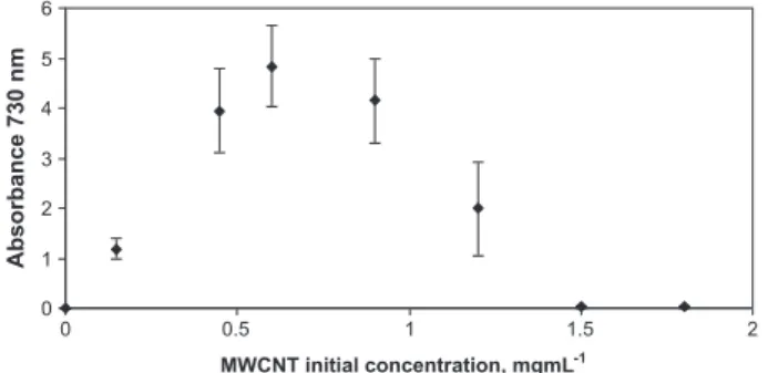
We found that the ratio of surfactant to CNT was crucial in order to optimise the dispersion[20]. An example of one of these dispersion curves is shown inFig. 3, showing MWCNTs dispersed with Lyso PC. In this dispersion curves the concen-tration of surfactant was kept constant and the amount of CNTs was varied in the dispersion mixture. The yield of dis-persion of CNTs was obtained by measuring the absorption value of the suspension at the selected wavelength, as it was stated above. As can be seen, as the amount of CNTs in the dispersion mixture increases the concentration of dis-persed CNTs increases until a maximum is reached where the optimum conditions for dispersion are obtained. Above this optimal concentration of nanotubes in the dispersion mixture, the yield of dispersed nanotubes decreases. This is likely to be due to limiting concentrations of surfactant being shared between large numbers of nanotubes such that insuf-ficient active surfactant is available for solubilizing each nanotube. This effect support the fact that the CNTs are actu-ally being dispersed by the surfactant as limiting concentra-tions of surfactant lead to not properly or not completely dispersed CNTs.
It was clear that with each of the surfactants tested, there was an optimum ratio of surfactant to CNTs for maximum solubilization as can be seen inTable 1. It was generally ob-served that the optimal mass of CNTs solubilized with low molecular weight surfactants tended to be higher than the optimum mass obtained with higher molecular weight sur-factants, attributed to the increased hydrophobicity of the low molecular weight compounds.
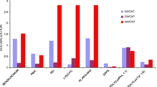
Fig. 4compares this efficiency when solubilization has been optimised for each surfactant. It can be observed that the effi-ciency of solubilization of three types of nanotubes was in the following type order of nanotubes MWCNTs > SWCNTs > DWCNTs for benzalkonium, PEI, PL-PEG-NH2and poly(Lys:Tyr,
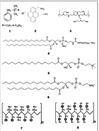
1:9), MWCNTs > DWCNTs > SWCNTs for Lyso PC, Fig. 1 – Surfactant structures: (1) benzalkonium chloride, (2)
pyrenemethylamine (PMA), (3) polyethylenimine (PEI), (4) 1,2-distearoyl-sn-glycero-3-phosphoethanolamine-N-[amino(polyethylene glycosl)2000] (PL-PEG-NH2), (5)
1-stearoyl-2-hydroxy-sn-glycero-3-phosphocholine (Lyso PC), (6) 1,2-dipalmitoyl-sn-glycero-3-phosphoethanolamine (DPPE), (7) Poly(Lys:Phe, 1:1), (8) Poly(Lys:Tyr, 1:9).
SWCNTs > MWCNTs > DWCNTs for PMA and DPPE, and DWCNTs > SWCNTs > MWCNTs for poly(Lys:Phe, 1:1). When comparing surfactants, the best conditions for solubilization of CNTs were obtained with phospholipids, followed by non-biological surfactants and finally polypeptides. When compar-ing the solubilization yield for the non-biological surfactants, PEI solubilized better than low molecular weight surfactants (benzalconium and PMA). When the excess surfactant was re-moved in the case of benzalkonium and PMA, the CNTs be-come not dispersed, indicating that solubilization with these surfactants requires free surfactant in equilibrium with the f-CNTs. PL-PEG-NH2is significantly more efficient than DPPE,
which differs primarily in the absence of a PEG group, suggest-ing that the PEG part of PL-PEG-NH2molecule plays an
impor-tant role in the solubilization process. Conversely, the high solubilization yield for Lyso PC compared well to DPPE suggest-ing that increassuggest-ing the number of acyl chains (in DPPE) de-creases the solubilization efficiency.
0 1 2 3 4 5 6 200 300 400 500 600 700 800 900 1000 1100 wavelength (nm) Absorbance a b c d e f g h i 0 0.5 1 1.5 2 2.5 0 2 4 6 8 10 12 MWCNT-PEI concentration gmL-1 A bs orba nc e 730 n m
Fig. 2 – Set of spectra at increasing concentrations of dispersed MWCNT-PEI. PEI concentration was kept constant at 0.3 mg mL 1and different volumes of functionalized MWCNTs were added. In the insert the absorption value at 730 nm as a function of the concentration of dispersed MWCNT-PEI is presented showing a linear relationship.
0 1 2 3 4 5 6 0 0.5 1 1.5 2 MWCNT initial concentration, mgmL-1 Absorbance 730 nm
Fig. 3 – These optimisation curves were performed with all the surfactants tested and here as an example the
solubilization curve for MWCNTs with LysoPC at a
concentration of 0.3 mg mL 1is shown. Data obtained from triplicates at each MWCNT initial concentration. A proper dilution was made to obtain an absorbance value in the linear range of the spectrophotometer.
Table 1 – Optimum CNT/surfactant ratio for the best dispersion. In this table the optimum CNT/surfactant (w/w) for each surfactant are given.
Surfactant SWCNTs DWCNTs MWCNTs Benzalkonium 5 1.5 5 PMA 6 5 6 PEI 5 3 6 PL-PEG-NH2 2 0.5 3 Lyso PC 0.5 0.5 2 DPPE 0.5 5 2 Poly(Lys:Phe, 1:1) 3 6 1.5 Poly(Lys:Tyr, 1:9) 2 6 2
3.2. Optimisation of DNA binding
To test the use of dispersed CNTs with the cationic surfac-tants as gene carriers, we studied the binding of plasmid DNA to these dispersed CNTs by agarose gel electrophoresis. The plasmid used for this study was the pGL3 plasmid (from Promega) that encodes the luciferase enzyme (lane 2 Fig. 5A). Binding of plasmid DNA to functionalized CNTs inhibits EtBr intercalation[21], as the DNA is in a condensed form. The level of binding can thereby be assessed by the measurement of the non-bound DNA. The CNTs dispersed by the non-covalent attachment of cationic surfactants de-scribed above complexed with DNA (CNT:DNA) were prepared for each surfactant at various mixing ratios to determine the effectiveness of DNA binding. In this way, a constant amount of plasmid DNA was incubated with decreasing concentra-tions of dispersed CNTs. After running the agarose gel, the ex-cess of plasmid DNA can be followed as a band for free plasmid DNA (Fig. 5A lanes 5–8). The dispersed CNTs that most effectively bound the DNA were the PL-PEG-NH2,
poly(Lys:Phe, 1:1), and PEI, whereas the other kind of
dispersed CNTs did not show any DNA binding (see Supple-mentary material, Fig. S6). A constant amount of plasmid DNA was also incubated with decreasing amounts of free sur-factants as a control (seeSupplementarymaterial, Table S1). It was observed that only PL-PEG-NH2, poly(Lys:Phe, 1:1),
and PEI surfactants were able to bind plasmid DNA. It was also found that the surfactant non-covalently attached to CNTs is more efficient to bind plasmid DNA. After determin-ing the amount to surfactant attached to CNTs (see Supple-mentary material), it was found that surfactant bound to CNTs leads to a better condensation of DNA. This conclusion makes the non-covalent attachment of cationic surfactants to CNTs a good method for the condensation and binding of DNA onto CNTs.
The DNA binding capacity of each form of dispersed CNTs can be estimated fromFig. 5by reference to the lowest con-centration of nanotubes that demonstrates detectable DNA binding (for instance, lane 5 inFig. 5A). By normalizing this value to the DNA concentration it is possible to obtain a DNA binding capacity of each f-CNT as shown inTable 2. It can be seen that the best results were obtained for PEI which
1 2 3 4 5 6 7 8
1 2 3 4 5 6 7 8
1 2 3 4 5 6 7 8
1 2 3 4 5 6 7 8
A
1 2 3 4 5 6 7 8
B
1 2 3 4 5 6 7 8
C
Fig. 5 – Agarose gel electrophoresis for the f-SWCNTs that effectively bind plasmid DNA: (A) PEI, (B) PL-PEG-NH2and (C)
poly(Lys:Phe, 1:1). Lane 1: ladder, lane 2: pGL3 plasmid alone 6.8 ng lL 1, lanes 3–8: f-SWCNT:plasmid DNA complexes with
plasmid 6.8 ng lL 1and different dilutions of f-SWCNTs from 1/1 to 1/105(1/1 refers to the best conditions found for solubilization of SWCNTs: 51 lg mL 1for PEI, 56 lg mL 1for PL-PEG-NH
2and 37 lg mL 1for poly(Lys:Phe, 1:1).
0 0.5 1 1.5 2 2.5 3 BENZ ALKO NIUM PMA PEI LYSO PC PL-PE G-NH2 DPPE POLY( LysP he, 1: 1) POLY( Lys:Ty r: 1;9) 0.5 1 1.5 2 2.5 3 BENZ ALKO NIUM PMA PEI LYSO PC PL-PE G-NH2 DPPE POLY( LysP he, 1: 1) POLY( Lys:Ty r: 1;9) SO LU BI LI ZATI O N SWCNT DWCNT MWCNT
Fig. 4 – Solubilization (expressed as the absorbance at 730 nm of the suspension) as a function of the surfactant used for the different kinds of CNTs: SWCNTs, DWCNTs and MWCNTs, in the optimal conditions found for solubilization (these optimal conditions refer to the optimum found when getting the solubilization curve as shown inFig. 3).
has 10 times more binding yield compared to poly(Lys:Phe, 1:1) and 100 times more than PL-PEG-NH2. The other f-CNTs
showed negligible DNA binding.
3.3. Functionalization of RNA-wrapped SWCNTs by a bilayer approach
We also examined functionalization of SWCNTs with biologi-cal molecules such as nucleic acids and proteins. RNA-wrapped CNTs are an attractive method of solubilizing CNTs because the RNA gives high solubilization yields and is non-cytotoxic [22]. However, RNA-wrapping confers negative charges on the carbon nanotubes which then makes them unsuitable for DNA binding. To overcome this problem we investigated the use of a cationic ion or molecule that can act as bridge between the negatively charged RNA wrapping
the CNT, and the negatively charged plasmid DNA (Fig. 6). The following cationic polymers were investigated: poly(-Lys:Phe, 1:1), PEI and polylysine (data not shown). The best re-sults were obtained using the cationic polymer polylysine as a bridging molecule. With poly(Lys:Phe, 1:1) and PEI it was ob-served a higher aggregation of the dispersed CNTs owing to the cationic molecules acting as ionic bridges between nega-tively charged RNA-wrapped CNTs. As this aggregation was lower for polylysine the studies with this functionalization method were carried out with this polymer. Furthermore, it was quantified the amount of plasmid DNA that polylysine on its own is able to bind as a control. This amount was deter-mined as 1.40 mg DNA per mg of polylysine which is higher than for PEI and poly(Lys:Phe, 1:1) (seeSupplementary mate-rial, Table S1). This property also makes polylysine a good choice for the development of this bilayer approach for DNA binding to carbon nanotubes.
The effect of concentration of cationic polymer on DNA solubilization was investigated by agarose gel electrophoresis (Fig. 7). The results showed that the complex between RNA-wrapped CNTs and polylysine is positively charged when the concentration of polylysine is high which is the best con-dition for DNA binding (seeFig. 7A) we observe. As the con-centration of polylysine is decreased, the binary complex becomes negatively charged because the RNA is in excess of the polylysine. There is also a RNA:polylysine ratio at which the binary complex becomes neutral. These effects on func-tionalized CNT surface charge can be observed during the electrophoresis process of the sample preparation (see Sup-plementary material, Fig. S7), negatively charged CNTs run to-wards the positive electrode and vice versa (although this can be seen only in the well as the CNTs are too long and rigid to
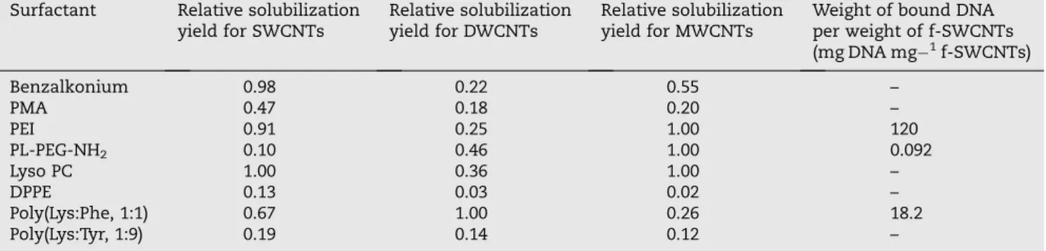
Table 2 – Properties of the f-CNTs. The relative solubilization yield were normalized to those obtained which the highest solubilization yield (PL-PEG-NH2for SWCNTs and LysoPC for DWCNTs and MWCNTs).
Surfactant Relative solubilization yield for SWCNTs
Relative solubilization yield for DWCNTs
Relative solubilization yield for MWCNTs
Weight of bound DNA per weight of f-SWCNTs (mg DNA mg 1f-SWCNTs) Benzalkonium 0.98 0.22 0.55 – PMA 0.47 0.18 0.20 – PEI 0.91 0.25 1.00 120 PL-PEG-NH2 0.10 0.46 1.00 0.092 Lyso PC 1.00 0.36 1.00 – DPPE 0.13 0.03 0.02 – Poly(Lys:Phe, 1:1) 0.67 1.00 0.26 18.2 Poly(Lys:Tyr, 1:9) 0.19 0.14 0.12 – RNA-WRAPPED CNTs CATIONIC POLYMER + + + + + + + + + + + + + + + + + + + + + + + + + + + + + + + PLASMID DNA
Fig. 6 – Bilayer approach with RNA-wrapped CNTs for plasmid DNA binding.
1
2
3 4
5 6
7 8
1
2
3 4
5 6
7 8
1
2
3 4
5 6
7 8
1
2
3 4
5 6
7 8
A
B
Fig. 7 – Agarose gel electrophoresis for f-SWCNTs with the bilayer approach with RNA-wrapped CNTs. (A) Effect of polylysine concentration in plasmid DNA binding: lane 1: ladder, lane 2: pGL3 plasmid 1.8 ng lL 1, lanes 3–8 RNA-wrapped CNTs
(34 lg mL 1) with different concentrations of polylysine from 1.5 mg mL 1to 0.015 lg mL 1. (B) Lane 1: ladder, lane 2: pGL3
plasmid 1.8 ng lL 1, lanes 3–8: RNA-wrapped CNTs–polylysine complexes at different dilutions from 1/1 to 1/105starting in the same conditions as lane 5 in gel A.
enter the agarose). InFig. 7B the plasmid DNA concentration is optimised. These studies show that the optimum DNA binding is 0.071 mg DNA per mg RNA-wrapped CNTs, when working with 45 lg polylysine per mg of RNA-wrapped CNTs. This data confirms that the condensation of plasmid DNA is more efficient in this bilayer approach than with polylysine on its own.
4.
Conclusions
In conclusion, we have compared the solubilization proper-ties of SWCNTs, DWCNTs and MWCNTs with different kinds of surfactants using non-covalent functionalization. The best conditions for solubilization are with the use of phospholip-ids with PL-PEG-NH2for SWCNTs and LysoPC for DWCNTs
and MWCNTs. Furthermore, the solubilization yields with the surfactants tested are in general higher for MWCNTs and SWCNTs than for DWCNTs. The solutions of f-CNTs ob-tained by the solubilization methods presented here are very stable (several months). The use of these functionalized CNTs for development of gene delivery systems was also studied. The best conditions for plasmid DNA binding were obtained with PEI, but, given its cytotoxicity, the best combination for solubilization and DNA binding is poly(Lys:Phe, 1:1), which is less toxic. Furthermore, a bilayer functionalization method based on RNA-wrapped CNTs and the use of cationic poly-mers shows that comparable solubilization and DNA binding can be achieved by this method. Overall, this study is impor-tant as good optimisation strategies for CNT functionalization for gene delivery are crucial if CNT are to be used in a health-care scenario.
Acknowledgements
This work has been performed in the framework of the FP6Marie Curie Research Training Network ‘‘CARBIO’’ (RTN-CT-2006-035616) funded by the European Union. We also acknowledge funding received from the EPSRC Portfolio Part-nership award.
R E F E R E N C E S
[1] Bianco A, Kostarelos K, Prato M. Applications of carbon nanotubes in drug delivery. Curr Opin Chem Biol 2005;9(6):674–9.
[2] Dumortier H, Lacotte S, Pastorin G, Marega R, Wu W, Bonifazi D, et al. Functionalized carbon nanotubes are non-cytotoxic and preserve the functionality of primary immune cells. Nano Lett 2006;6(7):1522–8.
[3] Pantarotto D, Briand J, Prato M, Bianco A. Translocation of bioactive peptides across cell membranes by carbon nanotubes. Chem Commun 2004;1:16–7.
[4] Kam NWS, Jessop TC, Wender PA, Dai H. Nanotube molecular transporters: internalization of carbon nanotube-protein conjugates into mammalian cells. J Am Chem Soc 2004;126(22):6850–1.
[5] Kostarelos K, Lacerda L, Pastorin G, Wu W, Wieckowski S, Luangsivilay J, et al. Cellular uptake of functionalized carbon nanotubes is independent of functional group and cell type. Nature 2007;2:108–13.
[6] Kam NWS, Dai HJ. Carbon nanotubes as intracellular protein transporters: generality and biological functionality. J Am Chem Soc 2005;127(16):6021–6.
[7] Liu Z, Cai WB, He LN, Nakayama N, Chen K, Sun X, et al. In vivo biodistribution and highly efficient tumour targeting of carbon nanotubes in mice. Nat Nanotech 2006;2:47–52. [8] Fu K, Sun YP. Dispersion and solubilization of carbon
nanotubes. J Nanosci Nanotech 2003;3(5):351–64.
[9] Huang W, Taylor S, Fu K, Lin Y, Zhang D, Hanks TW, et al. Attaching proteins to carbon nanotubes via diimide-activated amidation. Nano Lett 2002;2(4):311–4. [10] Kim WJ, Kang SO, Ah CS, Lee YW, Ha DH, Choi IS, et al.
Functionalization of shortened SWCNTs using esterification. Bull Korean Chem Soc 2004;25(9):1301–2.
[11] Lim SH, Elim HI, Gao XY, Wee ATS, Ji W, Lee JY, et al. Electronic and optical properties of nitrogen-doped multiwalled carbon nanotubes. Phys Rev B 2006;73(4):0454021–6.
[12] Kam NWS, Liu Z, Dai H. Functionalization of carbon nanotubes via cleavable disulfide bonds for efficient intracellular delivery of siRNA and potent gene silencing. J Am Chem Soc 2005;127(36):12492–3.
[13] Liu Z, Winters M, Holodniy M, Dai H. SiRNA delivery into human T cells and primary cells with carbon-nanotube transporters. Angew Chem 2007;46(12):2023–7.
[14] Zheng M, Jagota A, Semke ED, Diner BA, McLean RS, Lustig SR, et al. DNA-assisted dispersion and separation of carbon nanotubes. Nat Mater 2003;2:338–42.
[15] Nakashima N, Okuzono S, Murakami H, Nakai T, Yoshikawa K. DNA dissolves single-walled carbon nanotubes in water. Chem Lett 2003;32(5):456–7.
[16] Gao H, Kong Y, Cui D. Spontaneous insertion of DNA oligonucleotides into carbon nanotubes. Nano Lett 2003;3(4):471–3.
[17] Bachmatiuk A, Borowiak-Palen E, Rummeli MH, Gemming T, Kalenczuk RJ. Influence of the substrate loading on the quality and diameter distribution of SWCNT in alcohol-CVD. Phys Stat Sol (b) 2007;244(11):3925–9.
[18] Borowiak-Palen E, Bachmatiuk A, Rummeli MH, Gemming T, Ruszynska M, Kalenczuk RJ. Modifying CVD synthesised carbon nanotubes via the carbon feed rate. Physica E 2008;40(7):2227–30.
[19] Flahaut E, Basca R, Peigney A, Laurent C. Gram-scale CCVD synthesis of double-walled carbon nanotubes. Chem Commun 2003;12:1442–3.
[20] Vigolo B, Penicaud A, Coulon C, Sauder C, Pailler R, Journet C, et al. Macroscopic fibers and ribbons of oriented carbon nanotubes. Science 2000;290(5495):1331–4.
[21] Singh R, Pantarotto D, McCarthy D, Chaloin O, Hoebeke J, Partidos CD, et al. Binding and condensation of plasmid DNA onto functionalized carbon nanotubes: toward the
construction of nanotube-based gene delivery vectors. J Am Chem Soc 2005;127(12):4388–96.
[22] Jeynes JCG, Mendoza E, Chow DCS, Watts PCP, McFadden J, Silva SRP. Generation of chemically unmodified pure single-walled carbon nanotubes by solubilizing with RNA and treatment with ribonuclease A. Adv Mater